JOURNAL OFVIROLOGY, Jan. 1988, p. 91-99 Vol. 62, No.1 0022-538X/88/010091-09$02.00/0
Copyright X) 1988, American Society for Microbiology
Isolation and Characterization
of Herpes Simplex
Virus
Type
1 Host
Range Mutants
Defective in Viral
DNA
Synthesis
ELLEN P. CARMICHAEL, MARSHALL J. KOSOVSKY, AND SANDRAK. WELLER*
DepartmentofMicrobiology, University of Connecticut Health Center, Farmington, Connecticut 06032-9984 Received 23 July1987/Accepted 25 September 1987
Cell linesweregenerated by cotransfection of Vero cells with pSV2neo and a plasmid containing the herpes simplex virus type1(HSV-1)EcoRI D fragment (coordinates 0.086 to 0.194). One such cell line (S22) contained the genesfor alkaline exonuclease and several uncharacterized functions. Three mutant isolates of HSV-1 strain KOS which grew on S22 cells but not on normal Vero cells were isolated and characterized. All three mutants (hr27, hr48, and hr156) were defective in the synthesis of viral DNA and late proteins when grown in nonpermissive Vero cells. Early gene expression in cells infected with these host range mutants appeared to be normal at the nonpermissive condition. The mutations were mapped by marker rescue to a 1.5-kilobase fragment(coordinates 0.145 to 0.155). The mutation of one of these mutants, hr27, was more finely mapped to an800-base-pair region (coordinates 0.145 to 0.151).Thispositionof these mutations is consistent with the map location of a putative 94-kilodalton polypeptide as determined by sequence analysis (D. McGeoch, personal communication). Complementation studies demonstrated that these mutants formed a new complementation group, designated 1-36. The results presented in this report indicate that the 94-kilodalton gene product affected by these mutations may have a direct role in viralDNAsynthesis.
The herpes simplex virus (HSV) genome is large (160 kilobases [kb]) and encodes many of the gene products involved in the replication of its double-stranded DNA chromosome. HSV functions involved either directly in DNA synthesis or in nucleotide metabolism include the major DNA-binding protein (10, 28, 54), DNA polymerase (42), alkaline nuclease (18, 38), a 54-kilodalton (kDa) poly-merase-associated protein (51), ribonucleotide reductase (15), thymidine kinase (14),and dUTPase (39).
The most direct approach to identifying gene products essential for viral DNA replication has been to isolate
mutants which exhibit alterations in DNAsynthesis. Ofthe functions identified, only the major DNA-binding protein (ICP8)(10, 37, 54) and the HSV DNA polymerase (5, 6, 9, 22, 23, 36)have been shown by the analysis of temperature-sensitive(ts) and othermutantstobeabsolutely essentialfor viral DNA replication. Genetic studies indicate that under certain conditions alkaline nuclease(19, 32), ribonucleotide reductase (15, 40), and thymidine kinase (24) may also be required. To identify additional proteins required for viral DNA synthesis, DNA-negative mutations in genes whose functions are currently unknown have been isolated and studied. These include tsK13 andtsM19,representing
com-plementationgroup1-10, whose mutations maptothefar left end ofthe genome betweencoordinates0.095 and 0.108(53; S. K. Weller, E. P. Carmichael, D. P. Aschman, D. J. Goldstein, and P. A. Schaffer, Virology, in press). In addi-tion, the lesion in tsS38, a member of complementation group 1-26, has beenmapped to coordinates 0.126to 0.133 (Welleret al., in press). tsS38 produces small to moderate levels of viral DNAatthenonpermissivetemperature(8). In this case, it isnotclear whether the geneproductisessential forviral DNA synthesis since the ts mutation may beleaky (i.e., some residualactivity of the affected protein may be present at the nonpermissive temperature). To circumvent problemswithpotentially leakytsmutants,wehaveusedan alternative approach for theisolation of mutations in
func-*Correspondingauthor.
tions thought to be involved in viral DNA synthesis. This strategy, originally developed byBenjamin (2), involves the isolationof host rangemutantswhich can be propagatedon
complementing transformed cell lines. We have isolated
mutantsin a newcomplementation group whosemutations map to the left of alkaline nuclease within the EcoRI D fragment ofHSV-1 strainKOS (coordinates0.086 to 0.194). These host rangemutantsfail tosynthesizedetectable levels of viralDNA in nonpermissive cells.
MATERIALS AND METHODS
Cells and viruses. Procedures for the maintenance and growth of African green monkey kidney cells (Vero) have been described previously (53). S22 cells (seebelow) were
maintainedasabove, but with the addition oftheantibiotic geneticin (G418; 250 ,ug/ml; GIBCO Laboratories, Grand Island, N.Y.).
TheKOSstrain ofHSV-1wasusedasthewild-type (WT) virus. ts mutants used in this study are shown in Table 1. HSV-1 strain KOS ts mutantsweregenerouslyprovided by P. A. Schaffer (Dana-Farber Cancer Institute, Boston, Mass.).tsJ12containsa tsmutation inthestructuralgenefor theglycoproteingBwhich has beenmappedto coordinates 0.357to 0.360 (44). HSV-1 strain 17 mutants tsR,
tsS,
and tsX were kindly provided by V. Preston and H. Marsden(MRC
Institute of Virology, Glasgow, Scotland) (30). Vi-ruses werepropagatedandassayedasdescribed previously(45).
Plasmidsandbacteria. Allrecombinant clonescontaining HSV-1 DNA are shown in Fig. 1. pSG10, generously pro-vided by R. Sandri-Goldin (University of California at Ir-vine) and M. Levine(University ofMichigan, AnnArbor), contains theEcoRIDfragmentof HSV-1 strain KOS (coor-dinates 0.086to0.194) cloned into the EcoRI site ofpBR325 (20). Theconstruction ofpSG10-SB, pSG10-B2,and pSG10-BD2isdescribed elsewhere(Welleretal., inpress).
pSG10-XIIwasconstructed byligating anXhoII fragment
(coordi-nates 0.145 to 0.155) into the BamHI site of
pUC19.
pSG10-BD2XHwasconstructedasfollows.pSG10-BD2
was 91on November 10, 2019 by guest
http://jvi.asm.org/
92 CARMICHAEL ET AL.
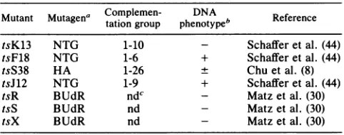
TABLE 1. HSV-1 ts mutants used in this study
Mutant Mutagena Complemen-tation groupphenotype1DNA ReferenceReference
tsK13 NTG 1-10 Schafferetal. (44)
tsF18 NTG 1-6 + Schafferetal. (44)
tsS38 HA 1-26 ± Chu et al. (8)
tsJ12 NTG 1-9 + Schaffer etal. (44)
tsR BUdR ndc - Matz etal. (30)
tsS BUdR nd - Matz et al. (30)
tsX BUdR nd - Matzetal. (30)
aHA,Hydroxylamine; BUdR, 5-bromodeoxyuridine; NTG,
nitrosoguani-dine.
bPhenotypes determined by Aron et al. (1) for tsF18 and by Weller et al. (53) for tsK13; others determined by authors indicated. Symbols indicate synthesis of viral DNA at the nonpermissive condition relative to that in the wild type: +, >20% of the wild-type level; ±,s20%; -, no detectable viral DNAsynthesized.
cnd,Notpreviously determined.
digested with XhoI and HpaI, and the ends were filled in withthelarge fragment of DNA polymerase I and religated. pSG10-P5 and pSG10-P6 were constructed by ligating the PstI fragments of EcoRI-D (coordinates 0.129 to 0.151 and 0.151 to 0.158, respectively) into the PstI site of pUC19. pSV2neocontainsthebacterialgene forneomycinresistance under the controlof the simian virus 40 early promoter (47). Recombinant plasmids were propagated in Escherichia coli JM83 orDH5 by standard procedures (29).
a b
UL
IL I-.
Viral and cellular DNA isolation. Viral DNA from host range mutants of HSV-1 strain KOS was prepared from partially purified virions as described by Parris et al. (35). CellularDNA wasisolated as described in Weller et al. (55). Transformation of Vero cells. Plasmids pSV2neo (5 ,ug) and pSGIO (3 ,ug) were coprecipitated in the presence of 16 ,ug of salmon sperm DNA in a total volume of 1.0 ml by the procedure ofGraham and van der Eb(21). Freshly trypsi-nized Verocells(4 x 10⁶)wereadded to theprecipitateand incubated at37°C for30 min withcontinuousagitation. This cell-DNAmixture wassuspendedin10 ml of culture medium andtransferredto a 100-mmpetridish. After 4 h at 37°C, the cells were shocked with 15% glycerol in HEPES (N-2-hydroxyethylpiperazine-N'-2-ethanesulfonic acid)-buffered saline,washedtwicewithHEPES-bufferedsaline,and incu-bated further at 37°C after the addition of medium. G418 selectionwascarriedout asdescribed by DeLuca et al. (12). The cells were grown to confluency (about2 days), trypsi-nized, and plated at a 1:10 dilution in medium containing G418(500,ug/ml). Afterapproximately2weeks at37°C(with periodic change of medium), individual G418-resistant colo-nies wereisolated, amplified, and screened as described in Results. Onepositive cell line, S22, was chosenfor further studies.
Analysis of cellular and viral DNA. Total cellular or viral DNA wasdigestedwithrestriction endonucleases, fraction-ated by agarose gel electrophoresis, and transferred to a GeneScreen Plus nylon membrane (New England Nuclear
b' a' c' Us c a
1 11 , l
M
0.1 0.2 0.3 0.4 0.5 0.6
11
2
S S a X IS SI x I H 55S X0o N X X a
, . .. .I.
D 0NDHKH D
pSG1O
-82
-XII
-SB
.BDI
-P.-PS
B3
-BD2
-BD2-XH
-K13, M19,FIB F43 S38
I
0.7 0.8 0.9 1.0
2.2-0kkbb
.0.9kb
- TRANSCRIPTS* - 2.3kb
3.9kb
4.5kbFIG. 1. Recombinant DNAplasmids and physical map locations of mutations in five ts mutants within theEcoRIDfragment. TheEcoRI
Dfragment (map coordinates 0.086to 0.194)has been expanded to showinternalcleavage sites: EcoRI (R),BgII (G),HindIII(H),KpnI(K),
HpaI(H), andXhoI (X).TheHSV-1DNAinsertionsin eachrecombinant plasmid described in the text are shown below the restriction map. Thephysical maplocations ofmutations infive HSV-1 strain KOSts mutants are shown in relation totranscripts which have been finely mapped within this region (11, 13, 52).
0.0
I
III I I I I I I
l
ll~~~~~~~~~~~~~~
_ _ _ _
;l~~~~~~~~~~~~~~1
COORDINATES
EcoRl D FRAGMENT
RECOMBINANT
CLONES
ts
MUTANTS
J. VIROL.
I
on November 10, 2019 by guest
http://jvi.asm.org/
[image:2.612.65.306.85.180.2]HSV-1 MUTANTS DEFECTIVE IN DNA SYNTHESIS 93 TABLE 2. Titers ofmutants at39°C
Titer(PFU/ml)
Strain Ratio,
Vero cells S22cells
S22/Vero
KOS 7.5 x 10⁸ 8.9 x 10⁸ 1.2
tsS38 8.5 x 10⁴ 1.7 x 10⁸ 2.1 x 10³
hr27 <102 1.5 x 10⁸ >1.5 x 10⁶
hr48 <102 2.2 x 10⁸ >2.2 x 10⁶
hr156 <102 5.8 x 10⁷ >5.8 x 10⁵
tsR <103 1.5 x 10⁷ >1.5 x 10⁴
tsS <103 1.8 x 10⁵ >1.8 x 10²
tsX <103 6.7 x 10⁶ >6.7 x 10³
Corp.) as suggested by the supplier. Recombinant DNAs used asprobesfor hybridizationwerelabeled bythe method ofFeinberg and Vogelstein(17).
Marker rescue analysis. Markerrescue experiments were performed by the procedure of Parris et al. (35) with the modifications described by Chiou et al. (7).
Complementation tests. Complementation tests were con-ductedasdescribedpreviously(45). Complementation indi-ces (CIs) were calculated from the formula CI = (A +
B)np/(A
+ B where A and B are two mutants. Virusyields from infections carried out in Vero cells at 39.6°C (nonpermissive conditions) were assayed forplaque forma-tion in S22 cellsat34°C (permissiveconditions).
Synthesis ofviral DNA in infected cells. Analysis of viral DNA synthesis wasperformedas described byAron et al. (1) except that proteinase K was used instead ofpronase. Growth inVerocells wasusedasthe nonpermissive condi-tionforhost range mutants, andgrowthinS22 cellswasused asthepermissive condition.
Viral protein labeling and gel electrophoresis. Labeling of HSV-infected Vero or S22 cells with [35S]methionine was performed as described previously (Weller etal., in press) except that label was addedtwice, first at 5 hpostinfection andagain at10 h postinfection. Addition of isotope at 10 h was found to improve labeling of viral late proteins. Cells werelysed inasolution containing0.05M Tris chloride, pH 7.2,0.15 M NaCl,0.1% sodium dodecyl sulfate, 1% sodium deoxycholate, 1% Triton X-100, and the proteaseinhibitors N-a-p-tosyl-lysine chloromethyl ketone (Sigma Chemical Co.) and phenylmethylsulfonyl fluoride (Sigma) at a final
concentration
of0.1 mM and 0.5 mM, respectively. The extracts were then sonicated for 45 s, and cell debris was removedby centrifugation for5min inanEppendorfmicro-centrifuge.
Anequal volume of sample buffercontaining0.37 M Tris chloride, pH 6.8, 10% glycerol, 5% 3-mercapto-ethanol, 10% sodium dodecyl sulfate,and0.001% bromphe-nol blue was added to each sample, which was then boiled for3 minand loaded onto a9%bisacrylamide-cross-linked
polyacrylamide gel (26). Gels were treated with Autofluor (NationalDiagnostics), dried, and exposedtoKodakXAR-5 filmat -70°C.RESULTS
Isolation of host cells. The HSV-1 EcoRI D fragment of strain KOS (16.5 kb; coordinates 0.086 to0.194) containsa numberofviral genes,several of whichhave beenimplicated in viral DNA synthesis (Fig. 1). Transcript mapping within theEcoRI D fragmentrevealed six mRNAs between coor-dinates 0.165 and 0.194, including the gene for alkaline nuclease(11, 13) (Fig. 1). tsmutantsofKOS whoselesions
map in the EcoRI D fragment include two DNA-positive mutants in complementation group 1-6 (tsF18 and tsF43, coordinates 0.095 to 0.108 and 0.112 to 0.118, respectively) andthree mutants representing twocomplementation groups whichwere defective in viral DNA synthesis. These include tsK13 and tsM19 (complementation group 1-10), whose mutations mapped to coordinates 0.095 to 0.108, and tsS38 (complementation group 1-26), whose mutation mapped to coordinates 0.126 to 0.133 (Fig. 1)(8, 44, 53; Weller et al., in press).
Tocarry out afunctional analysis of the EcoRI-Dregion, we constructed cell lines which contained the EcoRI D fragment. We anticipated that these cell lines could be used to complement both conditionally lethal and null mutations inthis region since thewild-type protein wouldbe
provided
by the cells in trans. Vero cells were cotransfected with plasmids pSV2neo and pSG10 (Fig. 1). Two weeks after transfection, G418-resistant colonies were isolated and tested at39.9°Cfor theabilitytocomplementthegrowthof themutanttsS38, whose mutation had been mapped within EcoRI-D. tsS38 formed plaques atthe nonpermissive tem-perature inone of the lines tested, S22cells, but did not form plaques efficiently in Vero cells (Table 2). This result con-firmsthatS22cells are capableofexpressingatleastoneof the viralproteins mapping withinthe EcoRI Dfragment.
To confirm the presence of the EcoRI D fragment and to determine theapproximatecopynumber, total cellular DNA isolated from S22 cells was analyzed by the method of Southern (46) (Fig. 2). After digestion with BamHI and Hindlll, S22 DNA was subjectedtoelectrophoresis, trans-ferredto anylon membrane, and probed with the 32P-labeled BamHI-HindIII fragment (coordinates 0.165 to 0.186) ob-tained frompSG10-BD2 (Fig. 1). Thisfragment, which lay within EcoRI-D, contained a large portion of the coding regionof thealkalinenucleasegene. The standards present
pSG10-BD2
M 1 5 10 50 100
4.36 1
3.26 am hd
2.67 _
2.30
1.86 1.69
S22
0=o :2- 0 In)
,-7r
FIG. 2. Analysis of HSV-1 DNA in S22 cells. BamHI-HindIII digests of S22DNA(5 and10 Fg)wereanalyzed bythemethod of Southern(46). pSG10-BD2wasdigestedwiththesameenzymesto
visualize1(10pg),5(50 pg),10(100pg),50(500pg),and 100(1ng) copies of viralDNAper 3 x 10⁹bp ofcellular DNA. The blotwas
probedwiththe 2P-labeled BamHI-HindIII fragmentfrom pSG10-BD2(Fig. 1).Numberstotheleftindicatesizesof molecularweight
markers(lane M).
VOL.62, 1988
on November 10, 2019 by guest
http://jvi.asm.org/
[image:3.612.330.543.440.649.2]TABLE 3. Marker rescue of hr27, hr48, and hr156
Map Marker rescue
efficiencyc
Plasmidacoordinatesb hr27 hr48 hr156
pSG10 0.086-0.194 399 316 196
pSG10-BD1 0.103-0.145 <0.05 <0.05 <0.05
pSG10-P5 0.129-0.151 328 ndd nd
pSG10-XII 0.145-0.155 40 1.0 1.0
pSG10-B2 0.145-0.165 2.0 nd 0.20
pSG10-P6 0.151-0.158 <0.05 nd nd
pSG10-B3 0.165-0.194 <0.05 <0.05 <0.05
None <0.05 <0.05 <0.05
a Plasmids were linearized before transfection. b Coordinates are in the P orientation.
Results areexpressed as platingefficiencies, which were determined by the formula[(PFU/mlverJ/PFU/mls22)I x 103. A value of<0.05 indicates no markerrescueoccurred.
d nd,Notdone.
on thegel (pSG10-BD2 digested with BamHI and HindIll) indicatedthatS22 cellscontained approximatelyfivecopies oftheBamHI-HindIII fragment per cell. Further Southern analysis ofthis DNA revealed that the cellscontainedDNA from approximately coordinates 0.118 to 0.194 (data not shown). Inaddition, the DNA appeared to be stably main-tained inthe cellsfor at least 25 passages (data not shown). Isolation of host range mutants.Our originalintentionwas to isolate deletion mutations in the genefor alkaline nucle-ase.Tothis end,S22 cells werecotransfected withwild-type KOSDNAand pSG10-BD2XH, whichcontainsadeletion of 300nucleotides in the alkaline nuclease gene (see Materials and Methods and Fig. 1). Three hundred plaques were isolated andtestedfor their abilityto grow on S22 and Vero cells. Sixteen host range mutants were identified which formed plaques on S22 but not on Vero cells. Data from titrations ofthreeofthese mutants (hr27, hr48, and hr156) are shown in Table 2. Restriction enzyme digestion and Southern analysis (46) of viral DNAs from each ofthe 16 mutants showed that all mutants lacked the 300-base-pair (bp) deletion in pSG10-BD2XH (datanotshown). Since S22 cells were constructed by introducing the entire 16.5-kb EcoRI D fragment into Vero cells, it is likely that several viralgenes could be expressed in these cells. We therefore feel that this class ofmutants most likely arose by sponta-neous mutation in one or moreofthese genes. Because of their interesting growth phenotypes, three mutants (hr27, hr48, and hr156) were plaque-purified three times, and stocks wereprepared on S22 cells.
Geneticanalysis. (i)Markerrescue. To determinewhether thelesions in hr27, hr48, and hr156werein the nucleasegene orin anothergenecontained in S22 cells, markerrescuewas
performed with cloned DNA fragments from wild-type HSV-1 KOS DNA (Fig. 1 and Table 3). As expected, plasmid pSG10, which contains HSV-1 EcoRI-D, efficiently rescued the lesions in all threemutants. Tomap the
muta-tionsmorefinely, subclones of EcoRI-Dwereused in marker
rescue tests. Plasmids pSG10-BD1, pSG10-B3 (which
con-tainsthe entire nucleasegene), andpSG10-P6didnotrescue
the mutations in hr27, hr48, and hr156; however, plasmids pSG1O-XII and pSG10-B2 rescued the mutation(s) in all three mutants.Thus, welocalized the mutationsto a1.5-kb
fragment,ataposition 1.5 to3.0 kbtothe left of the 3' end of the alkaline nucleasegene(coordinates 0.145to0.155).In addition, the mutation in hr27wasmorefinely mapped with
PstIclones pSG10-P5 and pSG10-P6 (Fig. 1). Since plasmid pSG10-P5 rescued the lesion in hr27, its mutation was
localized to an 800-bp fragment between coordinates 0.145
and 0.151.
(ii) Complementation studies. To determine the functional relationships between hr27, hr48, hr156 and other mutants
whose mutations have been mapped to the EcoRI D
frag-ment, complementation tests were conducted. Table 4
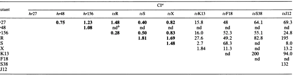
shows the results ofquantitativecomplementationtestswith the host range mutants and several ts mutants. hr27, hr48, and hr156 failed to complement each other for growth in Vero cells, indicating that they are members of the same
complementation group. Mutants in complementation
groups 1-6 (tsF18), 1-10 (tsK13), and 1-26 (tsS38) all
com-plemented the growth of the host range mutants. Another member ofcomplementation group 1-10 (tsM19) also
com-plemented themutantsefficiently (datanotshown).
Further-more,mutant tsJ12, whose mutation mapped between
coor-dinates 0.357 and0.360, was able to complement the host
range mutants. These results demonstrate that hr27, hr48, andhr156belongtoacomplementation groupdistinct from the previously isolated ts mutantsofstrain KOS. We have designated this complementation group 1-36 following the nomenclature establishedby Schafferet al. (45).
The lesions in three HSV-1 strain 17 mutants (tsR, tsS, andtsX) have been localized tothefar-left-handendof the long unique region between coordinates 0.145 and 0.167,to
a position overlapping the map location of the host range
mutations described in this study (30). In addition, these
mutants were capable of plaque formationon S22 cells but
[image:4.612.64.304.83.192.2]noton Verocells at the nonpermissive temperature (Table 2). These data ledustoexamine the functionalrelationships
TABLE 4. Complementation among 10 mutantsof HSV-1
cia
Mutant
hr27 hr48 hr156 tsR tsS tsX tsK13 tsF18 tsS38 tsJ12
hr27 0.75 1.23 1.48 0.40 0.82 15.8 48 64.1 69.3
hr48 1.08 ndb nd nd nd nd nd nd
hrlS6 0.28 0.50 0.83 16.0 52.3 55.1 24.8
tsR 1.81 1.69 27.6 49.2 82.8 195
tsS 1.48 2.7 68.3 nd 8.0
tsX 1.84 11.3 nd 13.2
tsK13 nd 200 94.0
tsF18 nd nd
tsS38 132
tsJ12
aNumbers in boldface areconsidered negative forcomplementation.
bnd,Not done.
on November 10, 2019 by guest
http://jvi.asm.org/
[image:4.612.71.560.578.704.2]HSV-1 MUTANTS DEFECTIVE IN DNA SYNTHESIS 95
S22 Cells
30
02
0L
50
40
30
20
10
0
Vero Cells
1.8 60 50
40
.1.7 30
20
10
.1.6 0
.1.8
1.7
1.6
[image:5.612.136.487.65.547.2]fraction
number
FIG. 3. Separation of viral and cellular DNAs by CsCl equilibrium centrifugation. Mutant- or wild-type-infected Vero or S22 cells
incubatedat34°Cwereexposedto10 p.Ci of[3H]thymidineperml from 6to24 hpostinfection. Lysates of these cellswerethensubjected
toequilibrium. centrifugationinneutralCsClgradientsasdescribedby Aronetal.(1).Cellular DNAcorrespondstoapeakwithabuoyant
densityof1.690, andviral DNAgivesapeakwithabuoyant density of1.725.
betweentsR, tsS,andtsXand the hostrangemutants. hr27, hr156, and hr48 failedtocomplementthe strain 17 mutants
(Table 4). We haveobservedthat CIsobtained with tsXare
reproducibly low; therefore, we do not feel that the 1.84 value obtained with tsX and tsK13 is significant. With this
oneexception,mutantsin all othercomplementationgroups
didcomplement tsR, tsS, and tsX efficiently. We conclude from these data thattsR, tsS, andtsX arealso members of complementationgroup 1-36.
Phenotypic analysis. (i) Synthesisof viral andcellular DNA by wild-typeand hostrangemutants. To determine whether the defect in these mutants was at the level of DNA
synthesis, the incorporation of [methyl-3H]thymidine into viral DNA in mutant-infected Vero or S22 cells was
mea-sured. The three host range mutants were analyzed inthis
way, and data fortwo are shown in Fig. 3. Wild-type and
mutant-infected cellswere labeled and total DNAwas har-vested as described by Aron et al. (1). Cellular and viral DNAs werethen resolved by equilibrium centrifugation in cesiumchloride. Figure3 showsthepattern ofincorporation of[3H]thymidine into viral and cellular DNA by the wild-typevirus(KOS), hr27,and hr156grownin Verocells and in S22 cells. S22 and Vero cells infected with KOS and S22 cells infected with hr27and hr156were capableofinducing
1.8
1 .7
.1.6
.1.8 >% .)
n
Oa
.1.7 C
Cu
0
.0-1.6
VOL.62, 1988
on November 10, 2019 by guest
http://jvi.asm.org/
TABLE 5. Viral DNA synthesis of wild-type and host
range mutants in Vero and S22cellsa
% ofwild-type DNAsynthesis Virus
Vero cells S22 cells
hr27 0 40
hr48 0 32
hr156 0 56
KOS 100 100
a Values under viral peaks were determined, and results are expressedas the percentage of viral DNA in wild-type virus-infected cultures. Values
representtheaverage oftwoseparate experiments.
significant levels of viral DNA. However, in Vero cells infected withhr27 and hr156, no detectableviral DNA was synthesized. The average values for viral DNA synthesis with the three mutants and wild-type virus are shown in Table 5. Based on this observation, it appears that the lesion(s)inthese mutants maybe inagenewhose product is involved in viralDNAsynthesis.
(ii) Viral protein synthesis at the permissive and nonpermis-sive conditions. Mutantswithablock in viralDNAsynthesis would be expected tobe defectivein induction of virallate
(yj
and_Y2) polypeptides (10).
Vero and S22 cells wereinfected with host range mutant or wild-type viruses at a multiplicityof 10PFU/cell. The patternof protein synthesis ofS22 cells infected withhr27, hr48, and hr156 resembled thatof cells infected with wild-type virus (Fig. 4, compare lanes 8, 9, and 10 with lanes 1 and 7). In contrast, hr27-, hr48-,andhr156-infectedVerocells (Fig.4, lanes 3, 4, and5) showednodetectablesynthesis oftruelate(_Y2)polypeptides suchas ICP15, ICP19/20, ICP33, and ICP43/44, and early-late
(-Yl)
polypeptides suchasICP5 and ICP25were synthe-sized in greatly reduced amounts compared with the wild type. The synthesis ofearlypolypeptides such as ICP8 and ICP36was not affectedinmutant-infected Vero cells, indi-cating that there was no grossdefect inearly gene expres-sion. The electrophoretic profile seen in mutant-infected Vero cells was quite similar to that seen in cells infected with wild-type virus in the presence of the viral replication inhibitor phosphonoacetic acid (Fig. 4, lane 2). Thus, we concludethatalthough earlygeneexpression isnotaffected in Vero cells infected with host range mutants,SYi
and -Y2 proteins are decreased, consistent with a defect in DNA synthesis.DISCUSSION
Wereport here theisolationandcharacterization ofa new groupofHSV-1 host range mutants defective inviralDNA synthesis. The isolation ofthese mutants wasmade possible by the availability of a permissive complementing cell line, S22. This cellline was isolated following transformation of Vero cells with a plasmid containing the EcoRI D fragment. Sixteen mutantswhose growth was supported by S22 cells were isolated, and three mutants (hr27, hr48, and
hrlS6)
were selected for further study. These mutants defined a newly designatedcomplementation group, 1-36, as shown by their ability to complement other mutants of strain KOS. The inability of these mutants tocomplement three DNA-negative mutants of strain 17, tsR, tsS, and tsX, indicates that allsixweremembers of the same group. Thelesions in the three host range mutants hr27, hr48, and
hrlS6
were localizedby marker rescue to a 1.5-kb fragment (coordinates 0.145 to 0.155) mapping to the left of the alkaline nucleasegene. Further finemapping has demonstrated that the lesion in hr27lies within an800-bp fragment between coordinates 0.145 and 0.151. Recent sequencing data reveal the existence of an open reading frame of 94 kDa in this region (D. McGeoch, personal communication). The mutation in mu-tant hr27 mapped to a position entirely within this open reading frame,and the mutations in hr48 and hr156 were also consistent withtheirbeingin this gene(Fig. 5).
Mutants hr27, hr48, and hr156 were defective in viral DNAsynthesis when grown in Vero cells. Furthermore, the patternof viralprotein synthesisinducedbythese mutants in Vero cells wasconsistent with their DNA-negative pheno-type in that they were defective in the synthesis of late polypeptideswhilesynthesis of early proteins appeared to be normal. These results suggest that the putative 94-kDa protein playsa rolein viral DNA synthesis.
Analternativeapproachhasrecentlybeen used toidentify gene products required for amplification of HSV origin-containing plasmids (4). It has been known for some time that HSV origin-containing plasmids can be amplified in Verocellsif HSV functions aresupplied in transby either cotransfectionwithHSVDNA orsuperinfectionwith intact
Virus WT PAA
1 2
Vero Cells S22 Cells
27 48 156 M WT 27 156 3 4 5 6 7 8 9
48
10
ICP1,2
ICP5 Am.-o
ICP6 Uz
ICP-.. a SE
lcP11
ICP15 _. I. .*
ICPI9,20
ICP25 ICP33
ICP36
am,
1CP43 _"
ICP44 4I Idg..dA. 00 mgl e6A
FIG. 4. Synthesis ofviralpolypeptidesinwild-type-and mutant-infectedcells. Monolayers of S22 andVerocellswereinfected with wild-type (WT)or mutantviruseshr27, hr48,orhr156. [3S]methi-oninewaspresentduring incubation from5to18 hpostinfection(see Results).At18 hpostinfection,total celllysateswereprepared for electrophoresis. The polypeptideswereseparated by electrophore-sis through a 9% polyacrylamide gel. Lane 2 represents KOS-infected Vero cells in the presence of 150 ,ugofphosphonoacetic acid(PAA)per ml. LaneM, Mock-infected cells.
on November 10, 2019 by guest
http://jvi.asm.org/
[image:6.612.323.558.309.637.2]HSV-1 MUTANTS DEFECTIVE IN DNA SYNTHESIS 97
a b
UL
11 J
b s c
US
c a0.0 0.1 0.2 0.3 0.4 0.5 0.6 0.7 0.8 0.9 1.0
I I I I I I I I I
to)
3 0) InIcl
Ie
le to 0n~~~~7
vr 7:@0 0
0o
9
?~~
I 9 0 -%H B S SS
BGG
X H XH X X RpI I II I ILB I I I1 I
p-4*--
VZO
Zi~-lu
.4 p
hr27
hr48, 156
tsR,S,X
I
I
KOS HOST RANGE
MUTANTS
STRAIN 17 MUTANTS
FIG. 5. Putative open reading frames in theright half ofEcoRI-D. TheHpaI-EcoRI restriction fragment has been expanded to show restriction sites (seelegendtoFig. 1). DNAsequence analysis (31;D.McGeoch, personal communication) revealed the presence of several openreading frames in this region. Shown below are the map locations of KOS host range mutants hr27, hr48, andhr156(describedin this paper) and strain 17 mutants tsR, tsS, and tsX (30). alk nuc, Alkaline nuclease gene; 94K, 94-kilodalton. Arrows indicate direction of transcript or open reading frame.
HSV virus (48, 49, 50, 55). Challberg (4) has found that certain combinations of HSVclones canbe usedtosupport origin-containing plasmid amplification. One clone that has been showntoberequiredinthissystem containsthe gene forthe94-kDaprotein (Challberg, personal communication). Thisobservation isconsistent withresults described herein demonstrating that the 94-kDa protein gene product is es-sential for viralDNAsynthesis in vivo.
Many putative DNA replication functions have been lo-calized to the left end ofthe viral genome. In addition to alkaline nuclease (32, 33) and the gene for the 94-kDa protein, twoother genes in thisregionhave beenimplicated inDNA synthesis. Mutants tsK13 and tsM19 (complemen-tationgroup1-10),whose mutationsmaptothefar left end of EcoRI-D (Fig. 1), failed to synthesize detectable levels of viral DNA at the nonpermissive temperature (Weller et al., in press). Inaddition,weandothers(8;E. P.Carmichaeland S. K. Weller, unpublished data) have shown that tsS38 (complementation group 1-26) produces small to moderate amounts of viral DNA at the nonpermissive temperature. However, thismutanthas been showntobeleakyat39.9°C. Anull mutation in the gene whichisdefective in tsS38 will be required toelucidate whetherthis gene productis essential
for DNA synthesis. It is anticipated that the use of host range mutants will greatly facilitate the analysis ofgenes whoseproducts areessential forDNAsynthesis.
At present, HSV isknownto encode atleast sevengene products involvedin DNA synthesisandnucleotide metab-olism. These include DNA polymerase (25, 41, 42),
DNA-binding protein (10, 51, 54), thymidine kinase (14), ribonu-cleotide reductase(15, 40),alkaline nuclease(18, 32, 33, 38),
a54-kDapolymerase-associated protein (51),andadUTPase (39). Otheractivitieshavebeenreportedtobe inducedafter HSV infection which may ultimately prove to be virus encoded. Theseincludeaproteinwhich
specifically
bindsan HSVorigin ofDNAsynthesis,oris
(16),topoisomerase (27, 34),uracylDNAglycosylase (3),and dCMPdeaminase(43). We might speculate that the virus encodes other activities essential for viral DNA synthesis, such as aprimaseand a DNAhelicase. Toidentifythe genesfor theseputativeDNA synthetic functions and to determine their role in viral replication, additional mutants which are defective in viral DNA synthesis will be necessary. Wefeel that host range mutants such as those described in this paper will be especially powerful in the identification of DNA synthesis functions. To avoidproblems
resulting
from spontaneous COORDINATESIORDINATES
] OPEN READING FRAMES VOL.62, 1988
%Ow
on November 10, 2019 by guest
http://jvi.asm.org/
[image:7.612.65.552.72.429.2]mutations,
cell linescanbeconstructed which containonlyoneviralgene. This will be facilitated
by
theavailability
of DNA sequence information.ACKNOWLEDGMENTS
WethankD.McGeoch forproviding unpublishedDNAsequence
information, V. Preston, H. Marsden, and P. Schaffer for
gener-ously providing ts mutants, and M. Challberg forcommunicating unpublisheddata. We also thankD. Coen,M. Osborn, M.
Deuts-cher, D. Goldstein, and D. Rowse for helpful comments on the
manuscript.
Thisinvestigation wassupported byPublic HealthService grant AI-21747 anda March of Dimes BasilO'Conner Research Award
(5-545). S.K.W. is the recipient of an American Cancer Society Junior Faculty Research Award. E.P.C. wassupported by a pre-doctoraltraininggrantfrom the National Institutes of Health (GM-0740).
LITERATURE CITED
1. Aron, G. M., D. M. Purifoy, and P. A. Schaffer. 1975. DNA
synthesisand DNApolymeraseactivityofherpes simplexvirus type1 temperature-sensitivemutants.J. Virol. 16:498-507. 2. Benjamin, T. L. 1970. Host range mutants ofpolyoma virus.
Proc. Natl. Acad. Sci. USA67:394-399.
3. Caradonna,S. J.,and Y. C. Cheng. 1981. Induction of uracil-DNA glycosylase and dUMP nucleotidohydrolase activity in
herpes simplex virus-infected human cells. J. Biol. Chem. 256:9834-9837.
4. Challberg, M. 1986. A method foridentifying the viral genes
required for herpes DNA replication. Proc. Natl. Acad. Sci. USA83:9094-9098.
5. Chartrand,P., C. S. Crumpacker,P. A. Schaffer, and N. M.
Wilkie. 1980. Physical andgenetic analysis of theherpes
sim-plexvirus DNApolymeraselocus. Virology103:311-326. 6. Chartrand,P.,N. D.Stow, M.C. Timbury,and N. M. Wilkie.
1979. Physical mapping of paar mutations ofherpes simplex virus type 1 and type 2 by intertypic markerrescue. J. Virol. 31:265-276.
7. Chiou,H.C.,S.K.Weller,and D. M.Coen. 1985.Mutations in the herpes simplex virus major DNA-binding protein gene
leading to altered sensitivity to DNA polymerase inhibitors.
Virology145:213-226.
8. Chu,C.T.,D.S.Parris,R.A. F.Dixon,F. E.Farber,and P. A. Schaffer. 1979.Hydroxylamine mutagenesisof HSVDNAand DNAfragments: introduction of mutations into selectedregions of the viral genome. Virology98:168-181.
9. Coen,D.M.,D. P.Aschman,P. T.Gelep,M.J.Retondo,S. K.
Weller, and P. A. Schaffer. 1984. Finemappingand molecular
cloningof mutations in theherpes simplexvirusDNA
polymer-aselocus. J. Virol. 49:236-247.
10. Conley,A.J.,D. M.Knipe,P. C.Jones,andB.Roizman. 1981. Moleculargeneticsofherpes simplexvirus. VII. Characteriza-tion of a temperature-sensitive mutant produced by in vitro
mutagenesisanddefective inDNAsynthesisandaccumulation ofy polypeptides. J. Virol. 37:191-206.
11. Costa,R.H.,K. G.Draper,L.Banks,K. L.Powell,G.Cohen,
R.Eisenberg, andE. K.Wagner.1983. High-resolution charac-terization ofherpessimplex virus type 1 transcripts encoding alkaline exonuclease and a 50,000-dalton protein tentatively identifiedasacapsid protein. J. Virol. 48:591-603.
12. DeLuca, N. A., A. M. McCarthy, and P. A. Schaffer. 1985. Isolation and characterization of deletion mutants of herpes simplex virus type 1 in the gene encoding immediate-early regulatoryproteinICP4. J. Virol.56:558-570.
13. Draper, K. G., G. Deri-Rao, R. H. Costa, E. D. Blair, R. L.
Thompson, and E. K. Wagner. 1986. Characterization of the genesencoding herpes simplexvirus type1 andtype2alkaline exonucleasesandoverlapping proteins.J. Virol.57:1023-1036. 14. Dubbs,D.R.,andS. Kit. 1964. Mutant strains ofherpessimplex deficient in thymidine kinase inducing activity. Virology 11:493-502.
15. Dutia,B. M. 1983.Ribonucleotide reductase inducedbyherpes
simplex virus has a virus-specified constituent. J. Gen. Virol. 64:513-521.
16. Elias,P., M. E. O'Donnell, E. S. Mocarski, and I. R. Lehman. 1986. ADNAbindingproteinspecific foranoriginofreplication ofherpes simplex virus type 1. Proc. Natl. Acad. Sci. USA 83:6322-6326.
17. Feinberg, A. P., and B. Vogelstein. 1983. A technique for radiolabeling DNA restriction fragmentstohigh specific activ-ity. Anal. Biochem. 132:6-13.
18. Francke, B.,and B. Garrett. 1982. The effect of temperature-sensitivelesion in the alkaline DNase ofherpes simplex virus type2onthesynthesis of viral DNA. Virology116:116-127. 19. Francke, B., H. Moss, M. C. Timbury, and J. Hay. 1978.
AlkalineDNase activity in cells infected with a temperature-sensitivemutantofherpes virus type 2. J. Virol. 26:209-213. 20. Goldin, A. L., R. M. Sandri-Goldin, M. Levine, and J. C.
Glorioso. 1981. Cloning of herpes simplex virus type 1
se-quencesrepresenting the whole genome. J. Virol.38:50-58. 21. Graham, F.L., and A. J.vander Eb.1973. Anewtechnique for
the assay ofinfectivity of human adenovirus 5 DNA. Virology 52:456-467.
22. Hay, J., and J. H. Subak-Sharpe. 1976. Mutants of herpes simplexvirus types 1 and 2 thatareresistanttophosphonoacetic acid induce altered DNApolymerase activities in infected cells. J. Gen. Virol. 31:145-148.
23. Honess, R. W., D. J. M. Purifoy, D. Young, R. Gopal, N. Commack, andP.O'Hare. 1984.Single mutations at many sites within the DNApolymerase locus of herpes simplex virusescan confer hypersensitivity to aphidicolinand resistance to phos-phonoacetic acids.J.Gen. Virol. 65:1-17.
24. Jamieson, A. T., G. A. Gentry, and J. H. Subak-Sharpe. 1974. Inductionof both thymidine and deoxycytidine kinaseactivity by herpes simplex virus. J. Gen. Virol. 24:465-480.
25. Jofre,J. T., P. A. Schaffer,and D.S.Parris. 1977.Genetics of resistance to phosphonoacetic acid in strain KOS of herpes simplex virus type 1. J. Virol. 23:833-836.
26. Laemmli, U. K. 1970. Cleavage of structural proteins during the assembly of the head ofbacteriophage T4. Nature (London) 227:680-684.
27. Leary, K.,and B. Francke.1984. Theinteractionof a topoisom-erase-like enzymefrom herpes simplex virus type 1-infected cells with non-viral circular DNA. J. Gen. Virol.65:1341-1350. 28. Lee, C. K., and D. M. Knipe. 1983. Thermolabile in vivo DNA-binding activity associated with a protein encoded by
mutantsofherpes simplex virus type1.J.Virol. 46:909-919.
29. Maniatis, T., E. F.Fritsch, andJ. Sambrook. 1982.Molecular cloning:alaboratory manual. Cold Spring Harbor Laboratory, ColdSpring Harbor,N.Y.
30. Matz, B., J. H.Subak-Sharpe,andV. G. Preston. 1983.Physical mappingoftemperature-sensitivemutations ofherpes simplex virustype1using cloned restriction endonuclease fragments.J. Gen. Virol. 64:2261-2270.
31. McGeoch, D. J., A. Dolan, and M. C. Frame. 1986. DNA sequence of theregion in thegenomeofherpes simplex virus type 1containing the exonuclease gene and neighboring genes. Nucleic Acids Res. 14:3435-3448.
32. Moss,H. 1986. Theherpes simplex virus type2alkaline DNase activityis essential forreplicationandgrowth. J. Gen. Virol. 67:1173-1178.
33. Moss, H., P. Chartrand, M. C. Timbury, and J. Hay. 1979.
Mutantofherpes simplexvirustype 2 with
temperature-sensi-tivelesionsaffectingvirionthermostability andDNaseactivity:
identification of the lethalmutationandphysical mapping ofthe
nuclesion. J. Virol. 32:140-146.
34. Muller,M.T.,C.Bolles,and D.S. Parris.1985.Associationof type 1 DNAtopoisomerase with herpes simplex virus. J. Gen.
Virol.66:1565-1574.
35. Parris,D.S.,R. A.F.Dixon,and P. A.Schaffer.1980. Physical mappingofherpes simplexvirustype1 ts mutantsbymarker
rescue: correlationof thephysicalandgenetic maps. Virology
100:275-287.
36. Powell, K., and D. Purifoy. 1981. Nonstructural proteins of
on November 10, 2019 by guest
http://jvi.asm.org/
HSV-1 MUTANTS DEFECTIVE IN DNA SYNTHESIS 99 herpes simplex virus. I. Purification of the induced DNA
polymerase. J. Virol. 24:618-626.
37. Powell, K. L., E. Littler, and D. J. M. Purifoy. 1981. Nonstruc-turalproteins of herpes simplex virus. II. Major virus-specific DNA-binding protein. J. Virol. 39:844-902.
38. Preston, C. M., and M. G. Cordingly. 1982. mRNA- and DNA-directed synthesis of herpes simplex virus-coded exonu-cleasein Xenopus laevis oocytes. J. Virol. 43:386-394. 39. Preston, V. G., and F. B. Fisher. 1984. Identification of the
herpes simplex virus type 1 gene encoding the dUTPase. Virology 138:58-68.
40. Preston, V. G., J. W. Palfreyman, and B. M. Dutia. 1984. Identification ofaherpes simplex type1polypeptidewhich is a component of the virus-induced ribonucleotide reductase. J. Gen. Virol. 65:1457-1466.
41. Purifoy, D. J. M., and K. L. Powell. 1981. Temperature-sen-sitivemutantsintwodistinct complementation groups of herpes simplex virus type 1 specifythermolabile DNApolymerase. J. Gen. Virol. 54:219-222.
42. Purifoy, D. J. M., R. B. Lewis, andK.Powell. 1977. Identifica-tion of the herpessimplex virus DNA polymerase gene. Nature (London) 269:621-623.
43. Rolton, H. A., and H. M. Keir. 1974. Deoxycytidylate
deami-nase:evidenceforanewenzyme incellsinfected by the virus of
herpessimplex. Biochem. J. 143:403-409.
44. Schaffer, P. A., G. M. Aron, N. Biswal, and M. Benyesh-Melnick. 1973. Temperature-sensitive mutants of herpes sim-plex type 1: isolation, complementation and partial characteri-zation.Virology 52:57-71.
45. Schaffer, P. A., V. C. Carter, and M. C. Timbury. 1978. Collaborative complementation study of temperature-sensitive
mutants ofherpes simplex virus types 1 and 2. J. Virol. 27:
490-504.
46. Southern, E. M. 1975. Detection of specific sequences among
DNAfragments separated by gel electrophoresis. J. Mol. Biol. 98:503-507.
47. Southern, P. J., andP. Berg. 1982.Transformation of mamma-lian cells to antibiotic resistance with a bacterial gene under control of the SV40 early region promoter. J. Mol. Appl.Genet. 1:327-341.
48. Spaete, R. R., and N. Frenkel. 1982. The herpes simplexvirus amplicon: a new eukaryotic defective-virus cloning amplifying vector. Cell 30:295-304.
49. Stow,N. D. 1982.Localizationofanorigin of DNAreplication within theTRs/1Rsrepeatedregion of the herpes simplex virus type 1 genome. EMBO J. 1:863-867.
50. Stow,N.D.,and E.C. McGonagle.1983.Characterizationof the
TRs/IRs repeated region of the herpes simplex virus type 1. Virology 130:427-438.
51. Vaughn, P. J., D. J. M. Purifoy, and K. L. Powell. 1985. DNA-binding protein associated with herpes simplex virus DNApolymerase. J. Virol. 53:501-508.
52. Wagner, E. 1985. Individual HSVtranscripts: characterization ofspecific genes, p. 45-104. In B. Roizman (ed.), Herpesvi-ruses, vol. III. PlenumPublishingCorp.,NewYork.
53. Weller, S. K.,D.P. Aschman, W. R.Sacks,D. M. Coen,and P. A. Schaffer. 1983.Geneticanalysis of temperature sensitive
mutantsof HSV-1: thecombineduseofcomplementationand
physical mapping for cistron assignment. Virology 130:290-305.
54. Weller, S. K., K. J. Lee, D. J. Sabourin, and P. A. Schaffer. 1983. Geneticanalysis oftemperature-sensitive mutantswhich define the gene for the major herpes simplex virus type 1 DNA-binding protein. J. Virol. 45:354-366.
55. Weller,S.K.,A.Spadaro, J.E.Schaffer,A. W.Murray,A. M. Maxam, and P. A. Schaffer. 1985. Cloning, sequencing and functionalanalysis of OriL,aherpessimplex virus type1origin ofDNAsynthesis. Mol. Cell. Biol. 5:930-942.
VOL. 62,1988