0022-538X/82/030813-08$02.00/0
Phosphorylation and Metabolism of the Transforming Protein
of Rous
Sarcoma Virus
BARTHOLOMEW M. SEFTON,l*TILOPATSCHINSKY,' CLAUDIEBERDOT,'TONY
HUNTER,'
ANDTHOMASELLIOTT2
TumorVirology Laboratory, The Salk Institute, San Diego, California92138,' and Departmentof Biology,
UniversityofCalifornia,SanDiego, LaJolla, California920932
Received 7 October1981/Accepted19 October1981
p60srC, thetransforming protein of Roussarcomavirus,wasfoundtocontain 0.5
to0.9mol oftotal phosphatepermolofpolypeptide. The proteinisknownto be
phosphorylatedat twosites,aserinein theamino-terminaldomain andatyrosine
in the carboxy-terminal domain. Becauseourindirectanalysis suggests that the
serine is phosphorylated toapproximately twice the extentof the tyrosine, we
estimate thatp6Osrc containsapproximately0.3 to0.6mol ofphosphoserine and
0.2to 0.3 mol ofphosphotyrosine per mol of polypeptide. p6O`rc was found to
represent approximately 0.02% ofthe total incorporated radioactivity in Rous sarcomavirus-transformed chick cells labeled with [35S]methionine for48 h. This
corresponds toapproximately 500,000 molecules of p60rc per cell. Pulse-chase
experiments revealed that the half-life ofp6Osrcranged from2 to 7h,dependingon
the strain of virus examined. The p6src of the Schmidt-Ruppin strain was
significantly morestable than thatof the Prague strain.
Thetransforminggeneof Roussarcomavirus
(RSV), src, encodesasingle polypeptide, p6Osrc
(1).This viralphosphoprotein hasanassociated
proteinkinaseactivity(3)whichphosphorylates
substratesontyrosine(11). Becausethisactivity
is retained through extensivepurification(5, 15),
andbecausep64Yrc produced byinvitro
transla-tion has the same novel proteinkinase activity
(11), it is very likely that this is an intrinsic
property of the viral protein. There is now
considerable evidence thatp60s7c functionsas a
protein kinase which phosphorylatestyrosine in
vivo and that this activity of the viralenzyme is
essentialtocellular transformation (22).
Asyet, ourknowledgeabout thepropertiesof p6OsrC is incomplete. Theprotein appears tobe
confined to the cytoplasm of the transformed
cell,whereasignificantfraction of it resides on thecytoplasmicface of theplasmamembrane(6,
14, 17, 25). It is phosphorylated at two sites,a
serine in the NH2-terminal domain of the
poly-peptide(4) andatyrosinelocated in the
COOH-terminal half (4, 11) at position 419 of the se-quence of
p6jsrc
of Schmidt-Ruppin RSV of subgroupA(SR-RSV-A)(23;T.Patschinsky,T.Hunter, F. S. Esch, J. A. Cooper, and B. S.
Sefton, Proc.Natl. Acad. Sci.U.S.A.,inpress)
asdeducedbyCzernilofsky etal. (7). Itisquite
possible that the phosphorylation of either or
both of these amino acids modulates the
enzy-matic activity ofp6Osrc. To evaluate this
possi-bility properly, one must know the extent to
which both of these sites are phosphorylated.
We havedetermined this in two ways: firstby
calculation of themoles of phosphatepresent in
ameasured amountofp6Osrc isolatedfrom cells
labeled tosteady statewith
32Pi
and second bymeasurement of the ratio of [3H]isoleucine
re-coveredin thephosphorylated and
unphosphor-ylated forms of the tryptic peptide containing the singlesiteoftyrosine phosphorylation in p6Osc.
Tocalculate the extentofphosphorylationof
p6srC,
we had to have an accurate way to measure the number of molecules present in agiven preparation of p6Osrc. This was done by
labeling cells to steady state with
[35S]methio-nine, determining the specific activity of the
cellular protein, and then quantitatively
immu-noprecipitating p6Jsrc with antibody in excess.
Sincethesesamples containedaknownamount
of p6Osrc and were obtained from a known
amount of cell lysate, they allowed us also to
determine the abundance atsteady state of the
p60src
polypeptide and to measurethe turnover number of the associated tyrosine protein kinaseactivity when itis assayed in the immune
com-plex. Our estimates forthe abundance ofp6JSrc
were significantly lower than those reported by
others who used cells labeled for 2 h with [35S]methionine. The discrepancy in these
re-sults could be attributed to the differences in
experimental design if the half-life of
p6)Stc
weresignificantly less thanthat ofbulk cellular
pro-tein.
Others have reported that the half-life of bulk cellular protein in chick cells isapproxi-mately 40 to 50 h. Although we knew that the
813
on November 10, 2019 by guest
http://jvi.asm.org/
p6Osrc
of SR-RSV-D wasmetabolically
stablewhen followed for2 h (19), the truehalf-life of
the polypeptide was uncertain. We therefore
repeated our pulse-chase experiments using
muchmore extendedperiodsof chase. MATERIALS AND METHODS
Cells and viruses. Cultures of chicken embryo cells
were prepared from eggs obtained from SPAFAS,
Roanoke,Ill. Prague RSV of subgroupB(PR-RSV-B)
andSchmidt-Ruppin RSV of subgroupD(SR-RSV-D)
originated in the laboratory of P. K. Vogt, University of Southern California. SR-RSV-A originated in the
laboratoryof H. Hanafusa, The Rockefeller University.
Preparation of infected cultures. Forinfection,
pri-marycultures were seededat adensity of 107 cells per
100-mmdishandsecondary cultures were seeded at a
density of 2 x 10⁶ cells per 100-mm dish in Dulbecco
modified Eagle medium (DMEM) supplemented with
2%tryptosephosphate broth,1% calf serum, and 1%
heat-inactivated chick serum. If the cells were to be
infectedwithPR-RSV-B or SR-RSV-D, Polybrene (2
,ug/ml) was included in themedium both before and
after infection. As soon asamajority of thecellshad
attached, the mediumwasremoved and the cellswere
infectedat ashighamultiplicityaspossible.
Adsorp-tion wasfor 30 min in 1 ml of medium. DMEM (10 ml)
supplemented with2%tryptosephosphate broth, 1%
calf serum, 1% heat-inactivated chick serum, and in
some cases Polybrene was then addedto eachdish,
and the cells wereincubated for 3 to 4 days at 41°C. In
most cases,thecellsweretransferred priorto use.The
cells were reseededat7x10'(uninfected cells)or1.5
x 106 (RSV-infected cells) cells per 35-mm dish in
DMEM supplemented with 2% tryptose phosphate
broth and 4% calfserum. The mediumwas changed
once ortwiceaday,and the cellswereusedno sooner
than 18 h andnolater than 72 h after transfer.
Immunoprecipitation and SDS-polyacrylamide gel
electrophoresis. Therabbit anti-RSVtumor serumand
the procedures for immunoprecipitation andsodium
dodecylsulfate(SDS)-polyacrylamide gel
electropho-resis have been described in detailpreviously(11, 19).
Determination of the abundance and the extent of
phosphorylation of p60.Chick cells transformedby
SR-RSV-D were labeled to steady state with
[35S]methionine (50
ICi/ml,
>500 Ci/mmol;Amer-sham/Searle)or32P,(750,uCi/ml,carrier-free; ICN)for 48 hat38°C. Inboth cases, labelingwas in DMEM
containing20%oofthenormal concentration of
methio-nine andsupplemented with 4% complete calfserum.
At 24h, the mediumwaschangedtofresh radioactive
medium. Thespecific activityof thephosphatein the
labeling medium was calculated from the measured
concentration of32P in the mediumatthe endof the
experiment(determinedby scintillationcounting
per-formed withBudget-Solv [ResearchProducts
Interna-tional Corp.] as scintillation fluid) and the known
concentration ofphosphate in DMEM (0.9 mM) and in
calf serum (1.5 mM). The specific activity of the
cellularproteinwasdeterminedbydissolvingasister
culture, labeled in parallel with [35S]methionine, in
Lowry C solution and measuring theprotein by the
method of Lowry et al. (16) and the incorporated
[35S]methionine
by precipitation withtrichloroaceticacid. Immunoprecipitationofp6Osrcwaswithantibody
in excess, and each sample was subjected to
SDS-polyacrylamide gel electrophoresis. The
[35S]methio-nine-labeled p6Osrc bands wereexcised, dissolved in
0.4ml of60%perchloric acid and 0.8 ml of 30% H202
by incubation at60°C for 5 h, and then counted by
scintillation counting performed with Aquasol (New
England Nuclear)asscintillation fluid. The
[35S]methi-onine-labeled trichloroacetic acid precipitates were
counted by the same procedure so as to minimize
errorsduetodifferences in counting efficiencies. The
32P-labeled p60src bands were excised and counted
directly byscintillation counting with 2,5-diphenyloxa-zole(PPO)dissolved in toluene as scintillation fluid.
Determinationof thehalf-life of the p6O'
polypep-tide.Chick cells transformed by either SR-RSV-D or
PR-RSV-Bwereincubated in methionine-free DMEM
supplemented with4%dialyzed calf serum for 20 to 25
minprior to labeling. This medium was then replaced
with 0.75 ml of the same medium (per35-mm dish)
containing[lS]methionine(400,uCi/ml). Labelingwas
for 60 minat41°C. The labeled cellswerethen either
lysedimmediately or washed twice withatleast 2 mlof
DMEM supplemented with 2% tryptose phosphate
broth and4% calf serum and then incubated further in
2mlof this medium. Cell lysis and
immunoprecipita-tion were exactly as described before. The same
fraction of each culturewasused for
immunoprecipita-tion ateach time point. Because the density of the
cultures doubled during the 24-h chase period, the
amountofp6Osrcin any given fraction of the culture
presumably also doubled. The quantity of anti-RSV
tumor serum used was such that it was in excess
throughout the chaseperiod. Equal fractions of each
sample were analyzed by SDS-polyacrylamide gel
electrophoresis. Detectionofp605rc wasenhanced by
fluorography,and the radioactivity inp60srcwas
quan-tifiedby scanningthefluorograms withahomemade
gelscanner.Half-liveswerecalculated from theareas
of the peaks whichwere measuredwith a
Hewlett-Packarddigitizer.
Measurement ofprotein kinase activity. The
mea-surement ofp6Osc.-associatedprotein kinaseactivity by immunoprecipitation withantibodyinexcess and
the assay in the immunecomplex were as described
(20, 21). The concentration of sodiumphosphate,pH
7.0,inthe kinase bufferwas0.01 M.(Thevalueof 0.1
M in reference 20 is a printing error.) To minimize
inactivation of p6OSrc, we performed cell lysis and
washing of the precipitates with phosphate-buffered
RIPAbuffer,which lacked bothsodiumdeoxycholate
andSDS (21). Allprocedureswereperformedat4°C,
and the celllysates wereneverfrozen. The
incorpo-ration ofradioactivity into thegel-purified heavychain
was determined by scintillation counting with PPO
dissolved in tolueneasscintillation fluid.
Determination of thehalf-lifeofenzymaticallyactive
p60"¢.Tomeasuretheturnover of the
p60src-associat-ed protein kinase activity, welysed cellspretreated
with emetine (5to 10,ug/ml) for various periodsat5x
106or107cells per mloflysis buffer and measured the
protein kinase activityremaining by
immunoprecipita-tion andassayinginthe immunecomplexasdescribed
before(21). The zero-time value in eachexperiment
wasderived fromsister culturestowhichnoemetine
wasadded rather than from cells harvestedatthetime
of addition of emetine to the experimental cultures.
This procedure was adopted because it minimized
on November 10, 2019 by guest
http://jvi.asm.org/
VOL. 41, 1982
variationsin the reproducibility of the kinase
measure-mnents.The cells treated withemetine did not multiply.
Correctionfor thiswas made bycountingthenumber
of cells in each culture, lysing all cultures at the same
concentration of cells permilliliterof lysisbuffer, and
using equal volumes of cell lysate in the assay.
Peptide mapping. Chick cells transformed by
SR-RSV-A, growing on 100-mm dishes, were labeled
overnightat 41°C with[3H]tyrosine (1 mCi/ml, 53 Ci/
mmol; Amersham/Searle) or[3H]isoleucine(2mCi/ml,
84 Ci/mmol; Amersham/Searle) in 5 ml of DMEM containing 5% of the normal concentration of tyrosine
orisoleucineand supplemented with4% complete calf
serum. Theproceduresfor the isolation of p60src and
digestion of the protein with trypsin have been de-scribed (11; Patschinsky et al., in press). Peptide
mapping oncellulosethin-layer plates involved
elec-trophoresis at pH 8.9 in the first dimension and ascending chromatography in modified buffer as de-scribed (11). The plates were dipped in molten 2-methylnaphthalene containing 0.4% diphenyloxazole prior to exposure to prefogged Kodak XAR-5 film. After sufficient exposure to allow detection of peptide 1, the 2-methylnaphthalene was allowed to sublime
completely, and thepeptide was recovered by
aspira-tionof the cellulose and elution of the peptide inwater.
Dephosphorylation of the peptide D labeled with
[3H]tyrosine
was accomplished byincubation of thepeptidein 3 ,ul of 20 mM Tris-hydrochloride, pH 7.4,
and 1 mM EDTAcontaining 2 U of bacterial alkaline
phosphatase(Bethesda Research Laboratories) for 60
min at 45°C. This dephosphorylated peptide [,B(-P)]
was shown to comigrate in two dimensions with a
tyrosine-containingtryptic peptide fromp6((rc synthe-sized in vitro. To obtain a reasonably dark fluoro-graphic image for the experiment shown in Fig. 1B, we included in the sample not only 100 cpm of the [3H]tyrosine-labeled dephosphorylated peptide P but
also 300cpm of the[3H]tyrosine-labeledpeptide
iden-tified above, isolated from a tryptic digest ofp6&rc
synthesizedin vitro. All theradioactivity comigrated
with the admixed synthetic peptide (src IV) (5 ±Lg)
Leu-Ile-Glu-AspAsnGluTyrThr-Ala-Arg
(corre-spondingtothetryptic peptide containingthetyrosine
phosphorylation site ofp60Yrc of SR-RSV-A), which
was detected by ninhydrin staining. For the
experi-mentinFig.1C, 48,000 cpm of a trypticdigestof
SR-RSV-A p6&rc labeled with [3H]isoleucine were re-solved in two dimensions in the presence of 5 ,ug of src
IV.Afterninhydrin stainingandfluorography, the
2-methylnaphthalenewasallowed tosublime
complete-ly. Areas corresponding to peptides 1 and
P(-P)
wereaspiratedandeluted withpH 1.9 buffer into scintilla-tion vials. After incubascintilla-tion for 30 min at room
tem-perature, thesampleswere counted in 10 ml of
Aqua-sol.
RESULTS
Abundance of p601. So as to be able to
calculate theamountof
p60src
in agivenprepara-tion,weisolatedthepolypeptideby
immunopre-cipitationfromcells whichhad beenlabeled for
48h with
[35S]methionine.
Because theseprecip-itates were prepared in antibody excess, we
couldalso determinetheabundance of the
poly-PHOSPHORYLATION OF pWrc 815
peptide.p60rcconstituted 0.01to0.08% of total
cellular protein in chick cells transformed by
SR-RSV-D (105to 8 x 10⁵ moleculespercell).
The variation in this value from experiment to
experiment probably reflects real variability in
themetabolic stabilityofp6&)rc since
quadrupli-cate determinations in any given experiment
gave essentially identical results. This
abun-dance is significantly less than that reported by
Collett etal. (2) and by Karess et al. (13). This
discrepancymayresultinpartfrom the fact that
weused cells labeled for48 hwith
[35S]methion-ine as the source of p6OSrc, rather than cells
which had been labeled for2 h,and thefact that
the half-life of p6Osrc is noticeably less (see
below)than thatof total cellular protein in chick
cells (9, 24). Alternatively, this discrepancy
could be duetodifferences in the efficiencies of
precipitation with different sera. Although such
afactor will affect the computation of the
abso-luteabundance of p6Osrc inatransformed cell, it
does not affect the calculation of either the
turnover number of the kinase activity in the
immune complex(seebelow)ortheextentof the
phosphorylation of the polypeptide(seebelow).
Finally, because all the estimates of abundance
have been made with transformed cultures of
primarychick cells rather than cloned celllines,
thepossibility exists that the discrepancycanbe
accounted for by cell variability.
Extent ofphosphorylationofp64'W. To deter-minetheextenttowhichp6Osrc is
phosphorylat-ed in vivo, we labeled cultures for 48 h with
either[35S]methionineor
32Pi
andisolatedp6Osrcby immunoprecipitation. The number of moles ofphosphate per mole ofp6QSrCwas calculated
with the assumption that after 48h the specific
activityof thephosphoaminoacids inp60Srcwas thesame asthatof thephosphatein thelabeling
medium and that the specific activity of the
methionine in p60srcwasthesame asthat inbulk cellularprotein. p6Osrcwasfound to contain0.5
to0.9mol ofphosphatepermol ofpolypeptide.
p6Osrc
is phosphorylated at two sites. The approach used here, however, reveals only thetotal moles of phosphate at both sites in the
polypeptide. Wetherefore examined the extent
ofphosphorylation of the single phosphorylated
tyrosineresidueemploying another method that hasbeen used to determine thedegree of
phos-phorylation ofathreonine residue in thelargeT
antigen of simian virus 40 (18). This involved
estimation oftherelative amount of
radioactiv-ity inthephosphorylatedandunphosphorylated
forms ofthetryptic peptidecorrespondingtothe
tyrosine phosphorylation site in
p6Osrc
insam-ples of
p6Osrc
labeled with3H-amino acids. We(Patschinsky et al., in press) and Smart and
colleagues (23) have deducedthatthe phospho-tyrosine-containing tryptic peptide of
p6Osrc
ofon November 10, 2019 by guest
http://jvi.asm.org/
SR-RSV-A, which we have termed ,B, has the
sequence
NH2-Leu
IleGluAsp
AsnGluTyr
Thr*Ala-Arg-COOH. The validity of this
conclu-sion was demonstrated by the fact that a
syn-theticpeptide with this sequence (T. Hunter, J.
Biol. Chem., in press)comigrated with
[3H]tyro-sine-labeled peptide
P
which had beendephorylated in vitro with bacterial alkaline phos-phatase (Fig. 1B). This synthetic peptide could
therefore be used to identify the
unphosphory-lated form of peptide
P.
For this experiment weanalyzed a mixture of a tryptic digest ofp6Osrc
labeled with [3H]isoleucine and the synthetic peptide. This was done because labeling of
p605src with [3H]isoleucine was more efficient
than labeling with[3H]tyrosineand because the
resultingpeptide map was simpler and thus the
chance of contamination of peptides was
re-duced. Peptide
P(-P)
contained approximatelynine times as much [3H]isoleucine (1,060 cpm)
as did its phosphorylated form, peptide
P
(120cpm). This suggested that this site was phos-phorylated in vivo to an extent of at least 0.1 mol of phosphate per mol of polypeptide.
Efficiency of the kinase assay in the immune
complex. Knowledge of how many moles of
p60src
were present in a givenimmunoprecipi-tate allowed us to measure the efficiency of the
A a
protein kinase assay in the immune complex.
Because p6Osrc is immobilized when assayed in
this manner, it seemed unlikely that any given
molecule could phosphorylate many heavy
chains. The efficiency of the assay in the
im-munecomplex was even lower than anticipated;
0.008 to 0.09mol of phosphate was incorporated
per mol of p60src. This value is similar to that
which we estimated previously (12) for the effi-ciency of the phosphorylation of the heavy chain byp60srcproduced by invitrotranslation.
Half-life of the p6Oslr polypeptide. The
appar-ent abundance ofthe p60src polypeptide which
wedetermined here by isolation of the protein
from cells labeled for 48 h is significantly less
thanthatreported by others whodetermined the
intracellular concentration of p60src with cells
labeled for 2 h. The difference in the labeling
interval could be the basis of this discrepancy if
the half-life ofp6Osrc is less than that of bulk
cellularprotein.We havepreviouslyshown that
p6osrc
ismetabolically quitestable whenstudied over a period of 2 h (19). To determine moreprecisely the half-life of this protein, we
per-formed pulse-chase experiments using
signifi-cantly longer chase periods. The half-life of
p6(Yrc
varied somewhat fromexperiment
toex-periment.
Weconsider thevariability
tobe realC
a.0
FIG. 1. Identification of the unphosphorylated form of peptide 1 of p60src. (A) Phosphorylated tryptic
peptides ofp60srcof SR-RSV-A. Atryptic digest ofp60Src labeledbiosynthetically with32p; waspreparedas
described in Materials and Methods, and740 cpmof thedigestwas spottedon acellulose thin-layerplate.
Separation of the peptides was by electrophoresis at pH 8.9 with the anode at the left and ascending chromatographyasdescribed(11). The exposure timewas18h.(B)Comigrationofdephosphorylated peptide P
withasynthetic peptide.[3H]tyrosine-labeledpeptidePwasdephosphorylatedinvitro,asdescribed in Materials
and Methods, andwasmixedwithacomigratingpeptidefrom[3H]tyrosine-labeled p60src synthesizedinvitro;
400 cpm of this mixturewasanalyzedasabove in the presenceof5,ug ofasyntheticpeptide,srcIV, which has
anamino acid sequencecorrespondingtothat deduced forpeptide13(Patschinskyetal.,inpress). Thepositionof
srcIVwasidentifiedbystainingwithninhydrin. Thefluorographic imageof thedephosphorylated peptide,P(-P),
is shown here. The exposure timewas28days. Thetwopeptidescomigrated.(C)Identification of
unphosphory-latedpeptideP.Atrypticdigestofp60frcof SR-RSV-A labeledbiosyntheticallywith[3H]isoleucinewasprepared
as described in Materials and Methods; 48,160 cpm of this digest was analyzed as described above in the
presenceof 5 ,ugof thesrcIVpeptide.Theunphosphorylated peptidewaslocatedby stainingthesynthetic peptide
withninhydrin. The exposure time was27days. The difference in thechromatographicmobility of
P(-P)
inpanelsBand C ismostprobably duetodifferences in the thickness of thethin-layer plates. Inallpanels theorigin is
indicated (o).
;.i4.-ji
..'T .4
fi(-P) '...iw
1% ..:....
on November 10, 2019 by guest
http://jvi.asm.org/
[image:4.491.59.452.374.519.2]PHOSPHORYLATION OF p6OsYC 817
and to reflect perhaps the health of the
trans-formedcells. Additionally, p6osrc of SR-RSV-D
was noticeably more stable than p64YrC of
PR-RSV-B. p6Osrc of SR-RSV-D had a half-life
which rangedfrom 7 to 11 h at 41°C, 7 h being a
typical value (Fig. 2). The half-life ofp6Osrc of
PR-RSV-B was somewhat more variable and ranged from 1.8 to 3 h at 41°C (Fig. 3), 2 h being atypicalvalue.
Metabolic stability of enzymaticatly active
p604Y'.
Thefact thatonly a very small fraction of the p6dsYc molecules which are present in an immunoprecipitate phosphorylate the immuno-globulinwhenincubated with ATPsuggested the possibility that only a subpopulation ofp6(Yrchadprotein kinaseactivity. If so, the half-lifeof
2 to 7 h which we determined for the whole
populationofp6Osrcbylabeling with [35S]methi-onine may not reflect that of those molecules
which areenzymatically active.
To determine the half-life of those molecules
ofp6Osrc whichhaveproteinkinaseactivity, we
treated cells with emetine and measured the
amountofp6Osrc which wasactive asaprotein
kinase in the immune complex as afunctionof
time after the inhibitionofprotein synthesis.For
reasons of convenience this experiment was
done by preincubating cells with emetine for
various times and preparing the cell extracts
from the treated and thecontrolcultures atthe
sametime. Like thehalf-life of
[35S]methionine-labeled
p6(src,
thehalf-life of theproteinkinaseactivity of p6Osrc was variable. This variation
was more pronounced than that observed with
biosyntheticallylabeledp6(yrc.This may well be
a result of toxicity ofthe emetine. Strikingly,
however, thehalf-life of theprotein kinase
activ-ity of
p6&src
was very much greater than thatwhich we had determinedfor the whole
popula-tionof the polypeptide (Fig. 4). The half-life of
thekinaseactivityofp6Osrc of SR-RSV-D ranged
from16 to 30h. A typicalvalue was 19 h. The
half-life oftheproteinkinaseactivity ofp6Osrcof
PR-RSV-B ranged from 12 to 50 h. This was
very much greater than the 2-h half-life ofthe
p6Osrc polypeptide
of PR-RSV-B. Theprotein
kinase activity of the cellular homolog of viral p6 src, p60"csrc had a half-life of approximately22 h in uninfected chick cells. Similar results
were obtained with cycloheximide (data not
shown).
Whyarethese half-lifevalues somuchgreater
than those determinedbyanalysis of
[35S]methi-onine-labeled p60src?Apparently, the inhibition
ofcellularprotein synthesiswithemetine
inhib-its the turnover of
p6(src.
When cellspulse-labeledwith
[35S]methionine
weresubjected to achase in the presence ofemetine, the apparent
half-life of the
[35S]methionine-labeled p6(src
wasincreasedtwo- to fivefold(Fig. 2).
Stabiliza-P
5
5
23
23
p
£180
4Pr76
U
AL...
S
a
p60
___m _p27
VW [image:5.491.253.446.65.466.2]ABCDEFGH
I J
FIG. 2. Half-lifeof
p6Osrc
of D.SR-RSV-D-transformed chick cells were labeled with
[35S]me-thionine for 60 min at41°Cand then were incubated for
5h or 23 h in unlabeled medium.Immunoprecipitation
waswith rabbit anti-RSV tumor serum and anti-tumor
serumpreabsorbedwithdisruptedSR-RSV-D virions.
Someof thecultures were chased in the presence of
emetine (12.5,ug/ml).Presented here is afluorogram of
analysisof the immunoprecipitates by
SDS-polyacry-lamidegel electrophoresis. Other experimental details
aredescribed in Materials and Methods. The number
180 indicates thegag-pol read-through product.A:
60-min label, no chase. B: 60-min label, no chase,
ab-sorbed serum. C: 5-h chase. D: 5-h chase, absorbed
serum. E: 5-h chase, emetinepresent. F: 5-h chase,
emetine present, absorbed serum. G: 23-h chase. H:
23-h chase, absorbed serum. I: 23-h chase, emetine
present. J: 23-h chase, emetine present, absorbed serum.
tion in thepresence of emetine is not unique to
p6Osrc.
Pr76'ag
is also rendered more stable bytheinhibition of protein synthesis with emetine.
VOL.41, 1982
on November 10, 2019 by guest
http://jvi.asm.org/
P)
e r Thelong
half-life of theprotein
kinaseactivity
of2
I1
p60,yc
in the presence of emetine is therefore m_
probably
notduetoarelatively
greaterstability
of anenzymatically active subpopulation butto
the greater stability of all molecules. The
half-|_j^*Xlives of the protein kinase activities reported
I3
here must therefore beregarded
asoveresti-mates. Theextentof overestimation is
approxi-mately
threefold.Pr76
=a
I'
--.~~~~Im
P6
X.:.:.: *"*as.:
DISCUSSION
Wehave found p6jsrc to contain between 0.5
and 0.9 mol of phosphate per mol ofpolypeptide
afterextraction from chick cells transformedby
SR-RSV-D and grown at 41°C. p6Xfc has two
major sites of phosphorylation, a serinelocated
somewhere near the NH2 terminus of theprotein
(4) and a tyrosine located in the COOH-terminal
domain of the protein (4, 11) at position 419(23;
Patschinsky et al., in press) in the numbering
systemofCzemilofsky et al. (7). The phosphate
we have measured here represents the sum of
that present at these two sites. We havefound,
both by peptide mapping and by partial acid hydrolysis(11), that theserine is phosphorylated to approximately twice the extent of the tyro-sine. A similar ratio is also obtained when the
amounts of radioactivity in the NH2- and
COOH-terminal fragments of 32P-labeled p60src generated by partial proteolysis with
Staphylo-coccus aureus V8 protease are measured (data
not shown). Although all these methods for determining the ratio of phosphoserine to phos-photyrosinein p6OsrC are subject to some draw-backs,weestimate thatp64Yrccontains, on
aver-age, 0.3 to 0.6mol of phosphoserine and 0.2 to
0.3 mol ofphosphotyrosinepermolof
polypep-tide. Because these are aggregate values, we
cannotestimatewhatfractionof thepopulation
of p60src molecules is unphosphorylated, what fraction is phosphorylated at only a single site,
and what fraction is phosphorylated at both
sites.
There isnecessarilysome uncertainty in these
values. They may be an underestimate if the specific activity of the phosphoamino acids in
p6o5rc
had notcome toequilibrium
with that of the phosphate in the labeling medium or ifp27
[image:6.491.53.245.53.660.2]A
B
C
D E
F
FIG. 3. Half-life of p60rc of PR-RSV-B. Chick cells transformed with PR-RSV-Bwere labeled with
1"S]methionine
for 60minandwere thenincubatedfor5or21h in unlabeled mediumat41°C. At each time
point immunoprecipitation wasdone with both neat
rabbit anti-RSV tumor serum and with this serum
preabsorbed with disrupted SR-RSV-D virions. All
otherexperimental detailsarepresented in Materials
andMethods. Presented here isafluorogram of
analy-sis of theimmunoprecipitatesby SDS-polyacrylamide
gelelectrophoresis. A:60-minlabel,nochase. B:
60-minlabel,nochase, absorbedserum.C: 5-h chase.D:
5-h chase, absorbed serum. E: 21-h chase. F: 21-h
chase,absorbedserum.
J. VIROL.
ap _..41
on November 10, 2019 by guest
http://jvi.asm.org/
PHOSPHORYLATION OF p6Osrc 819
A B
C
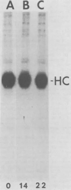
*S6-HC
[image:7.491.88.206.78.392.2]0
14
22
FIG. 4. Stability of the protein kinase activity of
p60"" of RSV-B. Chick cells transformed by
PR-RSV-Bwereincubated for 0,14,or22hat41°C in the
presenceof emetine (5 Lg/ml). Theamountofp60src_
associatedprotein kinase activity remainingwas
mea-sured by immunoprecipitation of p6&src with rabbit
anti-RSV tumor serum in excess and assaying the
kinase activity in the immune complex. All other
experimental details are described in Materials and
Methods. Presented here is an autoradiogram ofan
analysisof thephosphorylated products of the protein
kinase reaction by SDS-polyacrylamide gel
electro-phoresis. HC indicates the position of the heavy chain
of the immunoglobulin. A: No emetine. B: 14-h
pre-treatment with emetine. C: 22-h pretreatment with emetine.
cellular phosphatases were active during cell
lysis.Weattemptedtominimize these factorsby
labeling the cells for 48 hand by usinga
phos-phate-buffered RIPA buffer for cell lysis. The
fact that inclusion of NaF in the extraction buffer had no effect on the recovery of 32p_
labeled p60frc (data not shown) suggests that
marked enzymatic dephosphorylation was not
occurring. These values willtendtobe
overesti-matesifphosphorylation occurred after cell
ly-sis. We consider this unlikely because 2 mM
EDTAwaspresentin thelysisbuffer.
The apparent extent of thephosphorylationof
the tyrosine residue in p603rc revealed by this approach is somewhat greater than that obtained
from tryptic
peptide
mapping. We found that10% of the [ H]isoleucine-labeled tryptic
pep-tide which contains the site of tyrosine
phos-phorylation wasphosphorylated. Although this
approach does notinvolveassumptionsas tothe
specific activity of the cellular ATP pool, it too
is somewhat uncertain. The apparentextent of
phosphorylation measured this way, 0.1 mol of
phosphotyrosine per mol ofp6frc, isprobablya
minimum estimate. Both chemical
dephosphory-lation of the tyrosine and simple losses of the
phosphorylatedpeptidemay haveoccurred
dur-ing oxidationanddigestion of the protein. To estimatethe extent ofphosphorylation of
p6srC,
we had to label transformed cells tosteady state with[35S]methionine. Thisallowed
us to calculate the abundance of the p6fYrc
polypeptide in these cells. Typically, p6Ysrc of
SR-RSV-D constituted approximately 0.02% of
total cellular protein in cultures maintained at
41°C. Using the value of1 mg ofcellular
pro-tein per 4 x 10⁶ RSV-transformed chick cells,
we were able to calculate that this abundance
corresponds to approximately 500,000 mole-culesofp6Osrcpercell. The cellularhomolog of
viral p6fsrc, p60C"SC, is thought to be
approxi-mately 50- to 100-fold less abundant than the
viralprotein (2). This would correspond,
there-fore,toapproximately5,000 to10,000molecules ofp60C-srCperuninfected cell.
Our value for the abundance of p6Osrc is
somewhatlower than thatreported byothers(2,
13)who used cells labeled for 2 h. We suspect
thatthisdisagreementresultsin part from
differ-ences in thelengthof labeling of thetransformed
cellswith[35S]methionine. Totalcellularprotein
in chickcellsisreportedtohave an average
half-life of 40 to 50 h (9, 24). In contrast,p60src of
SR-RSV-D has a half-life of only 7 h at 41°C.p6Osrc
of PR-RSV-B is even less stable. In some
experi-mentsithad a half-life of 2 h.p6(Yrcistherefore
noticeably less stable thanthe average cellular protein in chick cells and may, as a result,
appear more abundantincellslabeledfor a short
time than in cells labeled to steady state.
Because we knew how many molecules of
p60srCwe hadimmunoprecipitated, we were able
tocalculate theturnovernumberof the
associat-edtyrosineprotein kinase activitywhenassayed
in the immune complex. The turnover number
wasvery low; 0.008 to 0.09 mol of phosphate per
mol ofp60src.Thisinefficiency probablyderives
in partfrom the immobilization of the enzyme. It
is also possible, however, that only a small
minority of the immunoglobulin heavy chains in
the immunoprecipitate are suitable substrates
for
p60src.
VOL.41, 1982
on November 10, 2019 by guest
http://jvi.asm.org/
We measured the half-life of the protein
ki-nase activity of
p60Src
by incubating transformedcelis
in the presence of inhibitors of protein synthesis and observing the decay of the activi-ty. The half-life of the protein kinase activity in such an experiment was three-to fourfold greater than that determined for the polypeptide by pulse-chase analysis. Although this couldsug-gest, in theory, that a stable subpopulation is
responsible for the protein kinase activity, it does not. The inhibition of protein synthesis by
emetine causes a
stabilization
ofp6Osrc.
Thehalf-life of the
p6&rc
polypeptide is increasedapproximately threefold in the presence of
eme-tine. This is not, however, unique to p64Yrc.
Pr76gar
is also stabilized. Additionally, a less pronounced but similar effect of inhibitors ofprotein synthesis on theturnoverof bulkcellular
protein has been observed (8-10).
The stability of the protein kinase activity under these conditions is notable. We measured half-lives of approximately 24 h when protein synthesis was inhibited. This suggests that the protein kinase activity is quite resistant to
sim-ple thermal inactivation in whole cells and that
the much more rapid rate ofturnovernormally is
due to the activity of some cellular enzyme rather than to spontaneous denaturation of the protein. The stability of the protein kinase
activ-ity of endogenous p60Jcsrc in uninfected chick
cells
is comparable to that of viral p60Orc when measured in the presence of emetine. Although the half-life measured this way, approximately 20 h, is almost certainly artifactually large, it does suggest that the protein kinase activities ofthe viral and thecellularenzymes have similar
half-lives.
ACKNOWLEDGMENTS
We thank Jon Cooper for helpful suggestions about the manuscript.
T.E. was supported by Public Health Service traininggrant CA 05274 from the National Cancer Institute, and T.P. was supported by a fellowship from theDeutsche Forschungs-gemeinschaft. This work was supported by Public Health Service grants CA 14195, CA17096,and CA 17289 from the National Cancer Institute.
LITERATURECITED
1. Brugge,J. S., and R. L.Erlkson.1977. Identification of a transformation-specific antigen induced by an avian sar-coma virus. Nature (London)269:346-348.
2. Collet, M. S., J. S. Brugge, and R. L. Erikson. 1978.
Characterizationof a normal avian cell protein related to avian sarcoma virus transforming gene product. Cell 15:1363-1370.
3. Collett, M. S., and R. L.Erikson. 1978. Protein kinase activity associated with the avian sarcoma virus src gene product. Proc. Natl.Acad. Sci. U.S.A. 75:2021-2024.
4. Colett, M. S., E. Erikson, and R. L. Erikson. 1979.
Structural analysis of the avian sarcoma virus transform-ingprotein: sites of phosphorylation. J. Virol.29:770-781.
5. CoJbtt, M.S., A. F. Purchlo,and R. L. Erikson. 1980.
Avian sarcoma virus-transforming protein,pp60&',shows protein kinase activity specific for tyrosine. Nature
(Lon-don)285:167-169.
6. Courtneldge, S. A., A. D. Levinson, and J. M. Bishop. 1980. The protein encoded by the transforming gene of aviansarcomavirus(jp60p)andahomologous protein in normalcells(pp6yP)'°)' areassociatedwith theplasma membrane. Proc. Natl. Acad. Sci. U.S.A. 77:3783-3787. 7. Czernilotsky, A. P., A. D.Levlnson,H. E.Varmus,J. M. Bishop, E. Tlcher, and H. M. Goodman. 1980.Nucleotide sequenceof an avian sarcoma virus oncogene (src) and proposed amino acidsequenceforgeneproduct.Nature (London)287:198-203.
8. Epstein, D., S. Elas-Bishko, and A. Hershko. 1975. Re-quirement forproteinsynthesis in the regulation of protein breakdown in cultured hepatoma cells. Biochemistry 14:5199-5204.
9. Hendil, K. B. 1977. Intracellular protein degradation in growing, in density inhibited, and in serum-restricted fibroblast cultures. J. Cell.Physiol.92:353-364. 10. Hershko, A., and G. M. Tompkins. 1971. Studies on the
degradation of tyrosine aminotransferase in hepatoma cells inculture. J.Biol. Chem.246:710-714.
11. Hunter, T., and B. M. Sefton. 1980. The transforming gene product of Rous sarcoma virus phosphorylates tyrosine. Proc. Natl.Acad. Sci. U.S.A.77:1311-1315.
12. Hunter, T., B. M. Sefton, and K. Beemon. 1979.Studies onthe structureand function of the avian sarcoma virus transforming gene product. Cold Spring Harbor Symp. Quant. Biol. 44:931-941.
13. Karess, R. E., W. S. Hayward, and H.Hanafusa. 1979. Cellular information in the genome of recovered avian sarcoma virus directs the synthesisoftransforming pro-tein. Proc. Natl. Acad. Sci. U.S.A. 76:3154-3158. 14. Kreuger, J. G., E. Wang, and A. R. Goldberg. 1980.
Evidencethat the src gene product of Roussarcoma virus is membrane associated. Virology 101:25-40.
15. Levinson, A. D., H.Oppermnn,H. E.Varmus,and J. M. BIhop. 1980. The purified product ofthe transforming gene of avian sarcomavirusphosphorylates tyrosine. J. Biol. Chem. 255:11973-11980.
16. Lowry, 0. H., N. J. Rosebrough, A.L. Farr,and R. J. Randall.1951. Proteinmeasurement withtheFolinphenol reagent. J. Biol. Chem. 193:265-275.
17. Rohrwchneider, L. R. 1980. Adhesion plaques of Rous sarcoma virus-transformed cells contain the src gene product. Proc. Natl.Acad.Sci.U.S.A.77:3514-3518. 18.Scheidtmann, K.-H., A. Kaiser, A. Carbone, and G.
Walter. 1981. Phosphorylation ofthreonine in the proline-rich carboxy-terminal regionofsimian virus 40 large T antigen. J. Virol.38:59-69.
19. Sefton,B. M., K.Beemon, and T. Hunter. 1978. Compari-son of the expression of the srcgene of Rous sarcoma virus in vivo and invitro. J.Virol. 28:957-971. 20. Sefton,B.M., T.Hunter,and K.Beemon.1979. Product of
in vitro translation ofRoussarcoma virus src gene has protein kinase activity. J.Virol.30:311-318.
21. Sefton, B. M., T. Hunter, and K.Beemon.1980. Tempera-ture-sensitivetransformation by Roussarcomavirus and temperature-sensitive protein kinase activity. J. Virol. 33:220-229.
22. Sefton, B. M., T. Hunter, K.Beemon,and W. Eckhart. 1980. Evidence that the phosphorylation of tyrosine is essential for cellular transformation by Rous sarcoma virus.Cell 20:807-816.
23. Smart, J. E., H. Oppermann, A. P. Czernllofsky, A. F.
Purchio, R. L.Erlkson,and J. M.Bishop. 1981. Character-ization ofsitesfortyrosine phosphorylationin the
trans-forming protein ofRoussarcomavirus(pp60v`5)and its normal cellularhomologue(pp60c-src). Proc. Natl. Acad. Sci. U.S.A.78:6013-6017.
24. Weber,M. J.1972.RibosomalRNA turnover in contact inhibitedcells. Nature(London)New Biol.235:58-60.
25. WlHngbam,M. C., G. Jay, and I.Pastan.1979.
Localiza-tion of the ASVsrc geneproductto theplasma membrane oftransformed cells byelectron microscopic immunocy-tochemistry. Cell18:125-134.
J. VIROL.
on November 10, 2019 by guest
http://jvi.asm.org/

![FIG. 2.thionineD-transformed Half-life of p6Osrc of SR-RSV-D. SR-RSV- chick cells were labeled with [35S]me- for 60 min at 41°C and then were incubated for](https://thumb-us.123doks.com/thumbv2/123dok_us/1464034.99058/5.491.253.446.65.466/thionined-transformed-half-osrc-chick-cells-labeled-incubated.webp)