JOURNAL OF VIROLOGY, Mar. 1976, p. 692-704 Copyrighti 1976 AmericanSocietyforMicrobiology
Vol. 17, No. 3 Printed inU.S.A.
Polyoma and Cell DNA Synthesis
in
Mouse L Cells
Temperature Sensitive for the Replication of Cell DNA
ROSE SHEININ
Ontario Cancer Institute, Toronto, Ontario M4X 1K9, Canada Received for publication 19 August 1975
Polyoma (Py) virus multiplies, at 34 and 38.5 C, in wild-type (WT-4) and in ts AlS9 mouse L cells, which are temperature sensitive for growth and for DNA replication (R.Sheinin, 1976; L. H. Thompson et al., 1970). De novo synthesis of double-stranded, fully covalently closed Py DNA has been shown to proceed by semiconservative replication in WT-4 and ts AlS9 cells at the permissive and nonpermissive temperatures. Cell DNA is made late during infection, by both cell types and atboth temperatures. Semiconservative replication of cell DNA proceeds in Py-infected WT-4 cells incubated at 34 or at 38.5 C and in Py-infected ts AlS9 cells incubated at 34 C. In virus-infected ts A1S9 cells incubated at 38.5 C, cell DNA synthesis appears to proceed almost entirely by a processanalogoustorepair replication. The inabilityoftsAlS9 cellstoproduce large-molecular-weight chromosomal DNA strands, at 38.5 C, by the normal mechanism is not overcomeby Py infection.
The transforming capacity of polyoma (Py) virus derives from the interactionofviralDNA synthesiswith the specific DNA metabolism of host cells, the final result being integration of viral into chromosomal DNA (14, 33). This interaction can take one of several pathways, depending uponthe physiology ofinfection (7,
15,37,38,40). Ofinterest inthe presentcontext
arethose eventswhichproceed inproductively infected cells. Early during infection, the
syn-thesis ofapparentlynormal cell DNAis stimu-lated or derepressed (7, 13, 15, 37, 38). In association with the onset ofPy DNA replica-tion, cell DNAsynthesis becomes aberrant(37, 40) suchthatincomplete,
single-stranded
DNA accumulates (10-12). Available evidence sug-geststhatthis phenomenonresultsfrom multi-ple initiation events, which ultimately tax the normal mechanism ofligation.Stimulation of cell DNA synthesis in Py-infected cellsprobablyresults from the
expres-sion of an earlyPy gene (17, 20, 31). Itseems
likely that most proteins which participate in
Py and in cell DNAsynthesis in virus-infected
cells are encoded in the cell genome. Py DNA
appears to carry little if any information in
excess of that required to code for the virion
polypeptides (cf. 17, 21, 33), the protein respon-sible for the temperature-sensitive ts a
func-tion (19, 20, 31) and the Py-specific Tantigen
(22, 31).
Little definitive information is available
re-garding either the proteins that participate in
thereplicationofPy DNAorthemechanismby which cell DNA synthesis is induced. One approach to this problem isto study Py
infec-tion of mutants ofpermissivemammalian cells defective in DNA synthesis. The present work describes suchexperimentswith a line of mouse
L cells that is ts for the replication of nuclear DNA (39). These ts AlS9 cells cannot convert newly made, small single-stranded DNA
seg-ments to large-molecular-weight chromosomal
DNA at the nonpermissive temperature. How-ever, they do support the multiplication of Py virus and normal Py DNA replication. Cell DNAsynthesis occurs, butitdoes notfollow the pattern seen inwild-type (WT-4) cellsor in ts
AlS9 cells infected at the permissive
tempera-ture.
MATERIALS AND METHODS Cells. WT-4 and ts AlS9 mouse L cells, the propertiesofwhich are described elsewhere (39, 43),
weregrownat34C,eitherinsuspensionor onasolid substratum, a a-minimum essential medium (42) lacking nucleosides but supplemented with 7.5%
(vol/vol) fetal calf serum (Reheis Chemical Co.). Suspension cultures (40-ml volumes in 15-by2.5-cm
tubes) wereincubatedin NewBrunswick Rollotherm cabinets.Theywereinoculated at 2 x 10⁴to5 x 104
cells/mland growntolatelogarithmicphasepriorto
subculture. In the case of cells grown on glass, confluent cultures were subcultured as follows. The
medium was decanted, and the cells were washed twice with phosphate-buffered saline (16) and then incubated at 34 C for 5 min with a solution of 692
on November 10, 2019 by guest
http://jvi.asm.org/
hyaluronidase and collagenase (50
Ag/ml
each; WorthingtonBiochemicals) in a volume just adequate to cover the culture surface. This solution was re-placed with 0.1% trypsin (Difco) in citrate saline (34) for an additional 5 min. The cells were then sus-pended in medium and plated at concentrations of about 5 x10'to 5 x 10'cells/ml (in volumes adequate tocoverthe growing surface of culture bottles).Virus. Py TSP1 (41) wasprepared by the proce-dure ofWinocour (47). A single stock having a titer of 6.8 x 10¹⁰ PFU/ml was used throughout. Plaque assays were performed on mouse embryo cultures (34).
Infection procedures. To infect cells grown on
glass, medium from subconfluent cultures was re-moved; virus (in a volume adequate to form a film over thecells) was added and allowed toadsorb for 1
h. Medium containing 1% (vol/vol) fetal calf serum was added, and the cultures were incubated at the appropriate temperature. In the case of suspension cultures, cells were collected by centrifugation (10 min,800 x g)and incubated with intermittent mixing for30min in 0.5 ml of virus suspension before medium wasadded and incubation at the desired temperature was continued. For mock infection, virus was replaced with anequalvolume of phosphate-buffered saline.
AntibodytoPy virus. AntiserumtoPy virus, with
a neutralization coefficientofK = 23 minm (1), was
raised in rabbits (36). In some experiments this antiserum wasused directly. In others, the antiviral gamma globulin coupled with fluorescein was used
for immunofluorescent staining of mock- or Py-infected cells grownoncoverslips (40, 46).
Experimental regimen for studying DNA
synthesis. Unlessotherwisenoted, thefollowing
pro-cedurewasusedforthestudyofDNAsynthesis. Cells weregrown at 34C through threetofourgenerations
to midlogarithmic phase in medium supplemented with 0.01 MiCi of [14C]thymidine (dThd) per ml (approximatespecific activity,60mCi/mmol; approx-imateconcentration, 0.2MM).Thismediumwasthen replaced by nonradioactive medium, after which the cellswereincubatedateither34 or38.5Cfor 16 to 24
h,aperiodadequatetopermit fullexpressionofthets
lesion of ts AlS9 cells (39, 43). [methyl-3H]dThd (approximatespecific activity, 20mCi/mmol;
approx-imate concentration, 0.05 MM) was then added, as
notedinindividualexperiments, tolabel DNAnewly madeunder test conditions.
Assessment of DNA synthesis. [14C]- and
[3'H]DNA
was examined by velocity sedimentationand by equilibrium centrifugation by procedures
previously described. For velocity sedimentation studies in neutral sucrose density gradients (6, 8), approximately 107cells,lysedin 2ml ofa 1%solution ofsodiumdodecylsulfateinSSC,werelayeredonto a 15to 30%(wt/wt) linear sucrose densitygradient (29 ml, in 0.01 M Tris [pH 7.4]-0.1 NaCl-0.001 M EDTA-0.5% sodium dodecyl sulfate) formed over a
5-ml cushion of70% sucrosein SSC andcentrifuged for 16hat 20C in aBeckman SW27rotor at22,000 rpm. One-milliliter fractions were collected with an
Isco density gradient fractionator and monitored for optical density at260nm, whichpermitted
identifica-tion ofthe internal sedimentation markers, 18S and 28S rRNA.
For analysis of alkaline-denatured DNA, approxi-mately5 x 10' to 5 x 101 cells (in 0.2 ml) were added to 2ml of 0.2 M NaOH-0.01 M EDTAlayered on a 5 to 20% sucrose density gradient (30 ml) in 0.9 M NaCl-0.3 M NaOH-0.001 M EDTA established overa
4-ml cushion of 70% sucrose (8). This was left at 2C for at least 12 hbefore being centrifuged at 22,000 rpm at 2 C for 9 h in a Beckman SW27 rotor. Purified "4C-labeled Py form I DNA (4) was run in parallel gradients toserve as marker. Fractions were collected as noted above. All fractions were analyzed for the presence oftrichloroaceticacid-precipitable radioac-tively labeled material. (Radioactive dThd was pur-chased fromAmersham-Searle Corp.). The amount of any given component was calculated by expressing the counts per minute in the corresponding peak as a percentage of the total acid-precipitable counts per minuterecovered from the gradient.
Equilibrium centrifugations were performed at2C
for48h at 48,000 rpm in a BeckmanSW50.1 rotor. For neutral gradient analysis (27), DNA samples were diluted to 5 ml with CsCl in 0.01 M Tris-0.01 M EDTA, pH 7.4 (final density, 1.70 g/cm'). Poly(dA:dT) and poly(dC:rG) (Miles Laboratories) were added as optical density markers. To prepare DNA for alkaline gradient analysis, samples were heated for 30 min at 50 C with one-tenth volumes of 1 M NaOH (26) andthen diluted to 5 ml with saturated CsCl solutionin0.01M Tris, pH8.0, to a final density
of 1.76 g/cm'. Poly(dA*dT) was used as optical
density marker. Fractions were collected from the bottomofeachtube and monitored for optical density at 260 nm, trichloroacetic acid-insoluble material, and refractive index. Calculations of density were made from the latter measurements.
DNA-DNA hybridization. To establish the na-ture of the 20S Py DNA made in infected L cells, DNA-DNA hybridization studies were performed as described previously (6, 8), using cellulose nitrate filters carrying either 0.1Mgofpurified form I Py DNA
or 10 Mg of L-cell DNA, isolated by established procedures (37). Prior to use, labeled DNA was sheared by sonic vibration, boiled at 100 C, and quick-cooledinice.
RESULTS
Growth ofPy virus in WT-4 and ts AlS9 cells. Several years ago it was reported that
certain strains ofPyvirusgrow in some lines of
mouse Lcells (2, 3, 23, 28). Initially,therefore,
experiments were performed to determine
whether WT-4 andtsAlS9cellswouldsupport thegrowthofPyTsP1. Asmay be seen from the
data presented inTable 1, virusmultiplication doesproceed inthese cells, atboth34and 38.5
C, even in thepresence of antiviral antiserum
added to inactive unadsorbed virus. This
anti-serum had little effect on virus formation of
WT-4 cellsor intsAlS9 cellsincubatedat 34C,
but it did reduce theyieldofPyfrom ts AlS9
on November 10, 2019 by guest
http://jvi.asm.org/
TABLE 1.Multiplication of Py TSPI in WT-4 and tsA1S9mouse Lcells
CellsTemp Antiserum Adsorbed Plaquetiter(PFU/mi)
Cells Temp antieru MOI
(C) added
~(PFU/cell)
2hp~i.
24hp.i. 48hp.i.WT-4 34.0 - 0.04 1.4x 10' 3.0x 10³ 6.2x 10⁴
+ 0.04 4.0x 10' 2.1 x 10⁵
38.5 0.03 8.0x 10' 3.1x 10⁹ 1.0x10⁶
+ 0.03 8.5 x 10³ 1.1 x 10⁶
tsAlS9 34.0 - 0.01 1.6x 10⁴ 1.5x 10' 1.4x 10⁵
+ 0.01 9.5x 10' 1.5 x 10⁵
38.5 0.01 8.0x 10³ 3.4x 10⁴ 1.2 x
106
+ 0.01 4.6x 10' 3.0x 10⁶
aReplicate cultures (in 2-oz [ca.
0.06-liter]
Brockway bottles) ofthe L cells, in mid-logarithmic phase ofgrowth, wereincubated for 24h at 34 or 38.5 C.They werethen infected with 0.2 ml of Py TSP1 (10 to 20
PFU/cell). After the adsorption period, one-half of the cultures were treated for 2 h at the appropriate
temperature with antiserum to virus, sufficient to inactivate unadsorbedvirus. At the intervals noted the cultures were washed, incubated in medium, and plaque assayed for virus on mouseembryo cells. L-cell cultures(including medium)weresubjectedtothreecyclesoffreeze-thawing priortotitrationby plaqueassay. Novirus was detected in control or mock-infected cultures. Theabsorbed multiplicityofinfection (MOI) was
calculatedastheamountofinfectious virus associated with the Lcells, releasedfrom tht glass, and counted
afterthe 2-hincubationwith or without antiserum.
cellsincubatedat38.5 C. Thismaybe relatedto
the increased fragility of the ts cells after
prolongedincubation atthistemperature.
The preceding growth experimentswere
con-ducted at low multiplicities of infection. The
biochemical experimentstobedescribed below
areperformedathighmultiplicitiesofinfection
toobtain totally infected cell populations.
Evi-dence that this objective was achieved was
obtained from two kinds ofexperiments. Data
from one experiment (Table 2) indicated that
all WT-4andtsAlS9 cells infectedateither low
or hightemperature with PyTSP1 at a
multi-plicity of infection of _2 x 10' PFU/cell plated
as infectious centers. Further evidence that all
cells incubated under such conditions were
infected wasobtained from studies of
immuno-fluorescent staining. WT-4 and ts AlS9 cells
were grown at34 C toabouttwo-thirds
conflu-ence in cultures containing cover slips (40).
Replicate cultureswereinfectedatanestimated
multiplicity of 2,000 PFU/cell and incubatedat
either 34or38.5 C. After 24and 48hthecover
slips were removed and stained with
fluo-rescein-conjugated antipolyoma gamma
glob-ulin (40, 46). Almost all nuclei of infected
cul-tures (butnot of mock-infected cells) exhibited
weak fluorescence at 48 h postinfection (p.i.),
indicating the presence of newly made virion
proteins (40,46).
DNA synthesis in Py-infected L cells.
Previous studies (39, 43) have shown that ts
[image:3.491.58.458.78.231.2]AlS9 cells are defective in DNA synthesis at
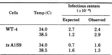
TABLE 2.Infection of WT-4 and tsAJS9cells by Py virus
Infectiouscenters Cells Temp(C) (x10-6)
Expected Observed
WT-4 34.0 2.7 2.4
38.5 1.2 2.9
tsAlS9 34.0 0.7 1.6
38.5 1.6 1.3
aReplicate cultures (in 4-oz
[ca.
0.12-liter]Brock-waybottles)inlate logarithmic phase wereincubated
for 24 h at 34 and 38.5 C. Individual cultures were then either infected withPy TSP1 (input multiplicity of infection, w2 x 103PFU/cell) ormock infected. Theywereincubated at the appropriate temperature with medium containingantibody to inactivate unad-sorbed virus. At 16 h p.i., cells were washed, har-vested, counted, and plated on indicator mouse em-bryo fibroblasts (cf. 34). The observed infectious centers were measured by this plaque assay. The
expected values are calculated from the actual cell
numbers counted prior toplating.
38.5C. After incubationatthistemperaturefor
16 to 24h, the incorporationofdThd into DNA
falls to a level about 1 to 5% of that observed
either with WT-4 cells incubated at 38.5 C or
with both WT-4 and ts AlS9 cells incubatedat
34 C. Such incorporation increases if ts AlS9
cells are incubated further at 38.5 C and can
approach 10% of control levels 48to72 h later.
694 SHEININ J. VIROL.
on November 10, 2019 by guest
http://jvi.asm.org/
[image:3.491.264.459.347.444.2]POLYOMA AND CELL DNA SYNTHESIS
Direct studies on the pattern of DNA
replica-tion indicate that this is notdue to restoration
of any capacity for normal semiconservative
DNA synthesis, but results from repair
replica-tion (cf. 39; R.
Sheinin,
manuscript inprepara-tion; and below).
Because the replicationofPyDNAis known
tobeclosely linked to cell DNA synthesis (cf. 6,
7, 17,38), itwasof interesttostudythepattern
offormation ofbothPy and cell DNA in tsA1S9
cells incubated at permissive and
nonpermis-sivetemperatures.
The first approach was to examine total Py
and cell DNA synthesis by velocity
sedimenta-tion analysis in neutral sucrose density
gradi-ents. tsAlS9 cells were grown to
midlogarith-micphase and thenincubatedat 34 or38.5 C for
24 h. The cultures were then infected with Py
virus and incubated further at the appropriate
temperature. Twenty-four hours p.i. (i.e., late
during the infectious cycle) cultures were
treated for 6 h with
[3H]dThd
to label newlymade DNA, which was analyzed by velocity
sedimentation inneutralsucrose density
gradi-ents as described in the legend toFig. 1. Two
majorcomponents wereobtained, one ofwhich
90
ts
AIS934WC
a)
70-N 50
> 30
X
18S 28S
4-
I.a
sedimented with thehigh-molecular-weight cell DNA of mock-infected cultures (data not
shown) andwas collected ontothe 70% sucrose
cushion at the bottom of the gradient (e.g.,
fractions 30-38 inFig. la and39-42 inFig. lb).
The second component wasfound in thesame
positionas marker(20S) ["4C]Py DNA isolated
frompurified virus particles.
In alkaline sucrose density gradients (dis-cussedbelow) the 3H-labeled 20S DNA isolated
forPy-infected tsAlS9 cells cosedimented with
the
[14C]Py
DNA marker at 53S (8). Similardatawereobtained with WT-4 cells.Essentially identical resultswereobtained with cellsgrown
in suspensionandonglass, labeled ateither24
or 48 h p.i. In all instances the sedimentation
profilesobtained resembled those observed with
otherproductively infected cells.
The finding that the 3H-labeled 20S DNAof
Py-infectedWT-4ortsAlS9 cells cosedimented
with "4C-marker Py DNA in neutral and
alka-line sucrose density gradients suggested that
theformer isdouble-stranded,covalentlyclosed
formIPy DNA (14,45). To further establish its identity, the following experiment was done. Cells were grown, infected, and labeled as
1s AIS9 38.50C b)
12
10
82iN
18S 28S
Fraction number
FIG. 1. Velocity sedimentation analysis in neutral sucrose density gradients of DNA synthesized in
Py-infectedtsAIS9cells.Replicatesuspension cultures were grown at 34 C to mid-logarithmic phase. One-half
ofthe cultureswasthenshiftedto38.5C,andincubationwascontinuedfor16h. Allsubsequentmanipulations ofcellswerecarriedoutatthe temperatureofincubation. The cellswere centrifugedat800 x gfor 10min,
infected with 0.5 ml ofaPy suspension (multiplicity of infection, 52,000 PFU/cell), resuspended to their
originalconcentration in mediumlackingnucleosides,andre-incubated. At0, 24,and48hp.i.,the cultureswere
sampled for plaque assay. At24hp.i., [3H]dThd(1 uCi/ml) wasaddedtoeachculture,and6hlater approxi-mately 2 x 10⁷ cells were harvested by centrifugation and washed once with SSC. The [3HJDNA was
analyzed byvelocity sedimentationin neutralsucrosedensitygradientsasdescribed in Materials andMethods.
Thearrowsmark thepositionof 18S and 28S rRNA. Theplaquetitersattimezero wereabout4x 106PFU/ml forall cultures.After48htheywere2.5x 107and 3.0x10⁷PFU/mlfortsAJS9cellsincubatedat34and38.5C, respectively.
VOL. 17, 1976
on November 10, 2019 by guest
http://jvi.asm.org/
[image:4.491.94.380.351.541.2]696 SHEININ
described in the legend to Fig. 1.The
[3H]DNA
was separated in neutral sucrose density gradi-ents. The material sedimenting at 20S (e.g., fractions 13-21 of Fig. la) was isolated and tested for its capacity tohybridize with Py and mouse cell DNA. The 3H-labeled 20S DNA isolated from WT-4 and ts AlS9 cultures in-fected and labeled at either 34 or 38.5 C hybridized with purified form I Py DNA with an efficiency equivalent to that exhibited by ['H
]Py
DNA extracted from infected mouse embryo cells (Table 3). Little or no hybridiza-tion wasdetectedbetween 'H-labeled20S DNA and cell DNA isolated fromuninfected mouse L cells (WT-4 or tsAlS9). It was therefore con-cluded that the 'H-labeled 20S DNA is in fact Py DNA.In some instances [3H]DNAsedimenting be-tween 21 to 28S (e.g., in fractions21-26 of Fig. la) was observed in Py-infected cultures. On the basis of our own studies (P. E. Branton and R. Sheinin, manuscript in preparation) and those of others (30), it has been assumed that this material is
['HjPy
DNAinthe replicating form.These experiments indicate that ts AlS9 cells, inwhich normal cellular DNAreplication is blocked at 38.5 C, are able to synthesize apparently normal Py DNA in amounts compa-rable to those produced by infected WT-4 cells (at 34 and 38.5 C) and at 34 C by infected ts AlS9 cells.
[image:5.491.62.254.493.622.2]Cell DNA synthesis in Py-infected WT-4 and ts AlS9 cells: size analysis by velocity sedimentation in alkaline sucrose density gradients. It has been demonstrated that, in
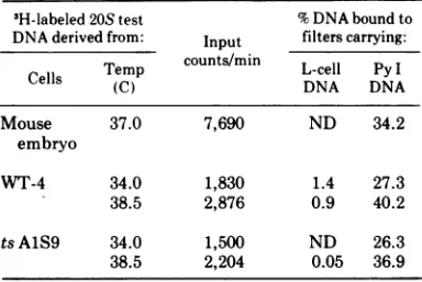
TABLE 3. Hybridizationof 3H-labeled20SDNA synthesized inPy-infected WT-4and tsAJS9cells,
with virus and cellDNAG
3H-labeled 20S test %DNAboundto DNAderivedfrom: Input filterscarrying:
Temp counts/min L-cell PyI
Cells (C) DNA DNA
Mouse 37.0 7,690 ND 34.2
embryo
WT-4 34.0 1,830 1.4 27.3
38.5 2,876 0.9 40.2
tsAlS9 34.0 1,500 ND 26.3
38.5 2,204 0.05 36.9
a
'H-labeled
20S DNA wasobtained from neutralsucrose density gradient fractions by precipitation with alcohol asdescribed elsewhere (8). L-cell DNA andPyI DNA werepurified using established proce-dures(4, 37).ND,Notdetectable.
cellsproductively infected with Py virus, cellu-lar DNA synthesis is stimulated several hours prior to the onset of viral DNA replication (13, 15, 37). Inaddition, it is known that the induced cell DNA synthesis becomes increasingly aber-rant afterviral DNA replication is initiated (6, 7, 10-12, 37). The abnormality is readily de-monstrable by velocity sedimentation analysis in alkaline sucrose density gradients. Since DNAreplication in ts AlS9 cells is defective at hightemperature, it was of interest to compare the patterns of cell DNA synthesis in virus-infected WT-4 and tsAlS9 cells at the permis-sive and nonpermispermis-sive temperatures.
Cells were grown at34 C with [4C ]dThd to labelcell DNA and were thenincubatedat 38.5 C for 24 h. They were then infected with Py virus(or were mock infected) and labeled with
['H JdThd
from 24to 30h p.i. The newly made[3'H]DNAwasanalyzed by velocity sedimenta-tion in alkaline sucrose density gradients (Fig. 2).
The data showninFig. 2a wereobtained with WT-4 cells. The mock-infected culture gave a sedimentation profileexpected for normal cells (10, 11). The majority (81.8%) of the
[3HIDNA
synthesized at 38.5 C was recovered along with marker
[4C
]DNA preformed at 34 C (data not shown) on the 70% sucrose cushion (fractions 31-36). In the case of Py-infected WT-4 cells, the sedimentation profile exhibited three com-ponents. One (52.6% ofthe total [3H]DNA) was recovered in the position of high-molecular-weight cell DNAatthe bottom of thegradient. Asecond (12.3%ofthetotal) sedimentedinthe position ofthemarker(53S) [4CJPyDNA. The third (19.9%ofthetotal) sedimented heteroge-neously between these two components (frac-tions 24-30). On the basis of the close qualita-tive and quantitative analogy between these data and those obtained from studies of Py-infected mouse embryo cells (10-12) and 3T3 mousefibroblasts (unpublished data), we have assumed that theheterogeneously sedimenting [3H]DNA
results from aberrant cell DNA syn-thesis, i.e., fromtheinhibitionofchainelonga-tionwhichoccurslateduring Pyvirusgrowth. The resultsobtained withtsAlS9 cells
incu-bated at 38.5C areshowninFig. 2b.Theyare,
in general, similartothoseobtained with WT-4 and other mouse cell systems. However, with both mock- and virus-infected cultures thereis more ['H
]DNA
that sedimentsheterogeneously throughout the gradient. The centrifugation profiles ofboth mock- and virus-infectedcul-turesexhibited ['H
]DNA
(21.3and21.0% ofthetotal, respectively), which sedimented as
high-molecular-weight DNA to the bottom of the
J. VIROL.
on November 10, 2019 by guest
http://jvi.asm.org/
2500
MW010 53S 2
1500 53S
500
-c'Pt
50 lo1510 25 30 35 0 5 10
1'52025
3035 40 [image:6.491.52.445.57.281.2]Froctionnumber
FIG. 2. Velocity sedimentation analysisinalkalinesucrosedensitygradients ofDNAsynthesizedin control
andPy-infectedmouseL cellsat38.5C. WT-4 andtsAJS9 cellsweregrowntomid-logarithmic phaseat34C in
Brockway bottles in medium containing [4CJdThd. The cells were washedfree ofradioactive medium and
incubatedfor 24 h at 38.5C. Theyweretheninfected withPyvirus(=2,000PFU/cell) labeled with ['H]dThd
from 24 to 30 hp.i., afterwhichtheywerewashed andlysedina1%solutionofsodiumdodecyl sulfateinSSC
andthelabeledDNA wasanalyzedby velocitysedimentation in alkalinesucrosedensitygradientsasdescribed
inMaterials and Methods. The arrow marks theposition ofmarker53S["4C]PyDNAruninparallelgradients.
Inallinstancesthe 14C-cellDNA preformed at 37 C was recovered on the70%sucrosecushion (e.g.,fractions
31-36in [a]). Only the data for'H-labeledDNA areshown. Symbols: 0, mock-infected cells;0,Py-infected
cells.
gradient. In thecaseofthePy-infected cells, a veryprominent3H-labeled 53Sviral DNApeak (12.8%ofthe total)wasdetected. The heteroge-neouslysedimentingcell DNA(fractions18-34) made up56.8% ofthe total.
Dataobtained from analysesofDNA
synthe-sized between 24 to 30 h p.i. in mock- and Py-infected WT-4 andtsAlS9 cellsat34Cwere
essentially identical to those illustrated inFig.
2a.
Cell DNA synthesis: pulse-chase studies. In our earlier work it was found that ts AlS9 cells are temperature sensitive in a step in-volved in the conversion ofnewly synthesized, short, single-stranded segments into large chro-mosomal DNA. The experiments described in
the foregoing section suggested that Py infec-tion might circumvent this ts lesion. It wasof interest, therefore, toexamine further the proc-ess ofPy-induced cell DNA synthesis in these cells under permissive and nonpermissive con-ditions.
WT-4 and ts AlS9 cultures, prelabeled with [4C]dThd, were incubated at 38.5 C for 16 h and thenwere either mock infectedorinfected
with Py virus. Some cultures ofeach set were
pulse-labeled for 5 min with [3H]dThd at 24 h
p.i. Others were pulse-labeled and then incu-bated for an additional 1 or 6 h in dThd-con-taining, nonradioactive medium. The ["4C]-and ['H
]DNA
ofeach culture wereanalyzed
by velocity sedimentation analysis in alkalinesu-crosedensity gradients.
Figure 3aand bpresentsresults from
experi-ments with mock- or Py-infected WT-4 cells
incubatedat38.5 C.Similardatawereobtained
in analyses of virus-infected WT-4 and tsAlS9 cells incubated at 34 C. In all instances the "4C-marker cell DNA was recovered primarily
as high-molecular-weight material, at the
bot-tomofthegradients.The [3H]DNA synthesized
at38.5C duringa 5-minpulseperiod
by
virus-ormock-infectedWT-4cells sedimented hetero-geneously throughout the whole gradient. The smallest components were about 10S; the
larg-est cosedimented with the "4C-marker DNA
(i.e., with S values of >300 [29]). In the Py-infected cultures no clear 53S component wasdetectable.After achaseof1h,the pattern of sedimentation ofthe [3H]DNA was altered greatly. Inmock-infected cultures(Fig. 3c), the pulse-labeled DNA was recovered as high-molecular-weight material atthebottomofthe gradient, with the
["4C]cell
DNA preformed aton November 10, 2019 by guest
http://jvi.asm.org/
698 SHEININ
0 5 10 I 20 25 30 35 40 0 5 10 15 20 25 30 35 40
Fraction nrumber
FIG. 3. Velocity sedimentation analysis inalkaline sucrose density gradients ofDNA synthesized during pulse and chase intervals by Py-infected WT-4 cells incubatedat38.5C. Replicate suspensioncultures ofWT-4 cellsweregrowntomid-logarithmic phaseat34 C in mediumcontaining['4C]dThd(0.1,Ci/m1).The cellswere
resuspended in fresh nonradioactive medium and incubatedat38.5C for16h. Theywerethen infected with Py
virus, ormock infected, asdescribed in the legendtoFig. 1.After24h the cultures received ['HJdThd (25
MCi/ml) for5min. The labeled DNAofonemock- andonevirus-infectedculturewasanalyzed immediatelyby
velocity sedimentation in analkaline sucrosedensity gradient. The other cultures received nonradioactive
dThd(0.05 mM) andwereincubatedfurther forI hprior toanalysis. Symbols: *, ['HJDNA;0, [14CJDNA
preformedat34 C. Thearrowsmark theposition ofI"C-labeled53S Py DNA,runinparallel gradients.
34 C. In the Py-infected WT-4 cells (Fig. 3d),
the sedimentation profile of the ['HJDNA
re-sembled thatshown inFig. 2a. Material(15.4%
ofthetotal) sedimentingat53S,intheposition
of Py DNA, was detected, as was a major
component (58.5% ofthe total) cosedimenting
with high-molecular-weight preformed
[I4CIDNA.
In addition, some of the [3HIDNA(28.2%) sedimented as heterogeneous material
between these two peaks. Insomeexperiments
Z
10
Xz
120
110
.
00
A:
90k)
80
70
60
50 40
30 20
10
0
J.- VIROL.
on November 10, 2019 by guest
http://jvi.asm.org/
[image:7.491.56.447.61.519.2]the chase interval was extended to 6 h. The sedimentation profiles observed after a 6-h chase resembled those showninFig.3and4for 1-h chase period.
Data obtained in pulse-chase studies with mock- and Py-infected tsAlS9cells incubated at38.5C are shown in Fig. 4. In all instances the [4C
]DNA
preformed at 34 C was recovered aslarge-molecular-weight material on the 70% sucrose cushion. In mock-infected tsAlS9 cells (Fig. 4a) the pulse-labeled [3H
]DNA
sedi-mented heterogeneously, with a majority (58.5%) being recovered as low-molecular-weight material at the top of the gradient. After a 1-h chase interval (Fig. 4c), there was a redistribution of3H-labeled material, withonly29.9% being recovered in this low-molecular-weight range. These observations, made 48 h after tsAlS9 cellswere shiftedto38.5C, arein accord with those made earlier (39) with cells incubated for only 16 to 24 h at the nonpermis-sive temperature.The tsAlS9cells differ from the WT-4 cells inthatthey are ts in the process of conversion ofnewly made small segments of DNA to chromosomalDNA strands (cf. 39).
The sedimentationprofile of the [8H DNA of ts AlS9 cells infected at 38.5 C and pulse-labeled for 5 min 24 h p.i. (Fig. 4b) closely resembled those obtained with material from mock-infected cultures. No 53S viral DNA was detected. However, after a 1-h chase interval
such
[3H]Py
DNA wasclearlyvisible (Fig. 4d).c)Ihr.chose: tsAIS9
-~~~~~~80
53S
70
-60
.50
40
30
20
d)Ihr.chose tsAIS9
Py-4
53S
i
~~~~35~~
0 5 10 15 20 25 30 35 40 0 5 10 15 20 25 30 35 40
Fraction number
FIG. 4. Velocity sedimentation analysis in alkaline sucrose density gradients of DNA synthesized during pulse and chase intervals in Py-infected tsA1S9cellsincubated at 38.5 C. Replicate cultures of ts AIS9 were processed as described in Fig. 3. Symbols:0, ['HJDNAsynthesized at 38.5 C 24 hp.i.;0,
["4C]DNA
preformedinuninfected cells at 34 C.
on November 10, 2019 by guest
http://jvi.asm.org/
[image:8.491.55.442.246.626.2]700 SHEININ
It made up 36.2% of the total DNA, of which about 20.6% cosedimented with preformed 14C-labeled chromosomal DNA. Approximately 11.5% of the
[3H]DNA
sedimented between these components, whereas the remainder was recovered as heterogeneously sedimenting ma-terial of molecular size smaller than53S.Essentially quantitative recovery of both
pre-formed
[14CIDNA
and newly synthesized[H
H]DNA
was achieved in such studies. Noevidence suggesting degradation and reutiliza-tion oflabeledDNA wasobtained. The experi-ments indicate that ts AlS9 cells incubated at 38.5 C for 48 h are seriously impaired in their capacity to synthesize chromosomal DNA. In contrast, Py-infected ts AlS9 appear able to make such large-molecular-weight DNA.
Cell DNA synthesis studied by BUdR la-beling. Under the experimental conditions em-ployed, i.e.,incubation of ts AlS9 cells for 48 to 54 h post-temperature shift, incorporation of
['H
JdThd
intocell DNA inuninfected cells wasabout 10to 15% ofthatobserved in WT-4 cells andabout8 to 10%ofthat detected in either ts AlS9 orWT-4 cells incubated continuously at
34 C. As a result of Py infection, such incorpora-tion was stimulated from 1.5- to 3.5-fold in ts AlS9 cells at the high temperature. In addition, the Py-infected cells synthesized some DNA which cosedimentedin alkalinesucrose density gradients with "4C-marker chromosomal DNA,," suggesting that the virus may have effected reversal of the ts lesion. The following experi-ment was performed to determine whether Py infection of tsAlS9 cells resultedinthe restora-tion ofnormal semiconservative DNA replica-tion.
Cultures of ts AlS9 cells, prelabeled with
["4C]dThd
at34C, wereincubatedat34 or 38.5C for16h, afterwhichtheywereeither infected with Py or mock infected. Twenty-four hours p.i., the cultures were treatedwith bromodeoxy-uridine (BUdR), 5'-fluorodeoxyuridine, and
[3H
JdThd
to mark the newly made DNA withboth a density and radioactive label. After 6 h
the cell DNA was separated by velocity sedi-mentation in neutral sucrose density gradients and then analyzed by equilibrium centrifuga-tion in neutral CsCl density gradients as de-scribed in the legendto Fig. 5.
Fractionnumber Fraction number
FIG. 5. Equilibrium centrifugation analysis ofCsCI ofcell DNA synthesizedin tsA1S9 cells. Suspension
cultures of ts A1S9 cells were grown to mid-logarithmic phase at 34 C with [14CldThd, incubated in nonradioactive medium at 34 or38.5 Cfor 16h,andtheneither mockinfected orinfectedwithPyvirusas described in the legend to Fig. 1. After 24 h BUdR (16MM), 5'-fluorodeoxyuridine (FUdR) (1 AM), and
['H]dThd(1MCi/ml)wereaddedtoeach culture. Six hourslater,the cellswereharvested,washed inSSC,and
lysed inSDS,and thePyand cell DNA wereseparatedbyvelocitysedimentationin neutralsucrosedensity
gradients (seeFig. 1). The cell DNAfractionwith maximum radioactive label(e.g.,fractions33or34,Fig. la)
wasfurtheranalyzed byequilibrium centrifugation inneutral CsClgradients asdescribed in Materials and
Methods.Symbols: 0,
['4C]DNA
preformedat34C;*, ['HJDNAmade inthe presenceofBUdR andFUdR;X,buoyant density. (a) Mockinfected,34C; (b)Py-infectedcells,34C; (c)Py-infected cells,38.5C.
J.- VIROL.
on November 10, 2019 by guest
http://jvi.asm.org/
[image:9.491.59.446.345.567.2]The [14C]DNA preformed in light medium by uninfected ts AlS9 cells was recovered as two components after the 6-h period of incubation at 34C inBUdR-containing medium (Fig. 5a). One had a buoyant density equal to that of normal mouse DNA (27), 1.706 g/cm3. The other had a density of 1.720 g/cm3. The data suggested that approximately 26.9% of the ["IC]DNA hadundergonesemiconservative rep-lication inthe presence ofBUdR in the experi-ment depicted. Approximately 70.7% of the
[3H]DNA
co-banded with this ["4C]DNA.In tsAlS9cells infected with Py (Fig.5b) at 34 C, about 15.1% of the preformed [14C]DNA wasrecovered at a heavier than normal density. About 58.5%ofthe [3H]DNA co-banded with it. Similar observations were made with WT-4 cells infected at34 or 38.5C.
Resultsobtained with cellDNA fromPy- and mock-infected tsAlS9 cells incubated at 38.5 C differed from those seen with WT-4 cells. Data from virus-infected cells are shown in Fig. 5c. The centrifugation profiles of both ["4C]- and [3H]DNA were broad, but exhibited a mean
buoyant density characteristic of unreplicated DNA, i.e., _1.71 g/cm3. A very small fraction (0.06%) ofthe [3H]DNA wasfound at a higher density, 1.745g/cm3.
Mode of Py replication in WT-4 and ts
A1S9
cells. The experiments described in ear-lier sections suggested that Py DNA replication proceeds normally in tsAlS9 cells incubated at the nonpermissive temperature. However, the results of the study outlined in the preceding section indicate that the cell DNA synthesis that proceeds in either uninfected or Py-infected ts A1S9 cells at 38.5 C is almost entirely abnormal. The mechanism of Py DNA replication was therefore re-examined using BUdR as density label.Once again WT-4 and ts AlS9 cells were
prelabeled with
["4C]dThd
at 34 C and incu-batedateither 34 or 38.5C for 16h.They were then infected with Pyvirus atthe appropriate temperature. They were density labeled with BUdRand radioactively labeled with [3H ]dThd at 24 h p.i. After 6 h, the 3H-labeled 20S Py DNA was isolated from neutral sucrose density gradients and analyzed in neutral CsCl density gradients, asdescribed in thelegend toFig. 6.The
'H-labeled
20S Py DNA produced byWT-4 and ts AlS9cells, at both 34 and 38.5 C,
in thepresence ofBUdR had a buoyant density heavier than that of the light-infecting virus
DNA (1.709
g/cm'
[24]). The centrifugation profiles were heterogeneous, suggesting that thefractionsexaminedcontainedPyDNAmol-ecules with varying degrees of BUdR
substitu-tion. Two major components appeared to be resolved in each case. Those from cells incu-bated at 34 C had buoyant densities of about 1.75 and 1.73g/cm';thosefrom cells incubated at 38.5 Cbanded at approximately 1.79 and1.75
g/cm3.
The 'H-labeled material banding at 1.75 g/cm' likely corresponds to Py DNA, in which only one strand is fully substituted with the density label, whereas that at 1.79 g/cm3 is probably Py DNA,which carriesBUdRinboth strands (24, 25). The material recoveredat 1.73
g/cm3 likely represents viral DNA moleculesin
which only partial substitution has occurred. A very small proportion of the 'H-labeled 20S Py DNAobtained from WT-4 cellsincubatedat 34 or38.5C, and from tsAlS9 cells incubated at 34 C, cosedimented at abuoyant density ofabout 1.704 g/cm3 with ["C]DNA preformed in light medium at 34 C. This isprobably pseudovirion DNA(44), comprisingcellular DNA molecules.
This study clearly establishes that Py DNA replicatesprimarilyinasemiconservative mode inWT-4 andintsAlS9cells at both34and 38.5 C. There is little evidence of repairreplication.
DISCUSSION
The experiments described here reveal that Py DNA is replicated by mouse cells, under conditions nonpermissive fortheduplicationof
theirownchromosomal DNA (see also39).Such DNAsynthesisnormallyproceedsbythe forma-tion of small, single-stranded DNA segments that are joined to produce intermediates of increasing molecular size, giving riseultimately
to large-molecular-weight chromosomal DNA (cf. 26). The evidence already obtained (39; Sheinin, manuscript in preparation) indicates that ts AlS9 cells can effect limited DNA synthesis and perform those ligating events involved in making the small-molecular-weight intermediates (up to :5 x 10⁶daltons) and in
repair replication. They are ts in that process that resultsinthejoining,orconversion, of such intermediates to chromosomal DNA strands. Clearly, the protein required for this latter cellularprocess is not essential for the replica-tion ofPy DNA form II and its maturation to thecovalentlyclosed form I DNA. These studies
leave unresolved the question of whether ring
closure of Py DNA, a process that requires continualprotein synthesis, is dependent upon
acellularprotein or one which is virus specified (5-9; P. E. Branton, Ph.D thesis, Univ. of Toronto, Toronto, Ont., 1972; Branton and Sheinin, manuscriptin preparation).
The multiplication of Py in mouse L cells is associated with a stimulation of cell DNA
on November 10, 2019 by guest
http://jvi.asm.org/
702 SHEININ
goo b)WT-4, 340C
i. 7n
I.)
I ,
bS.}
-I
'Z
0 5 10 1520 2530 35-0 0 5 IS5 20 25 30 35 40
[image:11.491.89.417.62.374.2]Fraction number
FIG. 6. Equilibrium centrifugation analysis of Py DNA synthesized in the presence of BUdR inPy-infected WT-4 and ts A1S9cells at 38.5C. Replicate cultures of WT-4 and ts AIS9 cells were grown in [14C]dThdat 34 C and then grownfor16 h at 38.5C andinfected with Py virus(ormockinfected)as described in the legend to Fig.
1. Twenty-four hoursp.i., the cultures were incubated withBUdR, 5'-fluorodeoxyuridine and [3H]dThd as
noted in Fig. 4. Six hours later, the labeled 20S DNA fraction was isolated from neutral sucrose density gradients and analyzed by equilibrium centrifugation in neutral CsCI density gradients. [14C]Py DNA,
analyzed in parallel gradients, was seen to have a buoyant density of 1.709 g/cm3. Symbols: *, [3HJDNA
synthesizedinthe presenceofBUdR;0, [''4CDNA, preformedat 34C in light medium; X, buoyant density.
synthesis. In this respect it resembles other permissive systems. In Py-infected mouse
em-bryo cells (10-12) or mouse 3T3 cells (unpub-lished data), the stimulated cell DNAsynthesis
occurs by the stepwiseelongation process,
dis-cussedabove.Asshownhere,cell DNA
synthe-sis in Py- or mock-infected WT-4 cells
incu-batedat 38.5 C occurs bythe semiconservative mechanism (26). Under similar incubation
con-ditions, the cell DNA made in virus- or mock-infected ts AlS9 cells appears toresult almost entirelyfrom aprocessakintorepairreplication
(18, 32; Sheinin, manuscript in preparation).
These observations suggestthat the Pygenome
does not carry information for a protein that
repairs themajor tsdefectof tsAlS9 cells.
CellDNAsynthesis, whichoccurslateduring Py infection of WT-4 and ts AlS9 cells, is
similar to that observed in Py-infected mouse
embryo cells (10-12). Itischaracterizedby the
accumulation of single-stranded segments of
molecularweightrangingfrom about 106to108.
This process, which results from premature or
multiple initiation, is accentuated in ts AlS9
cellsinfected at38.5C. Thedata indicatethat,
intsAlS9 cellsinwhich the temperature-sensi-tive lesion is well established, this process
proceeds primarilybyrepairreplication. Ifso, it
wouldsuggest that the abnormal DNA
synthe-sisthatoccurslateinPy-infected tsAlS9 cells
isinitiatedby endonucleolytic cleavageat chro-mosomal DNA loci other than normal sites of
origin. Such abnormal initiation may also
ex-plaintheaberrantpattern of cellDNAsynthesis observed lateduringproductiveinfectionby Py
virus in allpermissive cells.
J. VIROL.
on November 10, 2019 by guest
http://jvi.asm.org/
ACKNOWLEDGMENTS
The conscientious assistance of Pamela Darragh and Margaret Dubsky is gratefully acknowledged. I thank John S. Colter and L. Siminovitch for their critical editorial com-ments.
Thisworkwassupportedbythe Medical Research Council
ofCanada and the National Cancer InstituteofCanada. LITERATURE CITED
1. Adams, M. H. 1959. Bacteriophages. Interscience, New York.
2. Balduzzi, P., and H. R. Morgan. 1960. Growth of SE polyoma virus in primaryandestablished cell cultures. Proc. Soc. Exp. Biol. Med. 104:23-25.
3. Barski, G., and F. Cornefert. 1962. Responseof
differ-ent mouse cell strains to polyoma infectionin vitro. Latency and self-inhibition effectininfected cultures. J. Natl. Cancer Inst. 28:823-843.
4. Blackstein,M.E.,C. P.Stanners, and A. J. Farmilo. 1969. Heterogeneity ofpolyoma virus DNA: isolation and characterizationofnoninfectious smallsupercoiled molecules. J. Mol. Biol. 42:301-313.
5. Bourgaux, P., and D. Bourgaux-Ramoisy. 1972. Isa
specific protein responsible for the supercoiling of polyoma DNA? Nature (London) 235:105-107. 6. Branton, P. E., W. P. Cheevers, and R. Sheinin.1970.
The effect of cycloheximideonDNAsynthesis in cells
productively-infected with polyoma virus. Virology 42:979-992.
7. Branton, P. E., andR. Sheinin. 1968. Controlof DNA synthesis in cells infected with polyoma virus. Virology 36:652-661.
8. Branton, P. E., andR. Sheinin. 1973. Studiesonthe replicationofpolyoma DNA:physicochemical
proper-tiesof viral DNAsynthesizedwhenprotein synthesisis inhibited. Can. J. Biochem. 51:305-317.
9. Cheevers, W. P. 1973. Protein and messenger RNA requirementsforsuperhelicityofpolyomavirusDNA. Nature(London) New Biol.242:202-204.
10. Cheevers, W. P., P. E. Branton, and R. Sheinin.1970. Characterizationofabnormal DNA formed inpolyoma virus-infected cells.Virology40:768-772.
11. Cheevers, W. P., P. E. Branton, and R. Sheinin.1970. Formation of cellular deoxyribonucleic acid during productive polyoma virus infection. J. Virol. 6:573-582. 12. Cheevers, W. P., J. Kowalski, and K. K-Y. Yu. 1972. Synthesis ofhigh-molecular weight cellular DNA in productive polyoma virus infection J. Mol. Biol. 65:347-364.
13. Cheevers, W. P., and R. Sheinin. 1970.RNAsynthesis inpolyoma virus-infected cells.I.Pattern of formation
ofpolyribosome-associatedmessengerRNA during
pro-ductive infection. Can. J.Biochem.48:1104-1112. 14. Dulbecco, R. 1968. The state ofthe DNA ofpolyoma
virus and SV40 in transformed cells. Cold Spring HarborSymp. Quant. Biol. 33:777-783.
15. Dulbecco, R., L. H. Hartwell, and M. Vogt. 1965. InductionofcellularDNAsynthesis by polyoma virus. Proc. Natl. Acad. Sci. U.S.A. 53:403-410.
16. Dulbecco, R., andM.Vogt.1954.Plaque formation and isolation of pure lines with poliomyelitis viruses. J. Exp. Med.99:167-182.
17. Eckhart, W. 1974. Genetics of DNA tumour viruses. Annu.Rev. Genet.8:301-317.
18. Edenberg, H., andP. Hanawalt. 1972. Sizeofrepair patches in the DNA of ultraviolet irradiated HeLa cells.Biochim.Biophys. Acta272:361-372.
19. Francke, B., and W. Eckhart. 1973. Polyoma gene
function required for viral DNA synthesis. Virology
55:127-135.
20. Fried, M. 1970. Characterization ofa
temperature-sensi-tivemutantofpolyoma virus. Virology 40:605-617. 21. Friedmann, T. 1974. Geneticeconomyofpolyoma virus:
capsid proteins are cleavage products of same viral gene.Proc.Natl. Acad. Sci. U.S.A. 71:257-259.
22. Habel, K. 1966. Virustumorantigens: specific finger-prints? Cancer Res. 26:2018-2024.
23. Henle, G., H. C. Hinze, andW. Henle.1963.Persistent infection ofL cells with polyomavirus: periodic de-struction and repopulation of the cultures. J. Natl. Cancer Inst.31:125-141.
24. Hirt, B. 1966. Evidence for semiconservative replication of circular polyoma DNA. Proc. Natl. Acad. Sci. U.S.A. 55:997-1004.
25. Hirt, B. 1969. Replicating molecules of polyoma virus DNA. J. Mol. Biol.40:141-144.
26. Huberman, J. A., and H. Horwitz. 1973. Discontinuous DNA synthesisinmammaliancells. Cold Spring Har-borSymp. Quant. Biol. 38:233-238.
27. Kit, S. 1961. Equilibrium sedimentation in density gradients of DNA preparations from animal tissues. J. Mol. Biol. 3:711-716.
28. Lombardi, P. S., P. Balduzzi, J. D. Hare, and H. R. Morgan. 1970. Mechanism ofthe development ofa
stable carriersystemof L cells withpolyoma virus. J. Natl. Cancer Inst. 45:171-178.
29. McBurney, M. W., and G. F. Whitmore.1972. Molecu-lar weight analysis of mammalian DNA. Biochem. Biophys. Res. Commun. 46:898-904.
30. Meinke, W., and D. A. Goldstein.1971.Studiesonthe
structure andformation ofpolyoma DNAreplicative intermediate. J. Mol. Biol. 61:543-563.
31. Paulin, D., and F. Cuzin. 1975. Polyoma virus T antigen.I. Synthesisof modified heat-labile Tantigen
in cells transformed with the ts-a mutant. J. Virol. 15:393-397.
32. Rommelaere, J., A. Faures-Miller, and M. Errera. 1974. Isolation of replicating DNA segments from Chinesehamstercells by density equilibrium centrifu-gation. J. Mol. Biol. 90:491-508.
33. Sambrook, J. 1972. Transformation by polyoma virus and simian virus40.Adv. Cancer Res. 16:141-180. 34. Sheinin, R.1961. Arapid plaqueassayforpolyomavirus.
Virology 15:85-87.
35. Sheinin, R. 1962. Procedures for the purification of polyoma T virus. Virology17:426-440.
36. Sheinin, R. 1964. Studies on the effect of 5-fluoro-2'-deoxyuridineonpolyoma T formationinmouseembryo
cells.Virology22:368-376.
37. Sheinin,R. 1966.Deoxyribonucleic acid synthesisincells replicating polyoma virus. Virology 28:621-632. 38. Sheinin,R. 1967.Deoxyribonucleic acid synthesisincells
infected with polyoma virus.p.627-643.In J. S.Colter
and W. Paranchych (ed.), The molecular biology of viruses. Academic Press Inc.,NewYork.
39. Sheinin, R. 1976. Preliminary characterization of the temperature-sensitive defect in DNAreplication ina
mutantmouseL-cell. Cell7:49-57.
40. Sheinin, R., and P. A. Quinn. 1965.Effectof polyoma virusonthe replicative mechanismof mouseembryo
cells.Virology26:73-84.
41. Stanners, C.P. 1963. Studiesonthe transformation of
hamsterembryo cells inculture bypolyoma virus. II. Selective techniques for thedetection oftransformed cells.Virology 21:464-476.
42. Stanners, C. P., G.L. Eliceiri, and H. Green. 1971. Twotypesofribosomeinmouse-hamster hybrid cells. Nature (London)New Biol. 230:52-54.
43. Thompson,L. H., R. Mankovitz,R. M. Baker, J. E. Till, L. Siminovitch, and G. F. Whitemore. 1970.
on November 10, 2019 by guest
http://jvi.asm.org/
704 SHEININ
Isolation of temperature-sensitive mutantsofL-cells. Proc.Nati. Acad. Sci. U.S.A. 66:377-384.
44. Turler, H. 1975. Interactions of polvoma and mouse
DNAsIII.Mechanisms ofpolyomapseudovirion
forma-tion.Jl.Virol. 15:1158-1167.
45. Vinograd,J., J.Lebowitz, and R.Watson. 1968. Early and latehelix-coil transitions in closed circular DNA, the number of superhelical turns in polyoma DNA.
J.Mol. Biol. 33:173-197.
46. Williams, M. G., and R. Sheinin. 1961. Cytological studiesofmouse embryo cells infected with polyoma virus, using acridineorange and fluorescentantibody. Virology 13:368-370.
47. Winocour, E. 1963.Purificationofpolvoma virus.
Virol-ogyl9:158-161.
J. VIROL.
on November 10, 2019 by guest
http://jvi.asm.org/


![FIG. 5.culturesgradientsMethods.describednonradioactivewasbuoyantlysed['H]dThd Equilibrium centrifugation analysis of CsCI of cell DNA synthesized in ts A1S9 cells](https://thumb-us.123doks.com/thumbv2/123dok_us/1561668.108782/9.491.59.446.345.567/culturesgradientsmethods-describednonradioactivewasbuoyantlysed-equilibrium-centrifugation-analysis-csci-synthesized-cells.webp)
