Copyright i 1975 AmericanSociety forMicrobiology Printed in U.S.A.
Qualitative Complementation
Test for Temperature-Sensitive
Mutants of Herpes Simplex
Virus
CHIEN-TS CHU AND PRISCILLA A. SCHAFFER*
Department ofVirology and Epidemiology, Baylor Collegeof Medicine, Houston, Texas 77025
Received forpublication30June 1975
A simple, rapid, qualitative method for classifying temperature-sensitive
mutants of herpes simplex virus into functional complementation groups has
been developed.The positive reaction observed in thistestreflects theabilityof mutantpairstointeractby both complementation andrecombination.
For the complete genetic analysis of a virus,
mutantswith defects in allviral genes must be
available for study. The herpesvirus genome is
large and encodes approximately 100 genes; consequently, the task of saturating the genetic
map isconsiderable.
Studies with temperature-sensitive (ts)
mu-tants of herpes simplex virus types 1 (HSV-1)
and 2 (HSV-2) conducted in this laboratory
haveresulted in the identification of 15 cistrons
of HSV-1 (9) and 7 cistrons ofHSV-2 (5). In
other studieswithHSVtsmutants, Brown et al.
(1) haveidentified 8, and Hughes and Munyon
(Abstr. Annu. Meet. Am. Soc. Microbiol. 1975,
S322, p. 267) have identified 7 cistrons of
HSV-1; Timbury (13) and Halliburton and
Timbury (manuscript in preparation) have identified 13, and Koment and Rapp (6) have
identified 2cistrons ofHSV-2. Whetherany of
the complementation groups identified in the
various studies are the same has not yet been
determined. In view of its size, therefore, it is
clear that the number ofcistrons identified to
date accounts for only a small portion ofthe
totalcoding capacity of the HSVgenome.
Efforts to identify additional HSV cistrons should include combinedtestswith members of all existing complementation groups as well as
newly isolated mutants not yet subjected to
complementation analysis (7, 11, 14). Although conventional methods of quantitative
com-plementation are definitive in assigning
mu-tants to new and existing
complementation
groups, they are both laborious and
time-con-suming.
To simplify the identification of new HSV
cistrons, a
qualitative
complementation testhas been
developed.
The present reportde-scribes thetestand demonstrates its usefulness fortherapididentification ofnewHSV cistrons.
MATERIALS AND METHODS Viruses. The isolation and preliminary character-ization of the HSV-1 and HSV-2 ts mutants used in this study have been described previously (5, 8-10). Two new tsmutants of HSV-2 strain 186 were isolated after UV mutagenesis as described previously for ts mutants of HSV-1 (9). These mutants, designated
ts49 and ts314, were isolated from wild-type virus suspensions treated with UV light for 60 s and 30 s, respectively. Both mutants revert with low frequency andexhibit multiplicity-dependent leak. Plating effi-ciencies of these mutants (PFU/ml, 38 C/34 C) are 2
x 10' and 4.6 x 10-', respectively. Based on analytical cesium chloride density gradient centrifu-gationoflysates ofmutant-infected cells maintained
at both 34 and 38 C (9), ts314 is DNA- and ts49 is
DNA+.
Cells and cell culture. Monolayer cultures of serially propagated human embryonic lung fibro-blasts were used for the preparation of virus stocks. Verocell monolayers were used for virus titration and
for complementation tests. Both human embryonic
lung and Vero cells were grown in Eagle medium
supplemented with 10% fetal bovine serum and
0.075%
NaHCO,
for cultures in closed vessels and0.225%
NaHCO,
forculturesin open vesselsina 5%CO2 atmosphere.Tris-phosphatebufferatpH7.4was
usedforwashingmonolayers.
Virus stocks and assays. Virus stocks were pre-pared by infecting human embryonic lung cells in 100-mm petri dishesat multiplicitiesof0.001to0.01
PFU/cell. Infected cellswereincubated at34 Cfor4
to 6 days. When complete cytopathic effect was
evident, cells were scraped from dishes into the medium witharubberpoliceman.The cellsuspension
was sonically treated for 45 s in a Raytheon sonic
oscillator at 10 kc/sand clarified by low-speed
cen-trifugation, and supernatant fluids were stored in
aliquots at -90C. Virus infectivity was assayed on
Verocellmonolayers bya plaque method utilizinga
2% methyl cellulose overlay (3). Tris containing 1%
fetalbovineserum wasusedasvirus diluent.
Complementation tests. Standard
quantitative
complementation tests between pairs of ts mutants
1131
on November 10, 2019 by guest
http://jvi.asm.org/
1132 CHU AND SCHAFFER
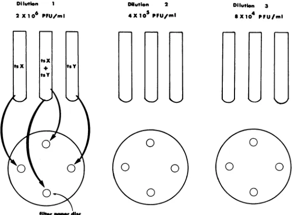
wereperformed as previouslydescribed (9).A simpli-fied qualitative complementation test is illustrated in Fig. 1. Three- to4-day-oldfully confluentmonolayers ofVero cells in 60-mm petri disheswereused for all tests. Three dilutions of each test mutant were
prepared to contain 2 x 10⁶, 4 x 10⁵, and 8 x 10'
PFU/ml. Equal volumes (0.5 ml) of single virus
suspensions at the same dilution were then mixed. Suspensions of each virus alone ateach of the three dilutionsservedascontrols. Vero cellmonolayerswere
washed oncewith Tris, leavingasmallvolume (0.1to
0.2 ml) ofbuffer on the monolayer to prevent cells fromdrying.Sterile filter paperdisks(Whatman filter paper no. 4) with diametersof6 mm weresaturated with virus suspensionanddrainedbeforebeing placed
onthe monolayer.Each disk contained about0.01ml
of solution when saturated with virus suspension. Four disks were placed on each 60-mm petri dish: duplicate test disks saturated with mixed virus
sus-pensionandtwocontroldisks each saturated withone
virus ofthe test pair (Fig. 1). Virus wasallowed to
adsorb to monolayers for 45 min at 37C in a CO2 incubator. Disks were then removed with sterile forceps, and8ml of Eaglemedium containing 5% fetal bovine serum and 2% methyl cellulose wasaddedto
eachplate. Plateswere incubatedat39C forHSV-1 and 38C for HSV-2 ts mutants. After 4 days of incubation, 5 ml of neutral red solution (1:20,000 in
Tris) was added to eachplate and monolayers were
observed for areas of clearingonthefollowing day. Progeny virus tests. Analysis of progeny from
Dilution 1 DUi
2 X10⁶ PFU/mI
mixed infections between HSV-1 ts mutants was conducted to determine whether the yield was com-posedofts and/or ts+ virus. For progeny analysis,no
neutral red was added to monolayers and clear areas were visualized by indirect light. Well-isolated plaques and clear, lysed areas of monolayers were picked after 4 days of incubation at 39 C. Plaque material was suspended in 0.8 ml of virus diluent and frozen at -90 C. Virus suspensions were then thawed, sonically treated, clarified by low-speed centrifuga-tion, and assayed on Vero cell monolayers at 39 and
34C.
RESULTS
Results of qualitative complementation
tests. Three characteristic reactions were
ob-served at sites on monolayers infected with
mixed suspensions of mutants (Fig. 2). (i)
Negativereactions were characterized by intact
monolayers; i.e., no evidence of lysis or clearing was observed (Fig. 2A). Mutant pairs that
exhibited this reaction were said to produce negative spots. (ii) The most frequently
ob-servedpositive reaction is illustrated in Fig. 2B.
The monolayer beneaththe site of infection was
totally lysed, and large plaques were evident in
thevicinity of the inoculation site (Fig. 2B-1).
(iii) The least frequently observed positive reac-tion is illustrated in Fig. 2C. The monolayer
lution 2 Xt05PFU/ml
Dilution 3
axle PFU/ml
fiberpepwdis
FIG. 1. Qualitative complementatoion test. Three fivefold dilutions of each mutant to be tested were preparedtocontain 2 x 10', 4 x 10⁵, and 8 x 10⁴PFU/ml.Equal volumes (0.5 ml) of two test mutants at the
samedilution were mixed. Filter paper disks were saturated with each virus suspension, drained, and placed on Vero cell monolayers. Disks containing each test mutant alone were also placed on monolayers as controls. After incubation for 45 min at 37 C in aCO2incubator, filter paper disks were removed and a methylcellulose overlay
wasadded. Plates were incubated at the nonpermissive temperature for 3 or 4 days, neutral red was added, and reactionswereread the following day.
J.VIROL.
4)
on November 10, 2019 by guest
http://jvi.asm.org/
[image:2.498.122.409.373.585.2]QUALITATIVE COMPLEMENTATION TEST FOR HSV 1133
2
FIG. 2. Characteristic results ofspot
complemen-tation tests with three pairs of HSV-1 ts mutants.
Virus dilutionswereasfollows: (1) 2 x 10⁶ PFU/ml; (2) 4 x 10⁵ PFU/ml; and (3) 8 x 10⁴PFUImI. (A)
tsB21 + tsB2, no reaction; (B) tsB21 + tsE6,
posi-tive reaction(tsandts+virus inyield);and(C) tsE5 + tsM19,positive reaction (tsvirusonly inyield).
beneath the site of infection characteristically
exhibited either complete lysis or a mottled
appearance. Occasionally, tiny plaques were
observed in the vicinity of the inoculation site.
Positive reactionswerelessprominentathigher
virus dilutions, demonstratingthat the
magni-tude of thereactionwasmultiplicity dependent.
Although monolayers infected with
suspen-sions ofsinglemutantswereusually intactatall
dilutions tested (Fig. 2A, B, and C, horizontal
positionsonmonolayersasillustrated in Fig. 1),
theyoccasionally exhibited minor mottling due
toleakatthehighest virusconcentration tested.
Progeny analysis of virus produced in
qualitative tests. To determine the
tempera-ture-sensitive phenotype of the progeny
pro-duced in mixed infections, virus was isolated
from individual plaques and from the lysed
monolayer (spot) directly beneath the
inocula-tion disk.Progeny virus wastested from mixed
infections with five HSV-1 ts mutant pairs
which exhibited reactions of thetypeillustrated
in Fig. 2B. Virus was also isolated from spots
produced bytwo mutant pairs which exhibited
the kind ofreactionillustrated inFig. 2C.Virus
was assayed onVerocell monolayers at34and
39C. Representative results ofthese tests are
shown in Table 1. When progeny produced in
the first type of reaction were tested, plating
efficiencies (39 C/34 C) of virus from isolated
plaques ranged from 0.5 to 1.2 PFU/ml and
from spots, 1.0 to 1.3 PFU/ml. Thus, progeny
virus plated nearly as well at 39C as at 34 C,
demonstrating that it was phenotypically ts+.
Sincesingle mutantcontrolswerenegative, this
virus was generated by recombination and not
reversion. Furthermore, since this type of
reac-tion was characteristic of nearly all positive
tests, recombination had occurred between
most mutant pairs.
Progeny virusisolatedfromspotsproduced in
the second type of positive reaction (Fig. 2C) produced plaques at 34 C but not at 39 C, indicating thatthisvirus was phenotypicallyts
and was generated by complementation. Comparison of results of qualitative and
quantitative
complementation tests with tsmutants of HSV-1 and HSV-2. Fifteen ts
mutants of HSV-1 representing 15 cistrons (A
through
0;
9) and 8HSV-2mutantsrepresent-ing 7 cistrons (Athrough G; 10) weretestedfor
their ability tointeract in thequalitative
com-plementation test. Results of these tests were
compared withpreviously determined resultsof
quantitative complementation and
recombina-tion tests (Table 2). Since recombinant virus
was produced in most cases, recombination frequencies are alsoshown (10). Two mutants, tsE5 and tsE6, in the same complementation
group as determined by negative results in
quantitative tests werenegative in the
qualita-tive test as well. In tests with mutants in two
other complementation groups, the results of
qualitative and quantitative tests also
agreed
well (i.e., tsE5 and tsE6 x tsC4, and tsE5 and tsE6 xts022).
Intestsof mutantsrepresenting
all 15cistrons,thequalitativetest wasnegative,
whereas the quantitativetest hadbeen positive
in only one case (tsC4 x
ts022).
Since moredefinitive quantitative tests had
previously
demonstrated that thetwo mutantsweredefec-tive indifferent cistrons, this reactionwasthus
a "false-negative" result.
InqualitativetestsofHSV-2tsmutants,
only
tsAl
and tsA8 exhibited anegative
reaction(Table
2). These mutants are in the samecomplementation
group as demonstratedby
quantitative tests, and they recombinepoorly.
All otherqualitativetests werebetweenmutantpairs previously shown to represent different
complementation groups by quantitative tests,
andallpairs gave positiveresultsin the
qualita-tive test. The great
majority
ofpositive
spotsproduced by HSV-2ts mutantpairs were also of
the"recombinant"type. In fact, the magnitude
of positive reactions produced by both HSV-1 andHSV-2mutant pairswasabetter reflection
oftheefficiencyofrecombination (10) than the efficiency ofcomplementation (5, 9).
VOI>.16, 1975
1
on November 10, 2019 by guest
http://jvi.asm.org/
[image:3.498.46.240.79.277.2]TABLE 1. Analysis of progenyfrom representative qualitative complementation tests betweents mutantsof HSV-la
Virusyield(PFU/ml) Efficiency of
Mutantpar Plaqueor _______________ plating Typeof interaction
Mutatpair spot no. 34C 39C (PFU/ml, between mutant pair
39C/34 C)
tsA16+tsB2 1 8 x 10³ 5 x 10³ 0.6 Recombination(and
2 1x 10⁴ 7x 10³ 0.7 complementation)
3 4x 10³ 2x 10' 0.5
4 1x 10⁴ 9 X 10³ 0.9
5 6x 10² 7x10² 1.2
sic 4x 10⁴ 5 x10' 1.3
S2 2x10² 2x 10² 1.0
tsE5+tsM19
SI
2x 10³ <101 <0.005 ComplementationS2 1 x 10² <101 <0.1
aSimilar resultswereobtained intests offour additionalrecombinant-containing spots andoneadditional recombinant-negative (complementation-initiated) spot.
"Individualplaques were isolated in the vicinity ofrecombinant-containing spots.
[image:4.498.61.457.308.563.2]CTotal virusin spots (S)was isolated and tested.
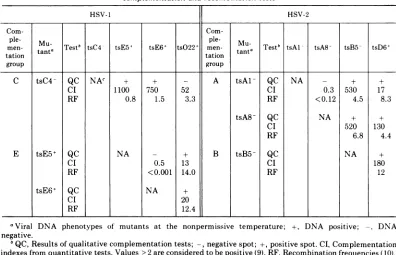
TABLE 2. Comparison ofrepresentative qualitative complementationdata with resultsof quantitative
complementatiors andrecombinationtests
HSV-1 HSV-2
Coin-
Coin-ple-
Mu.
ple-Mu-men- tata Test' tsC4 tsE5+ tsE6+ tsO22' men-
tnaTest'
tsAl tsA8 tsB5 -tsD6+tation tation tat
group group
C tsC4- QC NAc + + - A tsAl- QC NA - + +
CI 1100 750 52 CI 0.3 530 17
RF 0.8 1.5 3.3 RF < 0.12 4.5 8.3
tsA8-QC NA + +
CI 520 130
RF 6.8 4.4
E tsE5+ QC NA - + B tsB5 QC NA ±
CI 0.5 13 CI 180
RF <0.001 14.0 RF 12
tsE6+ QC NA +
CI 20
RF 12.4
aViral DNA phenotypes of mutants at the nonpermissive temperature;
+,
DNA positive; -, DNAnegative.
°QC,Results ofqualitative complementation tests; -, negative spot; +, positive spot. CI, Complementation indexesfromquantitativetests.Values>2areconsideredtobepositive (9). RF, Recombination frequencies (10).
cNA, Not applicable.
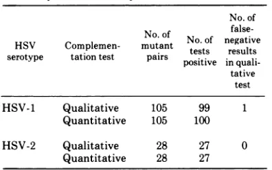
A summary ofthe results from allqualitative
and quantitative tests performed with HSV-1
and HSV-2 ts mutants is shown inTable3. Of
105 HSV-1 mutant pairs tested by both
methods, 99werepositiveinthequalitativetest
and 100 were positive in the quantitative test;
thus, false-negative results were observed in
only one instance. No false-negative results
were observed in tests of 28 HSV-2 ts mutant
pairs. False-positive reactions have not been
observed in from three to five replicate tests of
over 150mutantpairs.
Identification of new HSV cistrons. To evaluate the qualitative test as a means of* identifying new HSV cistrons, two recently isolated UV-induced ts mutants of HSV-2 were
1134 CHU AND SCHAFFER J. VIROL.
on November 10, 2019 by guest
http://jvi.asm.org/
QUALITATIVE COMPLEMENTATION TEST FORHSV 1135
tested by both qualitative and quantitative
tests (Table 4). Mutant ts314 complemented
mutants representing six of seven
complemen-tationgroupsinthequalitativetest and seven of
seven groups in the quantitative test. Repeat
tests of ts314 with tsAl and tsA8, which were
negative inthequalitativetest, werepositivein
the moresensitive quantitativetest. Therefore, by combined qualitative andquantitative
com-plementation tests, ts314 was shown to
repre-sent a newHSV-2 cistron. This cistron has been
designated
H, and the mutant ts314 has been renamed tsH10. Mutant ts49, on the other hand, failedtocomplement thegroup D mutantin both kinds of tests and was, therefore,
assigned tothat group;ts49hasbeen designated
tsD11.
DISCUSSION
Thispaperdescribes arapid, simple
qualita-tive complementation test for ts mutants of
herpes simplex virus. Two qualitative
com-plementationtests havebeen describedrecently
for tsmutants ofsimianvirus 40 (2, 12). Since recombination occurs rarely in the SV40 sys-tem, the positivereaction in bothtests isbased
upon the
ability
of mutant pairs to generateTABLE 3. Comparisonofresultsfromqualitativeand quantitativecomplementationtests
No. of No.of ~~false-HSV Complemen-Complemen-
outaft
No. oftests negativeresults serotype tationtest pairs posit insualipositive in quali-tative
test
HSV-1 Qualitative 105 99 1
Quantitative 105 100
HSV-2 Qualitative 28 27 0
Quantitative 28 27
progenyvirusby complementation. Aspot
com-plementation for ts mutants of
bacteriophage
T4D wasdescribed in 1964 by Edgaretal.
(4).
Recombinationoccursreadily in theT4 system,and the authors not surprisingly observed the presenceof recombinant virus in mixedyieldsof spot complementation tests. In part, it is a
misnomer to term the test described in this
report a complementation test since the
pri-maryeventobservedwasrecombination. This is
inevitable with herpes simplex viruses, how-ever, since
(i)
recombination is frequent withtheseviruses, (ii) asdevised, thetestmeasures
the yield after multiple rounds of
replication
resulting from complementation which favors recombination, and (iii) ts+recombinant virususuallyoutgrows tsmutantvirus.Althoughthe
majority of progeny in mixed infections
repre-sented recombinant virus, the less
frequently
observed result (Fig. 2C) probably reflects the
inability ofmutants in this mixed infection to
undergo multiplerounds ofreplication.
Hence,
in this case true complementation was the
consequence. Thegoodcorrelation betweenthe
qualitative and quantitative tests probably
re-flects the fact that mutants in the same
com-plementation grouprecombine inefficiently.
Ininitialattempts todemonstrate qualitative
complementation betweenHSVts mutants, we
observed that the use of thin agar overlays
inhibited thediffusion ofvirustotheunderlying
monolayer and inoculum spread freelyoverthe
surface of the agar. In contrast, the use of
virus-saturatedfilterpaperdisks insureddirect contactbetween cells and virus and resulted in ahighly localizedinfection. Diskswereremoved
afteradsorption sincethemostprominent
reac-tionwasevidentimmediately beneaththedisk.
This was especially true in the case of
mini-mally positive reactions.
Preliminary tests also indicated that the
clearest evidence for a positivereaction in the
qualitative test was obtained when both
mu-TABLE 4. Resultsofqualitativeandquantitativecomplementation betweentwonewlyisolated HSV-2ts mutants andeightestablished HSV-2ts mutants
Mu-
Testi
Complementation between ts mutant pairstant tsAl tsA8 tsB5 tsC2 tsD6 tsE7 tsF3 tsG4
ts314 QC [ ] + + + + + +
CI 23 23 5,333 588 1,233 1,400 1,017 108
ts49 QC + + + + 7
-
+ + +CI 6,700 209 231 122 0.5 2,421 4,483 141
ats314and ts49 arenewly isolatedts mutantsinduced with UVirradiation.
'QC,Resultsofqualitativecomplementationtests;-,negativespot;+,positivespot.CI,Complementation indexesfromquantitativetests(9). Values>2areconsideredtobepositive.
VOL. 16, 1975
on November 10, 2019 by guest
http://jvi.asm.org/
[image:5.498.44.242.389.513.2]1136 CHU AND SCHAFFER
tants in apairwereequally dilutedtocontain 4
x 10 to 2 x 10⁶
PFU/ml. Although
concentra-tions of 8 x 10' PFU/ml produced reactions consistent with those observed at higher
con-centrations, they were more difficult to
inter-pret.Thus, thetest wasapplicableover a0.7- to
1.5-log range of virus titers. Concentrations
below 10'
PFU/ml
always gave negative resultsin mixed infections-even in tests of mutant
pairs shown to complement in quantitative
tests.
The qualitative test was demonstrated to be slightly lesssensitive than thequantitativetest
since false-negative results were occasionally observed. The occasional appearance of
false-negative reactionsemphasizes the necessityfor
all negative results in qualitative tests to be tested by more definitive quantitative
com-plementation tests. False-positive
reactions,
onthe other hand,have neverbeen observedinthe qualitative test.
Theage ofthe Veromonolayerswasfoundto
influence the results of qualitative tests. Sub-confluent monolayers exhibited extensive leak and nonspecific
damage,
even when inoculatedwith single mutant control suspensions, thus
making results difficult to interpret. Conse-quently, fully confluent 3- to
4-day-old
mono-layers wereused.
Results were least
ambiguous
if tests wereevaluatedafter 4 or 5daysofincubation(4days
wasoptimal for HSV-2 and5dayswasoptimal
for HSV-1). After longer periods ofincubation
(as longas 7days), the interpretationofresults remained unchanged.
Mutants that exhibit moderate levels ofleak
andreversion are often the best mutants tobe
used in genetic and biochemical studies since those which fail to leak orrevert may contain
more thanone mutation. One advantageofthe
qualitative complementation test is that it is
applicable to mutants with low and moderate levels of leak
and/or
reversion. Mutants withleakorreversion levelsequalto orlessthanthe
lowest virusconcentration tested, i.e., 104 PFU/
ml, would be suitable for qualitative
com-plementation tests. Furthermore, for very leaky
mutants the highestconcentration of each
mu-tantthatfails toexhibitleak canbe determined
readily beforetesting.
The results presented in this report
demon-strate that the qualitative complementation
test offers a rapid and sensitive means for
assigning mutants ofHSV-1 and HSV-2 to new
and existing complementation groups and that
patterns of complementation obtained by this
method and the more laborious quantitative
method agree well. We estimate that the time
saved through the use of the qualitative test is
such that 1 year of quantitative tests can now be completed in 1 or 2 months. The test is cur-rently being used for screening new ts mutants
ofHSV and for comparative complementation
studies between groups of HSV-1 and HSV-2 ts
mutantsisolated in different laboratories.
ACKNOWLEDGMENTS
C.-T. C. was the recipient of a fellowship from the National Science Council oftheRepublicofChina, Taipei, Taiwan. Thiswork wassupported by PublicHealth Service research grant CA10,893from the National Cancer Institute.
LITERATURE CITED
1. Brown S. M., D. A. Ritchie, and J. H. Subak-Sharpe. 1973. Genetic studieswithherpes simplex type1.The isolation oftemperature-sensitive mutants, their ar-rangement intocomplementation groups and recombi-nationanalysis leadingto alinkagemap.J. Gen. Virol. 18:329-346.
2. Chou, J. Y., and R. G. Martin. 1974.Complementation analysis of simian virus 40 mutants. J. Virol. 13:1101-1109.
3. Dreesman, G. R., and M. Benyesh-Melnick. 1967. Spec-trum of human cytomegalovirus complement-fixing antigens. J. Immunol. 99:1106-1114.
4. Edgar, R.S., G. H. Denhardt, and R. H. Epstein.1964. A comparative genetic studyofconditional lethal muta-tionsofbacteriophage T4D. Genetics 49:635-648. 5. Esparza, J., D. J. M. Purifoy, P. A. Schaffer, and M.
Benyesh-Melnick. 1974. Isolation, complementation and preliminary phenotypic characterizationof tem-perature-sensitive mutants of herpes simplex virus type2.Virology 57:554-565.
6. Koment, R. W., and F. Rapp. 1975. Temperature-sensi-tivehost rangemutants ofherpes simplex virus type2. J. Virol. 15:812-819.
7. Manservigi, R. 1974. Method for isolation and selectionof temperature-sensitive mutants ofherpes simplex virus. Appl. Microbiol. 27:1034-1040.
8. Schaffer, P.,V. Vonka, R.Lewis, and M. Benyesh-Mel-nick: 1970. Temperature-sensitive mutants of herpes simplex virus. Virology 42:1144-1146.
9. Schaffer,P.A., G. M. Aron, N. Biswal, and M. Benyesh-Melnick. 1973. Temperature-sensitive mutants of herpes simplex type 1: isolation, complementation and partial characterization. Virology 52:57-71.
10. Schaffer, P. A., M. J. Tevethia, and M. Benyesh-Mel-nick. 1974.Recombination between temperature-sensi-tive mutants ofherpes simplex virus type 1. Virology 58:219-228.
11. Takahashi, M., and K. Yamanishi. 1974.Transformation of hamster embryo cells by temperature sensitive mutants of herpes simplex virus type 2. Virology 61:306-311.
12. Tevethia, M. J., L. W. Ripper, and S. S. Tevethia. 1974. A simple qualitative spot complementation test for temperature sensitive mutants ofSV40. Intervirology 3:245-255.
13.Timbury, M. C. 1971.Temperature-sensitive mutants of herpes simplex virus type 2. J. Gen. Virol. 13:373-376. 14. Zygraich, N., and C. Huygelen. 1973. Invivo behaviour of
atemperature sensitive (ts) mutant ofherpesvirus hom-inistype 2. Arch. Gesamte Virusforsch.43: 103-111.
J VIROL.