1983
The differentiation and characterization of canine
adenoviruses 1 and 2 that are used for vaccine
production in the United States
Cecelia Anne Whetstone
Iowa State University
Follow this and additional works at:
https://lib.dr.iastate.edu/rtd
Part of the
Animal Sciences Commons
, and the
Veterinary Medicine Commons
This Dissertation is brought to you for free and open access by the Iowa State University Capstones, Theses and Dissertations at Iowa State University Digital Repository. It has been accepted for inclusion in Retrospective Theses and Dissertations by an authorized administrator of Iowa State University Digital Repository. For more information, please contactdigirep@iastate.edu.
Recommended Citation
Whetstone, Cecelia Anne, "The differentiation and characterization of canine adenoviruses 1 and 2 that are used for vaccine production in the United States " (1983).Retrospective Theses and Dissertations. 8968.
This reproduction was made from a copy of a document sent to us for microfilming.
While the most advanced technology has been used to photograph and reproduce
this document, the quality of the reproduction is heavily dependent upon the
quality of the material submitted.
The following explanation of techniques is provided to help clarify markings or
notations which may appear on this reproduction.
1.The sign or "target" for pages apparently lacking from the document
photographed is "Missing Page(s)". If it was possible to obtain the missing
page(s) or section, they are spliced into the film along with adjacent pages. This
may have necessitated cutting through an image and duplicating adjacent pages
to assure complete continuity.
2. When an image on the film is obliterated with a round black mark, it is an
indication of either blurred copy because of movement during exposure,
duplicate copy, or copyrighted materials that should not have been filmed. For
blurred pages, a good image of the page can be found in the adjacent frame. If
copyrighted materials were deleted, a target note will appear listing the pages in
the adjacent frame.
3. When a map, drawing or chart, etc., is part of the material being photographed,
a definite method of "sectioning" the material has been followed. It is
customary to begin filming at the upper left hand comer of a large sheet and to
continue from left to right in equal sections with small overlaps. If necessary,
sectioning is continued again-beginning below the first row and continuing on
until complete.
4. For illustrations that cannot be satisfactorily reproduced by xerographic
means, photographic prints can be purchased at additional cost and inserted
into your xerographic copy. These prints are available upon request from the
Dissertations Customer Services Department.
5. Some pages in any document may have indistinct print. In all cases the best
available copy has been filmed.
Un
Whetstone, Cecelia Anne
THE DIFFERENTIATION AND CHARACTERIZATION OF CANINE
ADENOVIRUSES 1 AND 2 THAT ARE USED FOR VACCINE PRODUCTION IN
THE UNITED STATES
Iowa State University PH.D. 1983
University
Microfilms
In all cases this material has been filmed in the best possible way from the available copy.
Problems encountered with this document have been identified here with a check mark V .
1.
Glossy photographs or pages
2.
Colored illustrations, paper or print
3.
Photographs with dark background
4.
Illustrations are poor copy
5.
Pages with black marks, not original copy
6.
Print shows through as there is text on both sides of page
7.
Indistinct, broken or small print on several pages
8.
Print exceeds margin requirements
9.
Tightly bou nd copy with print lost in spine
10.
Computer printout pages with indistinct print
11.
Page(s)
lacking when material received, and not available from school or
author.
12.
Page(s)
seem to be missing in numbering only as text follows.
13.
Two pages numbered
. Text follows.
14.
Curling and wrinkled pages
15.
Other
production in the United States
by
Cecelia Anne Whetstone
A Dissertation Submitted to the
Graduate Faculty in Partial Fulfillment of the
Requirements for the Degree of
DOCTOR OF PHILOSOPHY
Interdepartmental Program: Immunobiology Major: Immunobiology
Approved:
In Charge of Ma^r Work
Professor-in-Chàage, Program of Immunobiology
For the Grad
Iowa State University
Ames, Iowa 1983
Signature was redacted for privacy.
Signature was redacted for privacy.
TABLE OF CONTENTS
Page
GENERAL INTRODUCTION iv
DISSERTATION FORMAT vii
PART I: RESTRICTION ENZYME ANALYSIS OF CANINE
ADENOVIRUSES 1 AND 2: DIFFERENTIATION OF ' STRAINS USED FOR VACCINE PRODUCTION IN THE
UNITED STATES 1
ABSTRACT 2
INTRODUCTION 3
MATERIALS AND METHODS 5
RESULTS 8
DISCUSSION 10
ACKNOWLEDGMENTS 17
LITERATURE CITED 18
PART II; CHARACTERIZATION OF THE CANINE ADENO
VIRUSES 1 AND 2 BY IMMUNOFLUORESCENCE, VIRUS NEUTRALIZATION, AND IMMUNOPRECIPITATION USING
MONOCLONAL ANTIBODIES 23
ABSTRACT 24
INTRODUCTION 25
MATERIALS AND METHODS 28
RESULTS 32
DISCUSSION 36
ACKNOWLEDGMENTS 54
Page
SUMMARY AND DISCUSSION 63
ADDITIONAL LITERATURE CITED 66
Canine adenovirus type 1 (CAV-1) was isolated by Cabasso et al. in
1954 from a dog with acute hepatitis and shown to be identical to the
infectious canine hepatitis (ICH) virus described by Rubarth in 1947
(7, 24). In 1962, Ditchfield et al. isolated a different canine adeno
virus from throat swabs of a dog with laryngotracheitis. This strain of
canine adenovirus type 2 (CAV-2) was called Toronto A26/61 (12). Several
strains of CAV-1 and CAV-2 have since been described, and differentiation
between the two has been based on studies of morphology (18),
cyto-pathology in cultured cells (1, 29), serology (4, 11, 26), and patho
genicity (2, 4, 27).
Morphology, serology, and in vitro cytopathology studies have left
ambiguities as to the distinctness of the two viruses. The greatest
difference shown between type 1 and 2 has been in the pathogenicity of
the two viruses. With CAV-1, the classical disease is infectious
hepatitis, but infections can range from inapparent to fulminating fatal
disease, including respiratory syndromes similar to those associated with
CAV-2 infection. The virus has an affinity for hepatic parenchymal,
Kupffer's, and endothelial cells. Virus has been isolated readily from
the liver, kidneys, and lymphoid organs. The viremia resulting from
infection with CAV-1 also leads to a phenomenon called "blue eye", a type
III hypersensitivity reaction in which immune complex formation resulting
from the release of virus brings about corneal endothelial damage and
with CAV-1 and to vaccination with modified live type 1 virus (10). The
pathogenesis of CAV-2, on the other hand, is largely confined to the
respiratory system, and is associated with a condition often referred to
as "kennel cough". The type 2 virus does not readily cause viremia, and
blue eye is not observed. Because of the potential problem of blue eye
associated with vaccination with CAV-1, many veterinary biologies firms
now market CAV-2 vaccines since a vaccine produced from either virus will
cross-immunize against the heterologous virus (3, 8, 9, 13).
The CAVs currently used for vaccine production in the United States
include the Cornell-1 and Lederle 255 strains of CAV-1 and the
Toronto A26/61 (Ditchfield) and Manhattan strains of CAV-2. Until now,
identification and classification of the master seed virus (the virus
from which subsequent vaccine is produced) have had to come from studies
on pathogenicity, morphology, cytopathology, and serology. Although
taken collectively, these data may provide an accurate evaluation of the
identity of a CAV type, there is currently no individual test, especially
in vitro, that can do this. Additionally, although differences have been
shown between CAV-1 and CAV-2 in pathogenicity (12, 27), morphology
(CAV-1 fiber is 25-27 nm as compared with CAV-2 fiber which is 35-37 nm)
(18), and antigenicity (11, 26,), these two viruses are still not
recognized, by definition, as two distinct adenovirus species (types)
(19, 34). Quantitative cross-neutralization studies with CAV-1 and CAV-2
have not shown immunologic distinctiveness by demonstrating either no
cross-reaction or homologous-to-heterologous titer ratios of >16 in both
be useful if such a method or methods could identify strains within a
given type so that vaccine and field strains could be discriminated.
Inherent to the success of this enterprise was the proof that CAV-1 and
CAV-2 are immunologically distinct species of CAV. The experiments that
were performed to accomplish these goals are described in the two papers
entitled, "Restriction enzyme analysis of canine adenoviruses 1 and 2:
differentiation of strains used for vaccine production in the United
States," and "Characterization of the canine adenoviruses 1 and 2 by
immunofluorescence, virus neutralization, and immunoprecipitation using
DISSERTATION FORMAT
This dissertation is presented as an alternate format which includes
two manuscripts to be submitted to scientific journals for publication.
Both will be submitted to the Journal of Virology. The manuscripts are
presented in the format required for the dissertation. References are
cited at the end of each manuscript and are in compliance with the
journal. The manuscripts are preceded by a general introduction. A
summary and discussion section follows the last manuscript. Literature
cited in those sections is listed after the summary and discussion.
The Ph.D. candidate, Cecelia Anne Whetstone, was the principal
investigator for each of the investigations and is the sole author for
PART I: RESTRICTION ENZYME ANALYSIS OF CANINE ADENOVIRUSES 1 AND 2;
DIFFERENTIATION OF STRAINS USED FOR VACCINE PRODUCTION IN THE
UNITED STATES
This manuscript has been submitted for publication to the Journal of
type 2 [CAV-2 (Manhattan, Toronto A26/61, also referred to as
Ditch-field)] were examined by restriction enzyme analysis. Each of these,
except Utrecht, is used for vaccine production in the United States. The
two types of adenoviruses could be readily differentiated on the basis of
restriction patterns obtained using either enzymes Bam HI, Eco RI, Kpn I
or Bgl II. Differences between strains within the same virus type,
however, were not as conspicuous. Among the CAV-1 strains, only
Cornell-l-PK showed restriction patterns that were distinct from the
other strains. Between the CAV-2 strains, small pattern dissimilarities
could be detected only with the use of the restriction enzymes Bam HI and
with acute hepatitis and showed this virus to be identical to the one
designated by Rubarth (1947) as infectious canine hepatitis virus
(9, 31). Subsequently, Ditchfield et al. (1962) isolated canine adeno
virus type 2 (CAV-2) from throat swabs of a dog with
laryngo-tracheitis (17). Although several isolates of CAV have since been
described, all have been identified by serology, pathology, clinical
signs, and virus morphology as either CAV-1 or CAV-2 (3, 6, 9, 15, 16,
17, 35, 37, 38, 39, 44). Although differences between the two types
occur in morphology, pathogenicity, and antigenicity (1, 3, 23, 37, 38),
almost all mammalian adenoviruses share group-specific determinants
located on the major capsid protein, hexon (10). Because of the shared
group-specific determinants, serologic responses in animals exposed to
one type of CAV are cross-reactive with the other type (3, 37, 38).
Moreover, immunity to either pathogen can be induced by vaccination with
homologous or heterologous CAV type (5, 11, 12, 13, 19, 40). Two strains
of CAV-1 (Cornell-1 and Lederle 255) and two strains of CAV-2
(Toronto A26/61 and Manhattan) are currently used for the production of
vaccine in the United States. Conventional in vitro detection of these
viruses with assays such as serum neutralization (SN) or hemagglutination
distinguishing CAV-1 from CAV-2. None of the above techniques can be
utilized for strain identification or differentiation.
Discrimination between types and even subtypes of human adenoviruses
has been accomplished with the use of restriction enzyme analysis of the
viral genomes (2, 26, 32, 41, 42, 43). In this study, therefore, antici
pating that this technique would also be useful in differentiating CAV
types and possibly strains of the same type, I examined the prototype of
CAV-1 and strains of each type that are used for vaccine production in
the United States. By using the restriction endonucleases Bam HI,
Eco RI, Kpn I, and Bgl II, I showed that it is possible to differentiate
CAV-1 from CAV-2. Furthermore, in some instances, strains of the same
type demonstrated similar but unique restriction profiles characteristic
MATERIALS AND METHODS
Virus and Cells. Primary dog kidney and Madin and Darby canine
kidney (MDCK) cells were maintained in Eagle's Minimum Essential Medium
(MEM) with Earle's salts, 2 mM L-glutamine, 10% heat-inactivated bovine
fetal serum (BPS), and gentamicln sulfate (50 mg/liter). The Utrecht and
Lederle 255 strains of CAV-1 and the Toronto A26/61 (Ditchfield) strain
of CAV-2 were obtained from the American Type Culture Collection,
passaged one time in primary dog kidney cells, and stored at -60 C. The
Cornell-1-66 and Cornell-l-PK strains of CAV-1 and the Manhattan strain
of CAV-2 were a gift from Dr. L. E. Carmichael of Cornell University. As
received, the Cornell-1-66 strain, a dog isolate (4), was at the third
passage in primary canine kidney cells; the Cornell-l-PK strain was the
16th passage of Cornell-1-66 in primary dog kidney cell cultures; and the
Manhattan strain was the fifth dog kidney passage. All three of these
viruses were passaged once in primary dog kidney cells and stored at
-60 C.
Virus Purification from Cell Culture. The MDCK cells were seeded at
5 2
a concentration of 2 x 10 cells/ml into 850 cm roller bottles contain
ing MEM, supplemented as described. After 48 hours, 2 ml of each virus
strain, diluted in MEM, were inoculated at a multiplicity of infection
(MOI) of 0.2 to 0.02 onto two roller bottles and allowed to adsorb for
one hour. Supplemented MEM was replaced and cells were incubated at 37 C
until the CPE was 100% (about 4 to 5 days post-inoculation). Rollers
250 X g for 10 minutes. The supernatant fluid was then centrifuged
through 40% sucrose (w/v in 0.2 M phosphate buffer, pH 7.5) at
100,000 X g for 11/2 hours and the resulting viral pellet was
resuspended in 2.0 ml of TE buffer (10 mM Tris, 1 mM EDTA, pH 7.6). A
small volume (0.1 ml) was removed from each sample, inoculated onto MDCK
cells, and examined by the FA technique for the presence of viable virus.
Viral DNA Extraction. The 2.0 ml viral pellet suspension was
treated with 20 jil sodium dodecyl sulfate [(SDS) 20%], 200 ul
proteinase K (1 mg/ml), and incubated at 37 C for 1 to 2 hours until
cleared. Extraction was performed using an equal volume of TE
buffer-saturated phenol (distilled) one time, 1/2 volumes each phenol and
chloroform one time, and an equal volume of chloroform two times, taking
care to save the interface until the last extraction. The DNA was then
ethanol precipitated, resuspended in TE buffer, and stored either at
-20 C or at 4 C until used.
Restriction Enzyme Analysis. Restriction enzymes [Bethesda Research
Laboratories, Inc. (BRL)] were stored, diluted, and used in running
buffers as prescribed by the company. Human adenovirus 2 (Ad 2) DNA
(from BRL) was used as a standard. Enzyme reactions were stopped with
10 yl of a mixture of 0.02% bromphenol blue in 60% sucrose [w/v in
Loening's buffer (36 mM Tris, 30 mM NaHgPO^, 1 mM EDTA, pH 7.7)].
Cleaved DNAs were electrophoresed in a horizontal chamber at a constant
45 volts for 16 to 17 hours in a 0.8% gel (SeaKEM ME agarose) in
Loening's buffer. The DNA was then stained with ethidium bromide
photographed through a red-orange filter. Molecular weights were
estimated graphically using Ad 2 as the molecular weight standard and
RESULTS
Selection and Purification of Viruses. The virus strains examined
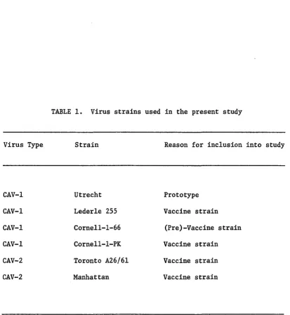
and the rationale for selecting those strains are presented in Table 1.
After virus titers were determined in MDCK cells, a low MOI was used to
culture viruses for DNA extraction so that the frequency of adenovirus
recombination and defective interference could be held to a minimum (18).
Viral CPE was 100% by days 4 to 5 post-inoculation, at which time cells
were harvested. Most of the cellular debris was removed by the
clarification step eliminating contamination with host cell DNA during
the extraction procedure. The presence of virus in each pellet was
confirmed by the FA test on MDCK cells inoculated with a small volume of
the resuspended pellet.
Extraction of Viral DNA. Samples were treated with SDS and
proteinase K to denature and degrade contaminating proteins. If one
treatment with these agents did not yield a translucent sample, the
treatment was repeated until the sample was clear. Because of the
hydrophobic nature of the reaction of phenol with proteins, DNA can get
trapped at the interface during extraction procedures. This problem is
solved by re-extraction. Therefore, care was taken not to discard the
interface until after the last extraction, which rendered noticeably
better yields of DNA than when this precaution was not taken.
Differentiation of Canine Adenoviruses by Restriction Enzyme
Analysis. The restriction endonucleases Bam HI, Eco RI, Kpn I, and
different six base pair sequence in DNA, and all have been used to map
the Ad 2 genome (28), the size marker used in this study. An 0.8% gel
was used because it resolves fragments from approximately 300 to
20,000 bp (33). Results of the restriction analysis are presented in
Fig. 1. All enzymes cut Ad 2 DNA as predicted (28). With all enzymes
used, CAV-1 and CAV-2 were easily differentiated. Among the CAV-1
strains, there was no difference between the prototype virus, Utrecht,
and the Lederle 255 and Cornell-1-66 strains. For the Cornell-l-PK
strain, however, the restriction enzymes cut differently than for the
other CAV-1 strains in all cases. Between the CAV-2 strains, there were
no pattern differences using the enzymes Kpn I or Bgl II. With Bam HI,
the Toronto A26/61 strain displayed an additional submolar band just
below the 14 kb fragment, and with Eco RI, this same strain showed a
small fragment just above the 21 kb fragment. Neither fragment was
present in the restriction patterns for the Manhattan strain. Although
the Eco RI pattern for Toronto A26/61 appeared to be a partial digest,
the anomally was repeatable. Genome size for all strains of CAV-1 and
CAV-2 tested were estimated from the gels at 20 to 21 x 10^ daltons,
DISCUSSION
Differentiation of CAV-1 from CAV-2 has previously been based on
studies of pathogenicity (3, 38), serology (6, 15, 37), cytopathology in
cultured cells (1), and morphology (23). Studies of cytopathology in
cultured cells have not readily differentiated CAV-1 from CAV-2, and
morphological studies differentiated the two viral types only on the
basis of fiber length, which is 25 to 27 nm for CAV-1 and 35 to 37 nm for
CAV-2 (22). Among the CAVs, the use of serology for type distinction and
strain identification has not been completely successful, because the
cross-reactivity of the group specific hexon, the major antigenic protein
for both viruses, masks any differences that might be present in other
antigenic proteins.
The greatest difference seen between type 1 and 2 has been shown in
the pathogenicity of the two viruses. With CAV-1, the classical disease
is infectious hepatitis, but infections can range from unapparent to
fulminating fatal disease, including respiratory syndromes similar to
those associated with CAV-2 infection. The virus has an affinity for
hepatic parenchymal, Kupffer's, and endothelial cells. Virus has been
isolated readily from the liver, kidneys, and lymphoid organs. The
viremia resulting from infection with CAV-1 also leads to a phenomenon
called "blue eye", a type III hypersensitivity reaction in which immune
complex formation resulting from the release of virus brings about
corneal endothelial damage and edema. The blue eye syndrome is attrib
modified live type 1 virus (14). The pathogenesis of CAV-2, on the other
hand, is largely confined to the respiratory system, and is associated
with a condition often referred to as "kennel cough". The type 2 virus
does not readily cause viremia, and blue eye is not observed. Because of
the potential problem of blue eye associated with vaccination with CAV-1,
many veterinary biologies firms now market CAV-2 vaccines because a
vaccine produced from either virus will cross-immunize against the
heterologous virus (5, 11, 13, 19). Until now, identification and
classification of the master seed virus (the virus from which subsequent
vaccine is produced) have had to come from studies on pathogenicity,
morphology, cytopathology, and serology. Restriction enzyme analysis
provides a relatively simple, direct, objective, and definitive method
for typing the master seed virus.
An advantage of restriction enzyme analysis over older serologic
classifications is sensitivity. Serologic methods rely on distinct
antigenic determinants that induce antibodies. The gene products carry
ing these antigenic determinants, however, represent only a few percent
of the total viral genome, meaning that analysis of cross-reactions
measured by serologic techniques gives information on only a few gene
products, and is not necessarily representative of the relatedness of
viral genomes. Sequence-specific endonucleases, such as the ones used in
this study, are second only to nucleotide-sequence analysis in the
detailed Information that can be gained on the homologies of related
genomes. This fact has been proven repeatedly with the human adeno
herpes-viruses (7, 8, 20, 21, 29, 34); parvoherpes-viruses (25); papovaherpes-viruses (36);
and poxviruses (27). The restriction patterns generated with Bam HI,
Eco RI, Kpn I, and Bgl II in this study all show that there are clearly
two distinct genome types of canine adenovirus that can be readily
differentiated from each other. Furthermore, strains of a given type
that are truly different from the prototype can be detected. This is
best illustrated with the Cornell-1 strains of CAV-1. The Cornell-1-66
strain, a virulent isolate from a dog suffering infectious canine hepa
titis (ICH), was lyophilized after three passages in primary dog kidney
cells: its restriction patterns are identical to Utrecht, the prototype
virus. The Cornell-l-PK strain is an attenuated version of the same
virus after 16 passages in porcine kidney cells. Its restriction pat
terns indicate that although it is still a type 1 CAV, it is now clearly
a subtype. The Lederle 255 strain appears to be identical to Utrecht.
There also seem to be small genetic differences between the CAV-2 strains
detectable by restricting with Bam HI and Eco RI.
Biologically, virus attenuation can be measured by reduced virulence
in the host animal. Genomically, a difference between virulent and
attenuated viruses has been shown with the porcine and equine herpes
viruses in which the restriction enzyme patterns of vaccine strains are
distinct from field isolates (21, 29, 34). Moreover, the stability of
restriction patterns from a variety of viral isolates is well-documented
(7, 8, 30, 36, 41). Since vaccines are limited to the number of times
they can be passed, the chance of a pattern changing would be very
repeated passage of that virus, but rather a reflection of a genomic
alteration that may be significant enough to be verified by restriction
enzyme analysis. This study supports the concept that attenuated
viruses, especially those that have been attenuated by passage in
heterologous species cell cultures, may yield unique restriction patterns
that provide a "fingerprint" of identification for that particular virus
strain, a factor that becomes important in the differentiation of vaccine
from field strains of virus.
Finally, according to the latest report (1982) of the International
Committee on the Taxonomy of Viruses, "a species (formerly type) of
adenovirus is defined on the basis of its immunologic distinctiveness, as
determined by quantitative neutralization with animal antisera. If
neutralization shows a certain degree of cross-reaction between two
viruses in either or both directions...distinctiveness of species is
assumed if...(ii) substantial biophysical/biochemical differences of the
DNAs exist" (24). As already discussed, the serologic cross-reactivity
of CAV-1 and CAV-2 has led to ambiguity on the definitive typing of CAV-2
as a distinct species from CAV-1. The data presented in this study
indicate that there are substantial differences between these two viruses
at the DNA level and that they should be recognized as separate CAV
TABLE 1. Virus strains used in the present study
Virus Type Strain Reason for inclusion into study
CAV-1
CAV-1
CAV-1
CAV-1
CAV-2
CAV-2
Utrecht
Lederle 255
Cornell-1-66
Cornell-l-PK
Toronto A26/61
Manhattan
Prototype
Vaccine strain
(Pre)-Vaccine strain
Vaccine strain
Vaccine strain
[image:28.580.80.494.72.521.2]MWxIO'
Daltons
Kpn I
0 b c d e f
Eco RI
0 b c d e f g
MWxlO®
Dallons
13.9—^-JBl 'y
Bam HI
0 b c d e f g
5.0 —
9.5-7.0-
•—
-—s#s
H'if
MACKNOWLEDGMENTS
I thank Patricia M. Gough and Thomas 0. Bunn for valuable critical
review of the manuscript, and Kathleen Kelderman for preparation of the
manuscript. I also extend appreciation to Leland E. Carmichael for
LITERATURE CITED
1. Adair, B. M. 1979. Differences in cytopathology between
canine adenovirus serotypes. Br. Vet. J. 135:328-330.
2. Aird, F., J. J. King, and H. B. Younghusband. 1983.
Identification of a new strain of adenovirus type 2 by restriction
endonuclease analysis. Gene 22:133-134.
3. Appel, M., S. I. Bistner, M. Menegus, D. A. Albert, and L. E.
Carmichael. 1973. Pathogenicity of low-virulence strains of two canine
adenovirus types. Amer. J. Vet. Res. 34:543-550.
4. Baker, J. A., M. G. Richards, A. L. Brown, and C. G. Rickard.
1950. Infectious hepatitis in dogs. Proc. 87th Ann. Mtg. Amer. Vet.
Med. Assoc. 242-248.
5. Bass, E. P., M. A. Gill, and W. H. Beckenhauer. 1980.
Evaluation of a canine adenovirus type 2 strain as a replacement for
infectious canine hepatitis vaccine. J. Amer. Vet. Med. Assoc.
177:234-241.
6. Binn, L. N., G. A. Eddy, E. C. Lazar, J. Helms, and T. Murnane.
1967. Viruses recovered from laboratory dogs with respiratory disease.
Proc. Soc. Exp. Biol. Med. 126:140-145.
7. Buchman, T. G., B. Roizman, G. Adams, and B. H. Stover. 1978.
Restriction endonuclease fingerprinting of herpes simplex virus DNA: a
novel epidemiological tool applied to a nosocomial outbreak. J. Infect.
8. Buchman, T. G., B. Roizman, and A. J. Nahmias. 1979.
Demonstration of exogenous genital reinfection with herpes simplex virus
type 2 by restriction endonuclease fingerprinting of viral DNA.
J. Infect. Dis. 140:295-304.
9. Cabasso, V. J., M. R. Stebbins, T. W. Norton, and H. R. Cox.
1954. Propagation of infectious canine hepatitis virus in tissue
culture. Proc. Soc. Exp. Biol. Med. 85:239-245.
10. Cepko, C. L., C. A. Whetstone, and P. A. Sharp. 1983.
Adenovirus hexon monoclonal antibody that is group specific and
potentially useful as a diagnostic reagent. J. Clin. Micro. 17:360-364.
11. Cornwell, H. J. C., G. Koptopoulos, H. Thompson, I. A. P.
McCandlish, and N. G. Wright. 1982. Immunity to canine respiratory
disease: a comparison of attenuated CAV-1 and CAV-2 vaccines. Vet. Rec.
110:27-32.
12. Cornwell, H. J. C. and H. Thompson. 1982. Vaccination in the
dog. Sm. Anim. Clin. Sept.:151-156.
13. Curran, J. M. and C. K. Cunninghan. 1983. Efficacy of an
inactivated canine adenovirus-type 2 vaccine. Vet. Med./Sm. Anim. Clin.
78:51-59.
14. Curtis, R. and K. C. Barnett. 1983. The 'blue eye'
phenomenon. Vet. Rec. 112:347-353.
15. Danskin, D. 1973. Isolation of canine adenovirus A26/61
(Toronto) using canine kidney (M.D.C.K.) cell line. Vet. Rec.
16. Dierauf, L. A., L. J. Lowenstine, and C. Jerome. 1981. Viral
hepatitis (adenovirus) in a California sea lion. J. Amer. Vet. Med.
Assoc. 11:1194-1197.
17. Ditchfield, J., L. W. Macpherson, and A. Zbitnew. 1962.
Association of a canine adenovirus (Toronto A26/61) with an outbreak of
laryngotracheitis ("Kennel Cough"). Can. Vet. J. 3:238-247.
18. Doerfler, W. 1982. Uptake, fixation, and expression of
foreign DNA in mammalian cells: the organization of integrated
adenovirus DNA sequences. P. 128-188 in T. Graf and R. Joenish (ed.)
Tumorviruses, neoplastic transformation and differentiation.
Springer-Verlag, New York, NY.
19. Emery, J. B., J. A. House, and N. R. Brow. 1978.
Cross-protective immunity to canine adenovirus type 2 by canine
adenovirus type 1 vaccination. Amer. J. Vet. Res. 39:1778-1783.
20. Engels, M., F. Steck, and R. Wyler. 1981. Comparison of the
genomes of infectious bovine rhinotracheitis and infectious pustular
vulvovaginitis virus strains by restriction endonuclease analysis. Arch.
Virol. 67:169-174.
21. Gielkens, A. L. J., and A. J. M. Berns. 1982. Differentiation
of Aujeszky's disease virus strains by restriction endonuclease analysis
of the viral DNA's. P. 3-13 in G. Whittman and S. A. Hall (ed.)
Aujeszky's disease. ECSC, EEC, EAEC, Brussels-Luxembourg.
22. Ginsberg, H. S. 1979. Adenovirus structural proteins.
P. 409-457 in H. Fraenkel-Conrat and R. Wagner (ed.) Comprehensive
23. Marusyk, R. G., E. Norrby, and U. Lundqvist. 1970.
Biophysical comparison of two canine adenoviruses. J. Virol. 5:507-512.
24. Matthews, R. E. F. 1982. Classification and nomenclature of
virus. Intervirology 17:59-61.
25. McMaster, G. K., J. Tratschin, and G. Siegl. 1981. Comparison
of canine parvovirus with mink enteritis virus by restriction site
mapping. J. Virol. 38:368-371.
26. Mulder, C., P. A. Sharp, H. Delius, and U. Pettersson. 1974.
Specific fragmentation of DNA of adenovirus serotypes 3, 5, 7, and 12,
and adeno-simian virus 40 hybrid virus Ad2+ND1 by restriction
endonucleases R'Eco RI. J. Virol. 14:68-77.
27. Muller, M. K., R. Witter, W. Schaffner, D. Schumperli, A.
Menna, and R. Wyler. 1977. Comparison of five poxvirus genomes by
analysis with restriction endonucleases Hind III, Bam I and Eco RI.
J. gen. Virol. 38:135-147.
28. O'Brien, S. J. (ed.). 1982. Genetics maps. Laboratory of
Viral Oncogenesis, National Cancer Institute, National Institutes of
Health, Frederick, MD.
29. Paul, P. S., W. L. Mengeling, and E. C. Pirtle. 1982.
Differentiation of pseudorabies (Aujeszky's Disease) virus strains by
restriction endonuclease analysis. Arch. Virol. 73:193-198.
30. Roizman, B., and M. Tognon. 1982. Restriction enzyme analysis
of herpesvirus DNA: stability of restriction endonuclease patterns.
31. Rubarth, S. 1947. An acute virus disease with liver lesions
in dogs (hepatitis contagiosa canis). A pathologico-anatomical and
etiological investigation. Acta Pathol. Microbiol. Scand. Suppl.
69:1-222.
32. Sambrook, J., M. Sleigh, J. A. Engler, and T. Broker. 1980.
The evolution of the adenoviral genome. Ann. NY Acad. Sci. 354:426-452.
33. Schleif, R. F. and P. C. Wensink (ed.). 1981. Practical
methods in molecular biology. Springer-Verlag, Inc, New York, NY.
34. Studdert, M. J., T. Simpson, and B. Roizman. 1981.
Differentiation of respiratory and abortigenic isolates of equine
herpesvirus I by restriction endonucleases. Science 214:562-564.
35. Studdert, M. J., and V. P. Studdert. 1972. Recovery of
infectious canine hepatitis virus from dogs with different clinical
syndromes. Aust. Vet. J. 48:554-557.
36. Summers, W. C. 1980. Molecular epidemiology of DNA viruses:
Applications of restriction endonuclease cleavage site analysis. Yale J.
Biol, and Med. 53:55-59.
37. Swango, L. J., G. A. Eddy, and L. N. Binn. 1969. Serologic
comparisons of infectious canine hepatitis and Toronto A26/61 canine
adenoviruses. Amer. J. Vet. Res. 30:1381-1387.
38. Swango, L. J., W. L. Wooding, and L. N. Binn. 1970. A
comparison of the pathogenesis and antigenicity of infectious canine
hepatitis virus and the A26/61 virus strain (Toronto). J. Amer. Vet.
PART II: CHARACTERIZATION OF
IMMUNOFLUORESCENCE,
PRECIPITATION USING
THE CANINE ADENOVIRUSES 1 AND 2 BY
VIRUS NEUTRALIZATION, AND
IMMUNO-MONOCLONAL ANTIBODIES
This manuscript has been submitted for publication to the Journal of
ABSTRACT
Monoclonal antibodies were produced against the Mirandola strain of
canine adenovirus type 1 (CAV-1) and the Manhattan strain of canine
adenovirus type 2 (CAV-2). The monoclonal antibodies were used in an
indirect fluorescence-antibody (IFA) test and in an vitro virus
neutralization (VN) assay to examine several strains of each viral type,
including those used for vaccine production in the United States. Out of
36 monoclonal antibodies produced against the Mirandola strain, 18 were
type-specific for CAV-1 by IFA and 13 of those neutralized the virus in
vitro. The other 18 antibodies bound both CAV-1 and CAV-2 by IFA;
however, 7 of those specifically neutralized only CAV-1. The 160
monoclonal antibodies made against the Manhattan strain of CAV-2 yielded
77 type-specific antibodies by IFA, of which 39 neutralized only CAV-2 in
vitro. The remaining 83 monoclonal antibodies recognized both CAV-1 and
CAV-2 by IFA, with 3 of those neutralizing both viral types, and none
neutralizing only CAV-2. Although type 1 CAV could be readily
differentiated from type 2 CAV by using monoclonal antibodies in the IFA
and VN tests, strains within each type could not be differentiated.
Viral proteins were examined by sodium dodecyl sulfate
(SDS)-poly-acrylamide gel electrophoresis (PAGE) and radioimmune precipitation (RIP)
with hyperimmune dog serum to each type. Monoclonal antibodies were also
used in RIP to identify the viral proteins associated with type
INTRODUCTION
Two types of canine adenovirus (CAV) have been described. Canine
adenovirus type 1 (CAV-1) was isolated by Cabasso et al. (1954) from a
dog with acute hepatitis and was shown to be identical to the infectious
canine hepatitis virus reported earlier by Rubarth (12, 43). Subse
quently, Ditchfield et al. (1962) isolated canine adenovirus type 2
(CAV-2) from throat swabs of a dog with laryngotracheitis (21). In the
early 1970s, several studies were done to characterize these two viruses
on the basis of morphology (32), pathogenicity in vivo (3, 8, 19, 21, 50)
and in vitro (1, 8, 53), and antigenicity (20, 21, 49, 50). Although
some differences were demonstrated between CAV-1 and CAV-2 in all cases,
the immunologic distinctness of these two viruses was not documented
because of the strong cross-reactivity displayed in quantitative
neutralization tests with animal sera (7, 20, 49, 50). In 1973, Tribe
and Wolff showed that dogs vaccinated with hexon antigen from
Toronto A26/61 CAV-2 virus were protected against virulent CAV-1 chal
lenge (52), and the thrust of research done on these two viruses in the
late 70s and early 80s centered around their immunologic relatedness in
that immunity to either pathogen could be induced by vaccination with
homologous or heterologous CAV types (4, 7, 14, 15, 18, 23).
Consequently, two strains of CAV-1 (Cornell-1 and Lederle 255) and two
strains of CAV-2 (Toronto A26/61 and Manhattan) are currently used for
the production of CAV vaccine in the United States. The identification
been pursued. Moreover, although a wealth of Information has been
published on the characterization of human adenoviruses both at the
protein (9, 10, 11, 24, 25, 26, 34, 35, 36, 39, 40, 41, 44, 51, 54, 60)
and DNA (22, 26, 40, 45, 51, 57) levels, the same has not been done for
the canine adenoviruses.
My intent in this study, therefore, was twofold. The first was to
characterize better the proteins of CAV-1 and CAV-2 so that a clearer
understanding of the biology, immunologic relatedness, and possibly
immunologic distinctiveness of the CAVs could be developed. The second
was to identify any differences that might exist among strains of CAV-1
and CAV-2 so that vaccine strains could be differentiated from field
strains.
To accomplish these goals, monoclonal antibodies were produced
against two virulent strains of CAV, the Mirandola strain of CAV-1 and
the Manhattan strain of CAV-2. These were then tested by the indirect
fluorescent-antibody (IFA) technique and by virus neutralization (VN)
against both the original virus from which each was prepared as well as
from the heterologous virulent virus. The IFA and VN tests showed
monoclonal antibodies with type as well as group specificities. Neutral
ization was associated with both type and group-specific antibodies.
Sodium dodecyl sulfate (SDS)-polyacrylamide gel electrophoresis (PAGE)
profiles were performed as well as radioimmune precipitation (RIP) with
35
dog origin CAV-specific antiserum to each [ Sjmethionine-labeled virus
to identify the major structural polypeptides. Monoclonal antibodies
were precipitated by antibodies that were type-specific, group-specific,
showed type or group specificity in neutralization, or were
group-specific but showed type-group-specific neutralization.
A bank of monoclonal ascites from each fusion were also tested by
IFA and VN against the Utrecht (prototype), Cornell-1-66, Cornell-l-PK,
and Lederle 255 strains of CAV-1 and the Toronto A26/61 strain of CAV-2
in order to identify virus strains useful in differentiating vaccine and
field virus strains. With the techniques used, however, no detectable
Virus and Cells. Primary dog kidney and Madin and Darby canine
kidney (MDCK) cells were maintained in Eagle's Minimum Essential Medium
(MEM) with Earle's salts, 2 mM L-glutamine, 10% heat-inactivated bovine
fetal serum (BFS), and gentamicin sulfate (50 mg/liter). The Utrecht and
Lederle 255 strains of CAV-1 and the Toronto A26/61 (Ditchfield) strain
of CAV-2 were obtained from the American Type Culture Collection,
passaged one time in primary dog kidney cells, and stored at -60 C. The
Cornell-1-66 and Cornell-l-PK strains of CAV-1 and the Manhattan strain
of CAV-2 were a gift from Dr. L. E. Carmichael of Cornell University. As
received, the Cornell-1-66 strain, a dog isolate (6), was at the third
passage in primary canine kidney cells; the Cornell-l-PK strain was the
16th passage of the Cornell-1-66 in primary pig kidney cell cultures; and
the Manhattan strain was the fifth dog kidney passage. All three of
these viruses were propagated in primary dog kidney cells and stored at
-60 C. The Mirandola strain of CAV-1 was from a stock culture kept at
the National Veterinary Services Laboratories (NVSL). The virus was at
passage level 31-35 in primary dog kidney cells and stored at -60 C.
Purified virus samples were prepared by centrifuging tissue culture
supernatant fluids through 40% sucrose (w/v in 0.2 M phosphate buffer,
pH 7.5) at 100,000 X g for 1-1/2 hours, then band purifying the pellet in
a 15-45% discontinuous sucrose gradient.
Monoclonal Antibody Production. The method used for the production
author (55). Briefly, Balb/c mice were inoculated intraperitoneally
(i.p.) with 0.5 ml of purified virus from either the Mirandola strain of
CAV-1 or the Manhattan strain of CAV-2. After 2-3 weeks, serum samples
from the mice were tested by IFA for the presence of antibody to CAV.
Three days prior to fusion, sero-positive mice were inoculated a second
time intravenously with 0.2 ml of the same virus. Spleen cells from
immunized mice were fused with nonsecretor sp2/0 myeloma cells and
culture fluids from the resulting hybridomas were screened by IFA for the
presence of antibody. Cloning was carried out in soft agar and the final
monoclonal antibody products were collected as ascites fluids from
pristane-primed Balb/c mice that had been injected i.p with selected
clones.
IFA Test. MDCK cells were prepared (2 x 10^ cells/ml) in 8-chamber
tissue culture slides, incubated in an humidified atmosphere of 5% CO^ at
37 C for 24 hours, inoculated with CAV at a multiplicity of infection
(MOI) of 0.5 - 0.1, and fixed at 24 hours post-inoculation in acetone.
Dilutions of 1:10, 1:100, 1:1000, and 1:10,000 of monoclonal ascites
(50 til) in phosphate-buffered saline were incubated on the cells for
30 minutes at 37 G in a humidified chamber. One well was used for each
dilution. Cells were washed in phosphate-buffered saline and stained
with a 1:150 dilution of fluorescein isothiocyanate (FITC)-labeled goat
anti-mouse immunoglobulin G (IgG) (heavy and light chain specific)
antibody (Cappel Laboratories) for 30 minutes. Controls included:
CAV-inoculated and uninoculated MDCK cells stained by direct FA with
FA with the goat anti-mouse conjugate; and uninoculated cells stained by
IFA with the monoclonal antibodies.
Virus Neutralization. The ability of the monoclonal antibodies to
neutralize CAV was assayed by a varying virus-constant antibody test
carried out in 96-well tissue culture plates. Monoclonal ascites were
diluted 1:50 or 1:100 in phosphate-buffered saline, mixed with an equal
volume of virus at each dilution (10 through 10 ), and inoculated
(50 yl) into each of four wells containing fresh monolayers of MDCK cells
in MEM supplemented with 10% BFS. Controls included uninoculated cells
and cells inoculated with virus titrations (25 yl/well) without antibody.
After 72 hours and at 6 days post-inoculation, cells were read for viral
cytopathogenic effect (CPE) and titers were calculated by the Reed and
Muensch 50% endpoint technique (42). A neutralization of 2,2.0 logs of
virus was considered indicative of specific activity.
Radiolabeling of Virus, RIP, and Polyacrylamide Gel Electrophoresis.
CAV-infected and sham-infected MDCK cells were radiolabeled with
35
[ Sjmethionine and analyzed by radioimmune precipitation (RIP) on a
sodium dodecyl sulfate (SDS)-10% polyacrylamide gel, in accordance with a
previously published method (28). MDCK cell monolayers growing in
2
150 cm flasks were infected at a high MOI. After 19 hours, the virus
35
and cells were labeled with 10 pCi/ml of [ Sjmethionine in
methionine-deficient medium for 4 hours. Cells were then sonicated, solubilized in
cold extraction buffer (1% NP-40, 0.1 mM phenylmethylsulfanyl fluoride,
pH 8.0), and clarified. Either 20 pi of dog anti-CAV serum or 5 ul of
with protein A-Sepharose CL-4B beads. Samples were dissolved in Laemmli
buffer (29) containing 2-mercaptoethanol and electrophoresed on SDS-10%
polyacrylamide gel with either 2.7% or 1.3% N,N'-methylene-bis-acrylamide
(bis) crosslinker. A monoclonal antibody to pseudorabies virus (PRV) was
used as a control and molecular size markers were run in the first and
last lanes of each gel.
Non-radiolabeled viruses from cell extracts were electrophoresed on
SDS-PAGE by the Laemmli method and stained with Coomassie Brilliant
RESULTS
Isolation and Characterization of Monoclonal Antibodies to CAV-1 and
CAV-2 by IFA and VN. Thirty-six monoclonal antibodies against the
Mirandola strain of CAV-1 and 160 monoclonal antibodies against the
Manhattan strain of CAV-2 were produced. For screening purposes all
antibodies were initially tested by IFA against only those two viruses
and uninoculated MDCK cells. Hybridomas with fluids positive on the
uninoculated controls were culled as secretors against tissue culture
antigens. Using CAV-specific antibodies, secreted by different
hybridomas, various staining patterns for CAV-infected MDCK cells were
observed. Although nuclear staining was always present, it varied from
large, lobular inclusions that were usually associated with the nuclear
membrane, to small, pinpoint inclusions that were diffused throughout the
nucleus. Some diffuse staining of the cytoplasm was also noted, usually
in conjunction with all types of nuclear staining except the diffuse,
pinpoint type.
After monoclonal ascites were produced, all antibodies were further
tested by VN against Mirandola and Manhattan. The 1:50 or 1:100 dilution
of each antibody that was used for VN was determined by its titer as
calculated from the IFA test. Monoclonal antibodies with IFA titers
21:1000 were used at 1:100, and those with a lower titer were used at
1:50.
Additionally, 20 of the CAV-1 monoclonal antibodies and 34 of the
Utrecht, Lederle 255, Cornell-1-66, and Cornell-l-PK strains of CAV-1 and
the Toronto A26/61 strain of CAV-2. Those virus strains were chosen
because they were representative of the strains used for vaccine produc
tion in the United States or because they were a prototype virus
(Utrecht). Among the strains within each type, either CAV-1 or CAV-2,
there were no differences detected by either assay. However, between the
two types, distinctions could be made both in the IFA and VN tests as
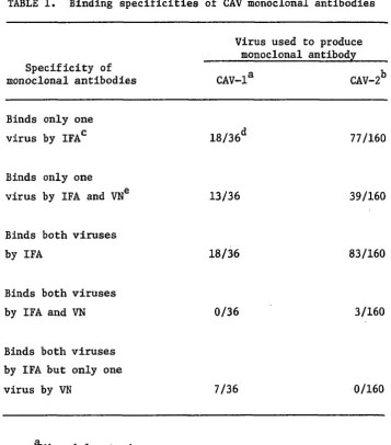
illustrated in Table 1.
There were 18 different IFA type-specific monoclonal antibodies to
CAV-1, 13 of which also neutralized the virus, and 77 type-specific
monoclonal antibodies to CAV-2, 39 of which were neutralizing. Of the
101 antibodies that bound both viruses in the IFA test, 3 of those also
neutralized both viruses. Those 3 monoclonal antibodies were from the
CAV-2 fusion. Also, among the antibodies with specificity for both
viruses by the IFA technique, there were 7 monoclonal antibodies from the
CAV-1 fusion that neutralized only CAV-1.
Characterization of the Structural Proteins of CAV-1 and CAV-2. The
results of SDS-PAGE analysis of whole virus are shown in Fig. 1. Based
on the exhaustive characterization that has been done on the human
adenoviruses (24, 26, 40, 63), tentative assignations were made for the
viral proteins: hexon (II), penton (III), fiber (IV), fiber-associated
protein (IVal), hexon-associated proteins (Ilia,VI,VIII), core proteins
(V,VII), and core-associated protein (IVa2). These are depicted more
35
CAV with dog CAV-1 and CAV-2 specific antisera. Estimated apparent
molecular weights (MW) for the major structural polypeptides were
calculated by plotting the relative rate of migration for the polypeptide
band against the log of the MW. These data are presented in Table 2, and
show good correlation with what has been reported for analogous poly
peptides from human adenoviruses (24, 26, 40, 51, 63). Although most of
the structural polypeptides were precipitated in the gel in Fig. 2, the
major antigenic proteins, i.e. hexon (II), penton (III), fiber (IV,IVal),
and hexon-associated peripentonal protein (Ilia), were not well-resolved.
In the gels shown in Fig. 3 and 4, therefore, the percent of bis
cross-linker was decreased from 2.7% to 1.3% and electrophoresis was carried
out for a longer period of time. The resulting gels gave good separation
of the higher MW components allowing for more detailed examination of
polypeptides precipitated in the RIPs.
Both homologous (Fig. 2, lane b; Fig. 3, lane b; Fig. 4, lane c) and
heterologous (Fig. 2, lane c; Fig. 3, lane c) dog origin CAV
type-specific antisera showed similar RIP bands to both CAV-1 and CAV-2. The
only dissimilarity noted was in Fig. 3 where there appears to be less
precipitation of some of the fiber and peripentonal hexon-associated
polypeptides of CAV-1 by CAV-2 dog antiserum. CAV-1 dog antiserum did
not precipitate any polypeptides (Fig. 4, lane b) from sham-infected
MDCK cells (Fig. 4, lane a).
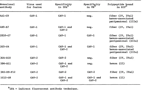
Representative CAV monoclonal antibodies that had shown different
specificities in the IFA and VN tests were chosen for use in the RIP.
35
presented In Table 3. In Fig. 3, [ S]methionine-labeled CAV-1 was used
35
as antigen and in Fig. 4, [ S]-CAV-2 was utilized. In lane d of each
gel, a PRV monoclonal antibody was used as a background control. The RIP
in lane e of each gel was carried out with type-specific monoclonals (by
IFA) that did not neutralize. In both cases, the monoclonal antibodies
bound to an antigenic determinant located on fiber polypeptides.
Additionally, the CAV-1 specific antibody detected the same antigen on
hexon-associated peripentonal protein (Ilia). In lane f of each gel,
where group-specific non-neutralizing monoclonal antibodies were used,
hexon (II) polypeptide was precipitated in CAV-2 but fiber (IV,IVal)
proteins were precipitated by CAV-1. The IV polypeptide in Fig. 3,
lane f, although only faintly visible in the autoradiogram, became more
evident upon longer exposure. Type-specific, neutralizing monoclonal
antibodies were utilized in lane g of each gel; for each virus the same
polypeptides were precipitated with both neutralizing (lanes g) and
non-neutralizing (lanes e) type-specific antibodies. In Fig. 3, lane h, a
group-specific monoclonal antibody that specifically neutralized CAV-1
was used. Again, fiber (IV, IVal) and hexon-associated peripentonal
(Ilia) polypeptides were precipitated. With CAV-2, Fig. 4, lane h, the
hexon (II) polypeptide was precipitated by a group-specific monoclonal
DISCUSSION
Since the early 1960s it has been believed that two types of canine
adenoviruses might exist. However, because quantitative
cross-neutralization studies with CAV-1 and CAV-2 could not, by definition,
separate these two viruses into distinct adenovirus species (types),
CAV-1 is still the only provisionally recognized species of CAV (33, 63).
And yet, studies of the pathology (3, 8, 19, 21, 50), morphology (32),
and antigenicity (20, 21, 49, 50) of these viruses indicate that there
are indeed significant differences between the two, similar to the types
of differences recognized in disparate species of human adenoviruses (48,
56, 57, 58).
It has been shown that monoclonal antibodies can be useful tools in
defining antigenic determinants (17, 30, 37, 46, 59) that can lead to
typing viruses (38), defining heterogeneity between serotypes and between
viruses which appear to be related according to neutralization data (5,
16, 27, 28, 31), and even differentiating between wild and vaccine
strains (47, 62). In order to validate the possible immunologic
distinctiveness of the CAVs, therefore, I prepared a bank of monoclonal
antibodies against the virulent Mirandola and Manhattan strains of CAV-1
and CAV-2 respectively. These two strains were chosen because they are
the standard CAV strains used at the NVSL to challenge the efficacy of
CAV vaccines licensed by the United States Department of Agriculture. I
through type-specific monoclonal antibodies, I also could differentiate
strains and, ultimately, separate vaccine and wild strains.
Eighteen of 36 monoclonal antibodies produced against CAV-1 and 77
out of 160 monoclonal antibodies produced against CAV-2 were
type-specific. This indicates the possibility that these two viruses are
different. In order for the two viruses to be recognized as different
species, however, several antibodies recognizing a significant number of
different epitopes must be derived. Epitope mapping of the CAVs using
neutralizing monoclonal antibodies remains to be done. However, fluores
cence patterns shown by monoclonal antibodies in the IFA test on MDCK
cells at the same stage of CAV infection, i.e. 24 hours post-inoculation,
displayed a wide diversity indicating that a variety of proteins were
being specifically recognized by antibodies from different hybridomas.
Although this does not preclude the possibility of shared determinants
between proteins, it does increase the likelihood that the antibodies are
binding to different epitopes.
The CAV-neutralizing monoclonal antibodies produced in this study
are significant for several reasons. Although monoclonal antibodies have
been produced against human adenovirus proteins (2, 13), none has been
reported to have neutralizing capabilities. These CAV antibodies then
provide a tool for studies on adenovirus neutralization, including
epitope mapping as well as mechanisms of neutralization. Thirteen CAV-1
and 39 CAV-2 monoclonal antibodies were type-specific, reinforcing the
concept of immunologic distinctiveness between these two viruses.
and 7 out of 18 group-specific CAV-1 antibodies were also involved in
neutralization, one might conclude that neutralization may be closely
linked to type specificity, but that more than one antigenic site may be
involved.
Prior to this study, protein analysis of the structural polypeptides
of CAV by SDS-PAGE had not been reported. In the characterizations
reported here, the Mirandola strain of CAV-1 and the Manhattan strain of
CAV-2 were utilized so that immune precipitations could also be performed
both with dog CAV strain-specific antisera and the CAV monoclonal anti
bodies. Structural polypeptides that correlated well with those reported
for human adenoviruses (26, 40, 63) were noted in both SDS-PAGE of whole
35
virus cell-culture extracts and in RIPs of [ S]methionine-labeled CAV
with dog strain-specific antisera (Table 2). The only notable difference
between the two viruses was the lack of detection of the hexon-associated
peripentonal (Ilia) polypeptide in CAV-2 by CAV-2 dog antiserum (Fig. 4,
lane c), whereas this same antiserum did precipitate the Ilia polypeptide
in CAV-1 (Fig. 3, lane c). A possible explanation for this is discussed
later.
Much has been written about the immunogenic properties of adeno
viruses, and models for type and group-specific binding and/or neutral
ization have been proposed (26, 36, 40, 51). Since a variety of binding
specificities were noted for the monoclonal antibodies in the IFA and VN
tests in this study (Table 1), a representative monoclonal from each
specificity group was selected and tested in RIPs to identify which poly
both neutralizing and non-neutralizing monoclonal antibodies bound to the
fiber polypeptides IV and IVal (Fig. 3 and 4, lanes e and g). Addition
ally, however, the CAV-1 monoclonal antibodies precipitated what appears
to be the hexon-associated peripentonal Ilia polypeptide. In referring
back to the RIPs with dog antisera, it was noted that the Ilia
polypeptide was not precipitated from CAV-2 by CAV-2 dog antiserum. From
this, one can propose that there are shared antigens on fiber and Ilia
polypeptide in CAV-1 and at least fiber but possibly also Ilia protein in
CAV-2 that are distinct and important in type specificity and
type-specific neutralization. The presence of some precipitation of Ilia
polypeptide from CAV-1 by CAV-2 dog serum (Fig. 3, lane c) supports the
idea of the shared antigen. Possibly, since the fiber of CAV-2 is longer
than that of CAV-1, the hexon-associated peripentonal protein of CAV-2 is
less accessible to the immune system and to antibodies. An RIP with
CAV-2 antibody and CAV-2 antigen, therefore, would not precipitate Ilia
polypeptide whereas an RIP with CAV-2 antibody and CAV-1 antigen, with
shorter fiber and less steric hindrance to antibody binding, would show
immunoprecipitation of Ilia polypeptide. If Ilia has a shared antigenic
determinant with another protein such as fiber, the CAV-2 antiserum would
contain antibody that would bind to Ilia, although probably in a lesser
quantity than occurs with CAV-1 antiserum (Fig. 3, lane c).
Group-specific monoclonal antibodies from the CAV-2 fusion, both
neutralizing and non-neutralizing, immunoprecipitated hexon (II) of CAV-2
(Fig. 4, lanes f and h). This supports prior evidence for the group
of many animal species (13, 36, 57). The interesting point here is that
type-specific neutralization is associated with fiber protein and
group-specific neutralization is tied to hexon protein.
With monoclonal antibodies from the CAV-1 fusion, a different
pattern emerges. The group-specific antibodies immunoprecipitated the IV
and IVal polypeptides of fiber instead of hexon (Fig. 3, lanes f and h).
Moreover, the neutralizing monoclonal antibody also immunoprecipitated
the Ilia polypeptide of the hexon-associated peripentonal protein. The
unique feature of this antibody is that it is group-specific in binding
both viruses in the IFA test, but type-specific in neutralizing only
CAV-1 in the VN test. The RIP results shown in Fig. 3 suggest that the
binding of the Ilia protein is important in neutralization of CAV-1.
Further testing with additional monoclonal antibodies needs to be done to
substantiate and perhaps further elucidate these ideas.
It is interesting to note that the group-specific neutralizing
antibodies from the CAV-2 fusion all neutralize both CAV-1 and CAV-2, but
that the group-specific neutralizing antibodies from the CAV-1 fusion
only neutralize CAV-1. Cross-protective immunity studies with the CAVs
have shown that vaccination with CAV-2 in dogs stimulates a serologic
response that is completely protective against challenge with either
virulent CAV-1 or virulent CAV-2 (7, 14, 15, 18). Dog vaccination with
CAV-1, on the other hand, produces a serologic response that is
completely protective against challenge with virulent CAV-1 but only
partially protects against challenge with CAV-2, with challenge virus