0022-538X/05/$08.00⫹0 doi:10.1128/JVI.79.21.13442–13453.2005
Copyright © 2005, American Society for Microbiology. All Rights Reserved.
Identification, Subviral Localization, and Functional Characterization
of the Pseudorabies Virus UL17 Protein
Barbara G. Klupp,
1Harald Granzow,
2Axel Karger,
1and Thomas C. Mettenleiter
1*
Institutes of Molecular Biology1and Infectology,2Friedrich-Loeffler-Institut, D-17493 Greifswald-Insel Riems, Germany
Received 24 January 2005/Accepted 8 August 2005
Homologs of the UL17 gene of the alphaherpesvirus herpes simplex virus 1 (HSV-1) are conserved in all three subfamilies of herpesviruses. However, only the HSV-1 protein has so far been characterized in any detail. To analyze UL17 of pseudorabies virus (PrV) the complete 597-amino-acid protein was expressed in
Escherichia coliand used for rabbit immunization. The antiserum recognized a 64-kDa protein in PrV-infected cell lysates and purified virions, identifying PrV UL17 as a structural virion component. In indirect immuno-fluorescence analyses of PrV-infected cells the protein was predominantly found in the nucleus. In electron microscopic studies after immunogold labeling of negatively stained purified virion preparations, UL17-specific label was detected on single, mostly damaged capsids, whereas complete virions and the majority of capsids were free of label. In ultrathin sections of infected cells, label was primarily found dispersed around scaffold-containing B-capsids, whereas on DNA-filled C-capsids it was located in the center. Empty intranu-clear A-capsids were free of label, as were extracellular capsid-less L-particles. Functional characterization of PrV-⌬UL17F, a deletion mutant lacking codons 23 to 444, demonstrated that cleavage of viral DNA into unit-length genomes was inhibited in the absence of UL17. In electron microscopic analyses of PrV-⌬ UL17F-infected RK13 cells, DNA-containing capsids were not detected, while numerous capsidless L-particles were observed. In summary, our data indicate that the PrV UL17 protein is an internal nucleocapsid protein necessary for DNA cleavage and packaging but suggest that the protein is not a prominent part of the tegument.
Herpesviruses express a multitude of viral proteins with strikingly different functions. Most of the proteins of the pro-totypic alphaherpesvirus herpes simplex virus 1 (HSV-1) are conserved in other alphaherpesviruses and ca. 50% also in beta- and gammaherpesviruses, indicating not only structural but also functional conservation (2, 9, 34). However, data on their function are primarily derived from studies of human herpesviruses, in particular, HSV-1, and only relatively few experiments have been performed to address functional rela-tionships and to ascertain the function of specific proteins in nonhuman herpesvirus systems. This relative lack of experi-mentation is mainly accounted for by the assumption that conservation of structure as deduced by amino acid homology also translates into conservation of function. However, this assumption may not necessarily be valid. Thus, there is a need for comparative functional analysis of herpesviral gene prod-ucts in different assay systems to elucidate similarities and differences in protein function.
The goal of our studies is to gain a complete picture and understand the role of each of the viral proteins in the repli-cation cycle of the porcine alphaherpesvirus pseudorabies virus (PrV) (37) and at the same time to compare PrV phenotypes with results from other herpesvirus-host systems to unravel similarities and differences in herpesvirus protein function. In recent years we concentrated on the role of viral structural proteins in tegumentation, primary and secondary
envelop-ment, and egress (reviewed in references 38 and 39), focusing on components of the viral envelope and tegument.
Virion components which are conserved throughout the three herpesvirus subfamilies include the products of genes homologous to HSV-1 UL17. Respective proteins in HSV-1 (51), human cytomegalovirus (HCMV) (59), and Epstein-Barr virus (EBV) (22) have been shown to be present in purified extracellular virions, and the HSV-1 homolog has been pro-posed to be located exclusively in the tegument (51) or asso-ciated with the capsid and tegument fractions (15, 57). The subviral localization of the HCMV and EBV homologous pro-teins has so far not been determined. Since the UL17 protein could thus play a role in tegumentation and envelopment ei-ther in the nucleus or in the cytoplasm, we set out to identify and characterize the PrV homolog in our standardized assay system.
The UL17 gene product of HSV-1 is required for cleavage and encapsidation of genomic viral DNA (51), a process which resembles the DNA maturation process of double-stranded DNA (dsDNA) bacteriophages such as P22, T7, and Lambda (44). In herpesviruses and bacteriophages, DNA which is cleaved from a concatemeric precursor into genome length is packaged into preformed icosahedral capsids by utilizing a two-subunit terminase which in HSV-1 consists of the UL15 and UL28 proteins (52); a dodecameric ring of connector or portal protein present at one vertex of the capsid which in HSV-1 consists of the UL6 protein (42, 44) and through which the viral DNA is translocated into the capsid; and a portal-capping protein which in HSV-1 is presumably formed by the UL25 gene product (36, 53) and seals the capsid after DNA is packaged. In the tailed bacteriophages the DNA-containing capsid serves as substrate for the addition of the preformed tail
* Corresponding author. Mailing address: Friedrich-Loeffler-Insti-tut, Federal Research Institute for Animal Health, Institute of Molec-ular Biology, Boddenblick 5A, D-17493 Greifswald—Insel Riems, Germany. Phone: 49 38351 7250. Fax: 49 38351 7151. E-mail: [email protected].
13442
on November 8, 2019 by guest
http://jvi.asm.org/
HCMV, and rhesus monkey rhadinovirus would occupy ap-proximately 90 to 92% of the total available space, suggesting that genomes are packed as naked DNA without associated histone-like basic proteins (61).
Besides a more spherical procapsid three types of icosahe-dral capsids can be differentiated in herpesvirus-infected cells in electron microscopic and biochemical analyses. They have been designated A-, B-, and C-capsids (14, 49). B-capsids, which contain an internal ringlike structure formed by the scaffold protein, have been considered precursors of A- and C-capsids, although they may actually represent dead-end structures (57). C-capsids lack the internal scaffold but contain viral genomic DNA and can mature into infectious virions. In contrast, A-capsids contain neither the internal scaffold nor DNA and are probably the result of a failed DNA packaging event (21). By using a panel of recombinant baculoviruses expressing the UL18, UL19, UL26.5, UL26, and UL38 genes, assembly of HSV-1 B-capsids has been successfully reconsti-tuted in vitro (43, 54, 58), showing that these proteins are sufficient for capsid formation.
Cleavage of viral concatemeric DNA into unit-length ge-nomes and stable packaging into the preformed capsid re-quires the products of the HSV-1 UL6, UL15, UL17, UL25, UL28, UL32, and UL33 genes (1, 3, 4, 21, 33, 46, 50, 55, 56) which are conserved in all subfamilies ofHerpesviridae(2, 9, 34). Mutants deficient in these genes show characteristic phe-notypes, with an intranuclear accumulation of concatemeric viral DNA and the concomitant absence of C-capsids (53). The only exception is a mutant defective in UL25 in which cleavage occurs and B- as well as A-capsids are found, indicating that UL25 plays a role in a later stage of the packaging process, which is in line with its proposed function as a portal-capping protein (36, 53). Of the seven proteins mentioned above, only UL6, UL17, and UL25 have been found in significant amounts in the mature HSV-1 virion.
The HSV-1 UL33 protein forms a complex with the UL15 and UL28 gene products, and it was therefore suggested that it constitutes a third component of the terminase (5, 6). HSV-1 UL32 is thought to be involved in efficient localization of capsids to replication compartments, directing preassembled capsids to the site of DNA packaging (33). A similar function has been suggested for the HSV-1 UL17 protein (55). Since this protein could not be detected as an integral part of capsids but was found in the tegument fraction, it was proposed that it constitutes a new class of DNA cleavage-encapsidation pro-teins (51). However, by immunoblot analysis of purified viral
into unit-length genomes, resulting in a block in capsid matu-ration (40). On the basis of the demonstmatu-ration that the HSV-1 UL15 protein can transport PrV UL28 from the cytoplasm into the nucleus, suggesting that both proteins form a complex, a terminase function for PrV UL15/UL28 can be assumed (31). The PrV UL25 protein is present in purified virions and was found associated with all forms of capsids (23), but no clear function has been assigned yet. Neither the gene products nor the function of PrV UL6, UL17, UL32, or UL33 has been investigated up to now.
For identification and functional characterization of the PrV UL17 gene product, we generated a monospecific antiserum and constructed and characterized a virus mutant lacking most of the UL17 open reading frame after mutagenesis of a bac-terial artificial chromosome (BAC) clone of PrV (30). We show that, like its HSV-1 homolog, PrV UL17 is essential for viral replication and necessary for cleavage and packaging of viral DNA. However, by immunoelectron microscopy the PrV UL17 protein was not detected in A-capsids but was found associated with the outside of B-capsids and within C-capsids. Thus, it appears to be incorporated into capsids concomitant with genomic DNA. Moreover, the UL17 protein was not found in the tegument of mature virions or in L-particles, indicating that the PrV UL17 gene product is an internal cap-sid protein.
MATERIALS AND METHODS
Viruses and cells.All PrV mutants analyzed were derived from PrV strain Kaplan (Ka) (24). Viruses were grown on rabbit kidney (RK13) or porcine kidney (PSEK) cells in minimum essential medium supplemented with 10% or
5% fetal calf serum, respectively. Generation of PrV-⌬UL28 (previously
desig-nated 332-31) has been described elsewhere (40).
Construction of PrV-⌬UL17F and complementing cell line. The complete UL17 open reading frame was excised from cloned BamHI fragment 3 by cleavage with BamHI and NcoI (Fig. 1) and inserted into BamHI- and EcoRI-digested pUC19 after Klenow treatment of the EcoRI and NcoI sites to obtain plasmid pUC-UL17. To delete the major part of the UL17 coding sequence, pUC-UL17 was digested with BmgBI, thereby removing codons 23 to 444 of the 597-codon open reading frame (Fig. 1). The deleted sequence was replaced with a Klenow-treated 1,258-bp BstBI fragment of pKD13 (11), which contains the
kanamycin resistance gene flanked byflprecognition target sites. The complete
insert of the generated plasmid pUC-⌬UL17kanF was amplified by PCR using
vector-specific primers M13 (-47) and M13 reverse (-48) (New England Biolabs,
Frankfurt am Main, Germany) andPfxDNA polymerase (Invitrogen, Karlsruhe,
Germany). The PCR product was used forRedrecombinase-mediated
mutagen-esis of the PrV BAC clone pPrV-⌬gB as described previously (30). After
isola-tion of kanamycin-resistant clones, the kanamycin cassette was removed usingflp
recombinase provided by plasmid pCP20 (10). The gB gene was finally restored
by cotransfection of BAC pPrV-⌬UL17F and plasmid pUC-B1BclI (16, 30).
on November 8, 2019 by guest
Several plaque isolates of the transfection progeny were characterized, and one
was randomly chosen and designated PrV-⌬UL17F. Correct deletion of UL17
sequences was verified by Southern blot analysis (data not shown).
For generation of a stably UL17-expressing cell line the complete UL17 open reading frame was cloned as a BamHI/EcoRI fragment from plasmid pUC-UL17 (see above) into appropriately cleaved eukaryotic pcDNA3 expression vector (Invitrogen). After transfection into RK13 cells by use of Superfect transfection reagent (QIAGEN, Hilden, Germany) G418-resistant cell clones were picked and tested by immunoblot and indirect immunofluorescence analysis for expres-sion of UL17 by use of monospecific anti-UL17 rabbit serum.
Preparation of UL17 antiserum.To generate a monospecific antiserum, the complete UL17 open reading frame, including 54 normally nontranslated
up-stream nucleotides, was cloned in frame with glutathioneS-transferase (GST)
into the prokaryotic expression vector pGEX-4T-1 (Amersham Biosciences, Freiburg, Germany). To this end, pUC-UL17 was digested with BamHI and EcoRI and cloned into appropriately cleaved pGEX-4T-1 vector. Correct inser-tion was verified by sequencing (data not shown). The ca. 90-kDa GST-UL17 fusion protein was eluted from a sodium dodecyl sulfate (SDS)–8% polyacryl-amide gel (32) and used for immunization of a rabbit as described recently (27, 29). Serum obtained after the fifth immunization was used throughout this study.
Virus purification and immunoblotting.Virus purification and preparation of infected cell lysates were performed as described previously (27). Parallel blots were incubated with monospecific antisera against the UL17 (1:50,000) (this study), UL16 (1:50,000) (28), UL37 (1:100,000) (27), and UL49 (1:100,000) (8) gene products and envelope glycoprotein H (gH) (25).
Indirect immunofluorescence and confocal laser scan microscopy.RK13 cells
were infected with PrV-Ka or PrV-⌬UL17F at a multiplicity of infection (MOI)
of 10 or were mock infected. At 6 h after infection, cells were fixed with cold acetone for 20 min at 20°C. Fixed cells were incubated with anti-UL17 serum (dilution, 1:300) for 1 h at room temperature followed by Alexa-488-conjugated anti-rabbit secondary antibody (Molecular Probes, Leiden, The Netherlands). Fluorescence was preserved in a 9:1 mixture of glycerol and phosphate-buffered saline containing 2.5 mg 1,4-diazabicyclooctane per ml. The slides were analyzed
with a confocal laser scan microscope (LSM510; Zeiss, Go¨ttingen, Germany).
Electron microscopy.Immunolabeling of purified virion preparations was per-formed as described previously (60). For analysis of ultrathin sections, RK13 and
RK13-UL17 cells were infected at an MOI of 1 with PrV-Ka or PrV-⌬UL17F
and incubated for 14 h at 37°C. Fixation for epoxy embedding in Glycid ether 100
(Serva, Heidelberg, Germany) was performed with 2.5% buffered glutardialde-hyde and 1% osmium tetroxide; for acrylic embedding in Lowicryl K4M (Lowi, Waldkraiburg, Germany), only 0.5% buffered glutardialdehyde (17) was used. Intermediate embedding in low-melting-point agarose (Biozym, Oldendorf, Ger-many), dehydration, and resin embedding were performed as described earlier (18–20). For postembedding labeling Lowicryl ultrathin sections were blocked at the surface with 1% cold-water-fish gelatin–0.02 M glycine–1% bovine serum albumin (Sigma, Deisenhofen, Germany) in phosphate-buffered saline for 2 h and then incubated with monospecific antisera for 2 h at room temperature. Labeling with 5- or 10-nm goat anti-rabbit colloidal gold particles (GAR 5 or GAR 10; British Biocell International, Cambridge, United Kingdom) was per-formed for 1 h at room temperature. Particles with more than three gold dots were considered positive. Ultrathin sections of epoxy-embedded cells and la-beled Lowicryl sections counterstained with uranyl acetate and lead salts were examined with a transmission electron microscope (EM 400 T or Tecnai 12; Philips, Eindhoven, The Netherlands).
One-step growth analysis and plaque assays.One-step growth analysis was performed as described previously (27). RK13 or complementing RK13-UL17
cells were infected with PrV-Ka or PrV-⌬UL17F at an MOI of 10, and cells and
supernatant were harvested 0, 4, 8, 12, 24, and 36 h postinfection (p.i.). Titers of progeny virus were determined on RK13-UL17 cells. For plaque assays RK13 or RK13-UL17 cells in six-well culture dishes were infected with approximately 100
PFU of PrV-Ka and PrV-⌬UL17F (for RK13-UL17) or 1,000 PFU of
PrV-⌬UL17F (for RK13). At 2 days postinfection, cells were fixed with ethanol and
stained with a monoclonal antibody against gC (B16-c8) (45). Fluorescent plaques were documented with a digital camera (C3040 Zoom; Olympus, Ham-burg, Germany).
Preparation of virus DNA and Southern blotting.RK13 or RK13-UL17 cells
were infected with PrV-Ka, PrV-⌬UL17F, or PrV-⌬UL28 (40) at an MOI of 1
and incubated until cells showed a clear cytopathic effect. Infected cells were collected by low-speed centrifugation, and DNA was extracted with phenol after lysis and pronase digestion and precipitated with ethanol. DNA was cleaved with BamHI and transferred onto nylon membranes by use of standard techniques. Filters were probed with radioactively labeled genome end-specific BamHI
frag-ments 13 and 14⬘(Fig. 1). Bound radioactivity was recorded using a
[image:3.585.113.471.68.286.2]phosphor-imager (FLA-3000; Raytest, Straubenhardt, Germany).
FIG. 1. Construction of PrV-⌬UL17F. (A) Map of the PrV genome with the unique long (UL), unique short (US), and inverted repeat (IR, TR) sequences. BamHI restriction sites are indicated, and fragments are numbered according to size. (B) Enlargement of the PrV UL15-UL17-UL16 gene region. The UL17 and UL16 genes are located within the intron of the spliced UL15 gene (dotted line), which is bracketed by exons I and II of the UL15 gene. UL17 and UL16 are transcribed into 3⬘coterminal mRNAs, which share a common polyadenylation signal (arrow pointing up at right), whereas the polyadenylation signal for the UL15 mRNA is located downstream of UL15 exon II (arrow pointing up at left). Relevant restriction sites are indicated. (C) The BamHI-NcoI fragment of BamHI fragment 3 was used for cloning into pro- and eukaryotic expression vectors and for generation of the UL17 deletion mutant. In mutant PrV-⌬UL17F, sequences between the BmgBI sites were deleted and replaced by one copy of aflprecognition target site.
on November 8, 2019 by guest
http://jvi.asm.org/
RESULTS
Identification of the PrV UL17 gene product.The sequence for the PrV UL17 gene has been determined only recently (26). As with other members of the herpesvirus family, the PrV UL17 gene is located within the intron of the spliced UL15 gene immediately upstream of the UL16 open reading frame (Fig. 1). The PrV UL17 gene encodes a protein of 597 amino acids with a predicted molecular mass of 64 kDa (26). The amino acid sequence contains a predicted nuclear localization signal (“RRRP” pat4) (http://psort.ims.u-tokyo.ac.jp) (41) lo-cated at amino acids 304 to 307. To identify the corresponding protein product the complete UL17 open reading frame, in-cluding 18 upstream normally nontranslated codons, was ex-pressed as a GST fusion protein inEscherichia coliand used for immunization of a rabbit. The generated antiserum specif-ically detected an approximately 64-kDa protein in lysates of PrV-Ka-infected cells (PrV-Ka L; Fig. 2) as well as in purified virion preparations (PrV-Ka V; Fig. 2), which identifies the PrV UL17 protein as a structural virion component. The size of the detected protein correlates well with the predicted mo-lecular mass. No specific signal was found in lysates of
mock-infected RK13 cells (mock; Fig. 2) or cells mock-infected with the UL17 deletion mutant (PrV-⌬UL17F L; Fig. 2). As a control, parallel blots were incubated with sera against tegument pro-teins UL37, UL16, and UL49 and envelope glycoprotein gH.
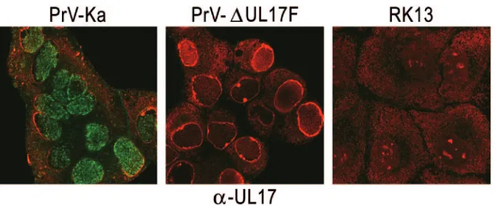
In indirect immunofluorescence analysis of PrV-infected cells, UL17-specific fluorescence was found predominantly in small dots in the nucleus. However, it was also detectable in punctate structures in the cytoplasm, which, however, were fewer in number and less intensely stained (Fig. 3). Cells in-fected with the UL17 deletion mutant or mock-inin-fected cells showed no specific fluorescence (Fig. 3).
Subviral localization of PrV UL17.HSV-1 UL17 has origi-nally been described as a tegument protein (51) but has re-cently been proposed to be localized in the capsid as well as tegument fractions (57). To directly locate the PrV UL17 pro-tein in the virus particle, immunoelectron microscopy was per-formed on purified virions. To this end, sucrose gradient-pu-rified PrV-Ka virions (Fig. 4A) were incubated with the anti-UL17 serum followed by negative staining (Fig. 4B to E). Labeling was scored positive when more than three gold grains were associated with the respective virus particle. Intact
[image:4.585.132.444.68.220.2]viri-FIG. 2. Identification of the PrV UL17 protein and characterization of PrV-⌬UL17F. Purified PrV-Ka virions (PrV-Ka V) or lysates of cells infected with PrV-Ka (PrV-Ka L) or PrV-⌬UL17F (PrV-⌬UL17F L) or mock-infected RK13 cells (mock) were separated on sodium dodecyl sulfate–10% polyacrylamide gels and incubated with monospecific antisera against the UL17, gH, UL37, UL16, and UL49 proteins. The locations of molecular mass markers are indicated on the left.
FIG. 3. Intracellular localization of PrV UL17. RK13 cells were infected with PrV-Ka or PrV-⌬UL17F at an MOI of 10 or mock infected and were fixed with ice-cold acetone 6 h later. Immunofluorescence analysis was performed by laser scanning microscopy using the UL17-specific antiserum and Alexa 488-conjugated secondary antibodies (green). Chromatin was counterstained with propidium iodide (red).
on November 8, 2019 by guest
[image:4.585.114.479.545.695.2]ons, virions with disrupted envelope but apparently undam-aged capsid, and most of the isolated capsids were not labeled (Fig. 4B to E; 24 clearly identified intact capsids were all not labeled). In contrast, heavy labeling was detected when the
[image:5.585.58.529.68.617.2]capsids were visibly damaged (Fig. 4B inset, 4C inset, and 4D and 4D inset; only 8 of 47 visibly damaged capsids were found unlabeled). UL17 label could sometimes also be found ema-nating from the capsid (Fig. 4B inset; see also Fig. 5B and C).
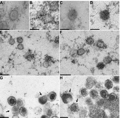
FIG. 4. Immunoelectron microscopy of purified PrV-Ka virions. Purified PrV-Ka virions (A) were incubated with monospecific antisera against the UL17 (B to E), UL19 (F), UL36 (G), UL48 (H), and UL11 (I) proteins or gB (J). Reactivity of the antisera was visualized after incubation with 10 nm-diameter-gold-tagged secondary anti-rabbit antibodies. Bars represent 100 nm.
on November 8, 2019 by guest
http://jvi.asm.org/
These data suggest that the UL17 protein is located within the capsid and is extruded when the capsid loses its integrity. This indicates that the bulk of the antigen may be masked by the capsid, which argues against a location of the major portion of virion-associated UL17 protein in the tegument that surrounds the capsid. However, specific label was also detected on single, seemingly intact capsids (Fig. 4B and E). For a control, the same virus preparation was labeled with antisera against the major capsid protein UL19 (Fig. 4F), the capsid-proximal teg-ument component UL36 (Fig. 4G), the major tegteg-ument tein UL48 (Fig. 4H), the membrane-associated tegument
pro-tein UL11 (Fig. 4I), and envelope glycopropro-tein B (Fig. 4J). Specific labeling for the tegument proteins UL11, UL36, and UL48 was found only on virus particles with partially disrupted envelope. The serum specific for the major capsid protein labeled all isolated capsids but not intact virions (Fig. 4F), whereas the serum against viral envelope glycoprotein B heavily labeled enveloped particles (Fig. 4J).
[image:6.585.53.534.67.543.2]The tight association of the PrV UL17 protein with DNA packaging is also evident in Fig. 5. For higher marker density only 5-nm gold particles were used for panels A and C, and counterstaining was omitted for better visualization of the
FIG. 5. Immunoelectron microscopy of PrV-Ka-infected cells. RK13 cells were infected with PrV-Ka as described above, and ultrathin sections were labeled with anti-UL17 serum (A to G) or anti-UL48 serum (H). Anti-rabbit sera conjugated with gold (10 nm [B, D, and E to H] or 5 nm [A and C]) were used. For better visualization of the smaller gold particles, counterstaining was omitted in the assays using 5-nm gold. Arrows in panels G and H point to L-particles. Bars represent 100 nm in panels A to D and 150 nm in panels E to H.
on November 8, 2019 by guest
marker. In ultrathin sections of PrV-Ka-infected RK13 cells UL17-specific labeling was detected in close proximity to scaf-fold-containing intranuclear B-capsids (Fig. 5A to C and 5F; 51 of 68 observed particles were labeled) and on DNA-containing C-capsids (Fig. 5D to F; 58 of 76 observed particles were labeled) and in mature virions (Fig. 5G). Interestingly, the label on B-capsids was found primarily around the capsid, while on C-capsids the gold particles were always found con-centrated in the center of the particle (Fig. 5D and F). None of the eleven identified A-capsids were labeled (Fig. 5E), imply-ing that UL17 capsid association is linked to the presence of DNA. On scaffold-containing intranuclear B-capsids UL17 la-beling was predominantly found on the outside of the capsid (Fig. 5A), sometimes in rows emanating from the capsid (Fig. 5B; see also Fig. 4B inset). In rare cases, the label appeared to extend toward the inside of a B-capsid (Fig. 5C). We speculate that this could be a capsid during DNA packaging. In the C-capsid shown in Fig. 5D, which had already packaged viral DNA (as demonstrated by its electron-dense interior), gold particles were concentrated in the center of the particle. Only complete virions but not L-particles, which were identified by their lack of a capsid structure, were labeled by the anti-UL17 serum (Fig. 5G), whereas labeling specific for the UL48 tegu-ment protein was detectable in complete virions and L-parti-cles (Fig. 5H). These data indicate that UL17 is packaged together with the newly replicated DNA into the capsid and suggest that the UL17 protein is a major internal capsid pro-tein but presumably not a prominent component of the tegu-ment.
Isolation and characterization of PrV-⌬UL17F. Since this particular ultrastructural localization of the PrV UL17 protein could correlate with a role in cleavage and encapsidation of viral DNA, as has been shown for the homologous protein of HSV-1 (51, 57), we functionally characterized PrV UL17 by isolation of a deletion mutant lacking amino acids 23 to 444 of the 597-amino-acid protein. The UL17 gene is located in the intron of the UL15 gene; to avoid any negative effects on the transcription of the adjacent UL16 gene (Fig. 1), site-specific mutagenesis of a recently described BAC clone of PrV (30) was performed. Infectious progeny could only be isolated on a cell line stably expressing the UL17 protein, indicating that the protein is required for viral replication. In immunoblot analysis (Fig. 2) and immunofluorescence (Fig. 3), absence of UL17 from PrV-⌬UL17F-infected RK13 cells could be verified. Im-munoblot analysis with a UL16-specific antiserum showed that expression of UL16 was not affected by the UL17 deletion (Fig. 2).
To investigate the defect in replication, one-step growth analyses (Fig. 6A) and plaque assays (Fig. 6B) were performed. After infection of RK13 cells with PrV-⌬UL17F, only very few infectious particles (not exceeding 103infectious units per ml)
were detectable, while on RK13-UL17 cells the deletion mu-tant replicated with kinetics similar to PrV-Ka kinetics, indi-cating that the defect is indeed solely due to deletion of UL17. Only single infected RK13 cells or very small syncytia were detectable at 2 days postinfection, indicating that cell-cell spread is significantly inhibited in the absence of PrV UL17 but that plaque formation could be fully rescued on RK13-UL17 cells (Fig. 6B).
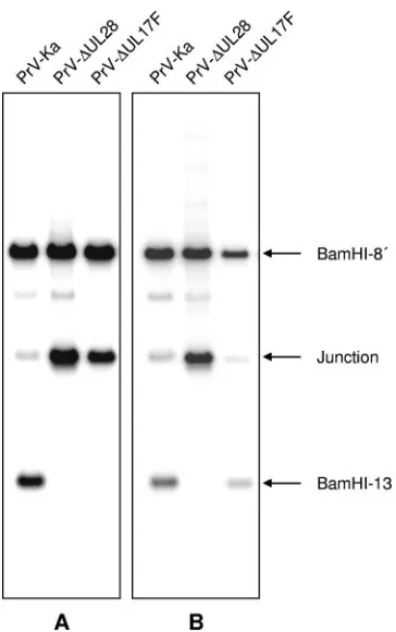
PrV-⌬UL17F does not cleave or package viral DNA.Since the HSV-1 UL17 protein is necessary for cleavage and encap-sidation of viral DNA (51), we investigated whether replicative concatemeric DNA is cleaved in PrV-⌬UL17F-infected cells. Whole infected-cell DNA was isolated, digested with BamHI, separated on a 0.8% agarose gel, blotted, and hybridized with the viral terminal genomic fragments BamHI-13 (Fig. 7) and BamHI-14⬘(data not shown). For control, DNAs of cells in-fected with the parental PrV-Ka or PrV-⌬UL28, which has been shown to be defective for DNA cleavage and encapsida-tion (40), were included. As is evident in Fig. 7A, the terminal BamHI fragment 13 was not detectable in PrV-⌬UL17F DNA, indicating that cleavage to unit-length genomes did not occur. In parallel, the amount of the junction fragment containing the fused genome end fragments 13 and 14⬘strongly increased. A similar finding was obtained when DNA of cells infected with PrV-⌬UL28 was analyzed. In contrast, a BamHI-13-specific signal was detectable in PrV-⌬UL17F DNA isolated from the UL17-expressing cells (Fig. 7B), while the defect in
PrV-⌬UL28, as expected, was not complemented on this cell line. In DNA isolated from PrV-Ka-infected cells, the terminal frag-ments could easily be identified.
In electron microscopic analyses of PrV-⌬UL17F-infected RK13 cells, extracellular virions or nucleocapsids in the cyto-plasm were not detectable (Fig. 8A). In the nucleus, only B-capsids, which were sometimes found in paracrystalline structures, were observed (Fig. 8B). The absence of C-capsids indicates that packaging is blocked before DNA is inserted into the capsid and before the scaffold protein is expelled. In the cytoplasm (Fig. 8A and C) and on the cell surface (Fig. 8A and D), numerous electron-dense L-particles were visible, indicat-ing that envelopment of cytoplasmic tegument occurs in the absence of UL17. Unimpaired virion morphogenesis and egress of mature virions was found on cells stably expressing UL17 (Fig. 8E), with C-capsids in the nucleus (Fig. 8F) and virus particles in transport vesicles in the cytoplasm (Fig. 8E) as well as complete virions on the cell surface (Fig. 8G). This correlates with the results obtained with the homologous HSV-1 protein and demonstrates that the two proteins execute similar functions in replication of the respective viruses.
DISCUSSION
In our endeavor to analyze individually the products of the 70 structural genes of the alphaherpesvirus PrV (26) in an isogenic strain background by use of standardized assay sys-tems, we studied the role of the UL17 protein. The HSV-1 UL17 gene product has been shown to be a nuclear protein required for cleavage and encapsidation of replicative concate-meric DNA (51, 57). Moreover, it has been proposed to be localized not only in the capsid, which would correlate with its role in genome packaging, but also in the tegument (51, 57). The latter could indicate (i) that it plays a role in virion mor-phogenesis beyond DNA packaging; (ii) that part of the tegu-ment is assembled in the nucleus and retained during passage of nucleocapsids through the nuclear membrane; and (iii) that the UL17 protein may be a candidate for interaction with the primary tegument protein UL31 and/or primary envelope pro-tein UL34 to direct intranuclear capsids to the primary envel-opment site (57).
on November 8, 2019 by guest
http://jvi.asm.org/
The salient findings in this paper are the following: (i) like the HSV-1 homolog, the PrV UL17 protein is a nuclear pro-tein required for cleavage and encapsidation of replicated con-catemeric viral DNA, which demonstrates a conserved func-tion for this conserved protein; (ii) the PrV UL17 protein is present in capsids, apparently in parallel with viral DNA; and (iii) the PrV UL17 protein has not been detected in tegument-containing L-particles. Thus, our studies corroborate a role for
UL17 in the formation of nucleocapsids but do not support a role for UL17 in virion morphogenesis beyond this step.
[image:8.585.110.473.64.570.2]PrV UL17 is a structural component of purified virions, as has been shown for several other herpesviruses such as HSV-1 (51, 57), EBV (22), and HCMV (59). However, its subviral distribution has only been analyzed in HSV-1 (15, 51, 57). Although HSV-1 UL17 was originally designated a tegument protein (51), recent data indicated that it is a component of the
FIG. 6. In vitro growth properties of PrV-⌬UL17F. (A) RK13 or RK13-UL17 cells were infected with PrV-Ka or PrV-⌬UL17F at an MOI of 10, harvested at the indicated times after infection, and titrated on RK13-UL17 cells. Average titers (PFU/ml) and standard deviations for three independent experiments are shown. (B) RK13 and RK13-UL17 cells were infected under plaque assay conditions with PrV-Ka and PrV-⌬UL17F and fixed 2 days postinfection. Infected cells were visualized by indirect immunofluorescence with a gC-specific monoclonal antibody.
on November 8, 2019 by guest
capsid which seems to be associated with all types of capsids (procapsids and A-, B-, and C-capsids), as judged by immuno-blot analyses of purified capsid preparations (57). Moreover, when L-particle fractions isolated by ultracentrifugation were used, the HSV-1 UL17 protein was also detected in L-particles. Our study was the first that directly visualized localization of the PrV UL17 protein in infected cells by immunoelectron microscopy using a highly potent monospecific anti-UL17 an-tiserum. This approach is not prone to misinterpretation due to the unavoidable contamination of purified virus particle frac-tions by other viral components, since it allows a direct iden-tification of the labeled virion structures. However, we realize that interpretation of electron microscopic images may be sub-jective. Moreover, limits of detection may be different. In any case, our studies show a high density of UL17 label on
nega-tively stained virion preparations, primarily at capsids with apparent damage, but not on intact virions, virions with intact capsids but disrupted envelope, or isolated capsids (Fig. 4B to E). This argues against a location of a major portion of the UL 17 protein in the tegument. Moreover, in thin sections label was detected on mature DNA-containing capsids, whereas no label was present at empty A-capsids, which lack the typical scaffold and probably result from abortive packaging attempts (Fig. 5E). In B-capsids, which contain a scaffold and which are considered able to package DNA, label was either absent or found dispersed around the particle (Fig. 5F). Although a more precise localization was not possible with the immuno-labeling technique due to the distance between the antigen and the antibody-bound gold particle, the clearly diverse labeling patterns indicate that UL17 is incorporated into the capsid in parallel with viral DNA. In the absence of UL17, DNA con-catemers are not cleaved into unit-length molecules and no mature capsids are found, which shows that UL17 is necessary for this process. The absence of A-capsids, which are thought to be the result of a failed packaging event, in PrV-⌬ UL17F-infected cells indicates that in this case the packaging reaction had not been initiated.
In rare cases label was also present around seemingly intact capsids in purified virion preparations or emanating from them (Fig. 4B and E). However, it is known that during purification herpesvirus capsids may specifically lose vertices due to the disintegration of penton (12). Thus, it is conceivable that in these cases capsids indeed lost part or all of their vertices with concomitant extrusion of the content.
The PrV UL17 protein is primarily located in the nucleus, as shown by immunofluorescence with our monospecific anti-serum (Fig. 3A). This correlates with its role in intranuclear capsid maturation. Nuclear localization is presumably medi-ated by a nuclear localization signal present in the amino acid sequence. However, punctate staining was also observed in the cytoplasm of infected cells, which is consistent with the dem-onstration of PrV-UL17 as a virion structural component.
Interestingly, we were unable to detect the PrV UL17 pro-tein in L-particles (Fig. 5G), which were identified by their lack of a capsid. L-particles mature by intracytoplasmic envelop-ment of teguenvelop-ment proteins without the need for inclusion of a nucleocapsid (35). L-particle formation is particularly promi-nent in cells which are infected with herpesvirus mutants de-ficient in several steps of secondary envelopment (see, e.g., reference 13). Thus, our results deviate from those obtained with HSV-1 (57) which demonstrated the presence of the UL17 protein in purified L-particle fractions by immunoblot-ting. This discrepancy could be due to actually different loca-tions of the two proteins despite a similar function in DNA packaging, which would again highlight the necessity for com-parative analyses of homologous gene products. Alternatively, the detection limits for the immunological procedures
(immu-FIG. 7. Defect in cleavage of concatemeric viral DNA in the ab-sence of PrV UL17. RK13 (A) or RK13-UL17 (B) cells were infected with PrV-Ka, PrV-⌬UL17F, or PrV-⌬UL28 at an MOI of 1. Cells were harvested at approximately 12 h p.i., and DNA was isolated, digested with BamHI, and separated on a 0.8% agarose gel. After transfer to nylon membranes, DNA was probed with labeled genome end-specific BamHI fragment 13. BamHI-13 hybridized to fragments BamHI-8⬘ and BamHI-13, which share homologous sequences since they are both derived from the inverted repeat regions (see Fig. 1), and the junction fragment composed of BamHI-13 and BamHI-14⬘, which is derived from head-to-tail concatemeric DNA.
FIG. 8. Electron microscopy of PrV-⌬UL17F-infected cells. RK13 (A to D) and RK13-UL17 (E to G) cells were infected with PrV-⌬UL17F at an MOI of 1 and processed for electron microscopy 14 h p.i. (A) Overview of an infected RK13 cell. (B) Pseudocrystalline aggregations of B-capsids in the nucleus. (C and D) L-particles in the cytoplasm (C) and on the cell surface (D). (E) Unimpaired virus morphogenesis on UL17-expressing cells, including production of C-capsids in the nucleus (F) and mature virions on the cell surface (G). Bars represent 2m in panel A, 500 nm in panels B and D, 250 nm in panels C, F, and G, and 1m in panel E.
on November 8, 2019 by guest
http://jvi.asm.org/
[image:9.585.72.254.65.355.2]on November 8, 2019 by guest
noblotting, immunoelectron microscopy) could be different so that in our studies the amount of L-particle-associated UL17 protein may have been below the detection level. However, the difference can also be explained by cross-contamination of preparations of the different viral structures after differential centrifugation (57), whereas our studies, which unambiguously visually identified single viral particles, were not prone to this confounding effect.
The location of the PrV UL17 protein within the mature capsid correlates with its function in DNA cleavage-encapsi-dation and parallels its predominant localization in the nucleus of infected cells. It also suggests that the PrV UL17 protein, being internally enclosed in the mature capsid, which is a substrate for primary envelopment, may not play a role in primary and/or secondary envelopment of nucleocapsids. B-capsids, which exhibited some label also around the capsid, normally do not undergo primary envelopment. Thus, so far the only tegument protein which has been detected unequivo-cally in both primary enveloped and mature PrV or HSV-1 particles is the US3 protein (18, 47).
There are striking similarities between dsDNA bacterio-phages and herpesviruses as concerns genome replication, cap-sid formation, and genome packaging (3). However, whereas genomic DNA of, e.g., bacteriophage T7 is present in the capsid associated with protein (48), packaged herpesvirus DNA has been proposed not to be protein associated, primar-ily on the basis of calculations of the available space in the herpesvirus capsid (61). Our data indicate that herpesvirus genomic DNA in the nucleocapsid may be associated with the UL17 protein, which would point to another intriguing parallel to the bacteriophages.
ACKNOWLEDGMENTS
This study was supported by a grant from the Deutsche Forschungs-gemeinschaft (DFG Me 854).
We thank Petra Meyer and Diana Werner for expert technical assistance, Mandy Jo¨rn for photographic help, and Egbert Mundt, Dorothee Wiesner, and Jutta Veits for help with the preparation of the antiserum.
REFERENCES
1.Al-Kobaisi, M. F., F. J. Rixon, I. McDougall, and V. G. Preston.1991. The herpes simplex virus UL33 gene product is required for the assembly of full
capsids. Virology180:380–388.
2.Baer, R., A. T. Bankier, M. D. Biggin, P. L. Deininger, P. J. Farrell, T. J. Gibson, G. F. Hatfull, G. S. Hudson, S. C. Satchwell, C. Seguin, P. Tuffnell, and B. G. Barrell.1984. DNA sequence and expression of the B95-8
Epstein-Barr virus genome. Nature (London)310:207–211.
3.Baines, J. D., and S. K. Weller.2005. Cleavage and packaging of herpes
simplex virus 1 DNA.InC. E. Catalano (ed.), Viral genome packaging
machines: genetics, structures, and mechanism. Landes Biosciences, George-town, Tex.
4.Baines, J. D., C. Cunningham, D. Nalwanga, and A. J. Davison.1997. The UL15 gene of herpes simplex virus type 1 contains within its second exon a
novel open reading frame that is translated in frame with the UL15 gene
product. J. Virol.71:2666–2673.
5.Beard, P. M., and J. D. Baines.2004. The DNA cleavage and packaging protein encoded by the UL33 gene of herpes simplex virus 1 associates with
capsids. Virology324:475–482.
6.Beard, P. M., N. S. Taus, and J. D. Baines.2002. DNA cleavage and
packaging proteins encoded by genes UL28, UL15, and UL33 of herpes
simplex virus type 1 form a complex in infected cells. J. Virol.76:4785–4791.
7.Booy, F. P., W. W. Newcomb, B. L. Trus, J. C. Brown, T. S. Baker, and A. C. Steven.1991. Liquid-crystalline, phage-like packaging of encapsidated DNA
in herpes simplex virus. Cell64:1007–1015.
8.Brack, A. R., B. G. Klupp, H. Granzow, R. Tirabassi, L. W. Enquist, and T. C. Mettenleiter.2000. Role of the cytoplasmic tail of pseudorabies virus
glycoprotein E in virion formation. J. Virol.74:4004–4016.
9.Chee, M. S., A. T. Bankier, S. Beck, R. Bohni, C. M. Brown, R. Cerny, T. Horsnell, C. A. Hutchinson, T. Kouzarides, J. A. Martignetti, E. Preddie, S. C. Satchwell, P. Tomlinson, K. M. Weston, and B. G. Barrell.1990. Analysis of the protein coding content of the sequence of human
cytomeg-alovirus strain AD169. Curr. Top. Microbiol. Immunol.154:125–169.
10.Cherepanov, P. P., and W. Wackernagel.1995. Gene disruption in Esche-richia coli: TcR
and KmR
cassettes with the option of Flp-catalyzed excision
of the antibiotic-resistance determinant. Gene158:9–14.
11.Datsenko, K. A., and B. L. Wanner.2000. One-step inactivation of
chromo-somal genes inEscherichia coliK-12 using PCR products. Proc. Natl. Acad.
Sci. USA97:6640–6645.
12.Davison, A. J., B. L. Trus, N. Cheng, A. Steven, M. Watson, C. Cunningham, R.-M. LeDeuff, and T. Renault.2005. A novel class of herpesvirus with
bivalve hosts. J. Gen. Virol.86:41–53.
13.Fuchs, W., H. Granzow, B. G. Klupp, M. Kopp, and T. C. Mettenleiter.2002. The UL48 tegument protein of pseudorabies virus is critical for
intracyto-plasmic assembly of infectious virions. J. Virol.76:6729–6742.
14.Gibson, W., and B. Roizman.1972. Proteins specified by the herpes simplex virus. VIII. Characterization and composition of multiple capsid-forms of
subtypes 1 and 2. J. Virol.10:1044–1052.
15.Goshima, F., D. Watanabe, H. Takakuwa, K. Wada, T. Daikoku, M. Yamada, and Y. Nishiyama.2000. Herpes simplex virus UL17 proteins is associated with B capsids and colocalizes with ICP35 and VP5 in infected cells. Arch.
Virol.145:417–426.
16.Graham, F. L., and A. J. van der Eb.1973. A new technique for the assay of
infectivity of human adenovirus 5 DNA. Virology52:456–467.
17.Granzow, H., F. Weiland, A. Jo¨ns, B. G. Klupp, A. Karger, and T. C. Mettenleiter.1997. Ultrastructural analysis of the replication cycle of
pseu-dorabies virus in cell culture: a reassessment. J. Virol.71:2072–2082.
18.Granzow, H., B. G. Klupp, and T. C. Mettenleiter.2004. Pseudorabies virus US3 protein is a component of primary and of mature virions. J. Virol.
78:1314–1323.
19.Granzow, H., B. G. Klupp, and T. C. Mettenleiter.2005. Entry of
pseudo-rabies virus: an immunogold labeling study. J. Virol.79:3200–3205.
20.Granzow, H., B. G. Klupp, W. Fuchs, J. Veits, N. Osterrieder, and T. C. Mettenleiter.2001. Egress of alphaherpesviruses: comparative
ultrastruc-tural study. J. Virol.75:3675–3684.
21.Homa, F. L., and J. C. Brown.1997. Capsid assembly and DNA packaging in
herpes simplex virus. Rev. Med. Virol.7:107–122.
22.Johannsen, E., M. Luftig, M. R. Chase, S. Weicksel, E. Cahir-McFarland, D. Illanes, D. Sarracino, and E. Kieff.2004. Proteins of purified Epstein-Barr
virus. Proc. Natl. Acad. Sci. USA110:16286–16291.
23.Kaelin, K., S. Deze´le´e, M. J. Masse, F. Bras, and A. Flamand.2000. The UL25 protein of pseudorabies virus associates with capsids and localizes to
the nucleus and to microtubules. J. Virol.74:474–482.
24.Kaplan, A. S., and A. E. Vatter.1959. A comparison of herpes simplex and
pseudorabies viruses. Virology7:394–407.
25.Klupp, B. G., and T. C. Mettenleiter.1999. Glycoprotein gL-independent infectivity of pseudorabies virus is mediated by a gD-gH fusion protein.
J. Virol.73:3014–3022.
26.Klupp, B. G., C. J. Hengartner, T. C. Mettenleiter, and L. W. Enquist.2004. Complete, annotated sequence of the pseudorabies virus genome. J. Virol.
78:424–440.
27.Klupp, B. G., H. Granzow, E. Mundt, and T. C. Mettenleiter.2001. Pseu-dorabies virus UL37 gene product is involved in secondary envelopment.
J. Virol.75:8927–8936.
28.Klupp, B. G., S. Bo¨ttcher, H. Granzow, M. Kopp, and T. C. Mettenleiter.
2005. Complex formation between the UL16 and UL21 tegument proteins of
pseudorabies virus. J. Virol.79:1510–1522.
29.Klupp, B. G., W. Fuchs, H. Granzow, R. Nixdorf, and T. C. Mettenleiter.
2002. The pseudorabies virus UL36 tegument protein physically interacts
with the UL37 protein. J. Virol.76:3065–3071.
30.Kopp, M., H. Granzow, W. Fuchs, B. G. Klupp, E. Mundt, A. Karger, and T. C. Mettenleiter.2003. The pseudorabies virus UL11 protein is a virion component involved in secondary envelopment in the cytoplasm. J. Virol.
77:5339–5351.
31.Koslowski, K. M., P. R. Shaver, X.-Y. Wang, D. J. Tenney, and N. E. Pederson.1997. The Pseudorabies virus UL28 protein enters the nucleus after coexpression with the herpes simplex virus UL15 protein. J. Virol.
71:9118–9123.
32.Laemmli, U. K.1970. Cleavage of structural protein during the assembly of
the head of bacteriophage T4. Nature227:680–685.
33.Lamberti, C., and S. K. Weller. 1998. The herpes simplex virus type 1 cleavage/packaging protein, UL32, is involved in efficient localization of
capsids to replication compartments. J. Virol.72:2463–2473.
34.McGeoch, D. J., C. Cunningham, G. McIntyre, and A. Dolan.1991. Com-parative analysis of the long repeat regions and adjoining parts of the long unique regions in the genomes of herpes simplex viruses type 1 and 2. J. Gen.
Virol.72:3057–3075.
35.McLauchlan, J., and F. J. Rixon.1992. Characterization of enveloped teg-ument structures (L-particles) produced by alphaherpesviruses: integrity of
on November 8, 2019 by guest
http://jvi.asm.org/
chem. Sci.24:34–35.
42.Newcomb, W. W., D. R. Thomsen, F. L. Homa, and J. C. Brown.2003. Assembly of the herpes simplex virus capsid: identification of soluble scaf-fold-portal protein complexes and their role in formation of
portal-contain-ing capsids. J. Virol.77:9862–9871.
43.Newcomb, W. W., F. L. Homa, D. R. Thomsen, Z. Ye, and J. C. Brown.1994.
Cell-free assembly of the herpes simplex virus capsid. J. Virol.68:6059–6063.
44.Newcomb, W. W., R. M. Juhas, D. R. Thomsen, F. L. Homa, A. D. Burch, S. K. Weller, and J. C. Brown.2001. The UL6 gene product forms the portal
for entry of DNA into the herpesvirus capsid. J. Virol.75:10923–10932.
45.Nixdorf, R., B. G. Klupp, A. Karger, and T. C. Mettenleiter.2000. Effects of truncation of the carboxy terminus of pseudorabies virus glycoprotein B on
infectivity. J. Virol.74:7137–7145.
46.Patel, A. H., F. J. Rixon, C. Cunningham, and A. J. Davison.1996. Isolation and characterization of herpes simplex type 1 mutants defective in the UL6
gene. Virology217:111–123.
47.Reynolds, A. E., E. G. Wills, R. J. Roller, B. J. Ryckman, and J. D. Baines.
2002. Ultrastructural localization of the herpes simplex virus type 1 UL31,
UL34, and US3 proteins suggests specific roles in primary envelopment and
egress of nucleocapsids. J. Virol.76:8939–8952.
48.Roeder, G. S., and P. D. Sadowski.1977. Bacteriophage T7 morphogenesis: phage-related particles in cells infected with wild-type and mutant T7 phage.
Virology76:263–285.
49.Roizman, B., and D. Knipe.2001. Herpes simplex viruses and their
replica-tion, p. 2399–2459.InD. M. Knipe and P. M. Howley (ed.), Virology, 4th ed.
Lippincott-Raven, Philadelphia, Pa.
50.Salmon, B., and J. D. Baines.1998. Herpes simplex virus DNA cleavage and
packaging: association of multiple forms of UL15-encoded proteins with B
56.Tengelsen, L. A., N. E. Pedersen, P. R. Shaver, M. W. Whatten, and F. L. Homa.1993. Herpes simplex virus type 1 DNA cleavage and encapsidation require the product of the UL28 gene: isolation and characterization of two
UL28 deletion mutants. J. Virol.67:3470–3480.
57.Thurlow, J. K., F. J. Rixon, M. Murphy, P. Targett-Adams, M. Hughes, and V. G. Preston.2005. The herpes simplex virus type 1 DNA packaging protein UL17 is a virion protein that is present in both the capsid and the tegument
compartments. J. Virol.79:150–158.
58.Thomson, D. R., L. L. Roof, and F. L. Homa.1994. Assembly of herpes simplex virus (HSV) intermediate capsids in insect cells infected with
re-combinant baculoviruses expressing HSV capsid proteins. J. Virol.68:2442–
2457.
59.Varnum, S. M., D. N. Streblow, M. E. Monroe, P. Smith, K. J. Auberry, L. Pasa-Tolic, D. Wang, K. Rodland, S. Wiley, W. Britt, T. Shenk, D. G. Camp, I. I., R. D. Smith, and J. A. Nelson.2004. Identification of proteins in human cytomegalovirus (HCMV) particles: the HCMV proteome. J. Virol.
78:10960–10966.
60.Veits, J., B. Ko¨llner, J. P. Teifke, H. Granzow, T. C. Mettenleiter, and W. Fuchs.2003. Isolation and characterization of monoclonal antibodies against
structural proteins of infectious laryngotracheitis virus. Avian Dis.47:330–
342.
61.Yu, X. K., C. M. O’Connor, J. Altanasov, B. Damania, D. H. Kedes, and Z. H. Zhou.2003. Three-dimensional structures of the A, B, and C capsids of rhesus monkey rhadinovirus: insights into gammaherpesvirus capsid
assem-bly, maturation, and DNA packaging. J. Virol.77:13182–13193.
62.Zhou, Z., D. Chen, J. Jakana, F. J. Rixon, and W. Chiu.1999. Visualization of tegument-capsid interactions and DNA in intact herpes simplex virus type
1 virions. J. Virol.73:3210–3218.