0022-538X/05/$08.00⫹0 doi:10.1128/JVI.79.11.6838–6847.2005
Copyright © 2005, American Society for Microbiology. All Rights Reserved.
Human Papillomavirus Type 31b Infection of Human Keratinocytes
Does Not Require Heparan Sulfate
Nicole A. Patterson, Jessica L. Smith, and Michelle A. Ozbun*
Department of Molecular Genetics and Microbiology, University of New Mexico School of Medicine, Albuquerque, New Mexico
Received 30 September 2004/Accepted 18 January 2005
Oncogenic human papillomaviruses (HPVs) are difficult to study experimentally as they replicate at low levels in vivo. This has precluded the purification of high-risk HPV virions from in vivo lesions. Virus-like particles (VLPs) and pseudovirions from low- and high-risk HPV types can emulate various aspects of HPV
virion attachment and infections. These studies suggest that HPV infection is mediated by␣6-integrin and/or
heparan-sulfonated receptors. However, whether VLPs and pseudovirions accurately reflect the infection process of HPV virions has not been verified. We generated infectious HPV31b virions from organotypic (raft) tissues and performed experimental infections in a variety of cells. Successful infection following viral attach-ment, internalization, and nuclear transport was assayed by detecting newly synthesized, spliced HPV tran-scripts using reverse transcription (RT)-PCR or RT-quantitative PCR. Most human epithelial cells were infected with HPV31b at a multiplicity of infection as low as 1 to 10 viral genome equivalents per cell. HPV31b infection was detected in other cell lines, including COS-7 monkey kidney cells, but higher viral multiplicities of infection were required. Heparin preparations of various molecular weights or heparinase I treatment of cells prevented HPV31b infection of COS-7 cells and C-33A human cervical cancer cells in reproducible and dose-dependent manners. However, these reagents were unable to block infection of human keratinocytes, including HaCaT and N/TERT-1 cells and low-passage human foreskin keratinocytes. These data suggest that HPV31b infection of human keratinocytes, the natural host cell for HPV infections in vivo, does not require a heparan-sulfonated receptor, whereas heparan sulfate is important for infection of some other cells.
Human papillomaviruses (HPVs) are small, nonenveloped icosahedral viruses with a double-stranded DNA genome of approximately 8 kilobases. HPVs display strict species and cell type specificity in nature, exclusively infecting human epithelial cells (44). The association between certain high-risk HPV types (16, 18, 31, 33, and 45) and human cancer is well estab-lished, especially for cervical cancer (58). Additional epithelial cancers are also linked to HPV infections, including other anogenital cancers, cancers of the head and neck, and possibly some types of nonmelanoma skin cancers (16, 20, 62).
The initial step in the life cycles of papillomaviruses is their interaction with cellular receptors that allow attachment and viral internalization. This presumably occurs in the basal layer of the epithelium in order to establish a persistent infection. Epidermal cells are not fully permissive for papillomaviruses at the onset of their differentiation process, but become permis-sive with increasing cellular differentiation (27, 33, 63). Pro-ductive in vivo HPV infections are thought to occur only in benign or lower grade genital lesions, and particles are made at very low levels (40). Although infectious particles can be ob-tained from growths caused by low-risk HPV types (46), high-risk HPV virions have never been purified directly from ano-genital lesions (27). In the past decade, the organotypic (raft) tissue culture system, which supports epithelial differentiation in vitro, has greatly improved our understanding of HPV life cycles (reviewed in references 39 and 53). Furthermore,
infec-tious virions have been cultivated in vitro from raft tissues harboring a number of high-risk HPV types, including types 16, 18, 31, and 45 and a chimeric 16/18 genome (26, 29, 32, 34, 37, 38).
Differentiated epithelial organotypic raft tissues grown from persistently HPV-infected keratinocytes can yield concen-trated virion stocks on the order of 108 to 109viral genome
equivalents per ml, corresponding to nearly 4⫻107viral
ge-nome equivalents per raft (37, 38). Even so, it remains a chal-lenge to investigate aspects of the early viral life cycles for a number of reasons. Raft-derived stocks typically lack the pu-rity, concentration, and/or absolute number of virus particles required for many receptor binding and trafficking studies. Furthermore, a highly sensitive, quantitative infectivity assay does not exist for HPVs. Infectivity systems based upon the final stages of the life cycle are not feasible (e.g., a plaque assay or an assay for viral particles) because papillomavirus life cy-cles are dependent upon differentiation, and the viruses do not display lytic properties. Moreover, there is a lack of robust antibodies that can detect the apparently low levels of viral early protein expression following infection. Bovine papilloma-virus types 1 and 2 can be titered in focus-forming assays (13), indicating a particle-to-infectious-unit ratio of 1:104(41).
How-ever, HPVs have not been found to induce cellular foci (13) and thus, a particle-to-infectious-unit ratio has not been deter-mined for HPVs.
Thus far, researchers studying experimental HPV infections in vitro have had to rely on quantal assays such as reverse transcription (RT)-PCR techniques for the detection of newly synthesized, spliced viral RNAs following infection (26, 29, 34, 37, 38, 50, 51). Nevertheless, these RT-PCR assays indicate
* Corresponding author. Mailing address: Department of Molecular Genetics and Microbiology, University of New Mexico School of Med-icine, Albuquerque, New Mexico 87131. Phone: 505-272-4950. Fax: 505-272-9912. E-mail: mozbun@salud.unm.edu.
6838
on November 8, 2019 by guest
http://jvi.asm.org/
only a small percentage of exposed cells become detectably infected, suggesting experimental infections in vitro are ineffi-cient (26, 29, 32, 34, 37, 38).
Much of our current understanding of HPV interactions with cells comes from the use of virus-like particles (VLPs) and pseudovirions. Overexpression of the L1 major capsid protein, or L1 plus the L2 minor structural protein results in efficient assembly of these virus-like structures (19, 23, 24, 41, 43, 55, 56). Although large numbers of VLPs can be purified and used for receptor studies, there is little way of determining whether the interactions might accurately reflect the function of true virions during the initiation of a productive infection. Pseudo-virions are composed of the HPV capsid proteins packaging or attached to a reporter gene whose subsequent expression is used to identify and quantify pseudoinfected cells. Thus, the readout requires not only receptor engagement, but also internalization, transport, and uncoating of the particles. Additionally, the pseudovirus reporter gene is typically carried on a plasmid that must be amplified for detection in specific cell types. For example, simian virus 40-derived reporters require the use of cells express-ing large T antigen such as COS-7 monkey kidney cells to amplify the input reporter DNA (3, 17, 55). However, COS-7 cells are not a natural host cell type for HPV infections.
The binding of VLPs to a variety of cells indicates that distribution of the papillomavirus receptor(s) is wide (36, 42), and the conservation of papillomavirus L1 sequences suggests the viruses may use the same cellular receptor(s). Treatment of HeLa cervical carcinoma cells with various compounds has demonstrated that a cell surface protein is involved in HPV11 and HPV33 VLP attachment (36, 57). The use of VLPs and pseudovirions to elucidate the cellular receptor for papilloma-viruses has yielded a number of candidates but also some incongruous findings.␣6 Integrin was proposed as an attach-ment receptor for HPV6b VLPs (14, 30), but is dispensable for HPV11 and HPV16 VLP binding, HPV33 pseudoinfection, and infection by bovine papillomavirus 4 and HPV11 virions (17, 22, 46, 48). Heparan sulfate and cell surface glycosamino-glycans also were identified as attachment moieties on the HaCaT human keratinocyte cell line for VLPs from HPV6, HPV11, and HPV16 (12, 22), on simian virus 40-immortalized keratinocytes for HPV16 VLPs and HPV11 virions (46), on A431 vulvar carcinoma cells for HPV11 and HPV40 virions (4), and on HeLa cervical carcinoma cells and COS-7 cells for HPV33 and HPV16 pseudovirions (17).
Additionally, there are conflicting data on the type of cellu-lar vesicles that are used for viral internalization. One study indicates bovine papillomavirus 1 uses caveolae (61), whereas other work indicates that clathrin-coated pits are employed for bovine papillomavirus 1 virion infection and HPV16 VLP in-ternalization (10). Although HPV31 pseudovirions appear to enter via a caveolar pathway, HPV16 and HPV58 pseudoviri-ons use a clathrin-mediated entry mechanism in simian virus 40 large-T-antigen-transformed COS-7 and 293T cells (3).
To date no studies have investigated the attachment re-quired to initiate infection by high-risk HPVs in natural host cells, human keratinocytes. In this study, we have used infec-tious HPV31b virions produced in the raft culture system to investigate the role of heparan sulfate in infection of human keratinocytes. We demonstrate that, although required for in-fection of some nonhost cell and transformed human cell
types, heparan sulfate does not appear to play an essential role in HPV31b infection of natural host human keratinocytes.
MATERIALS AND METHODS
Cell and tissue culture.The CIN-612 cell line was established from a cervical intraepithelial neoplasm (CIN) grade I biopsy (1). The CIN-612 clonal derivative 9E maintains the HPV31b genome episomally at an average of 50 copies per cell (21). CIN-612 9E cells were maintained in monolayer culture using E medium containing 5% fetal calf serum in the presence of mitomycin C-treated J2 3T3 fibroblast feeder cells as previously described (28, 31). Epithelial organotypic (raft) tissue cultures for in vitro differentiation were maintained as reported previously (28, 31, 33). Raft tissues were treated every other day with 10M 1,2-dioctanoyl-sn-glycerol (C8:0; Sigma) in E medium containing 5% fetal calf serum to promote differentiation. Epithelial tissues were allowed to stratify and differentiate at the air-liquid interface for 14 days.
The HaCaT cell line (a kind gift of N. Fusenig, Deutsches Krebsforschungs-zentrum) is a spontaneously immortalized epithelial line derived from normal adult skin (2). The C-33A cell line was established from an HPV-negative cervical carcinoma (59). C-33A and HaCaT cells were maintained in Dulbecco’s modified Eagle’s medium/F12-Ham’s Nutrient Mixture containing 10% fetal calf serum, 4⫻amino acids, 2 mM L-glutamine, 100 U/ml penicillin and 1g/ml streptomycin (Sigma). N/TERT-1 cells are human foreskin keratinocytes (HFKs) immortalized by the telomerase catalytic subunit and were generously provided by J. Rheinwald, Harvard University (11). Low-passage HFKs from a total of seven donors (strains 1 to 7) were a generous gift of C. Wheeler, University of New Mexico School of Medicine. N/TERT-1cells and HFKs were each main-tained in monolayer culture using E medium containing 10% fetal calf serum plus 10 ng epidermal growth factor per ml and were grown in the presence of mitomycin C-treated J2 feeder cells. COS-7 cells are a simian virus 40-trans-formed derivative of the CV-1 African green monkey kidney cell line (18). COS-7 cells were maintained in high glucose Dulbecco’s modified Eagle’s me-dium containing 10% fetal calf serum and 25g per ml gentamicin.
Virion purification and quantification.CIN-612 9E raft tissues grown for 14 days at the air-liquid interface were extracted following a modified protocol of Favre et al. using a biologically contained BeadBeater device (Biospec Products, Inc.) as previously reported (15, 38). Concentrated virion stocks were prepared from cleared raft tissue lysates by pelleting viral particles based upon a sedimen-tation coefficient for DNA-containing particles (38). Viral DNA containing par-ticles were quantified by DNA hybridization using a32
P-labeled probe in com-parison to genome copy number controls and are henceforth referred to as viral genome equivalents (37, 38). Typical yields were⬇107
viral genome equivalents per raft and resulted in virion stock concentrations of 108to 109viral genome
equivalents per ml. Four batches of virion stocks grown from separate groups of raft tissues were used in this study.
HPV31b infections and blocking assays.Cells were seeded at 2.5 to 4.0⫻105
cells per well in the absence of fibroblast feeder cells in 4 cm2wells and were
allowed to attach overnight. The monolayers were 60 to 80% confluent at the time of infection. Plating efficiency of cell lines was monitored visually just prior to infection. For low-passage HFKs, the plating of the pooled cells and individual strains was determined by counting cells of replicate plates prior to infection. Virion stocks were thawed from⫺80°C to room temperature and were sonicated for 20 seconds at 0°C. For all cell lines, virus dilutions were added to each well in 0.25 ml of normal HaCaT medium (see above). The plates were rocked at 4°C for 1 h; viral inocula were subsequently removed. The cells were then washed in an excess of their normal medium, and refed with their respective normal me-dium. Following infection N/TERT-1 cells and HFKs were cocultured with mitomycin C-treated J2 fibroblast feeder cells. The cells were moved to 37°C, and medium was changed every other day until the cells reached confluence and were expanded or were harvested for nucleic acid purification.
Heparin preparations of three different molecular weights (H-1027, low mo-lecular weight; H-3400, 3,000 Mr; and H-4784, 20,000 Mr; Sigma) were diluted to 10 mg per ml in sterile phosphate-buffered saline (PBS). The heparin prep-arations were further diluted in normal HaCaT medium (see above) to final concentrations of 3.0, 1.5, 0.5, or 0 mg per ml. Sonicated virion stocks diluted in HaCaT medium were combined with the heparin dilutions or with PBS and were incubated at 4°C for 1 h as reported (17). The heparin or PBS plus virion suspensions were added to cells, which were rocked at 4°C to allow viral attach-ment as described above. Inocula were removed and the cells were washed in an excess of medium and then refed with their respective usual medium and han-dled as described above.
For heparinase I treatments, monolayer cells were washed once with hepari-nase buffer (20 mM Tris-Cl, 50 mM NaCl, 4 mM CaCl2, 0.01% bovine serum
on November 8, 2019 by guest
http://jvi.asm.org/
albumin, pH 7.5). Heparinase I (Sigma) was diluted in heparinase buffer added at 20, 10, 5.0, 1.0 and 0 units per well in 0.25 ml to cells as previously reported (17, 22). Cells were incubated for 1 h at 37°C, washed twice on ice with cold PBS, and then infected with HPV31b at various multiplicities of infection (MOI) as described above or processed for flow cytometry.
Nucleic acid purification and qPCR analysis.Total RNA was extracted from cells using TRIzol reagent (Invitrogen Life Technologies) and nucleic acid con-centrations were determined by optical density measurement. RNA concentra-tions and quality were verified by electrophoresis through agarose gels containing ethidium bromide. RNA (1.5 to 4.0g) was reverse transcribed using random hexamer primers in a final volume of 20l. End-point and quantitative PCR were performed using GeneAmp RNA PCR reagents and AmpliTaq Gold DNA polymerase (Applied Biosystems) under conditions previously reported (37, 38). For quantitative PCR (qPCR) 5l of each cDNA reaction was analyzed in triplicate. HPV31b primer pairs E7.3A and E4.3B or E7.4A and E4B (37, 38) were used at 200 nM each with 2.5 mM MgCl2. For qPCR, the TaqMan probe
spanning the E1^E4 splice site (HPV31b nucleotides 3295 to 3325; 6FAM-CAG TGA CGA AAT ATC CTT TGC TGG GAT TGT T-TAMRA) was used at final concentration of 100 nM. Oligonucleotides were synthesized at Sigma Genosys. An ABI 5700 Quantitative PCR Machine was used for the PCR amplifications and data analysis. The qPCR thermocycling profile was as follows: 2 min at 50°C, 12 min at 95°C, 40 to 50 cycles at 95°C for 15 seconds and 65°C for 30 seconds. Copy number controls from 105to 101were made from serial tenfold dilutions of
a E1^E4 cDNA template from cloned sequences or from PCR amplicons. Acceptable slope values were between⫺3.2 and⫺3.5 and correlation values were between⫺0.9800 and⫺0.9999. Results are shown as the average of 2 to 3 values that fell between 0 to 4 standard deviations of the Ct value.
Flow cytometry analysis.HaCaT or COS-7 cells were seeded in monolayer at 106cells per 100-mm dish. The next day, cells were treated with heparinase I at
0, 1.0, or 10 units per dish for 1 h at 37°C as described above. Cells were washed one time with PBS and detached with 25 mM EDTA/PBS. Following detach-ment, the cells were pelleted and fixed in 3.7% formaldehyde/PBS for 10 min at room temperature. The cells were then pelleted, resuspended in a 1:200 dilution of an anti-heparan sulfate antibody (F58 to 10E4; Seikagaku America) in PBS-1% bovine serum albumin, and incubated for 30 min on ice. The cells were washed one time with PBS and incubated for 30 min on ice with 1:50 fluorescein isothiocyanate-anti-mouse immunoglobulin M (Zymed) in PBS-1% bovine se-rum albumin. The cells were then pelleted, resuspended in PBS, and analyzed by FACScan (Becton-Dickinson) with a 15-mW, 488-nm argon laser. An average of 10,000 to 20,000 cells were counted per sample. Data were analyzed using CellQuest software (Becton-Dickinson).
RESULTS
Reverse transcription and quantitative PCR assay for
ex-perimental HPV31b infections.In previous studies using RT
and end-point PCR to detect the presence of newly synthe-sized, spliced viral RNAs, we found that HPV31b reproducibly and efficiently infected HaCaT cells (37). To explore the va-lidity of using RT and quantitative TaqMan PCR (qPCR) as a means to compare the relative efficiencies of HPV31b infec-tions under various condiinfec-tions, as shown for HPV11 infection in cultured cells (9), HaCaT cells were infected with authentic HPV31b virions at various multiplicities of infection. At 4 days postinfection, the cells were harvested for total RNA, and the RNA was subjected to RT.
In earlier work we found that targeting the spliced HPV31b E1^E4 RNAs by RT-PCR was the most sensitive means of detecting HPV31b infection (37, 38). In Fig. 1 we compared the method of RT and end-point PCR to that of RT and qPCR for detecting HPV31b E1^E4 transcripts following infection of HaCaT cells. Subtle differences may be seen among the samples infected with HPV31b MOI from 1.0 to 100 viral genome equivalents per cell using RT and end-point PCR (Fig. 1A). However, the qPCR data clearly demonstrate a 10-fold variation in E1^E4 levels corresponding to the 10-fold serial dilutions of viral genome equivalents supplied to the cells,
indicating the accuracy and validity of using RT-qPCR to in-vestigate the relative levels of HPV31b infection.
Experimental HaCaT cell infection and quantification of HPV31b E1^E4 cDNA via qPCR was repeated a number of times with remarkable reproducibility. Additionally, -actin transcripts were targeted in RT plus end-point PCR to control for the presence and integrity of RNA in samples, for RT, and for PCR amplification (Fig. 1A). -Actin RNAs were also targeted in separate qPCR reactions to attempt to control for the amount of input cDNA; however, we were unable to per-form internal multiplex qPCR analysis of the two genes, and in separate reactions we found this to be an unreliable control for normalization. Instead, we chose to rely on the reproducible detection of HPV31b E1^E4 cDNAs analyzed in triplicate from each of multiple separate experimental infections.
Quantitative comparison of experimental HPV31b
infec-tions in cultured cells from various sources.Studies of HPV
[image:3.585.356.486.72.273.2]receptors and entry pathways have employed the binding of virus-like particles (VLPs) or the binding and internalization of pseudovirions to a number of nonhost cell lines including COS-7, 293T, C127, and CHO, as well as cell lines originating
FIG. 1. Endpoint RT-PCR and RT plus qPCR analysis of HPV31b transcripts following infection of HaCaT cells at various viral MOI. Subconfluent HaCaT monolayers were incubated with serial 10-fold dilutions of an HPV31b stock in normal medium at 4°C for 1 h. The inocula were aspirated, the cells washed, and the cultures refed. The cells were harvested for total RNA at 4 days postinfection; DNase I-treated total RNA was subjected to RT. RNA was analyzed from mock-infected HaCaT cells (M), HaCaT cells infected with MOI cor-responding to 0.1, 1.0, 10, and 100 viral genome equivalents per cell, and CIN-612 9E monolayers (9E; positive control). No RNA input (Ø RNA) served as a negative control. (A) Each RT reaction was divided into PCR amplifications of 45 cycles targeting the spliced transcripts of cellular-actin or HPV31b cDNAs. (Top) Primers E7.3A and E4.3B target a 498-bp amplicon derived from spliced E1^E4 RNA. The input RNA corresponded to 1.2g for the PCR. (Bottom) Primers -actin OA and-actin OB target a 641-bp amplimer derived from spliced -actin RNA. The input RNA corresponded to 380 ng. (B) Each RT reaction was divided into three qPCR amplifications of 45 cycles targeting the spliced transcripts of cellular-actin or HPV31b cDNAs. Primers E7.4A and E4B target an amplimer derived from spliced E1^E4 RNA. The input RNA corresponded to 0.5 to 1.0g for each qPCR replicate.
on November 8, 2019 by guest
http://jvi.asm.org/
from human epithelium such as C-33A, HeLa, HaCaT and simian virus 40-transformed human keratinocytes (10, 12, 14, 17, 22, 30, 46, 57, 60). Recent studies have used HPV11 virions in HaCaT keratinocytes and HPV11 and HPV40 virions in A431 vulvar carcinoma cells (4, 8, 46). Yet there have been no such investigations using bona fide high-risk HPV virions in human keratinocytes, the natural host cells targeted by HPV infections in vivo.
We wished to evaluate the properties of HPV31b infection among a few of the aforementioned cell lines. However, as papillomaviruses have a narrow host range and strict tissue tropism, which is thought to be dictated at the transcriptional level (52), we were unsure as to whether our transcription-based endpoint assay for measuring infection would permit such comparisons. We employed RT-qPCR to directly com-pare the relative efficiency and reproducibility of detecting HPV31b infection, as measured by E1^E4 transcript levels, among various cell lines and strains (Fig. 2).
COS-7 and HaCaT cells were concomitantly infected in monolayers with increasing MOI of HPV31b and maintained at 37°C until harvesting total RNA. Again, increased levels of HPV31b E1^E4 transcripts correlated with increased MOI in both COS-7 and HaCaT cells (Fig. 2A). However, qPCR anal-ysis of these samples demonstrated HPV31b E1^E4 RNA levels in HaCaT cells were 6 to 34 times higher than in COS-7 cells infected at the same MOI. We also directly compared
HPV31b infections among four replicate infections each in human keratinocyte cell cultures, including HaCaT cells, N/TERT-1 cells (an immortalized HFK cell line), and a mix-ture of low-passage HFKs from three donors. Concurrently, four replicates of each cell line were infected at an MOI of 20 viral genome equivalents per cell and the cells were maintained at 37°C until harvesting total RNA. The RNA was reverse transcribed and subjected to qPCR in triplicate.
[image:4.585.90.502.70.312.2]Infections were more readily detected and more reproduc-ible in HaCaT cells compared with the N/TERT-1 cells and HFKs (Fig. 2B). Specifically, levels of HPV31b E1^E4 RNAs were an average of 30 times greater in HaCaT cells compared with the N/TERT-1 cell replicates, and an average of 5 times greater in HaCaT cells versus HFKs. Replicate HaCaT cell infections varied only 1.3-fold from one another, whereas rep-licate infections of N/TERT-1 cells varied at least 2.6-fold and HPV31b infections of HFKs varied more than 2.3-fold with one replicate failing to show detectable infection. Each RT-qPCR analysis was repeated 2 to 3 times in triplicate to verify the results. Because different sources of copy number controls were used in experiments (see Materials and Methods), the absolute number of E1^E4 copies cannot be compared from one qPCR experiment to another. Rather, the relative levels of E1^E4 levels can only be compared within a qPCR experi-ment.
FIG. 2. Comparison of HPV31b infection in various cells and reproducibility of replicate infections as assayed by RT-qPCR. Subconfluent monolayer cells were incubated with serial dilutions of an HPV31b stock in normal HaCaT medium at 4°C for 1 h. The inocula were aspirated, the cells were washed, the cultures were refed, and the cells were maintained at 37°C until harvest. Total RNA was subjected to RT and RT reactions were divided into three replicate samples for qPCR. RNA from CIN-612 9E monolayer cells was analyzed as a positive control (not shown); no RNA input served as a negative control. (A) HaCaT cells and COS-7 cells were infected with serial dilutions of HPV31b. The samples were processed and analyzed concurrently for direct comparisons. Relative levels of E1∧E4 copies among samples were determined by comparison to serial dilutions of cloned E1∧E4 cDNA templates used as copy number controls. Error bars represent standard error of the mean. (B) Parallel cultures of HaCaT cells, N/TERT-1 cells, and a mixture of low-passage HFKs from three donors were each infected in quadruplicate with HPV31b at an MOI of 20 viral genome equivalents per cell. Individual replicate cultures are denoted 1, 2, 3, and 4. The samples were processed and analyzed together for direct comparisons. Relative levels of E1^E4 copies among samples were determined by comparison to serial dilutions of E1^E4 PCR amplicons used as copy number controls. Error bars represent the standard error of the mean.
on November 8, 2019 by guest
http://jvi.asm.org/
Investigation of a role for heparan sulfonated molecules in
HPV31b infections of various cells.In the past few years, much
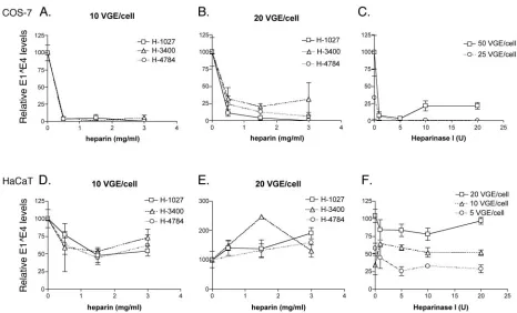
attention has been given to a potential role for heparan sulfo-nated molecules and glycosaminoglycans in papillomavirus in-fections (6, 12, 17, 22, 46). Yet most studies on virus attach-ment and infection pathways have simply investigated VLP binding to cells of various types or the use of pseudovirion infection of nonhost, nonkeratinocyte cells. Using reagents and methods previously reported to block HPV11 VLP binding to HaCaT cells as well as HPV33 and HPV16 pseudovirion in-fection of COS-7 cells (17, 22), we investigated the role of heparan-sulfonated molecules in HPV31b infection of cultured cells of human keratinocyte origins and COS-7 cells (Fig. 3).
HPV31b virions were incubated with various molecular weights of heparin and used to initiate infections as described in the Materials and Methods section and as reported by Gi-roglou et al. (17). For all experiments replicate PBS-treated virion controls were included, and E1^E4 levels were ex-pressed in percentages relative to those in the controls for each cell line. Consistent with previous reports using HPV33 and HPV16 pseudovirions (17, 45), heparin pretreatments vastly reduced the infectivity of HPV31b virions in COS-7 cells in a clear and dose-dependent manner (Fig. 3A and B). HPV31b
infection of COS-7 cells was reduced 70% to 100% when virions were pretreated with heparin compared with infections using control PBS-treated virions. However, no reproducible heparin dose-dependent reduction in HPV31b infectivity was observed in HaCaT human keratinocytes under the same con-ditions (Fig. 3D and E). The results in COS-7 cells are repre-sentative of four separate infections each analyzed 2 to 3 times in triplicate; experiments with HaCaT cells were repeated four times to verify the results. The data in Fig. 3D showing nearly 50% inhibition of HaCaT cell infection was the only analysis indicating possible heparin blocking in these cells.
[image:5.585.59.525.67.349.2]Removal of cell surface heparan sulfate also was performed to assess the requirement for interaction of HPV31b virions with heparan sulfate (Fig. 3C and F and Fig. 4). Heparinase I was used to remove heparan sulfate from COS-7 and HaCaT cells and removal was monitored by fluorescence-activated cell sorting (FACS) analysis. Treatment of COS-7 cells with 40 units per ml (10 U) of heparinase I reduced the cell surface expression of heparan sulfate by 55% compared to untreated cells (Fig. 4A), and this correlated with a⬎75% reduction of HPV31b infection of COS-7 cells (Fig. 3C). Although hepari-nase I treatment of HaCaT cells reduced cell surface expres-sion of heparan sulfate by 79% compared to untreated cells,
FIG. 3. Analysis of the role of heparan-sulfonated receptors in HPV31b infection of COS-7 cells and HaCaT human keratinocytes. Cells were allowed to attach overnight and were 60 to 80% confluent at the time of infection. HaCaT medium were used for all infections and mock-infected controls were included for each set of infections. (Panels A, B, D, and E) Virions were incubated for 1 h at 37°C with heparin preparations (0, 0.5, 1.5, and 3.0 mg per ml) of various molecular weights: low-molecular-weight H-1027 (open box); medium-molecular-weight H-3400 (average 3,000, open triangle); high-molecular-weight H-4784 (⬇20,000; open circle). Treated virions were used immediately for infection. Following infection cells were rinsed with medium, then fed and allowed to grow at 37°C before harvesting. (Panels C and F) Cells were treated with heparinase I (0, 1.0, 5.0, 10.0, or 20.0 units; Sigma H-2519) for 1 h at 37°C as previously described (17, 22) immediately prior to infection with the indicated viral MOI. Infected cells were harvested for total RNA at 2 to 4 days postinfection. RT-qPCR was performed as described in the Materials and Methods. The qPCR values were normalized to the PBS-treated controls for the triplicate samples. Error bars represent the standard error of the mean.
on November 8, 2019 by guest
http://jvi.asm.org/
negligible, if any effects were observed on HPV31b infection (Fig. 3F). The effect of heparinase I treatment of HPV31b infection of COS-7 cells was repeated three times with the same results; heparinase I effect on HaCaT cell infectivity was verified five times. These results are in full agreement with the heparin blocking experiments and indicate that although inter-action with heparan sulfate is important for HPV31b infection of the nonhost COS-7 cells, it is not essential for HPV31b infection of HaCaT human keratinocytes.
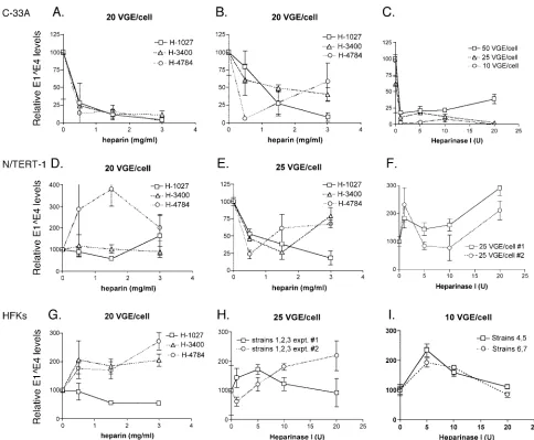
To further determine whether the interaction of HPV31b virions with heparan sulfate is important for infection of other biologically relevant human keratinocyte cell lines or low-pas-sage HFKs, we performed experiments using heparin treat-ment of virions and heparinase I treattreat-ment of cells in the C-33A cervical carcinoma cell line, the N/TERT-1 immortal-ized HFK cell line, and in pools of low-passage HFKs from a number of donors (Fig. 5). HPV31b infection in C-33A cervical cancer cells was reproducibly blocked in a dose-dependent manner when virions were incubated with heparin of various molecular weights (Fig. 5A and B). These results were similar in three separate infections. Likewise, heparinase I treatment of C-33A cells resulted in a substantial reduction in HPV31b infection (Fig. 5C) that was verified in four individual infec-tions.
From the data in Fig. 2B, we expected N/TERT-1 cells and low-passage HFKs to show more variable detection of HPV31b infections, and this held true in the heparin experiments as well. In many of these experiments we observed random infec-tion fluctuainfec-tions that were not heparin dose-dependent (e.g.,
see Fig. 5D and G). We attribute this to the more variable infectivity in these HFK-derived cells, likely due to the more heterogeneous nature of the cell populations (see Discussion). On more than one occasion both N/TERT-1 cells and low-passage HFKs exposed to virions treated with the low molec-ular weight heparin (H-1027) had greater than 50% infection reduction in a dose-dependent manner (e.g., Fig. 5E). Yet, considering the five separate experiments each testing heparin blocking in N/TERT-1 cells and HFKs, HPV31b infections of neither HFK-derived cell population were reproducibly blocked in a dose-dependent manner. Furthermore, hepari-nase I treatment of N/TERT-1 cells and three different pools of HFKs from seven donors (strains 1 to 7) failed to reduce HPV31b infection in these cells (Fig. 5F, H, I), and the tripli-cate RT-qPCR analyses were repeated to confirm the results. Taken together, our data suggest that heparan sulfonated mol-ecules play a role in HPV31b infection of COS-7 monkey kidney cells and the C-33A cervical carcinoma cell line, but that interaction with heparan sulfate is not essential for HPV31b infection of human keratinocytes, the host cell type for HPVs.
DISCUSSION
VLPs and pseudovirions have been used to study the re-quirement for heparan-sulfonated receptors in the binding to and internalization into a number of immortalized, trans-formed, and nonhost cell types for HPVs (6, 12, 17, 22, 46). However, to date there have been no reports investigating high-risk HPV infections in their natural host cells, nonimmor-talized human keratinocytes. The purpose of our study was to assess the requirement of heparan-sulfonated molecules as potential HPV31b attachment moieties resulting in authentic infection in cell types most relevant to infection in vivo. To this end we have purified infectious HPV31b virions from CIN-612 9E raft tissues and have obtained virion stocks containing⬇109
viral genome equivalents per ml.
Our previous work using RT and end-point PCR to detect spliced viral RNAs as a measure of HPV31b infection in a number of cell lines demonstrated HaCaT cell infection was most reproducibly and robustly detected (37, 38). These find-ings also held true in this study where we have modified our assay to include RT-qPCR to compare the relative levels of HPV31b infection among cells and cell lines as a function of viral E1^E4 transcript quantities. Using this assay to measure levels of spliced E1^E4 transcripts, HPV31b infections can be quantified and relative levels of infection compared, albeit not the absolute number of cells infected. Because the absolute levels of E1^E4 transcripts are different following infection of various cells (Fig. 2), we have not compared differences in transcription among cell lines as a basis for infection inhibition. Rather, we have compared the ability of potential blocking agents to inhibit HPV31b infection within a given cell line as normalized to untreated cells.
[image:6.585.46.277.69.305.2]We analyzed COS-7 monkey kidney cells and C-33A cervical carcinoma cells, two transformed cell lines, as controls for relation to other studies (6, 17, 42). We also investigated cells that more closely represent the target cell for HPV infection in vivo: HaCaT and N/TERT-1 cells, which are immortalized human keratinocytes that retain the ability to differentiate in
FIG. 4. Flow cytometry analysis of cell surface heparan sulfate ex-pression on HaCaT and COS-7 cells. Monolayer cultures of COS-7 cells (A) or HaCaT cells (B) were treated with 0, 1.0, or 10 units per well of heparinase I. Following detachment, cells were fixed and ana-lyzed for heparan sulfate surface expression by FACScan. Shaded area, autofluorescence control (secondary antibody only); thick solid line, 0 units heparinase I; dashed line, heparinase I at 4 U per ml; dotted line, heparinase I at 40 U per ml (17, 22).
on November 8, 2019 by guest
http://jvi.asm.org/
vitro (2, 11), and low-passage HFK pools. Using the methods of Giroglou and coworkers and Joyce et al. (17, 22), we found that interactions with heparan sulfate are required for HPV31b infection of COS-7 monkey kidney cells. Heparin treatment of virions or heparinase I treatment of COS-7 cells caused a 70 to 100% inhibition of HPV31b infection, even at the lowest chemical concentrations, and our findings using authentic HPV31b virion infection of COS-7 cells are in agreement with the previous studies exposing COS-7 cells to pseudovirions from HPV types 16, 18, 31, 33, 39, 45, 58, 59, and 68 (6, 17, 45). We found a similar requirement for heparan-sulfonated molecules with HPV31b infection of C-33A cervical carcinoma cells. However, heparan sulfate interaction was not required for efficient infection of low-passage HFKs and the immortal-ized but untransformed human keratinocyte cell lines HaCaT and N/TERT-1. The failure of heparin treatment of virions or heparinase I treatment of cells to block HPV31b infection was
clearest with HaCaT cells. The reproducibility studies shown in Fig. 2 indicated detection of infections in HFKs and N/TERT-1 cells were less consistent and varied two- to three-fold among the replicate samples. Thus, we believe that this explains the fluctuation observed in E1^E4 levels following the heparin or heparinase I treatments (Fig. 5), as dose-de-pendent blockage of HPV31b infection of HFKs or N/TERT-1 cells was not observed consistently. In a few experiments, there appeared to be a trend of low-molecular-weight heparin block-ing of HPV31b infection in HFK and N/TERT-1 cells, but the significance of this is presently unclear. The detection of more variable E1^E4 levels in HFK-derived N/TERT-1 cells or HFKs versus HaCaT cells could be a function of the lower expression of viral transcripts, which may reflect more cellular heterogeneity in the lower-passage HFK cells.
[image:7.585.48.532.67.466.2]We can envision at least three explanations as to why HPV31b infection of COS-7 and C-33A cells is heparan sulfate
FIG. 5. Analysis of the role of heparan-sulfonated receptors in HPV31b infection of C-33A cells, N/TERT-1 cells, and low-passage HFKs. The details of these experiments are the same as described in the legend to Fig. 3. Briefly, HPV31b virions were treated with heparin preparations as indicated (Panels A, B, D, E, and G) and used immediately for infection. (Panels C, F, H, and I) Cells were treated with heparinase I prior to infection. Whereas the HFK pool from three donors (strains 1 to 3) were maintained in culture for multiple passages (panels G and H), the cells from HFK strains 4 and 5 as well as those from 6 and 7 (panel I) were mixed in equal numbers upon seeding for treatment and exposure to virus.
on November 8, 2019 by guest
http://jvi.asm.org/
dependent, whereas infection of human keratinocytes does not require heparan sulfate interactions. These possibilities also address why our results differ from other studies indicating HPVs require heparan sulfate for cellular binding and/or in-fection. First, our study investigated HPV infection of human keratinocytes, rather than VLP binding to or pseudovirion infection of transformed cells or cells relevant to in vivo HPV infection. HPVs, like other viruses, may use distinct receptors to infect different cells. The heparan sulfate-dependent phe-notype could result from the fact that COS-7 monkey kidney cells are not the correct species or cell type for HPV infection, have been transformed by simian virus 40 large T antigen, and/or have undergone an indeterminate number of passages in culture.
Although we were surprised that C-33A cervical cells be-haved differently from HFKs, it is to be expected that these cells derived from a malignant carcinoma have lost a number of their epithelial characteristics. This is likely true also for other transformed cells like HeLa, A431, and KH-SV (simian virus 40-transformed human keratinocytes), which have been reported to display heparin-dependent phenotypes. Thus, the cells may not retain expression of a keratinocyte-specific HPV receptor(s). Furthermore, there may be a difference in the ratios of surface expression levels of nonspecific receptors (e.g., heparan sulfate) versus specific (␣6 integrin or as yet unidentified) receptors present on these different cell types. Our data and data from other labs are consistent with a model in which HPV may use heparan-sulfonated molecules as initial, nonspecific attachment receptors in COS-7 and C-33A cells. The virus may then be transferred to a second specific, hepa-rinase-resistant molecule that mediates entry into and produc-tive infection of cells as previously proposed (17, 45). In con-trast, human keratinocytes may express the specific receptor(s) at much higher levels, minimizing the requirement for an ini-tial heparan sulfate interaction.
Second, we have used significantly fewer virus particles for our infectivity assays (MOIs from 5 to 50 viral genome equivalents per cell) compared to most VLP binding or pseudovirion pseudoinfection experiments in which thousands to tens of thousands of particles per cell were used (6, 12, 17, 22, 45, 46). A disadvantage of using very high numbers of particles, especially in simple binding assays, is that the data could reflect bulk interactions resulting in nonproductive events, while masking the actual infectious pathway used by only a few virions as is expected in vivo. This has been observed with mouse polyoma pseudovirions where mass interactions can occur via a receptor-independent pathway that is distinct from the receptor-specific pathways required to successfully deliver DNA to the nucleus (25). A study using HPV16 VLPs also exemplifies this concern where VLP binding was com-pletely blocked in HaCaT cells using 10g per ml of heparin, whereas the more specific and sensitive measure of VLP in-ternalization required at least 3 mg per ml of heparin to achieve no more than 70% blocking (12). Interestingly, this work also points to a heparin independent uptake pathway in HaCaT cells consistent with our data as discussed above.
A third possibility is that HPV31 may use a receptor(s) different from other genital HPV types that have been tested. Data in support of this come from Bousarghin et al. who found that HPV31 pseudovirions use a caveolar-mediated entry
path-way in COS-7 or 293T cells, whereas HPV16 and HPV58 pseudovirions enter via a clathrin-dependent pathway (3). As viral receptors generally dictate the entry pathway (49), this suggests that different genital high-risk HPVs could use dis-tinct receptors. Nevertheless, the entry pathway of HPV31b could be distinct for large numbers of pseudovirions in non-relevant cells, and we are in the process of verifying the entry pathway in human keratinocytes.
Our data suggest that HPV31b can use separate receptors for infection of COS-7 cells and C-33A cells, both high passage transformed cell lines, versus the lower passage untransformed human keratinocytes (i.e., HaCaT cells, N/TERT-1 cells, and HFKs). Several viruses use heparan sulfate as an initial recep-tor, with specific secondary or tertiary receptors for viral up-take (5, 7, 35, 47, 54). Our work supports other data (17, 45) indicating that a heparin-independent receptor is used for HPV uptake, regardless of whether an initial heparan sulfate-dependent primary interaction is required.
An issue that remains to be addressed is whether COS-7 or C-33A cells versus normal keratinocytes use the same second-ary (specific) heparin-independent receptor. Another question is whether COS-7 and C-33A cells require heparan-sulfonated receptors because the expression of this putative secondary receptor is extremely low in these cells compared to keratino-cytes. Likewise, if virions employ different uptake pathways in these cells, the receptors may be different. This is consistent with a recent study where HPV11 virion entry and E1∧E4 expression kinetics were different in COS-7 cells compared to more relevant cell lines (8).
It is of great interest to test the role of heparan sulfate in other HPV infections. Although we have purified infectious HPV16 and HPV18 virions from raft tissues (26, 34), virions from these tissues are made at lower levels and early tran-scripts are more difficult to detect following experimental in-fections with HPV types 16 and 18. Thus, we are still in the process of optimizing a robust RT-qPCR assay capable of reproducibly distinguishing 2- to 10-fold decreases in infection levels, which is essential to our ability to obtain interpretable data from infection blocking experiments.
The continued study of infectious HPV virions in cell types relevant to in vivo infections will be imperative for uncovering the answers to many of the questions related to early infection events. For true biological relevance, infections will require the investigation of basal cells in the context of a differentiating epithelium.
ACKNOWLEDGMENTS
This work was supported by PHS grants CA-85747 and AI-52049 from the NIH (M.A.O.). J.L.S. is an Infectious Disease and Inflam-mation Fellow supported by NIH Training Grant T32 AI-07538.
We gratefully acknowledge the discussions of our colleagues during the completion of this work, including Martin Sapp, Patricia Day, Chris Buck, and John Schiller. Additional thanks to Martin Sapp for sharing details of unpublished protocols and thoughts on data inter-pretation throughout this work. We are indebted to Cosette M. Wheeler and Jesse Summers for helpful discussions and for critical comments on the manuscript. We are grateful to Norbert Fusenig for providing the HaCaT cell line, to James Rheinwald for the N/TERT-1 cell line, and to Cosette Wheeler for the gift of low-passage human foreskin keratinocytes.
on November 8, 2019 by guest
http://jvi.asm.org/
REFERENCES
1.Bedell, M. A., J. B. Hudson, T. R. Golub, M. E. Turyk, M. Hosken, G. D. Wilbanks, and L. A. Laimins.1991. Amplification of human papillomavirus genomes in vitro is dependent on epithelial differentiation. J. Virol.65:2254– 2260.
2.Boukamp, P., R. T. Petrussevska, D. Breitkreutz, J. Hornung, A. Markham, and N. E. Fusenig.1988. Normal keratinization in a spontaneously immor-talized aneuploid human keratinocyte cell line. J. Cell Biol.106:761–771. 3.Bousarghin, L., A. Touze´, P.-Y. Sizaret, and P. Coursaget.2003. Human
papillomavirus types 16, 31, and 58 use different endocytosis pathways to enter cells. J. Virol.77:3846–3850.
4.Christensen, N. D., C. A. Reed, T. D. Culp, P. L. Hermonat, M. K. Howett, R. A. Anderson, and L. J. D. Zaneveld.2001. Papillomavirus microbicidal activities of high-molecular-weight cellulose sulfate, dextran sulfate, and polystyrene sulfonate. Antimicrob. Agents Chemother.45:3427–3432. 5.Chung, C.-S., J.-C. Hsiao, Y.-S. Chang, and W. Chang.1998. A27L protein
mediates vaccinia virus interaction with cell surface heparan sulfate. J. Virol.
72:1577–1585.
6.Combita, A. L., A. Touze, L. Bousarghin, P.-Y. Sizaret, N. Munoz, and P. Coursaget.2001. Gene transfer using human papillomavirus pseudovirions varies according to virus genotype and requires cell surface heparan sulfate. FEMS Microbiol. Lett.204:183–188.
7.Compton, T., D. M. Nowlin, and N. R. Cooper.1993. Initiation of human cytomegalovirus infection requires initial interaction with cell surface hepa-ran sulfate. Virology193:834–841.
8.Culp, T. D., and N. D. Christensen.2004. Kinetics of in vitro adsorption and entry of papillomavirus virions. Virology319:152–161.
9.Culp, T. D., and N. D. Christensen.2003. Quantitative RT-PCR assay for HPV infection in cultured cells. J. Virol. Methods111:135–144.
10.Day, P. M., D. R. Lowy, and J. T. Schiller.2003. Papillomaviruses infect cells via a clathrin-dependent pathway. Virology307:1–11.
11.Dickson, M. A., W. C. Hahn, Y. Ino, V. Ronfard, J. Y. Wu, R. A. Weinberg, D. N. Louis, F. P. Li, and J. G. Rheinwald.2000. Human keratinocytes that express hTERT and also bypass a p16INK4a-enforced mechanism that limits life span become immortal yet retain normal growth and differentiation characteristics. Mol. Cell. Biol.20:1436–1447.
12.Drobni, P., N. Mistry, N. McMillan, and M. Evander.2003. Carboxy-fluo-rescein diacetate, succinimidyl ester labeled papillomavirus virus-like parti-cles fluoresce after internalization and interact with heparan sulfate for binding and entry. Virology310:163–172.
13.Dvoretzky, I., R. Shober, S. K. Chattopadhyay, and D. R. Lowy.1980. A quantitativein vitrofocus assay for bovine papilloma virus. Virology103:
369–375.
14.Evander, M., I. H. Frazer, E. Payne, Y. M. Qi, K. Hengst, and N. A. J. McMillan.1997. Identification of the␣6integrin as a candidate receptor for
papillomaviruses. J. Virol.71:2449–2456.
15.Favre, M., F. Breitburd, O. Croissant, and G. Orth. 1975. Structural polypeptides of rabbit, bovine, and human papillomaviruses. J. Virol.15:
1239–1247.
16.Gillison, M. L., W. M. Koch, R. B. Capone, M. Spafford, W. H. Westra, L. Wu, M. L. Zahurak, R. W. Daniel, M. Viglione, D. E. Symer, K. V. Shah, and D. Sidransky.2000. Evidence for a causal association between human papillomavirus and a subset of head and neck cancers. JNCI Cancer Spec-trum.92:709–720.
17.Giroglou, T., L. Florin, F. Schafer, R. E. Streeck, and M. Sapp.2001. Human papillomavirus infection requires cell surface heparan sulfate. J. Virol.75:
1565–1570.
18.Gluzman, Y.1981. SV40-transformed simian cells support the replication of early SV40 mutants. Cell23:175–182.
19.Hagensee, M. E., N. Yaegashi, and D. A. Galloway.1993. Self-assembly of human papillomavirus type 1 capsids by expression of the L1 protein alone or by coexpression of the L1 and L2 capsid proteins. J. Virol.67:315–322. 20.Harwood, C. A., J. M. McGregor, C. M. Proby, and J. Breuer.1999. Human
papillomavirus and the development of non-melanoma skin cancer. J. Clin. Pathol.52:249–253.
21.Hummel, M., J. B. Hudson, and L. A. Laimins.1992. Differentiation-induced and constitutive transcription of human papillomavirus type 31b in cell lines containing viral episomes. J. Virol.66:6070–6080.
22.Joyce, J. G., J.-S. Tung, C. T. Przysiecki, J. C. Cook, E. D. Lehman, J. A. Sands, K. U. Jansen, and P. M. Keller.1999. The L1 major capsid protein of human papillomavirus type 11 recombinant virus-like particles interacts with heparin and cell-surface glycosaminoglycans on human keratinocytes. J. Biol. Chem.274:5810–5822.
23.Kawana, K., H. Yoshikawa, Y. Taketani, K. Yoshiike, and T. Kanda.1998. In vitro construction of pseudovirions of human papillomavirus type 16: incor-poration of plasmid DNA into reassembled L1/L2 capsids. J. Virol.72:
10298–10300.
24.Kirnbauer, R., F. Booy, N. Cheng, D. R. Lowy, and J. T. Schiller.1992. Papillomavirus L1 major capsid protein self-assembles into virus-like parti-cles that are highly immunogenic. Proc. Natl. Acad. Sci. USA89:12180– 12184.
25.Krauzewicz, N., J. Stokrova, C. Jenkins, M. Elliott, C. F. Higgins, and B. E. Griffin.2000. Virus-like gene transfer into cells mediated by polyoma virus pseudocapsids. Gene Ther.7:2122–2121.
26.Lee, J. H., S. M. P. Yi, M. E. Anderson, K. L. Berger, M. J. Welsh, A. J. Klingelhutz, and M. A. Ozbun.2004. Propagation of infectious human pap-illomavirus type 16 by using an adenovirus and cre/loxP mechanism. Proc. Natl. Acad. Sci. USA101:2094–2099.
27.Lowy, D. R., and P. M. Howley.2001. Papillomaviruses, p. 2231–2264.In D. M. Knipe and P. M. Howley (ed.), Fields virology, 4th ed. Lippincott Williams & Wilkins, Philadelphia, Pa.
28.McCance, D. J., R. Kopan, E. Fuchs, and L. A. Laimins.1988. Human papillomavirus type 16 alters human epithelial cell differentiationin vitro. Proc. Natl. Acad. Sci. USA85:7169–7173.
29.McLaughlin-Drubin, M. E., S. Wilson, B. Mullikin, J. Suzich, and C. Mey-ers.2003. Human papillomavirus type 45 propagation, infection, and neu-tralization. Virology312:1–7.
30.McMillan, N. A. J., E. Payne, I. H. Frazer, and M. Evander.1999. Expression of the alpha6 integrin confers papillomavirus binding upon receptor-negative B-cells. Virology261:271–279.
31.Meyers, C.1996. Organotypic (raft) epithelial tissue culture system for the differentiation-dependent replication of papillomavirus. Methods Cell Sci.
18:201–210.
32.Meyers, C., J. L. Bromberg-White, J. Zhang, M. E. Kaupas, J. T. Bryan, R. S. Lowe, and K. U. Jansen.2002. Infectious virions produced from a human papillomavirus type 18/16 genomic DNA chimera. J. Virol.76:4723–4733. 33.Meyers, C., M. G. Frattini, J. B. Hudson, and L. A. Laimins.1992.
Biosyn-thesis of human papillomavirus from a continuous cell line upon epithelial differentiation. Science257:971–973.
34.Meyers, C., T. J. Mayer, and M. A. Ozbun.1997. Synthesis of infectious human papillomavirus type 18 in differentiating epithelium transfected with viral DNA. J. Virol.71:7381–7386.
35.Mondor, I., S. Ugolini, and Q. J. Sattentau.1998. Human immunodeficiency virus type 1 attachment to HeLa CD4 cells is CD4 independent and gp120 dependent and requires cell surface heparans. J. Virol.72:3623–3634. 36.Mu¨ller, M., L. Gissmann, R. J. Cristiano, X.-Y. Sun, I. H. Frazer, A. B.
Jenson, A. Alonso, H. Zentgraf, and J. Zhou.1995. Papillomavirus capsid binding and uptake by cells from different tissues and species. J. Virol.
69:948–954.
37.Ozbun, M. A.2002. Human papillomavirus type 31b infection of human keratinocytes and the onset of early transcription. J. Virol.76:11291–11300. 38.Ozbun, M. A.2002. Infectious human papillomavirus type 31b: purification and infection of an immortalized human keratinocyte cell line. J. Gen. Virol.
83:2753–2763.
39.Ozbun, M. A., and C. Meyers.1999. Human papillomavirus type 31b tran-scription during the differentiation-dependent viral life cycle. Curr. Top. Virol.1:203–217.
40.Pfister, H.1984. Biology and biochemistry of papillomaviruses. Rev. Physiol. Biochem. Pharmacol.99:111–181.
41.Roden, R. B. S., H. L. Greenstone, R. Kirnbauer, F. P. Booy, J. Jessie, D. R. Lowy, and J. T. Schiller.1996. In vitro generation and type-specific neutral-ization of a human papillomavirus type 16 virion pseudotype. J. Virol.70:
5875–5883.
42.Roden, R. B. S., R. Kirnbauer, A. B. Jenson, D. R. Lowy, and J. T. Schiller.
1994. Interaction of papillomaviruses with the cell surface. J. Virol.68:7260– 7266.
43.Rose, R. C., W. Bonnez, R. C. Reichman, and R. L. Garcea.1993. Expression of human papillomavirus type 11 L1 protein in insect cells: in vivo and in vitro assembly of viruslike particles. J. Virol.67:1936–1944.
44.Rowson, K. E. K., and B. W. J. Mahy.1967. Human papova (wart) virus. Bacteriol. Rev.31:110–131.
45.Selinka, H.-C., T. Giroglou, and M. Sapp.2002. Analysis of the infectious entry pathway of human papillomavirus type 33 pseudovirions. Virology
299:279–287.
46.Shafti-Keramat, S., A. Handisurya, E. Kriehuber, G. Meneguzzi, K. Slu-petzky, and R. Kirnbauer.2003. Different heparan sulfate proteoglycans serve as cellular receptors for human papillomaviruses. J. Virol.77:13125– 13135.
47.Shukla, D., J. Liu, P. Blaiklock, N. W. Shworak, X. Bai, J. D. Esko, G. H. Cohen, R. J. Eisenberg, R. D. Rosenberg, and P. G. Spear.1999. A novel role for 3-O-sulfated heparan sulfate in herpes simplex virus 1 entry. Cell99:13– 22.
48.Sibbet, G., C. Romero-Graillet, G. Meneguzzi, and M. S. Campo.2000. alpha6 integrin is not the obligatory cell receptor for bovine papillomavirus type 4. J. Gen. Virol.81:327–334.
49.Smith, A. E., and A. Helenius.2004. How viruses enter animal cells. Science
304:237–242.
50.Smith, L. H., C. Foster, M. E. Hitchcock, and R. Isseroff.1993.In vitro HPV-11 infection of human foreskin. J. Investig. Dermatol.101:292–295. 51.Smith, L. H., C. Foster, M. E. Hitchcock, G. S. Leiserowitz, K. Hall, R.
Isseroff, N. D. Christensen, and J. W. Kreider.1995. Titration of HPV-11 infectivity and antibody neutralization can be measuredin vitro.J. Investig. Dermatol.105:438–444.
on November 8, 2019 by guest
http://jvi.asm.org/
52.Steinberg, B. M., K. J. Auborn, J. L. Brandsma, and L. B. Taichman.1989. Tissue site-specific enhancer function of the upstream regulatory region of human papillomavirus type 11 in cultured keratinocytes. J. Virol.63:957– 960.
53.Stubenrauch, F., and L. A. Laimins.1999. Human papillomavirus life cycle: active and latent phases. Cancer Biol.9:379–386.
54.Summerford, C., and R. J. Samulski.1998. Membrane-associated heparan sulfate proteoglycan is a receptor for adeno-associated virus type 2 virions. J. Virol.72:1438–1445.
55.Unckell, F., R. E. Streeck, and M. Sapp.1997. Generation and neutralization of pseudovirions of human papillomavirus type 33. J. Virol.71:2934–2939. 56.Volpers, C., P. Schirmacher, R. E. Streeck, and M. Sapp.1994. Assembly of
the major and minor capsid protein of human papillomavirus type 33 into virus-like particles and tubular structures in insect cells. Virology200:504– 512.
57.Volpers, C., F. Unckell, P. Schirmacher, R. E. Streeck, and M. Sapp.1995. Binding and internalization of human papillomavirus type 33 virus-like par-ticles by eukaryotic cells. J. Virol.69:3258–3264.
58.Walboomers, J. M. M., M. V. Jacobs, M. M. Manos, F. X. Bosch, J. A.
Kummer, K. V. Shah, P. J. F. Snijders, J. Peto, C. J. L. M. Meijer, and N. Mun˜oz.1999. Human papillomavirus is a necessary cause of invasive cervical cancer worldwide. J. Pathol.189:12–19.
59.Yee, C., I. Krishnan-Hewlett, C. C. Baker, R. Schlegel, and P. M. Howley.
1985. Presence and expression of human papillomavirus sequences in human cervical carcinoma cell lines. Am. J. Pathol.119:361–366.
60.Yoon, C.-S., K.-D. Kim, S.-N. Park, and S.-W. Cheong.2001.␣6 integrin is the main receptor of human papillomavirus type 16 VLP. Biochem. Biophys. Res. Commun.283:668–673.
61.Zhou, J., L. Gissmann, H. Zentgraf, H. Mu¨ller, M. Picken, and M. Mu¨ller.
1995. Early phase in the infection of cultured cells with papillomavirus virions. Virology214:167–176.
62.zur Hausen, H.2000. Papillomaviruses causing cancer: Evasion from host-cell control in early events in carcinogenesis. J. Natl. Cancer Inst.92:690– 698.
63.zur Hausen, H., and A. Schneider.1987. The role of papillomaviruses in human anogenital cancer, p. 245–263.InN. P. Salzman and P. M. Howley (ed.), The papovaviridae, vol 2: the papillomaviruses. Plenum Press, New York, N.Y.