A DISSERTATION ON
“
ASSOCIATION OF CHOLELITHIASIS,
CHOLEDOCHOLITHIASIS AND
HYPOTHYROIDISM”
Dissertation submitted to
THE TAMIL NADU Dr.M.G.R.MEDICAL UNIVERISTY
CHENNAI
with partial fulfilment of the regulations
for the Award of the degree
M.S. (General Surgery- Branch- I)
DEPARTMENT OF GENERAL SURGERY
THANJAVUR MEDICAL COLLEGE
2
CERTIFICATE
This is to certify that the dissertation entitled “ASSOCIATION OF
CHOLELITHIASIS, CHOLEDOCHOLITHIASIS AND HYPOTHYROIDISM”
is a bonafide original work of Dr. V. L. Aishwarya in partial fulfilment of the requirements for M.S.Branch–I (General Surgery) Examination of the Tamil Nadu Dr.
M.G.R. Medical University to be held in APRIL 2016 under my guidance and supervision in 2014-2015
Prof Dr. K. Sathyabama, M.S Prof Dr. M. Elangovan, M.S
Professor of General Surgery, Director and Professor,
Guide and supervisor, Department of general surgery, Thanjavur Medical College Thanjavur- 613004
Dean,
3
DECLARATION
I Dr. AISHWARYA.V.L hereby solemnly declare that the dissertation titled “ASSOCIATION OF CHOLELITHIASIS, CHOLEDOCHOLITHIASIS AND HYPOTHYROIDISM” is done by me at Thanjavur medical College, Thanjavur
during 2014-2015 under the guidance and supervision of
Prof.Dr.K. SATHYABAMA, M.S. This dissertation is submitted to The Tamil Nadu Dr.M.G.R Medical University, Chennai towards the partial fulfillment of requirements
for the award of M.S.
Degree (Branch-I) in General Surgery.
Place: Thanjavur Dr. V. L. Aishwarya, Date:
5
6
ACKNOWLEDGEMENT
I express my heartful gratitude to the Dean, Dr. Thanjavur Medical College, Thanjavur for permitting me to do this study.
I am deeply indebted to Prof.Dr.M.ELANGOVAN,M.S., Director& Professor, Department of General Surgery, Thanjavur Medical College for his support and guidance.
I am very grateful to Prof.Dr.K.SATYABAMA M.S, Professor of Surgery, Department of General Surgery, and my Assistant Professors Dr.P. Vanathy M.S, Dr. Saravanan. R M.S, Dr. N. Priya M.S Thanjavur Medical College who guided me throughout the period of my study.
My sincere thanks to my former Chief, Prof. Dr. P. Shanthini M.S, and my then assistant professors Dr. Balasundaram M.S.,M.Ch., Dr. Senthilkumaran M.S, for their valuable guidance. I am grateful to all my professors and assistant professors, who have contributed in many ways towards the completion of this study
7
LIST OF ABBREVIATIONS
GB - - Gall Bladder
CBD - - Common Bile Duct SO - - Sphincter of Oddi CCK - - Cholecystokinin USG - - Ultrasonogram
CECT - - Contrast Enhanced Computed Tomography
MRCP - - Magnetic Resonance Cholangiopancreaticography ERCP - - Endoscopic Retrograde Cholangiopancreaticography PTC - - Percutaneous Transhepatic Cholangiography
TSH - - Thyroid Stimulating Hormone TH - - Thyroid Hormones
8
CONTENTS
S.No TITLE
1. INTRODUCTION
2. REVIEW OF LITERATURE
3. MATERIALS AND METHODS
4. RESULTS
5. DISCUSSION
6. LIMITATIONS OF THE STUDY
7. SUMMARY
8. CONCLUSION
9. RECOMMENDATIONS
9
CHAPTER 1
10
1.
BACKGROUND
Gallstones are the most common biliary pathology, can be divided into three main types: cholesterol, pigment (black, brown) or mixed stones. In the USA and Europe, 80%cholesterol or mixed stones, where as in Asia, 80% are pigment stones. Cholesterol or mixed stones contain 51 – 99 % cholesterol plus admixture of calcium salts, bile acids, bile pigment and phospholipids. Gallstones may be single or multiple, large or small those containing calcium salts are radio-opaque. Single stones are uncommon but usually consist mainly of cholesterol and arise due to a disorder of the physio-chemical equilibrium which normally maintains cholesterol in micellar form in the bile.
Many studies were done to identify risk factor for biliary lithiasis in the west have focused on hypersaturation of cholesterol in bile in nucleation process a critical step in the genesis of bile stone.
12
1.2 OBJECTIVES
The purpose of this study is
1. To study the prevalence of hypothyroidism in patients presenting with CHOLELITHIASIS/ CHOLEDHOCOLITHIASIS.
13
2.1 Anatomy of the biliary system
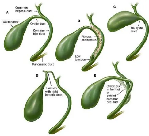
“The biliary tree consists of the system of vessels and ducts which collect and deliver bile from the liver parenchyma to the second part of the duodenum. It is conventionally divided into intrahepatic and extrahepatic biliary trees. The intrahepatic ducts are formed from the larger bile canaliculi which join together to form segmental ducts. These fuse close to the porta hepatis into right and left hepatic ducts. The extrahepatic biliary tree consists of the right and left hepatic ducts, the common hepatic duct, the cystic duct and gallbladder and the common bile duct.”
GALLBLADDER
14
15
[image:15.595.86.383.202.499.2]gallbladder, the so-called Phrygian cap: on ultrasound, this may be wrongly interpreted as an apparent septum within an otherwise normal gallbladder. Again, rarely, the gallbladder may be bifid or completely duplicated, usually with a duplicated cystic duct
Fig 1. Overall arrangement of the intrahepatic and extrahepatic biliary tree.
CYSTIC DUCT
16
[image:16.595.76.319.512.734.2]junction usually occurs near the porta hepatis but may be lower down in the free edge of the lesser omentum.
17
Mucosa of the cystic duct has 5–12 crescentic folds; continuous with the neck of the gallbladder. They project obliquely in regular succession; appearing to form a spiral valve when the duct is cut in longitudinal section. When the duct is distended, the spaces dilate and it appears twisted like neck of the GB.
HEPATIC BILE DUCTS
The main right and left hepatic ducts; emerge from the liver and unite near the right end of the porta hepatis as the common hepatic duct. The extrahepatic right duct, is short and nearly vertical while the left, is more horizontal and lies in segment IV. The common hepatic duct is joined on its right in an acute angle by the cystic duct to form the CBD. “The common hepatic duct lies to the right of the hepatic artery and anterior to the portal vein in the free edge of the lesser omentum.
COMMON BILE DUCT
18
cm from it: even when it is embedded in the pancreas, a groove in the gland marking its position can be palpated behind the second part of the duodenum.
Calot's triangle
The near triangular space, formed between the cystic duct, the
19
[image:19.595.73.331.381.664.2]opposed, there is an appreciable amount of loose connective tissue within the triangle. It is perhaps better described as a pyramidal ‘space' with one apex lying at the junction of the cystic duct and fundus of the gallbladder, one at the porta hepatis, and two closer apices at the attachments of the gallbladder to the liver bed. The base of the triangle, thus lies on the inferior surface of the liver. This space usually contains the cystic artery as it approaches the gallbladder, the cystic lymph node and lymphatics from the gallbladder, one or two small cystic veins, the autonomic nerves running to the gallbladder, and some loose adipose tissue. It may contain any accessory ducts which drain into the gallbladder from the liver. One must be aware of the variations in cystic duct anatomy so that we avoid accidently ligating the common bile duct.
20
VASCULAR SUPPLY AND LYMPHATIC DRAINAGE
21
These fine branches form a network which anastomoses with the vessels ascending around the common bile duct “and with the vessels from the liver parenchyma which descend with the right and left hepatic ducts.
Ductal arteries
The common bile duct and hepatic ducts are supplied by a fine network of vessels, which usually receive contributions from several sources; and which lie in close proximity to the ducts.” Disruption of the network during surgical exposure of the bile ducts over a long length frequently causes chronic ischaemia and resultant stenosis of the duct. Approaches which spare the network are necessary to avoid this complication.The common bile duct is fairly consistently supplied by two to four ascending and descending arteries which form long narrow anastomotic channels along the length of the duct: the most prominent of these are disposed into medial and lateral ‘trunks', although they may lie more anterolateral and posteromedial. The largest contributions, usually arise as one or two branches, from the retroduodenal branch of the gastroduodenal artery; as it crosses the anterior surface of the duct at the upper border of the duodenum. Three or four descending branches; supply this
22
Posteriorly, a retroportal artery often arises from the coeliac axis, superior mesenteric artery or one of their major branches close to the origin from the “
Lymphatics:
23
node lying in the anterior border of the free edge of the lesser omentum. Hepatic nodes collect lymph from vessels that accompany the hepatic ducts and the upper” part of the bile duct. The inferior hepatic and upper pancreaticosplenic nodes receive lymphatics from the lower part of Common bile duct.
INNERVATION
The gallbladder and the extrahepatic biliary tree are innervated by branches from the hepatic plexus. The retroduodenal part of the common bile duct and the smooth muscle of the hepatopancreatic ampulla are also innervated by twigs from the pyloric branches of the vagi.
Referred pain: In common with other structures of foregut origin, pain caused by stretch of the common bile duct or gallbladder is referred to the
epigastrium. Involvement of the overlying somatic peritoneum produces pain which is more localized to the right upper quadrant.
GALLBLADDER
The fundus of the gallbladder is completely covered by a
serosa, and the inferior surfaces and sides of the body and neck of the gallbladder are usually covered by a serosa. If the gallbladder possesses a mesentery the serosa
extends around the sides of the body and neck onto the superior surface and continues into the serosa of the mesentery, whereas the serosa is limited to the inferior surfaces only if the gallbladder is intrahepatic. Beneath the serosa is subserous loose
24
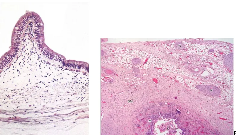
generally resembles that of the small intestine. The mucosa is yellowish-brown and elevated into minute rugae with a honeycomb appearance. In section, projections of the mucosa into the gallbladder lumen resemble intestinal villi, but they are not fixed structures and the surface flattens as the gallbladder fills with bile. The epithelium is a single layer of columnar cells with apical microvilli; basally, the spaces between epithelial cells are dilated. Many capillaries lie beneath the basement membrane. The epithelial cells actively absorb water and solutes from the bile and concentrate it up to ten-fold. There are no goblet cells in the epithelium. The thin fibromuscular layer is composed of fibrous tissue mixed with smooth muscle cells arranged loosely in longitudinal, circular and oblique bundles. Gall bladder does not have submucosal layer and a muscular layer.
BILE DUCTS
25
Fig 4: Low
power micrograph showing the gallbladder wall and the human common bile duct structure.
Sphincter of Oddi:
The sphincter of Oddi regulates flow of bile; into the duodenum, prevents the regurgitation of duodenal contents into the biliary tree, and diverts bile into the gallbladder. It is a “complex structure that is functionally independent from the duodenal musculature and creates a high-pressure zone between the bile duct and the duodenum.
[image:25.595.75.467.69.294.2]26
hormones and neurons acting on the smooth muscle cells. Relaxation occurs with a rise in CCK, leading to diminished amplitude of phasic contractions and reduced basal pressure, allowing increased flow of bile into the duodenum. During fasting, the sphincter of Oddi activity is coordinated with the periodic partial gallbladder emptying and an increase in” bile flow; that occurs during phase II of migrating myoelectric motor complex.
2.2 Physiology of biliary system
BILE:
The amount of bile secreted per day is 500- 1000ml. Bile serves two important “functions: First, bile plays an important role in fat digestion and
absorption, because bile acids in the bile do two things:
(1) Bile emulsifies fat into smaller particles which can be attacked by lipase enzyme secreted from the pancreas
(2) They aid in absorption of the digested fat end products through the intestinal mucosal membrane.
Second, bile serves as a means for excretion of several important waste products from the blood.” These include especially “bilirubin”, an end product of haemoglobin degradation; and excesses of cholesterol.
SECRETION OF BILE:
27
STORAGE OF BILE:
28
continually absorbed through the gallbladder mucosa, concentrating the remaining bile constituents that contain the bile salts, cholesterol, lecithin, and bilirubin.
Most of this gallbladder absorption is caused by active transport of sodium through the gallbladder epithelium, and this is followed by secondary absorption of chloride ions, water, and most other diffusible constituents. Bile is normally concentrated in this way about 5-fold, but it can be concentrated up to a maximum of 20-fold.
Comparison of Hepatic Duct Bile and Gallbladder Bile.
Hepatic Duct Bile Gallbladder Bile
Percentage of solids 2–4 10–12
Bile acids (mmol/L) 10–20 50–200
pH 7.8–8.6 7.0–7.4
30
ENTEROHEPATIC CIRCULATION:
31
some 17 times before being carried out in the feces. The small quantities of bile salts lost into the feces are replaced by new amounts formed continually by the liver cells. This recirculation of the bile salts is called the enterohepatic circulation of bile salts.
The quantity of bile secreted by the liver each day is highly dependent on the availability of bile salts—the greater the quantity of bile salts in the enterohepatic circulation (usually a total of only about 2.5 grams), the greater the rate of bile secretion. Indeed, ingestion of supplemental bile salts can increase bile
secretion by several hundred milliliters per day. If a bile fistula empties the bile salts to the exterior for several days to several weeks so that they cannot be reabsorbed from the ileum, the liver increases its production of bile salts 6- to 10-fold, which increases the rate of bile secretion most of the way back to normal. This demonstrates that the daily rate of liver bile salt secretion is actively controlled by the availability (or lack of availability) of bile salts in the” enterohepatic circulation.
2.3 GALLSTONES- PATHOPHISIOLOGY:
32
Cholesterol Stones:
Pure cholesterol stones “are uncommon and account for <10% of all
stones. They usually occur as single large stones with smooth surfaces. Most other cholesterol stones contain variable amounts of bile pigments and calcium, but are always >70% cholesterol by weight. These stones are usually multiple, of variable size, and may be hard and faceted or irregular, mulberry-shaped, and soft. Colors range from whitish yellow and green to black. Most cholesterol stones are radiolucent; <10% are radiopaque. Whether pure or of mixed nature, the common primary event in the formation of cholesterol stones is supersaturation of bile with cholesterol.
Therefore, high bile cholesterol levels and cholesterol gallstones are considered as one disease. Cholesterol is highly nonpolar and insoluble in water and bile. Cholesterol solubility depends on the relative concentration of cholesterol, bile salts, and lecithin (the main phospholipid in bile). Supersaturation almost always is caused by
cholesterol hypersecretion rather than by a reduced secretion of phospholipid or bile salts. Cholesterol is secreted into bile as cholesterol-phospholipid vesicles. Cholesterol is held in solution by micelles, a conjugated bile salt-phospholipid-cholesterol
complex, as well as by the cholesterol-phospholipid vesicles. The presence of vesicles and micelles in the same aqueous compartment allows the movement of lipids
33
cholesteroldense “zones develop on the surface of the cholesterol-enriched vesicles, leading to the appearance of cholesterol crystals. About one third of biliary cholesterol is transported in micelles, but the cholesterol-phospholipid vesicles” carry the
majority of biliary cholesterol.
Pigment Stones:
Pigment stones contain <20% cholesterol and “are dark
because of the presence of calcium bilirubinate. Otherwise, black and brown pigment stones have little in common and should be considered as separate entities. Black pigment stones are usually small, brittle, black, and sometimes spiculated. They are formed by supersaturation of calcium bilirubinate, carbonate, and phosphate, most often secondary to hemolytic disorders such as hereditary spherocytosis and sickle cell disease, and in those with cirrhosis. Like cholesterol stones, they almost always form in the gallbladder. Unconjugated bilirubin is much less soluble than conjugated bilirubin in bile. Deconjugation of bilirubin occurs normally in bile at a slow rate. Excessive levels of conjugated bilirubin, as in hemolytic states, lead to an increased rate of production of unconjugated bilirubin. Cirrhosis may lead to increased secretion of unconjugated bilirubin. When altered conditions lead to increased levels of
deconjugated bilirubin in bile, precipitation with calcium occurs.
34
form either in the gallbladder or in the bile ducts, usually secondary to bacterial infection caused by bile stasis. Precipitated calcium bilirubinate and bacterial cell bodies compose the major part of the stone. Bacteria such as Escherichia coli secrete β-glucuronidase that enzymatically cleaves bilirubin glucuronide to produce the insoluble unconjugated bilirubin. It precipitates with calcium, and along with dead bacterial cell bodies, forms soft brown stones in the biliary tree. Brown stones are typically found in the biliary tree of Asian populations and are associated with stasis secondary to parasite infection. In Western populations, brown stones occur as
primary bile duct stones in patients with biliary strictures or” other common bile duct stones that cause stasis and bacterial contamination.
35
Fig 6: Cholesterol and pigment stones
Choledocholithiasis:
Common “bile duct stones may be small or large and single or multiple, and are found in 6% to 12% of patients with stones in the gallbladder. The incidence increases with age. About 20% to 25% of patients above the age of 60 with symptomatic gallstones have stones in the common bile duct as well as in the
36
The causes of biliary stasis that lead to the development of primary stones include biliary stricture,” papillary stenosis, tumors, or other (secondary) stones.
Acute Cholecystitis:
Gall stones are the “initiating event in 90-95% of the cases. Tumour contributes to only 1% of the cases. Acalculous cholecytitis is found to occur as a part of systemic disease. Cystic duct gets obstructed by the gall stone, following which the gall bladder gets distended and the inflammatory process begin. Toxin lysolecithin, a product of lecithin, is released along with bile salts and platelet
aggregating factor, these are the mediators invloved in the inflammatory process. This process gets amplified by increase in prostaglandin synthesis. Secondary bacterial infection occurs in 15% to 30% of patients. In acute cholecystitis, GB wall becomes thickened and subserosal haemorrhages are seen. The mucosa is hyperaemic and patchy necrosis of the mucosa occurs. The gall stone dislodges and the inflammatory process subsides in” most of the cases, but in few cases the inflammation will progress leading to the necrosis of the entire gall bladder wall.
Chronic Cholecystitis:
About two thirds of patients “with gallstone disease present with chronic cholecystitis characterized by recurrent attacks of pain, often
37
pathologic changes, which often do not correlate well with symptoms, vary from an apparently normal gallbladder with minor chronic inflammation in the mucosa, to a shrunken, nonfunctioning gallbladder with gross transmural fibrosis and adhesions to nearby structures. The mucosa is initially normal or hypertrophied, but later becomes atrophied, with the epithelium protruding into the muscle coat, leading to the
formation of the” so-called Aschoff- Rokitansky sinuses.
2.4 Clinical Features
Gallstone disease is one of the most “common problems affecting the digestive tract. Autopsy reports have shown a prevalence of gallstones from 11% to 36%. The prevalence of gallstones is related to many factors, including age, gender, and ethnic background. Certain conditions predispose to the development of gallstones. Obesity, pregnancy, dietary factors, Crohn’s disease, terminal ileal resection, gastric surgery, hereditary spherocytosis, sickle cell disease, and
thalassemia are all associated with an increased risk of developing gallstones. Women are three times more likely to develop gallstones than men, and first-degree relatives of patients with gallstones have a” twofold greater prevalence.
Natural History
38
disease may progress to complications related to the gallstones. These include acute cholecystitis, choledocholithiasis with or without cholangitis, gallstone pancreatitis, cholecystocholedochal fistula, cholecystoduodenal or cholecystoenteric fistula leading to gallstone ileus, and gallbladder carcinoma.
Rarely, complication of gallstones is the presenting picture. Gallstones in patients without biliary symptoms are commonly diagnosed incidentally on ultrasonography, CT scans, or abdominal radiography or at laparotomy. Several studies have examined the likelihood of developing biliary colic or developing significant complications of gallstone disease. Approximately 3% of asymptomatic individuals become symptomatic per year (i.e., develop biliary colic). Once
symptomatic, patients tend to have recurring bouts of biliary colic. Complicated gallstone disease develops in 3% to 5% of symptomatic patients per year. Over a 20-year period, about two thirds of asymptomatic patients with gallstones remain symptom free. Because few patients develop complications without previous biliary symptoms, prophylactic cholecystectomy in asymptomatic persons with gallstones is rarely indicated. For elderly patients with diabetes, for individuals who will be isolated from medical care for extended periods of time, and in populations with increased risk of gallbladder cancer, a prophylactic cholecystectomy may be
39
Acute Cholecystitis:
About 80% of patients with acute cholecystitis give a history
compatible with chronic “cholecystitis. Acute cholecystitis begins as an attack of biliary colic, but in contrast to biliary colic, the pain does not subside; it is unremitting and may persist for several days. The pain is typically in the right upper quadrant or epigastrium and may radiate to the right upper part of the back or the interscapular area. It is usually more severe than the pain associated with uncomplicated biliary colic. The patient is often febrile, complains of anorexia, nausea,” and vomiting, and is reluctant to move, as the inflammatory process affects the parietal peritoneum.
On physical examination, focal tenderness and guarding are usually present in the right “upper quadrant. A mass, the gallbladder and adherent omentum, is occasionally palpable; however, guarding may prevent this. A Murphy’s sign, an inspiratory arrest with deep palpation in the right subcostal area, is
40
(Mirizzi’s syndrome). In elderly patients and in those with diabetes mellitus, acute cholecystitis may have a subtle presentation resulting in a delay in diagnosis. The incidence of complications is higher in these patients, who also have approximately 10-fold the mortality rate compared” to that of younger and healthier patients.
The differential “diagnosis for acute cholecystitis includes a peptic ulcer with or without perforation, pancreatitis, appendicitis, hepatitis,
perihepatitis (Fitz-Hugh–Curtis syndrome), myocardial ischemia, pneumonia, pleuritis, and herpes” zosterinvolving the intercostal nerve.
Chronic Cholecystitis
:The chief “symptom associated with symptomatic gallstones is pain. The pain is constant and increases in severity over the first half hour or so and typically lasts 1 to 5 hours. It is located in the epigastrium or right upper quadrant and frequently radiates to the right upper back or between the scapulae. The pain is severe and comes on abruptly, typically during the night or after a fatty meal. It often is associated with nausea and sometimes vomiting. The pain is episodic. The patient suffers discrete attacks of pain, between which they feel well. Physical examination may reveal mild right upper quadrant tenderness during an episode of pain. If the patient is pain free, the physical examination” is usually unremarkable.
41
of gallstone disease is common. Association with meals is present in only about 50% of patients. Some patients report milder attacks of pain, but relate it to meals. The pain may be located primarily in the back or the left upper or lower right quadrant.
Bloating and belching may be present and associated with the attacks of pain. In patients with atypical presentation, other conditions with upper abdominal pain should be sought out, even in the presence of gallstones. These include peptic ulcer disease, gastroesophageal reflux disease, abdominal wall hernias, irritable bowel disease, diverticular disease, liver diseases, renal calculi, pleuritic pain, and myocardial pain. Many patients with other conditions have gallstones. When the pain lasts >24 hours, an impacted stone in the cystic duct or acute cholecystitis should be suspected. An impacted stone without cholecystitis will result in what is called hydrops of the
gallbladder. The bile gets absorbed, but the gallbladder epithelium continues to secrete mucus, and the gallbladder becomes distended with mucinous material. The
gallbladder may be palpable but usually is not tender. Hydrops of the gallbladder may result in edema of the gallbladder wall, inflammation, infection, and perforation. Although hydrops may persist” with few consequences, early cholecystectomy is generally indicated to avoid complications.
Essentials of Diagnosis:
✓ Episodic abdominal pain.
✓ Dyspepsia
42
43
dilated common bile duct (>8 mm in diameter) on ultrasonography in a patient with gallstones, jaundice, and biliary pain is highly suggestive of common bile duct stones. Magnetic resonance cholangiography (MRC) provides excellent anatomic detail and has a sensitivity and specificity of 95% and 89%, respectively, at detecting
choledocholithiasis >5 mm in diameter. Endoscopic cholangiography is the gold standard for diagnosing common bile duct stones. It has the distinct advantage of providing a therapeutic option at the time of diagnosis. In experienced hands,
cannulation of the ampulla of Vater and diagnostic cholangiography are achieved in >90% of cases, with associated morbidity of <5% (mainly cholangitis and
pancreatitis). Endoscopic ultrasound has been demonstrated to be as good as ERCP for detecting common bile duct stones (sensitivity of 91% and specificity of 100%), but it lacks therapeutic intervention and requires expertise, making it less available. PTC is rarely needed in patients with secondary common bile duct stones but is frequently performed for both diagnostic and” therapeutic reasons in patients with primary bile duct stones.
2.5 Diagnostic Studies
Baseline Blood Investigations:
• Complete hemogram
• Differential Count
44
• Liver Function Test including liver enzymes
• Fasting Lipid Profile
Prothrombin time
When patients with “suspected diseases of the gallbladder or the extrahepatic biliary tree are evaluated, a complete blood count and liver function tests are routinely requested. An elevated white blood cell (WBC) count may indicate or raise suspicion of cholecystitis. If associated with an elevation of bilirubin, alkaline phosphatase, and aminotransferase, cholangitis should be suspected. Cholestasis, an obstruction to bile flow, is characterized by an elevation of bilirubin (i.e., the
conjugated form) and a rise in alkaline phosphatase. Serum aminotransferases may be normal or mildly elevated. In patients with biliary colic or chronic cholecystitis, blood tests will typically be normal”. A variety of diagnostic modalities; are available.
45
Fig7: Multiple gallstones in a plain X- ray Abdomen showing radio-opaque shadow
Ultrasonography:
An ultrasound is the initial investigation of any patient suspected of disease of the biliary tree. It is “ non-invasive, painless, does not submit the patient to radiation, and can be performed on critically ill patients. It is dependent upon the skills and the experience of the operator, and it is dynamic (i.e., static images do not give the same information as those obtained during the ultrasound investigation itself). Adjacent organs can frequently be examined at the same time. Obese patients, patients with ascites, and patients with distended bowel may be difficult to examine
46
indicate cholecystitis. The patient has acute cholecystitis if a layer of edema is seen within the wall of the gallbladder or between the gallbladder and the liver in
47
Fig 8: Ultrasound: gallbladder showing echogenic lesion. US should be done with change of position to find out movement of the lesion with posterior acoustic shadow to say it as gallstone. Otherwise it will be gallbladder polyp or sludge ball.
Oral Cholecystography:
48
Fig 9: Oral cholecystogram with smooth filling defect (Cystic duct stone).
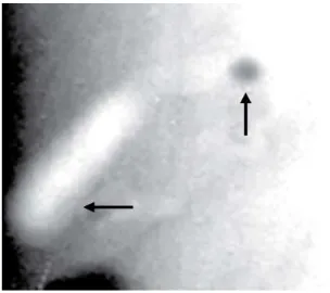
Biliary Radionuclide Scanning (HIDA Scan)
Biliary scintigraphy; provides a noninvasive evaluation of the liver, GB, ducts, and duodenum ; with anatomic and functional information.
49
as in critically ill patients and in patients receiving parenteral nutrition. Filling of the gallbladder and common bile duct with delayed or absent filling of the duodenum indicates an obstruction at the ampulla. Biliary leaks as a complication of surgery of the gallbladder or the biliary” tree can be confirmed and frequently localized by biliary scintigraphy.
Percutaneous Transhepatic Cholangiography:
50
cholangiography (PTC) has little role in the management of patients with uncomplicated gallstone disease but is particularly useful in patients with bile duct strictures and tumors, as it defines the anatomy of the biliary tree proximal to the affected segment. As with any invasive procedure, there are potential risks. For PTC, these are mainly bleeding, cholangitis, bile leak”, and other catheter-related problems
51
Fig 10: MRCP- This view shows the course of the extra- hepatic biliary tree marked by the arrow heads.
Fig 11A: Endoscopic retrograde cholangiography. A schematic picture showing the side-viewing endoscope in the duodenum.
[image:51.595.73.355.70.350.2]53
Endoscopic Ultrasound
Endoscopic ultrasound “requires a special endoscope with an
ultrasound transducer at its tip. The results are operator dependent, but offer noninvasive imaging of the bile ducts and adjacent structures. It is of particular value in the evaluation of tumors and their resectability. The ultrasound endoscope has a biopsy channel, allowing needle biopsies of a tumor under ultrasonic guidance. Endoscopic ultrasound also has been used to identify bile duct stones, and although it is less sensitive than ERC, the technique is less invasive as cannulation of the sphincter of Oddi is not necessary” for diagnosis of choledocholithiasis.
2.6 Management
Symptomatic Gallstones
Patients with symptomatic gallstones should be advised to
have “elective laparoscopic cholecystectomy.” While waiting for surgery; or if surgery has to be postpone; the patient should be advised to avoid dietary fats and large meals. Diabetic patients with symptomatic gallstones; should have a cholecystectomy promptly; as they are more prone to develop acute severe cholecystitis .
54
effective in children as well as in the elderly. Cholecystectomy, open or laparoscopic, for patients with symptomatic gallstones offers excellent long-term results.” About 90% of patients with “typical biliary symptoms and stones are rendered symptom free after cholecystectomy. For patients with atypical symptoms or dyspepsia (flatulence, belching, bloating, and dietary fat intolerance) the results are not” as favorable.
Acute Cholecystitis
55
approximately 2 months later. Approximately 20% of patients will fail to respond to initial medical therapy and require an intervention. For those unfit for surgery, a percutaneous cholecystostomy or an open cholecystostomy under local analgesia can be performed. Failure to improve after cholecystostomy usually is due to gangrene of the gallbladder or perforation. For these patients, surgery is unavoidable. For those who respond after cholecystostomy, the tube can be removed once cholangiography through it shows a patent ductus cysticus. Laparoscopic cholecystectomy may then be scheduled in the near future. For the rare patients who can’t tolerate surgery, the stones can be extracted” via the cholecystostomy tube before its removal.
Choledocholithiasis
For patients with; symptomatic gallstones and suspected
56
surgery to endoscopic treatment have documented less morbidity and mortality for endoscopic treatment in this group of patients. They do not need to be submitted for a cholecystectomy, as only about 15% will become symptomatic from their gallbladder stones, and such patients can be treated as the need arises” by a cholecystectomy.
Surgical Interventions in Biliary Disease:
Cholecystostomy:
This procedure is performed in patients who are unfit for anaesthesia. In this procedure under ultrasound guidance the distended gall bladder is decompressed by percutaneous drainage. Initially a guide- wire is inserted through the abdominal wall, liver and the gall bladder, then a catheter is passed over it. Once the patient’s condition has improved the catheter is removed and cholecystectomy can be done at a later date. This procedure is rarely performed nowadays.
Laparoscopic Cholecystectomy:
57
Open Cholecystectomy:
Due to the advent of laparoscopic technique the number of
open procedures has drastically come down. It is now performed under two settings, one following conversion of the laparoscopic approach and two as a step during another surgery like pancreaticoduodenectomy.
Common Bile Duct Exploration:
Laparoscopic techniques have been improvised in the recent years
58
[image:58.595.73.412.75.286.2]Fig 12: Lap and open techniques
2.7 Thyroid Function
Hypothyroidism:
Hypothyroidism is a common disorder; again especially in women,
59
irregularity. When THs are lacking in early childhood, the result is severe bodily and mental retardation. Clinical signs of hypothyroidism may be dry, coarse and cold skin, periorbital and peripheral oedema, coarse and thin hair, pallor of skin, thick tongue, slow speech, decreased reflexes, hypertension, bradycardia, pleural and pericardial effusions, ascites and vitiligo. In laboratory tests, e.g. hypercholesterolaemia and anaemia are associated findings.” The laboratory hallmark of primary hypothyroidism and the most sensitive test for detecting early thyroid failure is an “increased serum TSH concentration. The serum FT4 level is decreased in clinical hypothyroidism. In the subclinical form an increased serum TSH level is accompanied by a normal serum FT4 level, and the patient is asymptomatic.” The presence of thyroperoxidase antibody confirms chronic autoimmune thyroiditis as the cause of hypothyroidism. The treatment of hypothyroidism with levothyroxine is usually lifelong.
The effects of thyroid hormones:
The thyroid gland secretes both T4 and triiodothyronine (T3) into the circulation. “In extrathyroidal tissues, T4 is converted to T3, assumed to be the major active TH. Characteristic of THs is the multiplicity of the cellular functions they regulate in virtually every type of vertebrate tissue. The diverse responses to TH can be divided into two major categories:
Regulation of metabolic activity, energy consumption and muscular activity in adult mammals, and
60
Most actions of THs can be explained by their interaction with nuclear receptors. THs bind to specific intranuclear TH receptors (TR), TR1, TR1 or TR2. This ligand-receptor complex binds to TH response elements in the target genes to regulate the rate of synthesis of specific messenger RNAs. This results in a change in the amount or activity of the cognate proteins, which in turn alter the rate of the metabolic process. The TRs are expressed in a tissue- and development-stage-specific fashion. For example, TR1 is known to be highly expressed in brain, muscle and fat, and has been identified in frog, chicken, rat, mouse and humans; TR1 is highly expressed in liver and kidney, and has been demonstrated in frog, chicken, rat, mouse and humans. TR2, again, is highly expressed in the pituitary, and has been demonstrated in rat and mouse. The expression of TRs in the SO has not been studied. The genomic effects of THs necessarily require a finite period of time for protein synthesis and for the biological response. Acute response of a cell to THs is unlikely to involve a transcriptional mechanism but is rather a result of nongenomic mechanisms involving extranuclear sites of action. Extranuclear sites of TH action include the cell membrane, the cytoskeleton, the sarcoplasmic reticulum, the cytoplasm, the mitochondria, and in vascular smooth-muscle cells presumably the contractile elements.” For example, THs mediate sugar uptake, adenylate cyclase, and Ca2+- ATPase activity directly at the level of the plasma membrane of various tissues. The second messengers associated with the extranuclear actions of the THs are not yet known.
61
62
is significantly increased and the rate of bile secretion decreased. Biliary secretion of cholesterol is reduced in hypothyroidism compared to euthyroidism. However, when serum cholesterol values rise, bile may also become supersaturated with cholesterol and thus result in gallstone formation. An association has been reported between cholesterol gallstones and treated hypothyroidism in women.” It has also been reported that gallbladder stones may have been dissolved after T4 treatment.
2.8 Prevalence of Clinical Hypothyroidism in Gall Stone Patients
Several recent studies report an association between
63
mU/L), compared to only 1.4% in the control group. In women over 60 years, the prevalence of subclinical hypothyroidism was as high as 11.4% in the gall stone group” compared to 1.8% among” the control patients.
64
been initiated, the gall stones do indeed form or continue to grow. Earlier studies with subclinical hypothyroid patients have demonstrated that a positive effect on changes in the cholesterol level, cardiovascular effects, or neuromuscular symptoms may be achieved with early replacement treatment with thyroxine. It has also been reported that gallstones have dissolved after initiation of thyroxine therapy. It is possible that thyroxine replacement therapy is not sufficient at all times of the day, in all patients to maintain normal sphincter of Oddi function, causing the formation of gall stones. These interesting findings raise a question” about the mechanisms underlying the association, which is stronger between hypothyroidism and CBD stones than between hypothyroidism and gallbladder stones.
Mechanism of Formation of Gallstones in Hypothyroidism:
In general, the pathogenesis of gallstones is a complex process involving mechanisms affecting bile content and bile flow. There are several factors that may contribute to the formation of gall stones in hypothyroid patients. Based on the investigations currently available, it cannot be concluded whether hypothyroid individuals develop isolated gall stones or present with CBD stones in addition to gallbladder stones. “However, based on what is known about the effects of hypothyroidism on the formation of gallbladder and CBD stones, it seems likely that in hypothyroidism both
65
In hypothyroidism, the lack of thyroxine
➢ decreases liver cholesterol metabolism resulting in bile cholesterol supersaturation,
which in turn impairs the motility, contractility, and filling of the gallbladder, contributing to the retention of cholesterol crystals and to the nucleation and growth of gallstones.
➢ diminishes bile secretion from hepatocytes resulting in impaired clearance of
precipitates from the bile ducts.
➢reduces SO relaxation resulting in delayed bile flow and thus the formation and
accumulation of CBD stones.
66
Hypothyroidism Reduces Bile Flow into the Duodenum:
67
duodenum is reduced in the hypothyroid stage. This could be due to changes in bile composition and gallbladder motility, and because of changes” in the resistance to flow, that is, in the SO motility.
Hypothyroidism Leads to Impaired SO Relaxation:
The existence “of gastrointestinal hypoactivity in hypothyroidism has been well known for decades. For example, the effect of thyroxine has been documented in anal canal pressure and in lower esophageal sphincter pressure. The effect of THs on smooth muscle contraction depends on the smooth muscle type and the species studied. THs have a direct, relaxing effect on vascular smooth muscle contractility. This effect is mediated by intranuclear binding of TH to the TR, and partly by nongenomic mechanisms involving extranuclear sites of action. The potassium (K+) channel blocker glibenclamide attenuates triiodothyronine-induced vasodilatation in rat skeletal muscle arteries, and triiodothyronine-induced vasodilatation may thus be mediated by ATP-sensitive K+- channels. Since Sandblom et al. first demonstrated the hormonal action of cholecystokinin (CCK) on the SO in 1935; several other hormones have been shown to affect SO activity. In 2001, it was shown for the first time that thyroxine has a direct effect on SO contractility in physiological concentrations in pig experiments.”
68
relaxation seen in pregnancy, reduced not only the ACh- and Hist-induced but also
Mechanisms by Which Thyroxine Mediates SO Relaxation:
69
The passage of TH through cell membrane, cytoplasm, and nuclear membrane and binding to a nuclear protein (TR) is a relatively fast event, whereas the resulting transcriptional and translational regulation is time consuming, and probably explains why the relaxant effect is not immediate. Thus, the effect of thyroxine could be mediated by regulatory proteins partly synthesized as a result of thyroxine-induced gene expression. The prorelaxant effect of thyroxine is probably partly mediated via transporter proteins, and partly via binding to nuclear receptors, subsequently leading to the activation of K+ channels.” The opening of K+ channels is followed by hyperpolarisation, which closes cell membrane Ca2+ channels, reduces Ca2+ influx, and results in reduced contraction of the SO smooth-muscle cell in response to any specific stimulus.
Conclusions and Clinical Implications:
70
in bile and the reduced bile secretion rate, the deficiency of the pro relaxant effect of thyroxine on the SO appears to be a crucial factor leading to the reduced bile flow in hypothyroidism. The initial formation of bile cholesterol crystals may begin during the untreated period of hypothyroidism, and the stones may continue to develop or mature even after the thyroxine replacement therapy has begun. It is possible that thyroxine replacement therapy is not sufficient in all patients to maintain normal SO function, causing increased risk of CBD stone formation. Studies with subclinical hypothyroid patients have demonstrated that a positive effect on the changes in the serum cholesterol level, on cardiovascular effects, or on neuromuscular symptoms may be achieved with early replacement treatment with thyroxine, and it can be assumed that patients at risk of forming gall stones due to subclinical hypothyroidism may also benefit from such early treatment. In conclusion patients presenting with gallstones, especially females clinicians should be aware of the hypothyroid backround and
perform a thyroid profile.”
71
CHAPTER 2
MATERIALS AND METHOS
72
MATERIALS AND METHODS
3.1 Type of study: Prospective and observational study
3.2 Approval : Prior to conducting the study approval was
obtained from the ethical committee of Thanjavur
Medical College, Thanjavur.
3.3 Study Place: Thanjavur Medical College & Hospital,
Thanjavur – 613004.
3.4 Study Period : 2014 June to 2015 August
3.5 Sample size:
3.6 Inclusion Criteria:
1. Patients in age group of 18-70 yrs and
2. Patients with USG proven cholelithiasis/ choledocholithiasis
3.7 Investigation
1. USG abdomen
2. T3, T4, TSH
3.7 Study procedure:
73
patient admitted in surgery department with features suggestive of extrahepatic biliary lithiasis. Baseline investigations, as routinely required, were done, followed by imaging studies. Patients were then explained about their disease process and the possible line of management. All the necessary information regarding the study was explained to the patients or their valid guardian. Informed written consent was taken from the patients or their guardian willing to participate in the study. Detailed history was taken from the study group to establish proper diagnosis. Thorough physical examination was done in each case. Data collection sheets were filled in by the investigator himself. All of the preoperative factors related to the patient were noted down in the data sheet. After proper evaluation and preparation, patients who required surgical management were taken up for surgery. Strict aseptic precautions were followed during the operation. Meticulous techniques were practiced as far as possible. The operation procedure and related per operative factors were observed directly and recorded in the data collection sheet instantly. After completing the collection of data it was compiled in a systematic way.
3.8 Operational definitions:
Cholelithiasis:
a condition marked by presence of calculi in the gallbladderCholedocholithiasis:
a condition marked by presence of calculi in the common74
Hypothyroidism:
abnormally low activity of the thyroid gland develops when thethyroid gland fails to produce or secrete as much thyroxine as the body needs.
ERCP:
(short for endoscopic retrograde cholangiopancreatography) is a procedureused to diagnose and treat diseases of the gallbladder, biliary system, pancreas, and liver.
3.9 Variables Studied:
Dependent variable:
HypothyroidismIndependent variable:
1. Age 2. Sex
3. Co- morbidities: Diabetes, obesity. 4. USG findings
5. Surgeries performed
3.10 Ethical consideration
75
3.11 Data collection
Data were collected by pre-tested structured questionnaire. Data were collected from all the respondents by direct interview after getting informed written consent from them or from their legal guardian.
3.12 Data analysis
Data analysis was done both manually and by using
76
CHAPTER 3
RESULTS
77
RESULTS
[image:77.595.67.530.369.585.2]A prospective study was performed to determine the incidence of hypothyroidism in patients having cholelithiasis and choledocolthiasis. 68 patients were studied who fulfilled the inclusion criteria. All these patients were taken from the surgery department of Thanjavur Medical College, from 2014 June to 2015 August. All patients were evaluated clinically and only essential investigatons were performed before surgery, in addition patients underwent the thyroid function test. The results obtained are as follows.
TABLE 1: Age distribution of patients with gall stone
AGE/ SEX yrs
MALE FEMALE TOTAL 20-29 2 4 6 30-39 3 13 16 40-49 6 11 17 50-59 5 7 12 60-70 6 11 17 TOTAL 22(32.35%) 46(67.64%) 68
78
Table 2: Percentage of patient with co- morbidities
Co- Morbidity Number Percentage
Diabetes Mellitus 11 16
Hypertension 14 20.5
Both DM and HT 6 8
No co morbidity 49 72.05
Hypertension was the predominant co- morbidity in this study- in 20% of the patients.
0 2 4 6 8 10 12 14 16
20-29 30-39 40-49 50-59 60-70 Total
79
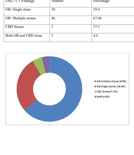
Table 3: USG / CT findings of patients
USG / CT Findings Number Percentage
GB- Single stone 20 29.4
GB- Multiple stones 46 67.64
CBD Stones 5 27.3
Both GB and CBD stone 3 4.4
80
Table 4: Management profile of gall stones
Surgery Performed Numbers Percentage
Open cholecystectomy 30 44.11
Lap Cholecystectomy 36 52.94
CBD exploration 4 5.8
Conservative 2 2.9
Lap Converted to open 4 5.8
36 Patients underwent laparoscopic cholecystectomy out of which 4 patients were converted from lap to open procedure.
0 5 10 15 20 25 30 35 40
Open
Lap
CBD exp
Conservative
81
Table 5: Thyroid Profile of the patients
Numbers Percentage
Hypothyroid 18 26.47
Euthyroid 50 73.52
p value <0.05 and it is statiscally significant.
26% of patients with cholelithiasis/ choledocolitiasis had hypothyroidism.
p value is < 0.05 and it is statistically significant.
74%
26%
82
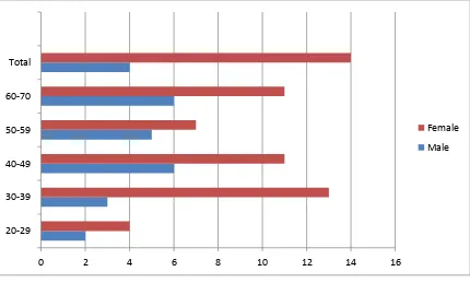
Table 6: Age and sex distribution of patients with hypothyroidism and gallstones/ CBD stones
AGE/ SEX yrs
MALE FEMALE
20-29 0 3(4.4%)
30-39 0 1(1.4%)
40-49 0 1(1.4%)
50-59 1(1.4%) 2(2.9%)
60-70 3(4.4%) 7(10.2%)
TOTAL 4(5.8%) 14(20.5%)
(Figures in parentheses indicates percentages)
Female sex and 50-70 yrs were the predominant group. More than 70% of the patients with hypothyroidism belong to this group.
0 2 4 6 8 10 12 14 16
20-29 yrs 30-39 yrs 40-49 yrs 50-59 yrs 60-70 yrs Total
[image:82.595.73.510.427.689.2]83
Table 7: Image findings of patients with hypothyroidism
USG Findings Numbers Percentage
GB- Single stone 2 11.11
GB- Multiple stones 14 77.77
CBD stone 3 16.6
Both 1 5.5
Total 18 26.47
Table 8: Management profile in gallstone patients with hypothyroidism
Surgeries Performed Numbers Percentage
Open Cholecystectomy 7 38.88
Lap Cholecysyecyomy 7 38.88
CBD exploration 3 16.66
Lap to Open 0 0
Conservative 1 5.5
[image:83.595.69.529.454.638.2]84
0 1 2 3 4 5 6 7 8
85
CHAPTER 4
86
DISCUSSION
This prospective study was conducted 68 selected patients with evidence of cholelithiasis or choledocholithiasis in Department of General Surgery, Thanjavur Medical College. The study was carried out with a view to determine the prevalence of hypothyroidism in patients with extra hepatic biliary lithiasis in view of determining its importance as a causative factor and to include thyroid function tests as part of routine workup in gallstone patients.
Age of these selected patients ranged from 20- 70 yrs, with mean age being 47.14 yrs. 32% were male patients and 68% were female patients. The male to female ratio is 1: 2. Females were the predominant group. On analysing the co- morbid factors HT was the predominant co- morbid factor followed by Diabetes. HT was found to be a co- morbid factor in 14 patients (20%). DM was found to be a co- morbid factor in 11 (16%). 6 patients had both DM and HT.
87
Patients were managed with both laparoscopic and open procedures. 36 patients underwent laparoscopic cholecystectomy out of which 4 patients were converted from lap to open procedure. The reasons for conversion were bleeding, adhesions and technical difficulties. Open procedure was done in 30 patients and 4 patients underwent CBD exploration in addition to cholecystectomy.
Thyroid function test was performed in all 68 patients out of which, 18 patients (26.4%) were found to have hypothyroidism and the rest i.e. 50 patients were euthyroid. Of these 18 patients only 2 were known hypothyroid, rest 16 patients were newly diagnosed hypothyroid patients.
Of these 18 patients, 16 patients were females rest were males. 50-70 yrs were the predominant group. More than 70% of the patients with hypothyroidism belong to this group.
88
LIMITATIONS OF THE STUDY
89
SUMMARY
Cholelithiasis and Choledocholithiasis are one of the commonly encountered diseases in a general surgery department. Multiple risk factors have been attributed to the development of gallstones which include age, female gender, obesity, high fat diet, family history etc. Patients usually present with right hypochondrial pain, dyspepsia, vomiting and occasionally fever or jaundice. Management includes diagnostic imaging studies followed by surgery, if indicated. Hypothyroidism is not an accepted or proven risk factor for gallstones as of yet. Several studies have postulated the possible pathophysiology behind the role of thyroxine in the normal physiology of the biliary system. No concurrence has been established
Age and Sex Distribution:
The most commonly involved age group in patients with gall stones was 50 - 70, with incidence increasing with age. This correlates with the general literature that gallstones is a disease of the elderly. Female sex, is in itself a risk factor for developing gallstones with more than seventy five percent of the involved patients belonging to the female gender.
Co Morbid Factors:
90 Diagnostic Studies:
USG was sensitive in detecting stones in nearly all patients with CECT abdomen and MRCP required only in few cases.
Management:
Laparoscopic and open Cholecystectomy were both perfomed. Conversion to open was seen in 4 patients. This can be attributed to technical problems and as the study was done in a teaching institute and a good percentage of surgeries was done by trainees.
Hypothyroidism and gallstones:
91
CONCLUSION
92
RECOMMENDATIONS
On the basis of the findings of the study, the following recommendations can be made:
1. Evaluation of thyroid profile should be a part of general workup in patients with both cholelithiasis and choledocholithiasis especially in female patients.
2. Proper evaluation and preoperative preparation in patients with hypothyroidism, anticipating complications.
93
BIBLIOGRAPHY
1. Healey J E, Schroy P C 1953 Anatomy of biliary ducts within the human liver; analysis of prevailing pattern of branchings and major variations of biliary ducts. Arch Surg 66: 599–616
2. Bioulac-Sage P, Le Bail B, Balabaud C 1991 Liver and biliary tract histology. In:
McIntyre N, Benhamou J-P, Bircher J, Rizzetto M, Rodes J (eds) Oxford textbook of clinical hepatology Vol 1.Oxford University Press: Oxford: pp 12–20
3. Suzuki et al., 2000. Suzuki M, Akaishi S, Rikiyama T, Naitoh T, Rahman MM, Matsuno S: Laparoscopic cholecystectomy, Calot's triangle, and variations in cystic arterial supply. Surgical Endoscopy 2000; 14:141-144.
4. IHPBA Brisbane, 2000. IHPBA Brisbane 2000 Terminology of Liver Anatomy and Resections
5. Marve GM: Nerves and hormones interact to control gallbladder function. News Physiol Sci 13:64, 1998.
6. Portincasa P, Di Ciaula A, vanBerge-Henegouwen GP:Smooth muscle function and dysfunction in gallbladder disease. Curr Gastroenterol Rep 6:151, 2004.
7. Russell DW: The enzymes, regulation, and genetics of bile acid synthesis. Annu
Rev Biochem 72:137, 2003.
94
9. Kidd JF, Thorn P: Intracellular Ca2+ and Cl- channel activation in secretory cells. Annu Rev Physiol 62:493, 2000.
10. Fuchs M: Bile acid regulation of hepatic physiology: III. Regulation of bile acid synthesis: past progress and future challenges. Am J Physiol Gastrointest Liver
Physiol 284:G551, 2003.
11. Chiang JY: Bile acid regulation of hepatic physiology: III. Bile acids and nuclear
receptors. Am J Physiol Gastrointest Liver Physiol 284:G349, 2003.
12. Brett M, Barker DJ. The world distribution of gallstones. Int J Epidemiol.
1976;5:335.
13. Nakeeb A, Comuzzie AG, Martin L, et al. Gallstones: genetics versus
environment. Ann Surg. 2002;235:842.
14. Brasca A, Berli D, Pezzotto SM, et al. Morphological and demographic associations of biliary symptoms in subjects with gallstones: findings from a population-based survey in Rosario, Argentina. Dig Liver Dis. 2002;34:577. 15. Attili AF, De Santis A, Capri R, et al. The natural history of gallstones: the GREPCO experience. The GREPCO Group. Hepatology. 1995;21:655.
16. Bellows CF, Berger DH, Crass RA. Management of gallstones. Am Fam
Physician. 2005;72:637.
95
Surg. 1998;2:109.
18. Stewart L, Oesterle AL, Erdan I, et al. Pathogenesis of pigment gallstones in Western societies: the central role of bacteria. J Gastrointest Surg. 2002;6:891. 19. Trowbridge RL, Rutkowski NK, Shojania KG. Does this patient have acute cholecystitis? JAMA. 2003;289:80.
20. Fletcher DR. Gallstones. Modern management. Aust Fam Physician. 2001;30:441.
21. Ko C, Lee S. Epidemiology and natural history of common bile duct stones and
prediction of disease. Gastrointest Endosc. 2002;56:S165.
22. Lilly MC, Arregui ME. A balanced approach to choledocholithiasis. Surg Endosc. 2001;15:467.
23. Ross SO, Forsmark CE. Pancreatic and biliary disorders in the elderly. Gastroenterol Clin North Am. 2000;30:531.
24. Lilly MC, Arregui ME. A balanced approach to choledocholithiasis. Surg Endosc. 2001;15:467.
25. Ross SO, Forsmark CE. Pancreatic and biliary disorders in the elderly. Gastroenterol Clin North Am. 2000;30:531.
26. Hunter JG. Acute cholecystitis revisited: get it while it’s hot. Ann Surg.
96
27. Lee HJ, Choi BI, Han JK, et al. Three-dimensional ultrasonography using the minimum transparent mode in obstructive biliary diseases: early experience. J Ultrasound Med. 2002;21:443.
28. Ralls PW, Jeffrey RB Jr, Kane RA, et al. Ultrasonography. Gastroenterol Clin
North Am. 2002;31:801.
29. Wexler RS, Greene GS, Scott M. Left hepatic and common hepatic ductal bile
leaks demonstrated by Tc-99m HIDA scan and percutaneous transhepatic
cholangiogram. Clin Nucl Med. 1994;19:59.
30. Breen DJ, Nicholson AA. The clinical utility of spiral CT cholangiography. Clin
Radiol. 2000;55:733.
31. Liu TH, Consorti ET, Kawashima A, et al. Patient evaluation and management
with selective use of magnetic resonance cholangiography and endoscopic
retrograde cholangiopancreatography before laparoscopic cholecystectomy. Ann
Surg. 2001;234:33.
32. Magnuson TH, Bender JS, Duncan MD, et al. Utility of magnetic resonance
cholangiography in the evaluation of biliary obstruction. J Am Coll Surg.
1999;189:63.
97
open cholecystectomy for acute and gangrenous cholecystitis. Lancet.
1998;351:321.
34. Lo CM, Liu CL, Fan ST, et al. Prospective randomized study of early versus
delayed laparoscopic cholecystectomy for acute cholecystitis. Ann Surg.
1998;227:461.
35. Chikamori F, Kuniyoshi N, Shibuya S, et al. Early scheduled laparoscopic
cholecystectomy following percutaneous transhepatic gallbladder drainage for
patients with acute cholecystitis. Surg Endosc. 2002;16:1704.
36. Patel M, Miedema BW, James MA, et al. Percutaneous cholecystostomy is an
effective treatment for high-risk patients with acute cholecystitis. Am Surg.
2000;66:33.
37. Tranter S, Thompson M. Comparison of endoscopic sphincterotomy and
laparoscopic exploration of the common bile duct.Br J Surg. 2002;89:1495
38. Rhodes M, Sussman L, Cohen L, et al. Randomised trial of laparoscopic
exploration of common bile duct versus postoperative endoscopic retrograde
cholangiography for common bile duct stones. Lancet. 1998;351:159.
39. Byrne MF, Suhocki P, Mitchell RM, et al. Percutaneous cholecystostomy in
98
Am Coll Surg. 2003;197:206.
40. Akhan O, Akinci D, Ozmen MN. Percutaneous cholecystostomy. Eur J Radiol.
2002;43:229.
41. Richards C, Edwards J, Culver D, et al. Does using a laparoscopic approach to
cholecystectomy decrease the risk of surgical site infection? Ann Surg.
2003;237:358.
42. Flum DR, Dellinger EP, Cheadle A, et al. Intraoperative cholangiography and risk
of common bile duct injury during cholecystectomy. JAMA. 2003;289:1639.
43. Biffl W, Moore E, Offner P, et al. Routine intraoperative ultrasonography with
selective cholangiography reduces bile duct complications during laparoscopic
cholecystectomy. J Am Coll Surg. 2001;193:272.
44. Halpin VJ, Dunnegan D, Soper NJ. Laparoscopic intracorporeal ultrasound versus
fluoroscopic intraoperative cholangiography: after the learning curve. Surg
Endosc. 2002;16:336.
45. Barwood NT, Valinsky LJ, Hobbs MS, et al. Changing methods of imaging the
common bile duct in the laparoscopic cholecystectomy era in Western Australia:
Implications for surgical practice. Ann Surg. 2002;235:41.
99
(1993):The thyroid. In: Cecil essentials of medicine, pp 470-481. Eds. TE
Andreoli, JC Bennett,CCJ Carpenter, F Plum and LH Jr Smith, W.B. Saunders
Company, Philadelphia, PA.
47. Woeber KA (1997): Subclinical thyroid dysfunction. Arch Intern Med
157:1065-1068.
48. Woeber KA (2000): Update on the management of hyperthyroidism and
hypothyroidism.Arch Intern Med 160:1067-1071.
49. Wuttke W (1989): The thyroid system. In: Human physiology, pp. 383-386. Eds.
RF Schmidt and G Thews, Springer-Verlag, Berlin, Germany.
50. Vanderpump MP, Tunbridge WM, French JM, Appleton D, Bates D, Clark F,
Grimley Evans J, Hasa