Int. J. Electrochem. Sci., 14 (2019) 238 – 249, doi: 10.20964/2019.01.10
International Journal of
ELECTROCHEMICAL
SCIENCE
www.electrochemsci.org
Electrode materials based on Micro-emulsion Polymerized
Polyaniline and Their Capacitive Property
Lijun Ren*, Gaini Zhang, Huiqin Li, Dengwei Hu, Shumei Dou
College of Chemistry and Chemical Engineering, Engineering Research Center of Advanced
Ferroelectric Functional Materials, Key Laboratory of Phytochemistry, Baoji University of Arts and Sciences, Baoji, 721013, P. R. China
*E-mail: [email protected]
Received: 14 September 2018 / Accepted: 22 October 2018 / Published: 30 November 2018
The conductive polyaniline (PANI) electrode materials with different morphologies were prepared by micro-emulsion polymerization with dodecylbenzene sulfonic acid (DBSA) as the dopant and surfactant, and ammonium persulfate (APS) as the oxidant. The concentration of DBSA was crucial in tailoring the morphology of PANI. Electrochemical tests showed that the capacitive properties of the electrode materials were influenced markedly by their morphologies and conductivities. At low DBSA concentration, the obtained PANI nanoribbon (PANI-1) electrode processed high specific capacitance (573 F g1 at a current density of 0.2 A g1) and excellent rate performance (78% capacitance retention from 0.2 to 10 A g1) but showed poor cycling stability. Furthermore, the specific capacitance and rate performance of the resultant PANI electrodes were decreased with increase in the concentration of DBSA. At high DBSA concentration, the specific capacitance and rate performance of the obtained PANI nanoparticle (PANI-4) electrode decreased to 205 F g1 (0.2 A g1) and63% (from 0.2 to 10 A g 1), respectively. Nevertheless, it possessed superior cycling stability with just 8% capacitance loss after 2000 charging/discharging cycles.
Keywords: Polyaniline; Dodecylbenzene sulfonic acid; Micro-emulsion polymerization; Electrode material; Supercapacitor
1. INTRODUCTION
are scaled down to the nano-level [5]. Therefore, it is necessary to develop reliable preparation methods that can precisely tailor the nanostructure of electrode materials. Many researches have been focused on the design and preparation of various kinds of electrode materials [6, 7]. However, compared to inorganic nanomaterials, it is intrinsically difficult to control the morphology of organic polymer nanomaterials whose dimensional stabilities drop dramatically at the nanoscale level [8].
Polyaniline (PANI) has become one of the attractive cathode candidate materials for SCs due to its high electrical conductivity, ease of synthesis, low cost, and reversibility between redox states through doping/dedoping processes [9]. It has been proven that the nanostructure of PANI can remarkably affect its capacitive property [10]. In recent years, a large number of nanostructured PANI (nano-fibers/nanorods/nanowires/nanotubes) were prepared by a variety of approaches including template preparation method, electrochemical deposition, interfacial polymerization, and micro-emulsion polymerization, which improved their electrochemical properties [5, 11-13]. Among these, micro-emulsion polymerization is known to be one of the most effective methods among various preparation methods [14, 15]. Surfactants are usually appear in the micro-emulsion polymerization system, which are amphiphilic molecules that are known to lower the surface tension of a liquid and to form micelles. Because of their nature, when incorporated into the micro-emulsion polymerization system, they effectively control the growth of particles [16].
Up to now, very less attention has been paid attention to the assembly behaviors of the PANI nanomaterials and the resulting capacitive properties. Here we report preparation of PANI electrode materials with different morphologies by micro-emulsion polymerization with dodecylbenzene sulfonic acid (DBSA) as the dopant and surfactant, and ammonium persulfate (APS) as the oxidant at 20 C. In the micro-emulsion polymerization experiment system, where aniline monomers in the cationic form formed complexation with DBSA micelles, which in turn determine the PANI morphology grown. The morphologies and sizes of PANI nanostructures could be tailored by the concentration of DBSA surfactant. The remarkable capacitance characteristics of the PANI nanostructures could be attributed to their morphologies and conductivities. To verify it, the analyses of molecular structures, aggregation processes and capacitive properties have been studied in detail.
2. EXPERIMENTAL SECTION
2.1 Materials preparation
Aniline (An, analytical grade) was distilled until color-less under reduced pressure before use. Other chemicals (analytical grade) were used as received without further treatment.
at 20 °C. The obtained precipitates were followed by washing with ultrapure water and ethanol, and dried in a vacuum at 60 °C for 10 h, the PANI electrode materials PANI-1, PANI-2, PANI-3, PANI-4 were finally obtained when the different concentrations of DBSA (0.0034, 0.0054, 0.0068, and 0.0136 mol/L) were used in the reaction.
2.2 Characterization
A SU8020 field-emission scanning electron microscopy (FESEM) was used to observe the morphology of the obtained materials. Infrared spectra (FTIR) were obtained by KBr method on a Fourier Transform Infrared Spectrometer EQUINX55. Raman spectra were measured on a Renishaw inVia Raman microscope with an excitation wavelength of 785 nm. The electrochemical properties of the electrode materials were studied on a CHI 660E (Chenhua, Shanghai) electrochemical workstation.
2.3 Electrochemical measurement
The electrochemical properties of the obtained electrode materials were studied by cyclic voltammetry (CV), galvanostatic charge-discharge (GCD) and electrochemical impedance spectroscopy (EIS) in a three electrode system. Electrodes were prepared by mixing electro-active materials, acetylene black and PTFE (polytetrafluoroethylene) in a mass ratio of 85:10:5 to form homogenous slurry. Then the slurry was spread onto the stainless steel cloth (Mesh sizes 500) (1 cm2) and dried at 60 C for 10 h. After drying, the coated mesh was pressed to form working electrodes. The loading mass of the active material was about 3 mg. The electrochemical tests were performed using 1.0 H2SO4 as electrolyte. The specific capacitance of the electrode was calculated from the galvanostatic discharge process according to the following equation: Cs = I t/(V m), where I is the discharge current (A), t is the discharge time (s), V is the voltage change (V) excluding the IR drop during the discharge process, and m is the total mass of the active material for both electrodes (g). The electrochemical impedance spectroscopy (EIS) was performed with an amplitude of 5 mV at a frequency range of 0.01 to 100 kHz.
3. RESULTS AND DISCUSSION
[image:4.596.112.485.69.348.2]
Figure 1. FESEM images of PANI electrode materials prepared using different concentrations of DBSA: (a) PANI-1 ([DBSA] = 0.0034 mol L1), (b) PANI-2 ([DBSA] = 0.0054 mol L1), (c) PANI-3 ([DBSA] = 0.0068 mol L1), and (d) PANI-4 ([DBSA] = 0.0136 mol L1)
Figure 2. Schematic illustration of the possible complexation morphologies between DBSA and aniline cationic to prepare PANI electrode materials
[image:4.596.119.480.426.663.2]
formed complexation with micelles of dodecylbenzene sulfonate anions. As a result, PANI with different morphologies were obtained. As shown in Figure S1, a rock-like structure was readily obtained without using of any structure directing DBSA agent.
[image:5.596.97.502.236.421.2]However, in the presence of DBSA, the morphologies of the resulting PANI materials could be tuned instead to nanoribbons, nanofibers or nanoparticles, which depending on the concentration of DBSA. Figure 1 exhibits the FESEM images of PANI nanostructures prepared using different concentrations of DBSA. It could be seen that the presence of the surfactant impacts the nanostructure of the final material significantly.
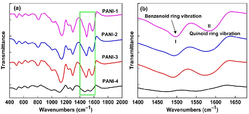
Figure 3. (a) FTIR spectra of PANI electrode materials and (b) the expanded view of 14001680 cm1 region
When the concentration of DBSA was 0.0034 mol L1, PANI nanoribbons with diameters of
500 nm and lengths of 3 m were formed. When the concentration of DBSA was increased to 0.0054 mol L1, a few of PANI nanofibers appeared. When the concentration of DBSA was twice of the original concentration, PANI nanofibers are formed with a diameter of 80 nm. With sufficiently high concentration of DBSA (0.0136 mol L1), PANI nanoparticles with a diameter of 120 nm are found. One of the key factors controlling the electrochemical behavior of conducting polymers is morphology, and modification of PANI morphology can lead to better SC performance [17].
[image:6.596.118.476.319.595.2]
Surfactants interfere with particle growth, which in turn influence the structures and properties of PANI electrode materials [16]. Their ability to induce structural changes could be useful to control the capacitive properties of the resultant PANI electrode materials. FTIR and Raman studies were carried out in order to characterize the molecular structures of PANI electrode materials. The FTIR spectra of the resultant PANI electrode materials prepared using different concentrations of DBSA surfactant are shown in Figure 3. It could be seen that characteristic bands for each PANI sample were almost the same. The spectra exhibited the characteristic bands at around 1575 and 1490 cm1, corresponding to the CC stretching vibrations of quinoid and benzenoid rings, respectively [18-20]. The other bands at 1295, 1130, and 805 cm1 could be assigned to the C−N stretching vibration, the C−H in plane bending vibration and the C−H out of plane bending vibration, respectively [21]. These characteristic bands indicated that all the four PANI electrode materials were identical to the emeraldine salt form. Particularly, the bands at 1010 and 505 cm1 could be assigned to the absorption of –SO3H group of doped DBSA [15].
Table 1. The intensity ratios of III/II in FTIR spectra of the PANI electrode materials
PANI samples PANI-1 PANI-2 PANI-3 PANI-4
III/II 0.77 0.79 0.88 0.92
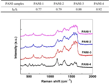
Figure 4. Raman spectra of PANI electrode materials
[image:7.596.62.543.150.420.2]
semiquinone radicals at 1334 cm1, C–H bending of quinoid or benzenoid ring at 1170 cm1 were observed for all samples, indicating the structure of PANI is in doping state [19, 22, 23].
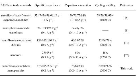
Table 2. Comparison of capacitive properties with reported values
PANI electrode materials Specific capacitance Capacitance retention Cycling stability References
nanofibers/nanosflowers
/nanorods/nanotubes
521/543/638/661 F g1
(1 A g1)
50/70/75/88%
(1~10 A g1)
58/59/58/63%
(1000 C) [5]
nanospheres/nanorods
/nanofibers
71/133/192 F g1
(0.1 A g1)
nearly 0%
(0.1~10 A g1) —— [8]
nanofibers/nanoparticles
/helixes
159/163/190 F g1
(0.5 A g1)
66/39/72%
(0.5~10 A g1)
72/66/79%
(2000 C) [10]
nanorods 279 F g
1
(0.5 A g1)
50%
(0.5~50 A g1)
45%
(2200 C) [26]
nanoribbons/nanofibers
/nanoparticles
573/405/205 F g1
(0.2 A g1)
78/69/63%
(0.2~10 A g1)
52/80/92%
(2000 C) This work
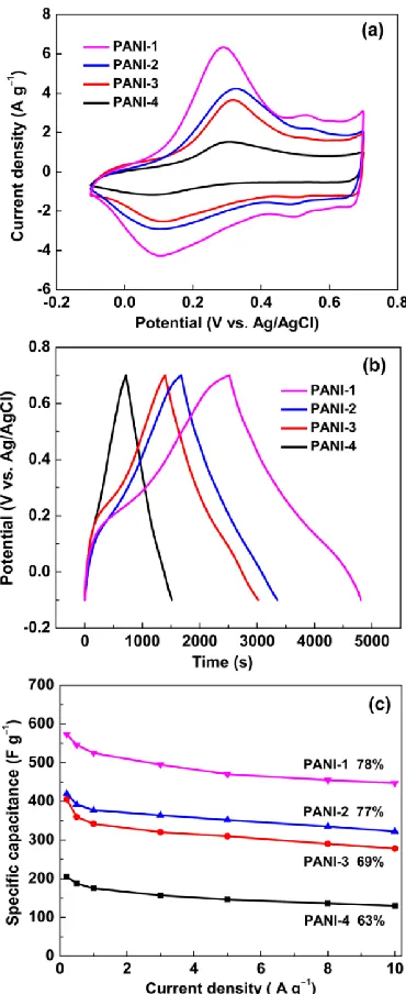
Figure 5. Capacitive properties of PANI electrodes in 1 M H2SO4 electrolyte: (a) CV profiles at 5 mV s1, (b) GCD curves at 0.2 A g1, and (c) capacitance variation from 0.2 to 10 A g1
[image:8.596.114.481.585.728.2]
With the increase of DBSA concentration, the discharging times of the PANI electrodes became shorter, which indicated that an increase in surfactant concentration resulted in poor redox reactions. The differences in the capacitance performance of the four electrodes could be assigned to the diverse morphologies of PANI nanostructure. The rate performance of the PANI electrodes were further investigated at different current densities from 0.2 to 10 A g1 (Figure 5c). It was evident that the specific capacitance gradually decreased with increase in current density. The capacitive retentions of four PANI electrodes from 0.2 to 10 A g1 were 78, 77, 69, and 63% for PANI-1, PANI-2, PANI-3, and PANI-4, respectively. This indicated that the electrodes with lower DBSA concentration had excellent rate performance. Moreover, the specific capacitance of PANI-1 electrode was always higher than those of other three electrodes. The better rate performance for PANI-1 electrode could probably be ascribed to its highly delocalized conjugated structure with high conductivity, which improved the rate of ion transportation. As shown in table 2, the specific capacitance and rate performance of PANI-1 were comparable to the reported PANI electrode materials [5, 8, 10, 26].
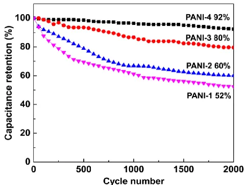
Figure 7. Cycling performance of PANI electrodes at 5 A g1 for 2000 cycles in 1 M H2SO4 electrolyte
Another important parameter of electrode materials for SCs is cycling performance. As exhibited in Figure 7, after 2000 consecutive cycles at a current density of 5 A g1, the specific capacitances of the PANI-1, PANI-2, PANI-3, and PANI-4 electrodes retained about 52, 60, 80, and 92% of their original capacitances, respectively. It was obvious that the better long-term electrochemical stability was achieved at higher DBSA concentration. In comparison with other three PANI electrodes, PANI-4 electrode showed relatively better cycling performance. This capacitance retention was also better than those of earlier reported PANI electrode materials. For example, Zhou’s group reported PANI helixe, nanofiber, and nanoparticle electrodes had 79, 72 and 66% of the original values retain after 2000 cycles, respectively [10], and the PANI nanofiber electrode reported by Du’s group retained only 45% value of initial specific capacitance after 2200 cycles [26]. The small nanoparticles of PANI-4 electrode could restrict the volume changes during insertation/deinsertation processes, which further improved the cycling stability [30]. The above results indicated that the conductivities and morphological differences of DBSA doped PANI electrode materials have great influence on their capacitance behavior, ie. specific capacitance, rate performance and cycling stability.
4. CONCLUSION
SUPPORTING MATERIAL:
Figure S1. FESEM image of PANI prepared without DBSA surfactant
ACKNOWLEDGEMENT
This work was supported by the National Natural Science Foundation of China (51702006), the Natural Science Foundation Research Project of Shaanxi Province (2018JQ2065 and 2018JQ2068), the Scientific Research Project of Shaanxi Province Office of Education (16JK1040), the Science and Technology Project of Baoji (2017JH2-04) and the Doctoral Scientific Research Starting Foundation of Baoji University of Arts and Science (ZK2017031 and ZK2017028).
References
1. K. S. Kumar, N. Choudhary, Y. Jung, J. Thomas, ACS Energy Lett., 3 (2018) 482−495.
2. N. Choudhary, C. Li, J. Moore, N. Nagaiah, L. Zhai, Y. Jung, J. Thomas, Adv. Mater., 29 (2017) 1605336−1605345.
3. H. Sun, Y. Zhang, J. Zhang, X. M. Sun, H. S. Peng, Nat. Rev. Mater., 2 (2017) 17023−17034. 4. J. E. Elshof, H. Yuan, P. G. Rodriguez, Adv. Energy Mater., 6 (2016) 1600355−1600388. 5. L. J. Ren, G. N. Zhang, J. F. Wang, L. P. Kang, Z. B. Lei, Z. W. Liu, Z. T. Liu, Z. P. Hao, Z. H.
Liu, Electrochim. Acta, 145 (2014) 99−108.
6. W. Liu, M. S. Song, B. Kong, Y. Cui, Adv. Mater., 29 (2017) 1603436−1603469. 7. B. M. Sánchez and Y. Gogotsi, Adv. Mater., 28 (2016) 6104−6135.
8. H. W. Park, T. Kim, J. Huh, M. Kang, J. E. Lee, H. Yoon, ACS Nano, 6 (2012) 7624−7633. 9. W. W. Li, F. X. Gao, X. Q. Wang, N. Zhang, M. M. Ma, Angew. Chem. Int. Ed., 55 (2016) 9196 –
9201.
10. C. Li, L. Yang, Y. Meng, X. J. Hu, Z. X. Wei, P. Chen, RSC Adv., 3 (2013) 21315−21319.
11. H. D. Tran, J. M. D’Arcy, Y. Wang, P. J. Beltramo, V. A. Strong, R. B. Kane, J. Mater. Chem. 21 (2011) 3534−3550.
12. D. Li, J. Huang, R. B. Kaner, Accounts Chem. Res., 42 (2009) 135−145.
14. Li Wei, Q. Chen, Y. J. Gu, J. Alloy. Com., 501 (2010) 313–316.
15. D. T. Ge, L. L. Yang, A. Honglawan, J. Li, S. Yang, Chem. Mater., 26 (2014) 1678−1685.
16. J. P. Vareda, P. Maximiano, L. P. Cunha, A. F. Ferreira, P. N. Simões, L. Durães, J Colloid Interf. Sci. 512 (2018) 64–76.
17. A. Eftekhari, L. Li, Y. Yang, J. Power Sources, 347 (2017) 86–107.
18. M. Q. Sun, G. C. Wang, X. W. Li, Q. L. Cheng, C. Z. Li, Ind. Eng. Chem. Res., 51 (2012) 3981−3987. 19. H. P. Cong, X. C. Ren, P. Wang, S. H. Yu, Energy Environ. Sci., 6 (2013) 1185–1191.
20. K. Zhou, Y. He, Q. C. Xu, Q. E. Zhang, A. A. Zhou, Z. H. Lu, L. K. Yang, Y. Jiang, D. T. Ge, X. Y. Liu, H. Bai, ACS Nano, 12 (2018) 5888–5894.
21. Z. Q. Tong, Y. N. Yang, J. Y. Wang, J. P. Zhao, B. L. Su, Y. Li, J. Mater. Chem. A, 2 (2014) 4642– 4651.
22. P. P. Yu, Z. M. Zhang, L. X. Zheng, F. Teng, L. F. Hu, X. S. Fang, Adv. Energy Mater., 6 (2016) 1601111–1601120.
23. F. Huang and D. Chen, Energy Environ. Sci., 5 (2012) 5833–5841.
24. L. J. Ren, G. N. Zhang, Z. Yan, L. Kang, H. Xu, F. Shi, Z. B. Lei, Z. H. Liu, Electrochim. Acta, 231 (2017) 705–712.
25. Z. S. Wang, Q. Zhang, S. C. Long, Y. X. Luo, P. K. Yu, Z. B. Tan, J. Bai, B. H. Qu, Y. Yang, J. Shi, H. Zhou, Z. Y. Xiao, W. J. Hong, S. Wang, H. Bai, ACS Appl. Mater. Interfaces, 10 (2018) 10437–10444.
26. S. P. Sasikala, K. E. Lee, J. Lim, H. J. Lee, S. H. Koo, I. H. Kim, H. J. Jung, S. O. ACS Nano, 11 (2017) 9424–9434.
27. M. M. Mohamed, M. A. Mousa, M. Khairy, A. A. Amer, ACS Omega, 3 (2018) 1801–1814. 28. P. C. Du, L. Lin, H. X. Wang, D. Liu, W. L. Wei, J. G. Li, P. Liu, Mater. Design, 127 (2017) 76–
83.
29. K. Zhang, L. L. Zhang, X. S. Zhao, J. Wu, Chem. Mater., 22 (2010) 1392–1401. 30. C. C. Chang and T. Imae, ACS Sustainable Chem. Eng., 6 (2018) 5162−5172.
![Figure 1. FESEM images of PANI electrode materials prepared using different concentrations of DBSA: (a) PANI-1 ([DBSA] = 0.0034 mol L1), (b) PANI-2 ([DBSA] = 0.0054 mol L1), (c) PANI-3 ([DBSA] = 0.0068 mol L1), and (d) PANI-4 ([DBSA] = 0.0136 mol L1)](https://thumb-us.123doks.com/thumbv2/123dok_us/1754471.128975/4.596.119.480.426.663/figure-fesem-images-electrode-materials-prepared-different-concentrations.webp)