DOI: 10.1102/1470-7330.2013.0034
R E V I E W
Neuroendocrine tumours of the head and neck:
anatomical, functional and molecular imaging and
contemporary management
Navaraj Subedia, Robin Prestwichb, Fahmid Chowdhurya,c, Chirag Patela,c, Andrew Scarsbrooka,c
a
Department of Radiology, Leeds Teaching Hospitals NHS Trust, Leeds, UK;bDepartment of Clinical Oncology, St Jamess Institute of Oncology, Leeds, UK;cDepartment of Nuclear Medicine, Leeds Teaching Hospitals NHS Trust, Leeds, UK
Corresponding address: Dr Andrew Scarsbrook, Department of Nuclear Medicine, Level 1, Bexley Wing, St Jamess University Hospital, Beckett Street, Leeds LS9 7TF, UK.
Email:andrew.scarsbrook@leedsth.nhs.uk
Date accepted for publication 6 June 2013
Abstract
Neuroendocrine tumours (NETs) of the head and neck are rare neoplasms and can be of epithelial or non-epithelial differentiation. Although the natural history of NETs is variable, it is crucial to establish an early diagnosis of these tumours as they can be potentially curable. Conventional anatomical imaging and functional imaging using radio-nuclide scintigraphy and positron emission tomography/computed tomography can be complementary for the diag-nosis, staging and monitoring of treatment response. This article describes and illustrates the imaging features of head and neck NETs, discusses the potential future role of novel positron-emitting tracers that are emerging into clinical practice and reviews contemporary management of these tumours. Familiarity with the choice of imaging techniques and the variety of imaging patterns and treatment options should help guide radiologists in the management of this rare but important subgroup of head and neck neoplasms.
Keywords: Neuroendocrine carcinoma; head and neck malignancy; magnetic resonance imaging; positron emission tomography/ computed tomography; somatostatin receptor scintigraphy; [123I]meta-iodobenzylguanidine scintigraphy.
Introduction
Neuroendocrine tumours (NETs) of the head and neck region are a rare and diverse group of tumours. A wide range of nomenclature has been used to describe these neoplasms, with limited consensus[1,2]. NETs arising in the head and neck can be divided into two broad groups: (1) those with epithelial differentiation including typical carcinoid (well differentiated), atypical carcinoid (mod-erately differentiated, including large cell carcinoma) and small cell carcinomas (poorly differentiated, including composite small cell carcinoma), and (2) neurally derived tumours, including paragangliomas and olfactory neuroblastomas[3]. Merkel cell carcinoma is an uncom-mon primary cutaneous small cell cancer, with a predi-lection for the head and neck region. Rarely, neuroendocrine cancers from non-head and neck sites can metastasize to the head and neck region; this should be considered in the differential diagnosis
particularly if there is a previous history of neuroendo-crine cancer[4]. For the purposes of this article, medullary thyroid cancer and other neuroectodermal tumours, including Ewing sarcoma, primitive neuroectodermal tumours and mucosal melanomas, which have been extensively reviewed elsewhere, are not considered further.
The aim of this article is to discuss current anatomical and functional imaging techniques in head and neck NETs, explore novel molecular imaging techniques, par-ticularly those using highly specific positron-emitting radiopharmaceuticals and the role they may play in the future, describe and illustrate the multimodality imaging appearances of head and neck NETs and review contem-porary patient management in each of the different tumour types. Anatomical (cross-sectional) and func-tional imaging using single photon and positron-emitting radiopharmaceuticals have distinct and complementary roles in the evaluation of head and neck NETs.
This paper is available online athttp://www.cancerimaging.org. In the event of a change in the URL address, please use the DOI provided to locate the paper.
An understanding of the range of NETs found in the head and neck with their highly variable natural history, in conjunction with a familiarity with the range of ima-ging and treatment options available, will be invaluable to help guide radiologists in the management of these rare tumours.
Imaging techniques
Anatomical imaging
Computed tomography (CT) and magnetic resonance imaging (MRI) are the most commonly used techniques when imaging head and neck tumours. Ultrasonography (US) is also widely used as the initial imaging technique in the assessment of neck masses, largely due to its avail-ability and avoidance of ionizing radiation, but it has a relatively limited role in this patient cohort with the exception of patients with head and neck paragangliomas.
Multislice CT facilitates rapid and detailed evaluation of the entire neck with the ability to produce multiplanar reformatted images. A bone algorithm, in addition to a standard soft tissue algorithm, is particularly important for tumours that may involve the skull base. Intravenous contrast administration is essential to delineate the mass, or lymphadenopathy, from adjacent normal structures. Enhancement patterns may be helpful in characterizing some masses, such as paragangliomas.
MRI has superior soft tissue resolution, which makes it an ideal technique for imaging head and neck masses. It is superior to CT in defining intracranial extension of tumours. A head or neck coil is usually required and generally the study should include a combination of axial and coronal T2-weighted fast spin echo (FSE) sequences, T2-weighted fat suppression or inversion recovery sequences and an unenhanced T1-weighted FSE or spin echo (SE) sequence[5]. Further fat-saturated T1-weighted SE sequences after gadolinium administra-tion often improve characterizaadministra-tion of the mass. The addition of contrast-enhanced MR angiography (CE-MRA) has incremental value in patients with head and neck paragangliomas[6]. There is emerging evidence that diffusion-weighted imaging (DWI) may also be a diagnos-tic adjunct in this patient cohort but data are inconclusive at present[7].
Functional Imaging
Functional imaging using single photon radiopharmaceu-ticals has been used to evaluate head and neck NETs for many years. These techniques are often complementary to anatomical imaging, as a result of their superior spe-cificity and the ability to perform total-body imaging, which can identify multifocal disease or distant metasta-ses that may otherwise evade detection. More recently, advances in imaging hardware technology have led to the development of hybrid scanners capable of performing
anatomical and functional imaging sequentially in the same session. Single photon emission computed tomogra-phy (SPECT)/CT has many established clinical applica-tions in the field of oncology, particularly in patients with endocrine malignancies[8]. The use of SPECT/CT in patients with head and neck NETs provides incremental diagnostic value with convenient and accurate co-registra-tion of anatomical and funcco-registra-tional data.
There have also been stepwise advances in the poten-tial range of functional imaging techniques that can be used to evaluate patients with NETs. In particular, there has been significant development of a range of highly sensitive and specific positron-emitting radiopharmaceu-ticals, which show great promise as highly accurate molecular imaging probes for detection and characteriza-tion of NETs[9]. There have been simultaneous advances in positron emission tomography (PET) scanner technol-ogy and combined PET/CT scanners are now widely available.
Tracers used in functional and molecular imaging of head and neck NETs target three distinct cellular aspects:
(1) Somatostatin receptors: these are frequently overex-pressed by NETs and the conventional agent used is [111In]DTPA-octreotide ([111In]pentetreotide, OctreoScan). PET tracers targeting this receptor include 68Ga-labelled somatostatin analogue pep-tides (68Ga-DOTA-TOC, 68Ga-DOTA-NOC, 68 Ga-DOTA-TATE)[10].
(2) Catecholamine synthesis and storage pathways: these are relatively unique to neural crest tumours and the conventional scintigraphic technique uses [123I]- or [131I]-meta-iodobenzylguanidine (MIBG). Novel PET tracers that also target this pathway include [18F]fluorodopamine (FDA) and [18F]fluorodihydroxyphenylalanine (FDOPA)[11]. (3) Glucose metabolism: [18F]fluoro-2-deoxy-D-glucose
(FDG) is the most widely used PET radiotracer in clinical use today. The metabolism and biodistribu-tion of FDG have been extensively described. FDG is a non-physiologic glucose analogue accumulated in tumour cells with an overexpression of glucose transporters, which undergoes phosphorylation and is trapped intracellularly. Uptake of FDG is inver-sely related to the degree of tumour differentiation and correlates with biological behaviour. FDG-PET/ CT has a specific role in imaging poorly differen-tiated NETs with more biologically aggressive behaviour.
There is an emerging treatment paradigm for NETs guided by molecular imaging using some of the diagnos-tic radionuclides described above. In patients with inop-erable or metastatic NETs demonstrating receptor positivity, a possible therapeutic option is to replace the diagnostic radionuclide with a therapeutic one. For exam-ple, 111In or 68Ga can be replaced with 90Y or 177Lu (both emit beta radiation) and labelled with the same
peptides. Therefore, molecular imaging and diagnosis of the disease can be effectively followed by personalized treatment using the same molecular imaging vectors, a process referred to as theranostics. Consequently, soma-tostatin receptor imaging particularly using68Ga peptide PET tracers shows great promise in guiding individua-lized patient dosimetry by pre- or posttherapeutic imaging and assessment of therapy response using quantitative imaging[12]. There is a paucity of data on the use of this treatment in patients with head and neck NETs at present but there is a substantial evidence base on its efficacy in more common NETs[13].
Laryngeal neuroendocrine carcinoma
Epithelial malignancies with neuroendocrine differentia-tion can occur in the larynx, as in any other organ[14]. True laryngeal NETs are a rare and heterogeneous group. Laryngeal NETs originate from laryngeal mucosal pleur-ipotent cells and account for less than 1% of laryngeal tumours. There is a male predilection (male/female ratio of 3:1)[14-16]. As with squamous cell carcinoma of the larynx, smoking is a recognized risk factor[17]. The typi-cal clinitypi-cal presentations of these tumours are hoarseness of voice and dysphagia.
Histological identification of different tumour types is important to understand the tumour behaviour, prognosis and treatment planning[18]. Atypical carcinoid is the most frequent type[14,19]. Both typical and atypical carcinoid tumours commonly involve the supraglottic compartment of the larynx, whereas small cell neuroen-docrine carcinoma typically has a more homogeneous involvement of the larynx[20,21]. Paragangliomas are of neural origin and rarely arise in the larynx; they are dis-cussed later in this review.
Anatomical imaging of laryngeal NETs typically demonstrates an enhancing mass arising from the vocal cords (Fig. 1). CT and MRI are useful to demonstrate local extension of the disease. The findings are non-spe-cific and cannot lead to a definitive diagnosis. Small pri-mary NETs may only be localized by endoscopic assessment and not by conventional anatomical imaging. Due to the rare nature of laryngeal NETs, there is a paucity of data on the value of functional imaging with conventional scintigraphic agents and no data to guide the use of emerging PET tracers described above. There may be a role for FDG-PET/CT in patients with small cell carcinoma of the larynx to facilitate more accurate staging of the disease and assessment of prognosis[22,23]. The management of laryngeal NETs is determined by histological type along with clinical and radiological stag-ing. The preferred treatment options for localized disease are:
(1) Carcinoid tumours of the larynx: surgical excision is the treatment of choice for localized carcinoid tumours of the larynx; a subtotal laryngectomy
may be suitable depending on tumour size. Lymph node metastases are not usually seen, and hence a neck dissection is not required[19]. Radiotherapy is not considered an effective modality of treatment[1]. (2) Atypical carcinoid tumours of the larynx: surgical excision is the treatment of choice for atypical car-cinoids. By contrast with carcinoid tumours, the incidence of neck node metastases is high[19,24] and an elective neck dissection is justified. Radiotherapy and chemotherapy have not been con-sidered as effective treatment modalities[1,24] although this has been challenged by a small more recent series[25].
(3) Small cell tumours of the larynx: the prognosis of these tumours is very poor, with over 90% of patients developing distant metastatic disease[26]. It can be considered a systemic disease in a similar manner to small cell lung cancer. Treatment is with a combination of chemotherapy and radiotherapy.
Non-laryngeal neuroendocrine
carcinoma
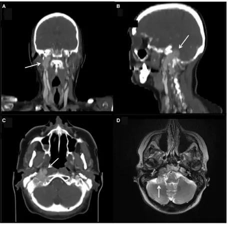
[image:3.595.313.527.47.337.2]neck are sparsely reported in the literature, occurring most commonly in the paranasal sinuses[27]. NETs can rarely arise in other sites, including the salivary glands and mucosa[2]. Like laryngeal NETs, classification is based on the degree of tumour differentiation. Correct pathological classification is challenging; for example in the paranasal sinuses, sinonasal neuroendocrine cancers of epithelial origin must be differentiated from sinonasal undifferentiated carcinomas and olfactory neuroblasto-mas. Large cell neuroendocrine cancers, with pathologi-cal features similar to those arising in the lung, have been reported in salivary glands and arising from the mucosa[2].
Paranasal sinus neuroendocrine cancer represents only about 5% of malignancies arising in this site, and demon-strate aggressive behaviour with a propensity for regional and distant metastases[28]. The ethmoid sinuses and nasal cavity are the most common sites of origin within
the paranasal sinuses[29]. Presenting symptoms overlap with those of benign sinus disease, including epistaxis and nasal obstruction. Partly as a result, presentation is typically with advanced disease[28,29]. Defining an opti-mal treatment strategy is currently impossible due to the lack of data, with surgery, radiotherapy and chemother-apy all potential options. The use of imaging to define adverse features including skull base involvement helps guide an individualized choice of treatment. Regional recurrence rates, reported to be in the order of 25%[28], mandate careful imaging assessment and consideration of prophylactic treatment of the node-negative neck.
[image:4.595.69.527.46.410.2]Anatomical imaging typically demonstrates a heteroge-neously enhancing mass within one of the paranasal sinuses with underlying bony destruction and extension into the adjacent anatomical spaces (Fig. 2). Appearances are often non-specific but the presence of expansion as well as erosion of sinus walls is more Figure 2 Right ethmoid sinus neuroendocrine carcinoma with nodal spread. (A) Axial contrast-enhanced CT and (C) coronal short tau inversion recovery MRI demonstrates the NET (white arrows) centred in the right ethmoidal complex without tumour extension outside the ethmoidal air cells; there is associated expansion of the sinus and bony erosion best appreciated on CT. (B) Axial contrast-enhanced CT and (D) axial fat-suppressed T1-weighted image on Ga MRI showing a right retropharyngeal lymph node (white arrows), which is much more conspicuous on MRI.
suggestive of a NET than the altogether more common squamous cell carcinoma[27]. Somatostatin receptor scin-tigraphy (SRS) improves specificity and positive cases are usually well-differentiated/moderately differentiated tumours and have better clinical outcome with treatment (Fig. 3). Conversely, lack of uptake on octreotide scinti-graphy does not exclude the diagnosis. There are no data in the literature on the use of other functional imaging techniques in this rare subtype of head and neck NETs.
Paragangliomas of the head and neck
Paragangliomas are rare vascular tumours accounting for less than 1% of head and neck tumours[30]. These neu-roectodermal origin tumours arise from a group of tissues (paraganglia), which in the head and neck region migrate along the branchiomeric (of the branchial mesoderm) distribution. Paragangliomas within the head and neck arise mainly from four primary sites: the carotid body at the common carotid artery bifurcation (carotid body tumours), the jugular foramen (glomus jugulare), along the vagus nerve (glomus vagale), and from the tympanic branch of the ascending pharyngeal artery within the middle ear (glomus tympanicum). Other sites, including the larynx[1], are rare. Median age at diagnosis is around 50 years, although paragangliomas can present at any age. Most paragangliomas are sporadic, with only 79% having a familial cause[31]. Presenting symptoms are typ-ically due to cranial nerve dysfunction and/or a slowly enlarging neck mass. Only 25% of paragangliomas
secrete catecholamines[32]and less than 5% of paragan-gliomas are malignant[33]. Malignancy is defined by the presence of regional or distant metastases and cannot be predicted histologically[34]. Biopsy is not usually advised due to the risk of bleeding, and appropriate radiology is essential for diagnosis.
Paragangliomas demonstrate early neural or blood vessel involvement and a propensity for skull base inva-sion and intracranial involvement. CT is the study of choice to investigate bone involvement, whereas MRI defines soft tissue detail, intracranial, neural and dural involvement. On MRI, all paragangliomas exhibit a high signal on T2-weighted imaging and a low signal on T1-weighted imaging (Fig. 4). As with CT, they demonstrate avid contrast enhancement. The classic salt and pepper appearance seen on MRI relates to the presence of hyper-intense foci (salt) interspersed with multiple areas of signal void (pepper) due to high flow in vascular chan-nels (Fig. 4)[35]. This feature is only reliably seen in tumours over 1 cm in size[36].
[image:5.595.74.530.46.264.2]candidates for radionuclide therapy with [131I]MIBG[38]. As a cautionary note, however, a recent study reported a high incidence of false-negative MIBG scans in patients with malignant head and neck paragangliomas that sub-sequently metastasized; this was particularly prevalent in patients with the familial form of the disease[39].
SRS with [111In]DTPA-octreotide has been reported to have a higher sensitivity than MIBG imaging in head and neck paragangliomas[40]. Sensitivity is reduced when there is lack of somatostatin receptor type 2 expression, such as that seen in poorly differentiated malignant para-gangliomas, which are less common in the head and neck and usually familial rather than sporadic in nature. A pitfall of this technique is the potential for false-positive uptake related to inflammation or infection, which can lead to interpretative errors[13]. Newer 68Ga-labelled somatostatin analogue peptides (68Ga-DOTA-TOC, 68
Ga-DOTA-NOC, 68Ga-DOTA-TATE) have shown favourable characteristics in PET/CT imaging, with a high affinity for somatostatin receptors and a stable
process of labelling[13]. They have shown promising results in imaging of nonhead and neck NETs com-pared with conventional SRS but there are no data on their usefulness in paragangliomas. Imaging can be com-pleted within 2 h with no need for delayed imaging on a subsequent day, as opposed to SRS, which routinely requires imaging at 4 h and at 24 h. However, despite the obvious strengths, this technology has been slow to be adopted into clinical routine in the United Kingdom due to a combination of financial considerations, regula-tory hurdles and logistical challenges. It is hoped these issues can be addressed in the near future and, if so, these agents may gradually replace the older agents for func-tional imaging of NETs[9].
[image:6.595.71.526.43.365.2]There has been sustained development and evaluation of several PET ligands with the goal of providing a more holistic molecular fingerprint of neural crest tumours that encompasses other biomarkers such as tumour aggres-siveness and molecular differentiation, which may have an important prognostic and therapeutic significance Figure 4 Glomus vagale in a patient with SDHD genetic mutation. (A) Maximum intensity projection PET image from a half-body FDG-PET/CT scan demonstrates avid FDG uptake within the right parapharyngeal region (black arrow). (B) Axial images from the same study: CT (top), PET (middle) and fused PET/CT (bottom) showing an FDG-avid paraganglioma within the right posterior parapharyngeal space (white arrow). (C) Coronal T1 (top) and short tau inversion recovery (bottom) images from an MRI examination in the same patient demonstrates a 2.5-cm mass situated immediately posterior to the carotid vessels in the right parapharyngeal space consistent with a glomus vagale tumour (paraganglioma). The bottom image shows a heterogeneous signal within the lesion with high signal interspersed with signal void, the so-called salt and pepper appearance.
particularly when combined with genetic and biochemi-cal features. FDOPA-PET/CT is a promising, highly spe-cific imaging technique for evaluation of head and neck paragangliomas. A recent study in patients with non-familial paragangliomas reported a sensitivity of 98% (62/64 lesions) and specificity of 100% for FDOPA-PET/CT, compared with MIBG sensitivity of only 53% (34/64 lesions) and specificity of 91%[41]. Patients with negative MIBG studies were found not to express vesic-ular monoamine transporter expression (VMAT-1) likely as a consequence of the poorly differentiated nature of these tumours. Another study reported that FDOPA-PET had a superior sensitivity of 90%, compared with MIBG-SPECT (65%) and CT and MRI (67%)[42]. However, more limited sensitivity of FDOPA-PET for familial and metastatic paragangliomas has also been reported[43].
Familial paraganglioma is associated with mutations in the succinate dehydrogenase (SDH) gene-mitochondrial complex involved in electron transfer and the Krebs cycle[44]. There are four subunits (A to D) that form the enzyme complex, and these are associated with dif-ferent geno-phenotypic expressions of disease. Patients with SDHB mutations are prone to malignant head and neck paragangliomas with a high propensity for metasta-sis, whereas SDHD mutations typically manifest with multiple, benign head and neck paragangliomas with very rare occurrence of metastatic disease[45]. Up to 10% of head and neck paragangliomas are reported to be related to these hereditary mutations[46]. Limited data exist concerning the clinical and imaging features that distinguish sporadic from familial paragangliomas. A recent study reported that young age, large tumour volume, greater rate of metastatic and multifocal para-gangliomas, higher intralesional metabolic activity on FDG-PET, and increased CT enhancement were observed in SDHB-related head and neck paraganglio-mas[47]. The authors suggested that these findings may warrant genetic screening for SDH mutations and because SDHB-positive patients demonstrate more supra-diaphragmatic lesions, whole-body functional imaging may be of particular value in these patients. In another study that evaluated 30 patients with SDHB germline mutation-related metastatic neural crest tumours, FDG-PET had a sensitivity of 100%, which exceeded that of FDOPA (88%), MIBG (80%), and SRS sensitivity (81%). A large proportion (90%) of the lesions negative on FDOPA and MIBG were localized with FDG-PET sug-gesting it to be the imaging study of choice in patients with SDHB germline mutations[43]. Others have reported that FDA-PET had the highest sensitivity (90%) for detection of bone metastases in patients with metastatic phaeochromocytomas and paragangliomas, followed by bone scintigraphy (82%), CT or MRI (78%), FDG-PET (76%) and MIBG (71%). However, in the subgroup with SDHB mutation, the optimal imaging approaches for bone metastases were CT and MRI (96%), bone
scintigraphy (95%), and FDG-PET (92%)[48]. It is not possible to predict the best combination of anatomical and functional imaging in individual patients with head and neck paragangliomas but, in general, FDG;PET/CT should be considered in the diagnostic work-up of SDHB mutation carriers to provide the most accurate staging (Fig. 5). FDOPA- or FDA-PET may provide the highest accuracy in non-SDHB patients although there are no cost-effectiveness data available and these tracers are lim-ited to highly specialized centres at present.
The presentation and imaging characteristics of each of the different sites of head and neck paraganglioma are summarized below:
Carotid body tumours
The most common type arises from the carotid body and accounts for over 60% of head and neck paraganglio-mas[49]. The epicentre of this tumour is typically the posteromedial wall of the carotid bifurcation but growth along the wall of the external or internal carotid arteries has also been reported[49]. They typically splay the internal and external carotid arteries. On further dis-ease extension, these tumours encase the carotid arteries and extension into the skull base/intracranial cavity is recognized[30]. The most common clinical presentation of a carotid body paraganglioma is an insidiously enlar-ging lateral neck mass often associated with bruit. Other symptoms include hoarseness, stridor, tongue paresis, vertigo, and mild dysphagia[30]. The typical CT appear-ance of a carotid body tumour is an avidly enhancing soft tissue mass located in the infrahyoid neck splaying the internal and external carotid arteries (Fig. 6).
Glomus jugulare tumours
Glomus vagale tumours
Vagal paragangliomas typically occur within or below the inferior ganglion (nodose ganglion) or within the supe-rior ganglion (jugular ganglion)[54]. These are the third most common type of paragangliomas in the head and neck. Typically, patients present with a painless insidious lateral neck mass behind the angle of the mandible. Lower cranial nerve palsies occur late in the disease pro-cess[55]. On CT, vagal paragangliomas appear similar to carotid body tumours but displace both internal and external carotid arteries anteromedially (Fig. 4). In addition, extension into the suprahyoid carotid space is
seen in approximately two-thirds of vagal paraganglio-mas.
Glomus tympanicum tumours
[image:8.595.83.509.48.469.2]The glomus tympanicum tumour is typically a small dis-crete lesion of the cochlear promontory confined within the tympanic cavity[56,57]. Unlike with glomus jugulare, ossicular chain destruction is unusual[56]. These tumours, however, may spread to the mastoid air cells, Eustachian tube and nasopharynx[58]. Pulsatile tinnitus is a common clinical manifestation of this tumour.
Figure 5 Metastatic head and neck paraganglioma in SDHB genetic mutation. (A) Maximum intensity projection PET image from a half-body FDG-PET/CT scan demonstrates a plaque-like area of extremely intense FDG uptake in the right neck around the carotid sheath (top black arrow). There are multiple foci of abnormal uptake elsewhere including the lungs (lower black arrow) and axial skeleton, including the right scapula, ribs, sternum and numerous vertebrae. (B) Axial images from the same study: CT (top), PET (middle) and fused PET/CT (bottom) showing a large FDG-avid bone metastasis within the left side of the sacrum; there is little evidence of structural abnormality on the CT component (white arrow, top).
Management of paragangliomas
Most head and neck paragangliomas demonstrate an indolent growth pattern. One imaging study demon-strated a median growth rate of 1.0 mm/year with a median tumour doubling time of 4 years[59]. Death from paraganglioma is rare[60]and the aim of treatment is to minimize/reduce morbidity rather than to improve survival. Options for treatment include observation for selected cases, surgery or radiotherapy. Traditionally, sur-gery has been the preferred method of primary treatment, with radiotherapy reserved for unresectable disease or less fit patients. The vascularity and skull base location of many paragangliomas make surgical management very challenging. Preoperative embolization has been used to reduce intraoperative blood loss and facilitate complete resection[61]. High local control rates of series of external beam radiotherapy and radiosurgery has challenged this approach. A recent systematic literature review[62] sug-gested that external beam radiotherapy or stereotactic radiosurgery offered similar tumour control compared with surgery, with lower risks of morbidity. Similarly, a meta-analysis[63] of tumour control rates and treatment-related morbidity for glomus jugulare tumours found that radiosurgery offered superior local control with a lower rate of cranial nerve palsy compared with surgery. Laryngeal paragangliomas are generally treated by local excision or partial laryngectomy[1].
Radiological investigations are key to planning both surgical and non-surgical approaches. Choosing the
correct surgical approach requires knowledge of the extent of invasion of adjacent structures. Similarly, the location of steep dose gradients provided by modern highly conformal radiotherapy or stereotactic radiosur-gery allowing sparing of adjacent normal structures requires excellent definition of the soft tissue and bony extent of disease. Radiotherapy is routinely planned on CT imaging in the treatment position. CT/MRI co-regis-tration can be used to directly incorporate the soft tissue definition offered by MRI into the radiotherapy planning process[64].
Olfactory neuroblastoma
Olfactory neuroblastoma (ONB) (also known as esthe-sioneuroblastoma) is a rare malignant neuroectodermal tumour arising from the olfactory epithelium of the olfac-tory ring of the superior nasal mucosa. Neuroepithelial sensory cells found in the upper nasal cavity are thought to be the cellular origin of these tumours[65]. Neuroectodermal carcinoma of the sinonasal tract is also thought to arise from these specialized sensory cells of the nasal mucosa. ONB accounts for about 2% of all sinonasal tract tumours[66]. A bimodal age distri-bution in the 2nd and 6th decades of life has been described, although ONB may occur at any age. No gender predilection has been found[66].
[image:9.595.75.531.47.303.2]and hence unilateral nasal obstruction and epistaxis can occur in the early stage of the disease. However, patients often present with locally advanced disease, due to local spread through the cribriform plate into the skull base. Symptoms of more advanced disease include headache, rhinorrhea or visual disturbances. Occlusion of the ipsi-lateral nasolacrimal duct may lead to epiphoria. There are isolated cases reports of ONB presenting with Cushing syndrome[68]. Clinical assessment may reveal a reddish-grey pedunculated mass.
[image:10.595.69.525.46.496.2]Cross-sectional imaging with CT to assess the extent of bone destruction and MRI are recommended to define the local extent of disease[69]. Anatomical imaging clas-sically reveals a dumbbell-shaped mass centred at the cribriform plate containing intracranial and nasal cavity components (Fig. 8). Speckled calcification and bone erosion are often seen on CT; the tumour exhibits intense contrast enhancement. MRI demonstrates the local extent of the soft tissue component. These tumours are hypointense on T1 and hyperintense on T2 with marked Figure 7 Glomus jugulare tumour. Coronal (A), sagittal (B) and axial (C) images from a contrast-enhanced CT scan demonstrate a bilobed, expansile, hypervascular mass (white arrows) within the right base of the skull extending through the jugular foramen into the right cerebellopontine angle. On the axial image (C), there is clear evidence of involvement of the adjacent bones. (D) Axial T2-weighted image from an MRI scan in the same patient showing heterogeneous signal within the lesion (white arrow) with high signal interspersed with signal void, the so-called salt and pepper appearance that is typical of a paraganglioma (glomus jugulare).
homogeneous contrast enhancement. Both obstructed secretions in the adjacent sinuses and areas of cystic degeneration appear hyperintense[70]. Anatomical ima-ging cannot reliably differentiate these tumours from other more common sinonasal tumours, including sino-nasal undifferentiated carcinoma and squamous cell carcinoma[65].
ONB usually expresses somatostatin receptors and SRS can be used as a diagnostic adjunct[71]. However, several brain tumours including meningiomas, gliomas and pituitary adenomas also express somatostatin
receptors and demonstrate octreotide uptake[72]. This radionuclide imaging is therefore not ideal for the defin-itive diagnosis of ONB. By contrast, MIBG scintigraphy has superior accuracy due to specific uptake and storage mechanisms in neuroblastomas[72]. The sensitivity and specificity of MIBG scintigraphy is very high in assess-ment of extracranial neuroblastomas[73-76]. At present, there are no data available on the use of novel PET agents for assessment of ONB.
[image:11.595.74.530.43.504.2]essential for planning treatment. The most widely recog-nized staging system is the Kadish classification; stage A is confined to the nasal cavity, stage B extends into the sinuses and stage C includes skull base and intracranial involvement and distant disease. Limited retrospective series are the best evidence to guide the management of these rare tumours. Outcomes have improved with the advent of craniofacial resections[77]. Craniofacial resection with clear margins may be adequate treatment for early-stage disease[67]. Adjuvant radiotherapy seems to improve local control and is routinely used for more advanced tumours[78]. Recent case reports suggest a potential role for neoadjuvant chemoradiotherapy before craniofacial resection for advanced disease[79].
ONB can spread to the regional lymph nodes either at presentation or as a site of relapse. The first echelon lymph nodes are in the lateral retropharyngeal region, levels Ib and II[80]. In a recent report of 26 patients treated without elective neck radiotherapy, 7 (27%) relapsed in the neck after a median time of 74 months; half of these neck recurrences were associated with dis-tant disease[80]; the authors recommended elective neck treatment for Kadish stages B and C. Therefore, careful radiological assessment of regional lymph nodes is essen-tial at diagnosis; any additional role of octreotide or MIBG-based imaging in this assessment is uncertain.
Merkel cell carcinoma
First described by Toker in 1972[81], Merkel cell carci-noma (MCC) is an uncommon neuroendocrine (small cell) tumour of the dermis that is characterized by aggres-sive regional nodal invasion, distant metastases, and a high rate of recurrence. Although it remains controver-sial, the widely accepted origin of the tumour is the Merkel cell, which is within the basal layer of the epider-mis usually functioning as a chemoreceptor[82,83].
MCC commonly occurs in sun-exposed skin with over half of these involving the head and neck region, often in elderly whites with no reported sex predilection[84,85]. It commonly manifests as a rapidly growing non-tender nodule in a sun-exposed area.
The clinical manifestation determines the classification of the disease: stage 1, cutaneous involvement; stage 2, regional nodal invasion; stage 3, systemic metastases. The definitive diagnosis of this tumour is based on histo-pathology. Imaging has been shown to be beneficial in staging, surgical guidance, therapeutic management, and follow-up[86,87]. Clinically it is difficult to differentiate stage 1 and 2 disease as up to two-thirds of patients have regional lymphadenopathy at presentation and only 731% of patients with stage 2 disease present with palpable lymphadenopathy[88,89].
The role of lymphoscintigraphy to localize the sentinel nodes that may be involved due to micrometastases from MCC has been examined[90-92]. The sentinel node is the first node to drain 99mTcfiltered sulphur colloid,
0.250.50 mCi (9.2518.5 MBq) of which is injected intradermally around the MCC site. As in melanoma; the presence of sentinel nodes seems to be a strong indi-cator of regional MCC disease[89,93,94]. These studies indicate a high rate of subclinical metastases; in a small meta-analysis, 33% of patients had a positive sentinel node biopsy[90]. The outcome of sentinel node-negative patients managed without adjuvant therapy seemed favourable in this meta-analysis, although with very lim-ited follow-up[90]. Due to the very limited size of the studies in this area, larger prospective studies are required to establish the role of sentinel node biopsy in the routine management of MCC[95].
The use of imaging to exclude distant metastases is essential before aggressive locoregional treatment with curative intent. When pathology demonstrates a cutane-ous small cell carcinoma, all patients should undergo chest imaging to determine whether this is a manifesta-tion of a metastatic small cell carcinoma of the lung or to identify lung metastases. About one-third of patients pres-ent with distant metastases[86]. The metastases can be evaluated with cross-sectional imaging, which may show hypervascular lesions with avid contrast enhancement but the findings are non-specific (Fig. 9). SRS can be used with greater reported sensitivity compared with ana-tomical imaging[96] but may be limited in assessing metastases in organs with physiological uptake of octreo-tide such as the liver, kidneys and spleen.
The additional benefit of FDG-PET/CT compared with conventional imaging has not yet been clearly defined in patients with MCC. However, emerging evidence has suggested that FDG-PET/CT may be more accurate in staging disease compared with conventional imaging methods including functional imaging with SRS[97-99]. The reported sensitivity and specificity of FDG-PET/ CT is as high as 89% and 100%, respectively[97]. FDG-PET/CT may be particularly useful in restaging patients with recurrent disease and in patients with possible metastases at presentation[100].
For locoregionally confined disease, treatment is with surgical excision. Achieving the recommended wide mar-gins of 23 cm is particularly difficult in the head and neck region[101]. This does not appear to be necessary with the use of adjuvant wide-field radiotherapy[102]. There is a high risk of subclinical nodal disease. In the case of MCC in the head and neck region, adjuvant radiotherapy can be used to encompass the primary site, potential in-transit dermal metastases, and regional lymph node beds[95].
Conclusion
In summary, the diverse tissues of the head and neck can give rise to a wide assortment of rare NETs. Anatomical and functional imaging is often complementary in this patient group and has a key role in diagnosis, staging and guiding management decisions. With the advent of
innovative techniques and novel tracers, the role of func-tional and molecular imaging in this patient cohort is likely to expand.
Conflict of interest
The authors have no conflicts of interest to declare.
References
[1] Ferlito A, Silver CE, Bradford CR, Rinaldo A. Neuroendocrine neoplasms of the larynx: an overview. Head Neck 2009; 31: 16341646. PMid:19536850.
[2] Kusafuka K, Ferlito A, Lewis JS, Jr. et al. Large cell neuroendo-crine carcinoma of the head and neck. Oral Oncol 2012; 48: 211215. PMid:22024350.
[image:13.595.72.532.45.540.2][3] Barnes L, editor. Neuroendocrine tumours. Lyon: IARC Press; 2005.
[4] Salama AR, Jham BC, Papadimitriou JC, Scheper MA. Metastatic neuroendocrine carcinomas to the head and neck: report of 4 cases and review of the literature. Oral Surg Oral Med Oral Pathol Oral Radiol Endod 2009; 108: 242247. PMid:19615663.
[5] Hermans R, De Keyser F, Vandecaveye V, Carp L. Imaging tech-niques. In: Hermans Reditor. Head and neck cancer imaging. 2nd ed. Berlin, Heidelberg: Springer-Verlag; 2012.
[6] Neves F, Huwart L, Jourdan G, et al. Head and neck paragan-gliomas: value of contrast-enhanced 3D MR angiography. AJNR Am J Neuroradiol 2008; 29: 883889. PMid:18339724. [7] Aschenbach R, Basche S, Vogl TJ, Klisch J. Diffusion-weighted
imaging and ADC mapping of head-and-neck paragangliomas: initial experience. Klin Neuroradiol 2009; 19: 215219. PMid:19705076.
[8] Chowdhury FU, Scarsbrook AF. The role of hybrid SPECT-CT in oncology: current and emerging clinical applications. Clin Radiol 2008; 63: 241251. PMid:18275863.
[9] Wong KK, Waterfield RT, Marzola MC, et al. Contemporary nuclear medicine imaging of neuroendocrine tumours. Clin Radiol 2012; 67: 10351050. PMid:22633086.
[10] Al-Nahhas A, Win Z, Szyszko T, et al. Gallium-68 PET: a new frontier in receptor cancer imaging. Anticancer Res 2007; 27: 40874094. PMid:18225576.
[11] Taieb D, Timmers HJ, Hindie E, et al. EANM 2012 guidelines for radionuclide imaging of phaeochromocytoma and paraganglioma. Eur J Nucl Med Mol Imaging 2012; 39: 19771995. PMid:22926712.
[12] Baum RP, Kulkarni HR, Carreras C. Peptides and receptors in image-guided therapy: theranostics for neuroendocrine neo-plasms. Semin Nucl Med 2012; 42: 190207. PMid:22475428. [13] Kwekkeboom DJ, Kam BL, van Essen M, et al.
Somatostatin-receptor-based imaging and therapy of gastroenteropancreatic neuroendocrine tumors. Endocr Relat Cancer 2010; 17: R5373. PMid:19995807.
[14] Alujevic A, Juric G, Separovic R, Kruslin B. Unusual features of metastatic atypical carcinoid of the larynx. Eur Arch Otorhinolaryngol 1998; 255: 318321. PMid:9693930. [15] Soga J, Osaka M, Yakuwa Y. Laryngeal endocrinomas
(carci-noids and relevant neoplasms): analysis of 278 reported cases. J Exp Clin Cancer Res 2002; 21: 513. PMid:12071530. [16] Mills SE. Neuroendocrine tumors of the head and neck: a
selected review with emphasis on terminology. Endocr Pathol 1996; 7: 329343. PMid:12114805.
[17] Kasantikul V, Keelawat S, Maneesri S, Panichabhongse V. Moderately differentiated neuroendocrine carcinoma (atypical carcinoid) of the larynx. J Med Assoc Thai 1997; 80: 396401. PMid:9240015.
[18] Ferlito A, Rinaldo A. The spectrum of endocrinocarcinomas of the larynx. Oral Oncol 2005; 41: 878883. PMid:16154516. [19] Ferlito A, Barnes L, Rinaldo A, Gnepp DR, Milroy CM. A review
of neuroendocrine neoplasms of the larynx: update on diagnosis and treatment. J Laryngol Otol 1998; 112: 827834. PMid:9876371.
[20] Machens A, Holzhausen HJ, Dralle H. Minimally invasive surgery for recurrent neuroendocrine carcinoma of the supraglottic larynx. Eur Arch Otorhinolaryngol 1999; 256: 242246. PMid:10392299.
[21] Wenig BM, Gnepp DR. The spectrum of neuroendocrine carci-nomas of the larynx. Semin Diagn Pathol 1989; 6: 329350. PMid:2692106.
[22] Pasquali C, Rubello D, Sperti C, et al. Neuroendocrine tumor imaging: can 18F-fluorodeoxyglucose positron emission tomogra-phy detect tumors with poor prognosis and aggressive behavior? World J Surg 1998; 22: 588592. PMid:9597933.
[23] Rambaldi PF, Cuccurullo V, Briganti V, Mansi L. The present and future role of (111)In pentetreotide in the PET era. Q J Nucl Med Mol Imaging 2005; 49: 225235. PMid:16172568.
[24] Goldman NC, Katibah GM, Medina J. Carcinoid tumors of the larynx. Ear Nose Throat J 1985; 64: 130134. PMid:3979328. [25] Gillenwater A, Lewin J, Roberts D, El-Naggar A. Moderately
differentiated neuroendocrine carcinoma (atypical carcinoid) of the larynx: a clinically aggressive tumor. Laryngoscope 2005; 115: 11911195. PMid:15995505.
[26] Gnepp DR. Small cell neuroendocrine carcinoma of the larynx. A critical review of the literature. ORL J Otorhinolaryngol Relat Spec 1991; 53: 210219. PMid:1653928.
[27] Kanamalla US, Kesava PP, McGuff HS. Imaging of nonlaryngeal neuroendocrine carcinoma. AJNR Am J Neuroradiol 2000; 21: 775778. PMid:10782795.
[28] Mitchell EH, Diaz A, Yilmaz T, et al. Multimodality treatment for sinonasal neuroendocrine carcinoma. Head Neck 2012; 34: 13721376. PMid:22052583.
[29] Smith SR, Som P, Fahmy A, Lawson W, Sacks S, Brandwein M. A clinicopathological study of sinonasal neuroendocrine carci-noma and sinonasal undifferentiated carcicarci-noma. Laryngoscope 2000; 110: 16171622. PMid:11037813.
[30] Bishop GB, Jr. Urist MM, el Gammal T, Peters GE, Maddox WA. Paragangliomas of the neck. Arch Surg 1992; 127: 14411445. PMid:1365691.
[31] Mendenhall WM, Amdur RJ, Vaysberg M, Mendenhall CM, Werning JW. Head and neck paragangliomas. Head Neck 2011; 33: 15301534. PMid:21928426.
[32] Lieberson RE, Adler JR, Soltys SG, Choi C, Gibbs IC, Chang SD. Stereotactic radiosurgery as the primary treatment for new and recurrent paragangliomas: is open surgical resection still the treatment of choice? World Neurosurg 2012; 77: 745761. PMid:22818172.
[33] Rinaldo A, Myssiorek D, Devaney KO, Ferlito A. Which para-gangliomas of the head and neck have a higher rate of malig-nancy? Oral Oncol 2004; 40: 458460. PMid:15006616. [34] Martin TP, Irving RM, Maher ER. The genetics of
paraganglio-mas: a review. Clin Otolaryngol 2007; 32: 711. PMid:17298303. [35] Olsen WL, Dillon WP, Kelly WM, Norman D, Brant-Zawadzki M, Newton TH. MR imaging of paragangliomas. AJR Am J Roentgenol 1987; 148: 201204. PMid:3024473. [36] Vogl TJ, Juergens M, Balzer JO, et al. Glomus tumors of the skull
base: combined use of MR angiography and spin-echo imaging. Radiology 1994; 192: 103110. PMid:8208919.
[37] Ilias I, Divgi C, Pacak K. Current role of metaiodobenzylguani-dine in the diagnosis of pheochromocytoma and medullary thy-roid cancer. Semin Nucl Med 2011; 41: 364368. PMid:21803186.
[38] Avram AM, Fig LM, Gross MD. Adrenal gland scintigraphy. Semin Nucl Med 2006; 36: 212227. PMid:16762612. [39] Fonte JS, Robles JF, Chen CC, et al. False-negative
(1)(2)(3)I-MIBG SPECT is most commonly found in SDHB-related pheo-chromocytoma or paraganglioma with high frequency to develop metastatic disease. Endocr Relat Cancer 2012; 19: 8393. PMid:22167067.
[40] Koopmans KP, Jager PL, Kema IP, Kerstens MN, Albers F, Dullaart RP. 111In-octreotide is superior to 123I-metaiodobenzyl-guanidine for scintigraphic detection of head and neck paragan-gliomas. J Nucl Med 2008; 49: 12321237. PMid:18632829. [41] Fottner C, Helisch A, Anlauf M, et al.
6-18F-fluoro-L-dihydroxy-phenylalanine positron emission tomography is superior to 123I-metaiodobenzyl-guanidine scintigraphy in the detection of extraadrenal and hereditary pheochromocytomas and paragan-gliomas: correlation with vesicular monoamine transporter expression. J Clin Endocrinol Metab 2010; 95: 28002810. PMid:20371665.
excess. J Clin Endocrinol Metab 2009; 94: 39223930. PMid:19622618.
[43] Timmers HJ, Hadi M, Carrasquillo JA, et al. The effects of carbi-dopa on uptake of 6-18F-fluoro-L-DOPA in PET of pheochromo-cytoma and extraadrenal abdominal paraganglioma. J Nucl Med 2007; 48: 15991606. PMid:17873132.
[44] Kantorovich V, King KS, Pacak K. SDH-related pheochromocy-toma and paraganglioma. Best Pract Res Clin Endocrinol Metab 2010; 24: 415424. PMid:20833333.
[45] Timmers HJ, Gimenez-Roqueplo AP, Mannelli M, Pacak K. Clinical aspects of SDHx-related pheochromocytoma and para-ganglioma. Endocr Relat Cancer 2009; 16: 391400. PMid:19190077.
[46] Grufferman S, Gillman MW, Pasternak LR, Peterson CL, Young WG. Jr. Familial carotid body tumors: case report and epidemiologic review. Cancer 1980; 46: 21162122. PMid:7000334.
[47] Venkatesan AM, Trivedi H, Adams KT, Kebebew E, Pacak K, Hughes MS. Comparison of clinical and imaging features in suc-cinate dehydrogenase-positive versus sporadic paragangliomas. Surgery 2011; 150: 11861193. PMid:22136839.
[48] Zelinka T, Timmers HJ, Kozupa A, et al. Role of positron emis-sion tomography and bone scintigraphy in the evaluation of bone involvement in metastatic pheochromocytoma and paragan-glioma: specific implications for succinate dehydrogenase enzyme subunit B gene mutations. Endocr Relat Cancer 2008; 15: 311323. PMid:18310297.
[49] Lack EE, editor. Tumors of the adrenal gland and extra-adrenal paraganglia. Washington, DC: American Registry of Pathology; 1997.
[50] Gulya AJ. The glomus tumor and its biology. Laryngoscope 1993; 103(11 Pt 2 Suppl 60): 715. PMid:8231596.
[51] Dickens WJ, Million RR, Cassisi NJ, Singleton GT. Chemodectomas arising in temporal bone structures. Laryngoscope 1982; 92: 188191. PMid:6298515.
[52] Brown JS. Glomus jugulare tumors. Methods and difficulties of diagnosis and surgical treatment. Laryngoscope 1967; 77: 2667. PMid:4289339.
[53] Kliewer KE, Wen DR, Cancilla PA, Cochran AJ. Paragangliomas: assessment of prognosis by histologic, immunohistochemical, and ultrastructural techniques. Hum Pathol 1989; 20: 2939. PMid:2912871.
[54] Glenner GG, editor. Tumors of the extra-adrenal paraganglion system (including chemoreceptors). Washington DC: Armed Forces Institute of Pathology; 1974.
[55] Tannir NM, Cortas N, Allam C. A functioning catecholamine-secreting vagal body tumor. A case report and review of the lit-erature. Cancer 1983; 52: 932935. PMid:6347360.
[56] Swartz JD, Harnsberger HR, Mukherji SK. The temporal bone. Contemporary diagnostic dilemmas. Radiol Clin North Am 1998; 36: 819853, vi. PMid:9747191.
[57] Valavanis A, Schubiger O, Oguz M. High-resolution CT investi-gation of nonchromaffin paragangliomas of the temporal bone. AJNR Am J Neuroradiol 1983; 4: 516519. PMid:6308990. [58] Chakeres DW, LaMasters DL. Paragangliomas of the temporal
bone: high-resolution CT studies. Radiology 1984; 150: 749753. PMid:6320257.
[59] Jansen JC, van den Berg R, Kuiper A, van der Mey AG, Zwinderman AH, Cornelisse CJ. Estimation of growth rate in patients with head and neck paragangliomas influences the treat-ment proposal. Cancer 2000; 88: Jun 1528112816. PMid:10870065.
[60] van der Mey AG, Frijns JH, Cornelisse CJ, et al. Does interven-tion improve the natural course of glomus tumors? A series of 108 patients seen in a 32-year period. Ann Otol Rhinol Laryngol 1992; 101: 635642. PMid:1497267.
[61] Tasar M, Yetiser S. Glomus tumors: therapeutic role of selective embolization. J Craniofac Surg 2004; 15: 497505. PMid:15111818.
[62] Suarez C, Rodrigo JP, Bodeker CC, et al. Jugular and vagal paragangliomas: systematic study of management with surgery and radiotherapy. Head Neck 2013; 35: 11951204. PMid:22422597.
[63] Ivan ME, Sughrue ME, Clark AJ, et al. A meta-analysis of tumor control rates and treatment-related morbidity for patients with glomus jugulare tumors. J Neurosurg 2011; 114: 12991305. PMid:21029039.
[64] Prestwich RJ, Sykes J, Carey B, Sen M, Dyker KE, Scarsbrook AF. Improving target definition for head and neck radiotherapy: a place for magnetic resonance imaging and 18-flu-oride fluorodeoxyglucose positron emission tomography? Clin Oncol (R Coll Radiol) 2012; 24: 577589.
[65] Argani P, Perez-Ordonez B, Xiao H, Caruana SM, Huvos AG, Ladanyi M. Olfactory neuroblastoma is not related to the Ewing family of tumors: absence of EWS/FLI1 gene fusion and MIC2 expression. Am J Surg Pathol 1998; 22: 391398. PMid:9580174. [66] Broich G, Pagliari A, Ottaviani F. Esthesioneuroblastoma: a gen-eral review of the cases published since the discovery of the tumour in 1924. Anticancer Res 1997; 17: 26832706. PMid:9252701.
[67] Morita A, Ebersold MJ, Olsen KD, Foote RL, Lewis JE, Quast LM. Esthesioneuroblastoma: prognosis and management. Neurosurgery 1993; 32: 706714; discussion 714715. PMid:8492845.
[68] Arnesen MA, Scheithauer BW, Freeman S. Cushings syndrome secondary to olfactory neuroblastoma. Ultrastruct Pathol 1994; 18: 6168. PMid:8191648.
[69] Dulguerov P, Allal AS, Calcaterra TC. Esthesioneuroblastoma: a meta-analysis and review. Lancet Oncol 2001; 2: 683690. PMid:11902539.
[70] Oskouian RJ Jr, Jane JA, Sr. Dumont AS, Sheehan JM, Laurent JJ, Levine PA. Esthesioneuroblastoma: clinical presenta-tion, radiological, and pathological features, treatment, review of the literature, and the University of Virginia experience. Neurosurg Focus 2002; 12: e4. PMid:16119902.
[71] Ramsay HA, Kairemo KJ, Jekunen AP. Somatostatin receptor imaging of olfactory neuroblastoma. J Laryngol Otol 1996; 110: 11611163. PMid:9015433.
[72] Lamszus K, Meyerhof W, Westphal M. Somatostatin and soma-tostatin receptors in the diagnosis and treatment of gliomas. J Neurooncol 1997; 35: 353364. PMid:9440032.
[73] Wafelman AR, Hoefnagel CA, Maes RA, Beijnen JH. Radioiodinated metaiodobenzylguanidine: a review of its biodis-tribution and pharmacokinetics, drug interactions, cytotoxicity and dosimetry. Eur J Nucl Med 1994; 21: 545559. PMid:8082671.
[74] Lumbroso JD, Guermazi F, Hartmann O, et al. Meta-iodobenzyl-guanidine (mIBG) scans in neuroblastoma: sensitivity and speci-ficity, a review of 115 scans. Prog Clin Biol Res 1988; 271: 689705. PMid:3261424.
[75] Rufini V, Giordano A, Di Giuda D, et al. [123I]MIBG scintigra-phy in neuroblastoma: a comparison between planar and SPECT imaging. Q J Nucl Med 1995; 39(4 Suppl 1): 2528. PMid:9002745.
[76] Troncone L, Rufini V, Montemaggi P, Danza FM, Lasorella A, Mastrangelo R. The diagnostic and therapeutic utility of radio-iodinated metaiodobenzylguanidine (MIBG). 5 years of experi-ence. Eur J Nucl Med 1990; 16: 325335. PMid:2351179. [77] Howard DJ, Lund VJ, Wei WI. Craniofacial resection for tumors
of the nasal cavity and paranasal sinuses: a 25-year experience. Head Neck 2006; 28: 867873. PMid:16823871.
[78] McLean JN, Nunley SR, Klass C, Moore C, Muller S, Johnstone PA. Combined modality therapy of esthesioneuroblas-toma. Otolaryngol Head Neck Surg 2007; 136: 9981002. PMid:17547995.
the literature. J Clin Oncol 2011; 29: e358e361. PMid:21282533.
[80] Demiroz C, Gutfeld O, Aboziada M, Brown D, Marentette LJ, Eisbruch A. Esthesioneuroblastoma: is there a need for elective neck treatment? Int J Radiat Oncol Biol Phys 2011; 81: e255e261. PMid:21676553.
[81] Toker C. Trabecular carcinoma of the skin. Arch Dermatol 1972; 105: 107110. PMid:5009611.
[82] Tang CK, Toker C. Trabecular carcinoma of the skin: an ultra-structural study. Cancer 1978; 42: 23112321. PMid:719609. [83] Winkelmann RK. The Merkel cell system and a comparison
between it and the neurosecretory or APUD cell system. J Invest Dermatol 1977; 69: 4146. PMid:68982.
[84] Brissett AE, Olsen KD, Kasperbauer JL, et al. Merkel cell carci-noma of the head and neck: a retrospective case series. Head Neck 2002; 24: 982988. PMid:12410532.
[85] Brenner B, Sulkes A, Rakowsky E, et al. Second neoplasms in patients with Merkel cell carcinoma. Cancer 2001; 91: 13581362. PMid:11283937.
[86] Gollub MJ, Gruen DR, Dershaw DD. Merkel cell carcinoma: CT findings in 12 patients. AJR Am J Roentgenol 1996; 167: 617620. PMid:8751663.
[87] Eftekhari F, Wallace S, Silva EG, Lenzi R. Merkel cell carcinoma of the skin: imaging and clinical features in 93 cases. Br J Radiol 1996; 69: 226233. PMid:8800866.
[88] Yiengpruksawan A, Coit DG, Thaler HT, Urmacher C, Knapper WK. Merkel cell carcinoma. Prognosis and manage-ment. Arch Surg 1991; 126: 15141519. PMid:1842182. [89] Marenda SA, Otto RA. Adnexal carcinomas of the skin.
Otolaryngol Clin North Am 1993; 26: 87116. PMid:8433844. [90] Mehrany K, Otley CC, Weenig RH, Phillips PK, Roenigk RK,
Nguyen TH. A meta-analysis of the prognostic significance of sentinel lymph node status in Merkel cell carcinoma. Dermatol Surg 2002; 28: 113117; discussion 117. PMid:11860419. [91] Gupta SG, Wang LC, Penas PF, Gellenthin M, Lee SJ, Nghiem P.
Sentinel lymph node biopsy for evaluation and treatment of patients with Merkel cell carcinoma: the Dana-Farber experience and meta-analysis of the literature. Arch Dermatol 2006; 142: 685690. PMid:16785370.
[92] Hitchcock CL, Bland KI, Laney RG 3rd, Franzini D, Harris B, Copeland EM 3rd. Neuroendocrine (Merkel cell) carcinoma of
the skin. Its natural history, diagnosis, and treatment. Ann Surg 1988; 207: 201207. PMid:3277546.
[93] Hill AD, Brady MS, Coit DG. Intraoperative lymphatic mapping and sentinel lymph node biopsy for Merkel cell carcinoma. Br J Surg 1999; 86: 518521. PMid:10215828.
[94] Zeitouni NC, Cheney RT, Delacure MD. Lymphoscintigraphy, sentinel lymph node biopsy, and Mohs micrographic surgery in the treatment of Merkel cell carcinoma. Dermatol Surg 2000; 26: 1218. PMid:10632680.
[95] Veness MJ, Palme CE, Morgan GJ. Merkel cell carcinoma: a review of management. Curr Opin Otolaryngol Head Neck Surg 2008; 16: 170174. PMid:18327038.
[96] Kwekkeboom DJ, Hoff AM, Lamberts SW, Oei HY, Krenning EP. Somatostatin analogue scintigraphy. A simple and sensitive method for the in vivo visualization of Merkel cell tumors and their metastases. Arch Dermatol 1992; 128: 818821. PMid:1599271.
[97] Maury G, Dereure O, Du-Thanh A, Mariano-Goulart D, Guillot B. Interest of (18)F-FDG PET-CT scanning for staging and management of Merkel cell carcinoma: a retrospective study of 15 patients. J Eur Acad Dermatol Venereol 2011; 25: 14201427. PMid:21366705.
[98] Peloschek P, Novotny C, Mueller-Mang C, et al. Diagnostic imaging in Merkel cell carcinoma: lessons to learn from 16 cases with correlation of sonography, CT, MRI and PET. Eur J Radiol 2010; 73: 317323. PMid:19108971.
[99] Concannon R, Larcos GS, Veness M. The impact of (18)F-FDG PET-CT scanning for staging and management of Merkel cell carcinoma: results from Westmead Hospital, Sydney, Australia. J Am Acad Dermatol 2010; 62: 7684. PMid:20082888. [100] Iagaru A, Quon A, McDougall IR, Gambhir SS. Merkel cell
carcinoma: Is there a role for 2-deoxy-2-[F-18]fluoro-D-glucose-positron emission tomography/computed tomography? Mol Imaging Biol 2006; 8: 212217. PMid:16724293.
[101] Allen PJ, Bowne WB, Jaques DP, Brennan MF, Busam K, Coit DG. Merkel cell carcinoma: prognosis and treatment of patients from a single institution. J Clin Oncol 2005; 23: 23002309. PMid:15800320.