Heparan Sulfate Proteoglycan Is an
Important Attachment Factor for Cell
Entry of Akabane and Schmallenberg
Viruses
Shin Murakami,
aAkiko Takenaka-Uema,
bTomoya Kobayashi,
aKentaro Kato,
a,cMasayuki Shimojima,
a,dMassimo Palmarini,
eTaisuke Horimoto
aDepartment of Veterinary Microbiology, Graduate School of Agricultural and Life Sciences, The University of Tokyo, Tokyo, Japana; Department of Infection Control and Disease Prevention, Graduate School of Agricultural and Life Sciences, The University of Tokyo, Tokyo, Japanb; National Research Center for Protozoan Diseases, Obihiro University of Agriculture and Veterinary Medicine, Obihiro, Japanc; Department of Virology I, Special Pathogens Laboratory, National Institute of Infectious Diseases, Tokyo, Japand; MRC-University of Glasgow Centre for Virus Research, Glasgow, United Kingdome
ABSTRACT
Akabane virus
(AKAV) and Schmallenberg virus (SBV) are members of
the genus
Orthobunyavirus
, which are transmitted by arthropod vectors with a broad
cellular tropism
in vitro
as well as
in vivo
. Both AKAV and SBV cause
arthrogryposis-hydranencephaly syndrome in ruminants. The main cellular receptor and attachment
factor for entry of these orthobunyaviruses are unknown. Here, we found that AKAV
and SBV infections were inhibited by the addition of heparin or enzymatic removal
of cell surface heparan sulfates. To confirm this finding, we prepared heparan sulfate
proteoglycan (HSPG)-knockout (KO) cells by using a clustered regularly interspaced
short palindromic repeat (CRISPR)-Cas9 system and measured the quantities of
bind-ing of these viruses to cell surfaces. We observed a substantial reduction in AKAV
and SBV binding to cells, limiting the infections by these viruses. These data
demon-strate that HSPGs are important cellular attachment factors for AKAV and SBV, at
least
in vitro
, to promote virus replication in susceptible cells.
IMPORTANCE
AKAV and SBV are the etiological agents of arthrogryposis-hydranencephaly
syndrome in ruminants, which causes considerable economic losses in the livestock
industry. Here, we identified heparan sulfate proteoglycan as a major cellular
attach-ment factor for the entry of AKAV and SBV. Moreover, we found that heparin is a
strong inhibitor of AKAV and SBV infections. Revealing the molecular mechanisms of
virus-host interactions is critical in order to understand virus biology and develop
novel live attenuated vaccines.
KEYWORDS
Akabane virus, heparan sulfate, Schmallenberg virus, cell entry
A
kabane virus
(AKAV) and Schmallenberg virus (SBV) belong to the Simbu serogroup
of the arthropod-borne
Orthobunyavirus
genus of the family
Bunyaviridae
. AKAV
and SBV are phylogenetically closely related, and they also possess similar biological
characteristics: (i) both cause abortion, stillbirth, premature birth, and congenital
deformities in cattle, sheep, and goats; (ii) both primarily infect the central nervous
system (CNS) of the fetus; and (iii) both are difficult to control, because they are
transmitted by biting midges of the genus
Culicoides
(1). Both AKAV and SBV cause
“abortion storms” that result in considerable economic losses to the livestock industry
(2–4).
Despite these similarities, there are important features that distinguish these two
viruses. AKAV is endemic throughout Australia, Southeast Asia, East Asia, the Middle
East, and Africa, whereas SBV has emerged and has dispersed across a large area of
Received28 March 2017Accepted10 May 2017
Accepted manuscript posted online24 May 2017
CitationMurakami S, Takenaka-Uema A, Kobayashi T, Kato K, Shimojima M, Palmarini M, Horimoto T. 2017. Heparan sulfate
proteoglycan is an important attachment factor for cell entry of Akabane and Schmallenberg viruses. J Virol 91:e00503-17.
https://doi.org/10.1128/JVI.00503-17.
EditorTerence S. Dermody, University of Pittsburgh School of Medicine
Copyright© 2017 American Society for Microbiology.All Rights Reserved. Address correspondence to Shin Murakami, [email protected], or Taisuke Horimoto, [email protected].
crossm
August 2017 Volume 91 Issue 15 e00503-17 Journal of Virology jvi.asm.org 1
on November 7, 2019 by guest
http://jvi.asm.org/
Europe since 2011. AKAV comprises four genogroups (I to IV), whereas SBV comprises
a single genotype. No differences in the pathogenicities of different SBV strains have
been described. On the other hand, the OBE-1 strain of AKAV [AKAV(OBE-1)]
group II) causes severe fetal malformation, whereas the Iriki strain [AKAV(Iriki)]
(geno-group I) also causes fatal nonsuppurative encephalomyelitis in newborn cattle.
Molec-ular determinants distinguishing the pathogenicities of these two different strains are
unknown (5).
Orthobunyaviruses carry a tripartite, single-stranded, negative-sense RNA genome.
The L segment encodes the L protein, a viral RNA-dependent RNA polymerase; the S
segment encodes the N protein and the nonstructural protein NSs, both transcribed
from an overlapping open reading frame; and the M segment encodes NSm and the
two major viral envelope proteins, Gn and Gc (Gn/Gc), which form heterodimeric spikes
on the virus particle. Gn/Gc are the proteins on the surface of the virion that bind to cell
surface molecules in the initial step of orthobunyavirus infection (6, 7). Relatively few
studies have investigated orthobunyavirus entry. La Crosse and Germiston neurotropic
orthobunyavirus entry into the cell has been described to be promoted by DC-SIGN (8,
9). However, DC-SIGN is probably not the main attachment factor of ruminant
orthobu-nyaviruses, because it is expressed on macrophages and dendritic cells, not in the CNS.
Heparan sulfate proteoglycan (HSPG), one of major negatively charged transmembrane
protein-linking glycosaminoglycans, is expressed by almost all cells, including neural
cells. HSPG is involved in cell attachment of many viruses (e.g., herpes simplex virus [10,
11], adenovirus [12], respiratory syncytial virus [13, 14], human papillomavirus [15],
foot-and-mouth disease virus [16], hepatitis B virus [17], hepatitis C virus [18], Ebola
virus [19, 20], dengue virus [21], and human immunodeficiency virus [22]). In addition,
HSPG is involved in cell attachment of phleboviruses in the family
Bunyaviridae
,
including Rift Valley fever virus and Toscana virus (23–25). In a previous study,
hem-agglutination of AKAV was inhibited by the addition of heparin, a form of heparan
sulfate (26). Therefore, it is possible that HSPGs are involved in AKAV and/or SBV
infection, similarly to other viruses.
In this study, we examined the role of HSPGs in AKAV and SBV replication.
RESULTS
Heparin or heparinase treatment inhibits AKAV and SBV infections.
Heparin is
a highly sulfated form of heparan sulfate (27) and is a known inhibitor of infection by
various viruses. To test whether heparin inhibits AKAV or SBV replication, AKAV and SBV
were incubated with different concentrations of heparin for 30 min, and the titers of the
viruses neutralized by heparin were measured by plaque reduction assay in HmLu-1
cells (Fig. 1A), which support efficient replication of the Akabane and Schmallenberg
viruses and abundantly express heparan sulfate. The number of plaques induced by
either AKAV or SBV was reduced in a heparin concentration-dependent manner. These
data suggest that heparin affects the replication cycles of both viruses.
Next, we pretreated HmLu-1 cells with heparinase to remove HPSG from the cell
surface and subsequently infected them with AKAV(OBE-1), AKAV(Iriki), and SBV. We
also used VSV-ΔG-GFP, a vesicular stomatitis virus (VSV)-based vector expressing green
fluorescent protein (GFP) that can complete only a single replication cycle within the
cell (28), because this virus does not utilize HSPG during the early events of infection
(23, 29). At 8 h postinfection (hpi), cells were fixed and stained with anti-AKAV or
anti-SBV antibodies for the detection of AKAV- or SBV-infected cells, respectively.
GFP-positive cells were instead counted to determine the number of
VSV-ΔG-GFP-infected cells. As shown in Fig. 1B, VSV-ΔG-GFP was not susceptible to heparinase
treatment, whereas the numbers of cells infected by AKAV and SBV were reduced in a
heparinase concentration-dependent manner. SBV was more susceptible to heparinase
treatment than the AKAVs (
P
⬍
0.05 at 20 and 78
U/
l of heparinase). These data
suggest that HSPG plays important roles in AKAV and SBV infections.
AKAV and SBV replication in HSPG-KO cells.
In order to further validate the data
shown above, we established HSPG-knockout (HSPG-KO) cells using a clustered
on November 7, 2019 by guest
http://jvi.asm.org/
larly interspaced short palindromic repeat (CRISPR)-Cas9 system (30) disrupting the
EXT2
gene, which encodes one of the HSPG-synthesizing enzymes (27). We designed
two guide RNA (gRNAs) (EXT2-1 and EXT2-2) targeting different positions in the
EXT2
gene and obtained three clones for each target (EXT2KO-1-1, -1-2, -1-3, -2-1, -2-2, and
-2-3 cells). We also established control “random-KO” HmLu-1 cells by introducing a
20-nucleotide (nt) random target sequence in the gRNA with the CRISPR-Cas9 system
and obtained three clones of the random-KO cells (random-KO-1, -2, and -3). Lack
of HSPG expression in HSPG-KO cells, but not in random-KO or wild-type cells, was
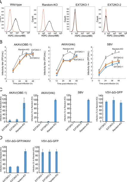
confirmed by flow cytometry analysis (Fig. 2A). In the HSPG-KO cells, titers of
AKAV(OBE-1) and AKAV(Iriki) were about 100-fold lower than that in random-KO cells at
24 hpi but were at similar levels at later time points (Fig. 2B). SBV titers were instead
between 10 and 1,000-fold lower in HSPG-KO cells than in random-KO cells throughout
the course of the experiment (Fig. 2B). Next, we examined AKAV and SBV infectivity in
the EXT2-KO cells and random-KO cells. EXT2-KO cells or random-KO cells were infected
with AKAV(OBE-1), AKAV(Iriki), SBV, or VSV-ΔG-GFP (multiplicity of infection [MOI] of
0.1). At 8 hpi, AKAV antigen-positive cells, SBV antigen-positive cells, or GFP-positive
cells were counted (Fig. 2C). Control VSV-ΔG-GFP-infected cell numbers were not
significantly different between random-KO and HSPG-KO cells. Five to 10 times lower
numbers of AKAV and SBV antigen-positive cells were detected in EXT2-KO cells than
random-KO cells. To eliminate the possibility that the replication step of AKAV affected
the results shown in Fig. 2C, we used a VSV-pseudotyped virus bearing AKAV
glyco-proteins (VSV-ΔG-GFP/AKAV). VSV-ΔG-GFP/AKAV or VSV-ΔG-GFP was inoculated into
EXT2-KO cells or random-KO cells. At 8 hpi, GFP-positive cells were counted (Fig. 2D).
As shown in Fig. 2C, control VSV-ΔG-GFP-infected cell numbers did not show a
significant difference between random-KO and HSPG-KO cells. However,
three-times-fewer VSV-ΔG-GFP-infected cells were detected in EXT2-KO cells than in random-KO
cells (
P
⬍
0.01). These data indicated that HSPG was important for AKAV and SBV
infections.
Quantification of AKAV and SBV bound to the HSPG-KO cell surface.
To
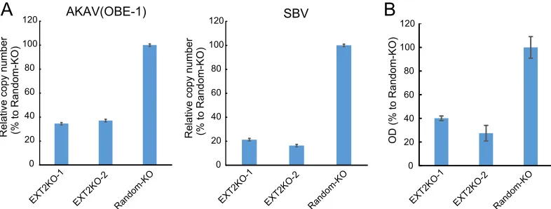
deter-mine whether HSPG is important for AKAV and SBV cell surface attachment, we
quantified the amounts of cell surface-bound viruses. AKAV(OBE-1) and SBV were
incubated with EXT2-KO-1, EXT2-KO-2, or random-KO cells for 1 h at 4°C, and
virus-bound cells were collected. RNA was extracted from the cells, and AKAV or SBV RNA (S
AKAV(OBE-1)
AKAV(Iriki)
SBV
0 20 40 60 80 100 120
0 125 250 375 500 625 750 875 1000
P
laque
fo
rm
at
ion i
n
hi
bi
ti
on (
%
)
Heparin (μg/mL) AKAV(Iriki)
SBV AKAV(OBE-1)
A
B
VSV-ΔG-GFP
0 20 40 60 80 100 120
R
e
la
ti
v
e
in
fe
c
tiv
it
y
(
%
)
0 20 78 313 1250 5000
Heparinase (μU/μL)
FIG 1Effects of heparin and heparinase treatment on AKAV and SBV infections. (A) Effects of heparin on AKAV and SBV plaque reduction. Various concentrations of heparin were incubated for 30 min at room temperature with 100 PFU of AKAV(OBE-1), AKAV(Iriki), or SBV, and the ability of heparin to reduce plaque formation was assessed. (B) Effects of heparinase treatment on AKAV and SBV infectivity. HmLu-1 cells were treated with various concentrations of heparinase II, followed by AKAV or SBV infection. Cells were stained for AKAV or SBV antigen, and positive cells were counted under a fluorescence microscope. For VSV-ΔG-GFP-infected cells, GFP-positive cells were counted under a fluorescence microscope. Results are expressed in percentages relative to cells that were not treated with heparinase. The data are reported as the mean value with standard deviations for three independent experiments.
August 2017 Volume 91 Issue 15 e00503-17 jvi.asm.org 3
on November 7, 2019 by guest
http://jvi.asm.org/
[image:3.585.41.403.74.245.2]0 1 2 3 4 5 6 7 8 9
0 12 24 36 48
0 1 2 3 4 5 6 7 8
0 12 24 36 48
0 1 2 3 4 5 6 7 8 9
0 12 24 36 48
Time post-infection (h) Time post-infection (h) Time post-infection (h)
Infectivity titer (PFU/mL) Infectivity titer (PFU/mL) Infectivity titer (PFU/mL)
Random-KO EXT2KO-1 Random-KO Random-KO
EXT2KO-1
EXT2KO-1 EXT2KO-2
EXT2KO-2
EXT2KO-2
AKAV(OBE-1)
AKAV(Iriki)
SBV
100 200 500 400 300 100 200 500 400 300 600 700 100 200 500 400 300 100 200 500 400 300 600 700 900 800
0 102 103 104 105 0 102 103 104 105 0 102 103 104 105 0 102 103 104 105
Co unt Co unt Co unt Co unt
Wild-type
Random-KO
EXT2KO-1
EXT2KO-2
HSPG (Alexa488) HSPG (Alexa488) HSPG (Alexa488) HSPG (Alexa488)
A
B
0 20 40 60 80 100 120 0 20 40 60 80 100 120 0 20 40 60 80 100 120 140 EXT2KO-1EXT2KO-2 Random-KOInfection (% to Random-KO) Infection (% to Random-KO) Infection (% to Random-KO)
AKAV(OBE-1)
AKAV(Iriki)
SBV
C
VSV-ΔG-GFP
0 20 40 60 80 100 120Infection (% to Random-KO)
EXT2KO-1EXT2KO-2
Random-KO EXT2KO-1EXT2KO-2Random-KO EXT2KO-1EXT2KO-2Random-KO
140
D
EXT2KO-1EXT2KO-2 Random-KO EXT2KO-1EXT2KO-2 Random-KO 0 20 40 60 80 100 120Infection (% to Random-KO)
0 20 40 60 80 100 120
Infection (% to Random-KO)
VSV-ΔG-GFP/AKAV
VSV-ΔG-GFP
FIG 2AKAV and SBV growth kinetics and infectivity in HSPG-KO HmLu-1 cells. (A) Flow cytometric analysis of EXT2-KO HmLu-1 cells. CRISPR-Cas9-mediated EXT2-KO cell clones (EXT2-1 and EXT2-2) were labeled with anti-heparan sulfate mouse monoclonal antibody (10E4) (black) or with isotype control (red) and analyzed by flow cytometry (FACSVerse; BD Biosciences). Representative data (one out of three clones of random-KO, EXT2KO-1, and EXT2KO-2) are shown. (B) Growth kinetics of AKAV or SBV in HSPG-KO cells. AKAV(OBE-1),
(Continued on next page)
on November 7, 2019 by guest
http://jvi.asm.org/
[image:4.585.46.473.74.686.2]segment) was quantified by quantitative reverse transcription-PCR (qRT-PCR) (Fig. 3A).
AKAV and SBV RNAs were significantly lower in HSPG-KO cells than in random-KO cells
(
P
⬍
0.01). We also measured cell-bound AKAV by quantifying N protein using an
N-detecting sandwich enzyme-linked immunosorbent assay (ELISA) (Fig. 3B). Cell
surface-bound N proteins were significantly lower in EXT2-KO-1 and EXT2-KO-2 cells
than in random-KO cells, confirming the qRT-PCR results. These data suggest that HSPG
is an important molecule for AKAV and SBV cell surface attachment.
AKAV and SBV replication is restored in cells with
EXT2
added back.
We next
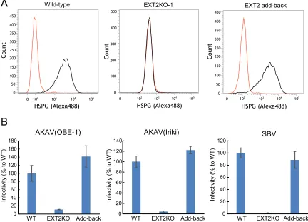
transduced the EXT2-KO cells with the
EXT2
gene in order to confirm that AKAV and SBV
replication could be restored by simply reintroducing the
EXT2
gene back to EXT2-KO
cells. We first confirmed that HSPG expression was restored in these cells by flow
cytometry (Fig. 4A). Next, we examined AKAV and SBV infectivity in the
EXT2
gene
transduced EXT2-KO cells and wild-type cells. EXT2-KO cells or wild-type cells were
infected with AKAV(OBE-1), AKAV(Iriki), or SBV. At 8 hpi, AKAV- and SBV-infected cells
were detected (Fig. 4B). As expected, infectivity of AKAV(OBE-1), AKAV(Iriki), and SBV
was restored in the cells with
EXT2
added back.
DISCUSSION
AKAV and SBV infect neurons (2, 4, 31) and a broad range of cells in the infected
hosts. This suggests that AKAV and SBV use a cellular receptor(s) or attachment factor(s)
FIG 2Legend (Continued)
AKAV(Iriki), or SBV was inoculated onto three clones of random-KO, EXT2KO-1, and EXT2KO-2 cells at a multiplicity of infection of 0.01. Virus titers were determined by plaque assay in normal HmLu-1 cells. The data are reported as the mean titer for three clones of each KO cell (EXT2KO-1, EXT2KO-2, or random-KO) with standard deviations. (C) Infectivities of AKAV and SBV in HSPG-KO cells. Random-KO or HSPG-KO cells were infected with AKAV(OBE-1), AKAV(Iriki), SBV, or control VSV-ΔG-GFP. Cells were stained for AKAV or SBV antigen, and positive cells were counted under a fluorescence microscope. For VSV-ΔG-GFP-infected cells, GFP-positive cells were counted under a fluorescence microscope. Results are expressed as percentages relative to the number of positive random-KO cells. The data are reported as the mean value for three clones of each KO cell (EXT2KO-1, EXT2KO-2, or random-KO) with standard deviations. (D) Infectivities of VSV pseudotyped with AKAV Gn/Gc (VSV-ΔG-GFP/AKAV) in HSPG-KO cells. Random-KO or HSPG-KO cells were infected with VSV-ΔG-GFP/AKAV or control VSV-ΔG-GFP. GFP-positive cells were counted under a fluorescence microscope. Results are represented as percentages relative to the number of positive random-KO cells. The data are shown as the mean value for three clones of each KO cell (EXT2KO-1, EXT2KO-2, or random-KO) with standard deviations.
0 20 40 60 80 100 120
0 20 40 60 80 100 120
0 20 40 60 80 100 120
AKAV(OBE-1)
SBV
OD (% to Random-KO)
Relative copy number (% to Random-KO) Relative copy number (% to Random-KO)
A
B
EXT2KO-1 EXT2KO-2 Random-KO EXT2KO-1 EXT2KO-2 Random-KO EXT2KO-1 EXT2KO-2 Random-KO
FIG 3AKAV and SBV binding assays in HSPG-KO cells. (A) Real-time RT-PCR for the quantification of cell surface-attached viruses. AKAV(OBE-1) or SBV was incubated with HSPG-KO cells at 4°C. After a washing step, total RNAs were extracted. AKAV or SBV S RNAs were quantified by one-step real-time RT-PCR. For relative quantification, standard curves of AKAV or SBV S RNA and GAPDH were prepared by serial dilution of a mixture of total RNA from uninfected HmLu-1 cells and RNA extracted from AKAV(OBE-1) or SBV-containing supernatants. Results are expressed as the percentages relative to the levels in random-KO cells. The results are representative of three different experiments. (B) Sandwich ELISA for the detection of N proteins of AKAV attached to cell surfaces. AKAV(OBE-1) was inoculated onto HSPG-KO or random-KO HmLu-1 cells and left for 1 h at 4°C. After a washing step, the cells were lysed, and the lysates were added to the anti-AKAV N monoclonal antibody (5E8)-coated wells of 96-well ELISA plates (Maxisorp, Nunc), followed by incubation with biotinylated anti-AKAV mouse polyclonal antibody. Subsequently, the wells were incubated with avidin-biotinylated horseradish peroxidase (HRP) complex (Vectastain ABC kit; Vector Laboratories). A 3,3=,5,5=-tetramethylbenzidine (TMB) substrate solution was used for detection, and optical density values were measured. Results are expressed as percentages relative to the number of positive random-KO cells. The results are representative of three independent experiments.
August 2017 Volume 91 Issue 15 e00503-17 jvi.asm.org 5
on November 7, 2019 by guest
http://jvi.asm.org/
[image:5.585.43.436.73.222.2]that is expressed on a variety of cells. DC-SIGN was previously shown to promote
La Crosse and Germiston neurotropic orthobunyavirus entry (8, 9). However, the
distribution of DC-SIGN is not consistent with the tropisms of either AKAV or SBV,
because DC-SIGN is expressed on a limited number of cell types (i.e., dendritic cells and
macrophages). Here, we demonstrated that HSPG plays an important role in AKAV and
SBV infections as an attachment factor. Cell surface HSPGs are expressed ubiquitously
throughout the body (27), including on neuronal cells (32). Therefore, our data support
the correlation between HSPG distribution and AKAV and SBV tropism. However, AKAV
and SBV show a strong preference for neuronal cells
in vivo
, and there are likely other
reasons for this. AKAV and SBV were still able to replicate in HSPG-KO cells, although at
lower levels than those in HSPG-expressing cells. These data suggest the presence of
another cellular factors(s) that defines the tissue tropisms of AKAV and SBV. Further
studies are needed to identify the neuronal cell-specific receptors or reveal other
defining steps after cell entry.
In AKAV(Iriki)-infected HSPG-KO cells, after the virus titer reached a plateau at 24
and 36 hpi, a second AKAV(Iriki) growth wave was observed at 48 hpi (Fig. 2B). During
the AKAV(Iriki) replication in HSPG-KO cells, the virus may have acquired a mutation(s)
in the receptor-binding site of the glycoproteins, possibly leading to adaptation to
HSPG-KO cells. Thus, to investigate whether the AKAV(Iriki) recovered from HSPG-KO
cells at a later time point of infection included mutations in the glycoproteins, we
sequenced the entire M segment at 60 hpi but found no mutations. Hence, the
mechanism of the second growth wave of AKAV(Iriki) still remains unknown.
Continu-0 20 40 60 80 100 120 140 160 180
WT EXT2KO Add-back
AKAV(OBE-1)
0 20 40 60 80 100 120 140
WT EXT2KO Add-back
AKAV(Iriki)
0 20 40 60 80 100 120
WT EXT2KO Add-back
SBV
100 200
50 400
300
150
Co
unt
0 102 103 104 105 HSPG (Alexa488)
0 102 103 104 105
HSPG (Alexa488) 0
102 103 104 105 HSPG (Alexa488) 100
200 400
300 250
350 450
100 200
50 400
300
150 250 350 450
0 0
0
500
Co
unt
Co
unt
Wild-type EXT2KO-1 EXT2 add-back
Infectivity (% to WT) Infectivity (% to WT) Infectivity (% to WT)
A
B
FIG 4Rescue of AKAV and SBV infectivities by adding back theEXT2gene to EXT2-KO cells. (A) Flow cytometric analysis of adding back theEXT2gene in EXT2-KO-1 HmLu-1 cells. Cells were labeled with anti-heparan sulfate mouse monoclonal antibody (10E4) and analyzed by flow cytometry (FACSVerse; BD Biosciences). (B) Infectivities of AKAV and SBV in cells withEXT2added back. The cells were infected with AKAV(OBE-1), AKAV(Iriki), or SBV. Cells were stained for AKAV or SBV antigen, and positive cells were counted under a fluorescence microscope. Results are expressed as percentages relative to the number of wild-type cells. The data are reported as the mean value with standard deviations for three independent experiments.
on November 7, 2019 by guest
http://jvi.asm.org/
[image:6.585.41.473.76.387.2]ous virus passage in HSPG-KO cells may result in mutations to adapt to another cellular
attachment factor(s) or receptor(s).
Repetitive passage of some viruses in tissue culture induces one or two amino acid
mutations in their glycoproteins, which increases their affinity toward HSPGs (33–38).
This adaptation also induces viral attenuation
in vivo
(33–38). A previous study showed
that cell culture-derived SBV showed slightly slowed replication in cattle; however, the
involvement of HSPG underlying this is unknown (39). Rift Valley fever virus (23),
dengue virus (40), and human T-lymphotropic virus 1 (HTLV-1) (41) do not acquire any
mutations after passage in cell culture, which enhances their affinity toward HSPGs. This
suggests that the species of the virus determines whether the virus adapts to cell
culture and uses HSPGs for entry. AKAV(OBE-1), AKAV(Iriki), and SBV, used in the present
study, were passaged several times in tissue culture before being cloned into reverse
genetics plasmids. However, AKAV(Iriki) maintained its pathogenic potential against
goat fetuses and mice (42, 43), implying that it did not undergo changes during
passage in the cell culture. However, it is unclear whether serial passages in cell culture
induced mutations in Gn/Gc proteins of AKAV and SBV to give them higher affinity to
HSPG. Therefore, further studies are needed to compare the HSPG-binding affinities
of the viral strains examined with those of our lab strains and isolates from clinical
specimens which have original sequences.
In this study, we showed that the heparinase susceptibly of SBV was greater than
that of AKAVs. In addition, SBV replication in HSPG-KO cells was limited compared to
that of AKAVs. Moreover, AKAV(OBE-1) exhibited slightly lower HSPG dependency than
AKAV(Iriki) did for virus infection and growth. One possible explanation for the
differ-ence in HSPG dependency is that AKAV(OBE-1), AKAV(Iriki), and SBV possess different
sequences in their binding domains and probably in Gn or Gc. Although
HSPG-binding domains have been identified in other viral glycoproteins as well as in cellular
proteins (44), we could not find known HSPG-binding motifs in AKAV and SBV
glyco-protein sequences (data not shown). HSPG-binding domains are not simply defined by
the secondary sequences of HS-binding proteins (44). The tertiary structure of these
proteins and heparan sulfate interactions are also important for binding. Determining
the three-dimensional (3D) structure of Gn/Gc proteins is likely required to define the
binding site more precisely. Although the orthobunyavirus Gn/Gc 3D structure is not
available currently, determining the 3D structure of Gn/Gc proteins is likely required to
define the HSPG binding site.
Here, we clearly show that AKAV and SBV utilize HSPG for their initial cell surface
attachment in gene-edited HSPG-KO cells. These findings further our understanding of
the orthobunyavirus life cycle. Molecules inhibiting orthobunyavirus and HSPG
inter-actions may be effective antivirals.
MATERIALS AND METHODS
Cells.Baby hamster kidney cells stably expressing T7 RNA polymerase (BHK/T7-9 cells) (45) were kindly provided by Naoto Ito (Gifu University, Japan) and cultured in Dulbecco’s modified Eagle’s minimum medium (DMEM) supplemented with 5% fetal calf serum (FCS) and 10% tryptose phosphate broth at 37°C. Golden hamster lung (HmLu-1) cells were cultured at 37°C in DMEM supplemented with 5% FCS. Human embryonic kidney (HEK293T) cells were maintained in DMEM supplemented with 10% FCS.
Viruses.AKAV strains OBE-1 and Iriki (42, 46) and SBV (31) were generated by reverse genetics. Briefly, to recover AKAV(Iriki), 1.2 g of pT7riboSM2/IL, 0.6 g of pT7riboSM2/IM, and 1.2 g of pT7riboSM2/IS plasmids were mixed in 200l of Opti-MEM (GIBCO, Grand Island, NY, USA) with 9l of Trans-IT LT1 transfection reagent (Mirus Bio, Madison, WI, USA), incubated at room temperature for 15 min, and then added to BHK-T7 cells (45) grown in 6-well plates. To recover AKAV(OBE-1), 1.2g of pT7riboSM2/OL, 0.6g of pT7riboSM2/OM, and 1.2g of pT7riboSM2/OS plasmids were transfected into BHK-T7 cells as described for AKAV(Iriki) generation. To recover SBV, 1g each of pUCSBVST7, pUCSB-VMT7, and pUCSBVLT7 plasmids and 2g of a plasmid expressing T7 polymerase under the control of the chicken-actin promoter (pCAGGS-T7pol) were mixed in 300l of Opti-MEM (GIBCO) with 15l of Trans-IT 293 transfection reagent (Mirus), incubated at room temperature for 15 min, and then added to HEK293T cells grown in 6-well plates. At 3 days posttransfection, the culture supernatant of transfected cells was harvested and added to HmLu-1 cells. Viruses were propagated in HmLu-1 cells cultured in DMEM supplemented with 2% FCS. Nonspreading vesicular stomatitis virus (VSV-ΔG-GFP) pseudotyped with its own glycoprotein G was propagated following transfection of the VSV G protein-expressing
August 2017 Volume 91 Issue 15 e00503-17 jvi.asm.org 7
on November 7, 2019 by guest
http://jvi.asm.org/
plasmid (pCAGGS-VSVG) into HEK293T cells (28). VSV-pseudotyped virus with AKAV Gn and mutant Gc was also propagated in HEK293T cells following transfection of the mutant Gn/Gc/NSm of AKAV(OBE-1), which lacks a 10-amino-acid region in the C-terminal of the wild-type Gc. This mutant was made since VSV-pseudotyped virus with Gn and mutant Gc lacking the C termini of cytoplasmic tail glycoproteins of Crimian-Congo hemorrhagic fever (CCHF) virus, a member ofBunyaviridae, showed higher titer than VSV-pseudotyped virus with wild-type CCHF Gn/Gc proteins (47).
Production of PAbs to AKAV or SBV.An anti-AKAV mouse polyclonal antibody (PAb) was prepared by two intraperitoneal injections of sucrose gradient-purified AKAV(OBE-1) into 6-week-old female ICR mice (Japan SLC, Hamamatsu, Japan) at 2-week intervals. An anti-SBV mouse PAb was prepared by two intraperitoneal injections of sucrose gradient-purified SBV in 6-week-old female ICR mice (Japan SLC) at 2-week intervals. Reactivities and specificities of both anti-SBV and anti-AKAV PAbs were confirmed by immunofluorescence assay, using AKAV(OBE-1)- or SBV-infected and mock-infected HmLu-1 cells.
Plaque assay.A standard plaque assay was used to determine the infectivities of AKAV and SBV. After virus adsorption to HmLu-1 cells for 1 h at 37°C, the inocula were removed, and cells were overlaid with DMEM containing 0.6% agarose and 2% FCS. After incubation for 3 days, cells were stained with neutral red before counting PFU.
Plaque reduction assay.One hundred microliters of serial 2-fold dilutions of heparin solution were prepared in minimal essential medium (MEM) containing 0.3% bovine serum albumin (BSA/MEM). An equal volume of the suspensions containing 100 PFU/100l of AKAV(OBE-1), AKAV(Iriki), or SBV was added to each dilution. After incubation for 30 min at room temperature, 200l of each virus-heparin mixture (containing 100 PFU of viruses) was titrated by the plaque assay.
Heparinase treatment.Cells were seeded in 48-well plates at 24 h prior to infection. Medium was removed, and the cells were incubated with serial 4-fold dilutions of heparinase II (New England BioLabs) in BSA/MEM with 2 mM CaCl2for 1 h at 37°C and then washed with BSA/MEM. The plates were
immediately transferred on ice to suppress the synthesis and transport of HSPG. We then added the appropriate amounts of AKAV(OBE-1), AKAV(Iriki), or SBV to 100l of BSA/MEM and incubated the mixture for 1 h on ice. The supernatant was removed, and the cells were washed twice with BSA/MEM and with and DMEM with 5% FCS before incubation for 8 h at 37°C. The cells were then fixed with 4% paraformaldehyde for 15 min at room temperature. After removing paraformaldehyde, the cells were permeabilized with 0.1% Triton X-100 and incubated with an anti-AKAV N mouse monoclonal antibody (MAb) (5E8) (48) for AKAV-infected cells or an anti-SBV mouse PAb for SBV-infected cells, followed by incubation with Alexa Fluor 488-conjugated anti-mouse IgG antibody. The fluorescence-positive cells were counted as AKAV- or SBV-infected cells under fluorescence microscopy (Vert. A1; Carl Zeiss).
EXT2-KO cells.EXT2-KO cells were established using the CRISPR-Cas9 system.EXT2gene target sequences (EXT2-1 [CTATCCCCTGAAAAAGTACG] or EXT2-2 [CTACACGGATGACATCAGCC]) containing oligonucleotides were introduced into the guide RNA (gRNA) expression cassette of the plentiCRISPR vector (a gift from Feng Zhang; Addgene plasmid 52961) (30). A random target sequence (N20) containing
oligonucleotides was also introduced into the gRNA expression cassette of the plentiCRISPR vector. One microgram of the plentiCRISPR plasmid containing each target gRNA sequence was transfected into HmLu-1 cells (1 ⫻105cells) with TransIT-LT1 (Mirus). One day after transfection, the medium was
replaced with 10g/ml of puromycin-containing medium for 5 days of selection. Surviving cells were passaged, diluted, and inoculated onto fresh dishes for colony formation. Each colony was picked, propagated, and genotyped. The genomic region surrounding the CRISPR-Cas9 target site for each gene in the cloned cell was PCR amplified with KOD-FX neo (Toyobo), and the PCR products were gel extracted and sequenced using a 3130 Genetic Analyzer (ABI). Primer sequences are available upon request. Clones with indels introduced at the targeted site were picked. Genotyped clones were detached with phosphate-buffered saline (PBS) containing EDTA and incubated with anti-heparan sulfate MAb (10E4) (USBio) (49), followed by incubation with Alexa Fluor 488-conjugated anti-mouse IgM antibody (AbCam). IgM clone MOPC 104E (Sigma, St. Louis, MO, USA) was used as an isotype control. Labeled cells were analyzed by flow cytometry (FACSVerse, BD Biosciences).
Real-time RT-PCR for the quantification of cell surface-attached viruses.For virus absorption on cells, 1⫻107PFU/ml of AKAV(OBE-1), AKAV(Iriki), or SBV was inoculated onto HSPG-KO or random-KO
HmLu-1 cells and left for 1 h at 4°C. After 1 h of incubation, unbound viruses were washed three times with ice-cold BSA/MEM. Virus-bound cells were lysed, and the total RNA was extracted with Isogen (Nippon Gene). The extracted RNAs were assayed using RNA-direct SYBR green real-time PCR master mix (Toyobo), according to the manufacturer’s instructions, in a Thermal Cycler Dice Real Time System (TaKaRa). One microgram of the extracted RNAs was amplified using the AKAV S RNA-specific primer set (forward, 5=-CCACAACCAAGTGTCGATCT-3=; reverse, 5=-AGATGCGGTGAAGCGTAAA-3=) or the SBV S RNA-specific primer set (forward, 5=-GGCCAAGATGGTCCTACATAAG-3=; reverse, 5=-TGTCTGGCACAGGATT TGAG-3=). The RNA was normalized to host GAPDH (glyceraldehyde-3-phosphate dehydrogenase) mRNA using a golden hamster GAPDH-specific primer set (forward, 5=-AAGGTCATCCCAGAGCTGAA-3=; reverse, 5=-CTGCTTCACCACCTTCTTGA-3=). For relative quantification, a standard curve of AKAV or SBV S RNA and GAPDH was prepared by serial dilution of the mixture of total RNA extracted from uninfected HmLu-1 cells and viral RNA extracted from AKAV(OBE-1)- or SBV-containing supernatants.
Sandwich ELISA for detection of N proteins of cell surface-attached AKAV.For virus absorption on cells, 1⫻107PFU of AKAV(OBE-1) was inoculated onto HSPG-KO or control HmLu-1 cells in 6-well
plates and left for 1 h at 4°C. After 1 h of incubation, unbound viruses were washed three times with ice-cold BSA/MEM. Virus-bound cells were then lysed with lysis buffer containing 10 mM Tris-HCl (pH 7.4), 0.5% Triton X-100, 150 mM NaCl, and 1 mM EDTA for 10 min on ice. The lysates were collected and clarified by centrifugation (10,000⫻gfor 5 min at 4°C). Supernatants were then added to the anti-AKAV
on November 7, 2019 by guest
http://jvi.asm.org/
N MAb (5E8)-coated wells of 96-well ELISA plates (MaxiSorp; Nunc) and incubated for 30 min at room temperature. After washing with PBS– 0.1% Tween 20 (PBS-T), biotinylated (biotin labeling kit-NH2;
Dojindo) anti-AKAV mouse PAb was added to the wells and incubated for 30 min at room temperature. After washing with PBS-T, avidin-biotinylated horseradish peroxidase (HRP) complex (Vectastain ABC kit; Vector Laboratories) was added to the wells and incubated for 30 min at room temperature. A 3,3=,5,5=-tetramethylbenzidine (TMB) substrate solution was used to read the assay.
EXT2 gene reintroduction into HSPG-KO cells.The golden hamsterEXT2 open reading frame sequence (GenBank accession numberXM_013118841) was amplified from a pool of cDNA, which was a product of reverse transcription of total RNA from HmLu-1 cells, using anEXT2-specific primer set. A pS lentivirus transfer vector under the control of the spleen focus-forming virus promoter was prepared by removing the Venus gene from the pSVenusfull vector (50) by BamHI and NotI digestion. AmplifiedEXT2
cDNA was cloned into the pS lentivirus vector and designated pS-EXT2. The lentivirusEXT2expression vector was produced in HEK293T cells by cotransfection of the transfer vector pS-EXT2 and two lentivirus packaging plasmids, p8.9QV (51) and pCAGGS-VSVG. The lentivirus vector was concentrated by ultra-centrifugation and inoculated onto EXT2-KO HmLu-1 cells. HSPG expression was confirmed by flow cytometry as described above.
Statistical analysis. All samples were compared by Student’sttest with two-tailed analysis to determine statistically significant differences.
ACKNOWLEDGMENTS
This work was supported by a Grant-in-Aid for Young Scientists (B) (grant number
15K19104), a Grant-in-Aid for Scientific Research (A) (grant number 26252048), and a
Grant-in-Aid for Challenging Exploratory Research (grant number 26660227) from the
Japan Society for the Promotion of Science (JSPS), Japan.
REFERENCES
1. Jennings M, Mellor PS. 1989. Culicoides: biological vectors of Akabane virus. Vet Microbiol 21:125–131. https://doi.org/10.1016/0378-1135 (89)90024-2.
2. Kirkland PD. 2015. Akabane virus infection. Rev Sci Tech 34:403– 410.
https://doi.org/10.20506/rst.34.2.2366.
3. Wernike K, Elbers A, Beer M. 2015. Schmallenberg virus infection. Rev Sci Tech 34:363–373.https://doi.org/10.20506/rst.34.2.2363.
4. Wernike K, Conraths F, Zanella G, Granzow H, Gache K, Schirrmeier H, Valas S, Staubach C, Marianneau P, Kraatz F, Höreth-Böntgen D, Reimann I, Zientara S, Beer M. 2014. Schmallenberg virus-two years of experi-ences. Prev Vet Med 116:423– 434.https://doi.org/10.1016/j.prevetmed .2014.03.021.
5. Kobayashi T, Yanase T, Yamakawa M, Kato T, Yoshida K, Tsuda T. 2007. Genetic diversity and reassortments among Akabane virus field isolates. Virus Res 130:162–171.https://doi.org/10.1016/j.virusres.2007.06.007. 6. Albornoz A, Hoffmann AB, Lozach PY, Tischler ND. 24 May 2016. Early
bunyavirus-host cell interactions. Viruses 8:143.https://doi.org/10.3390/ v8050143.
7. Elliott RM. 2014. Orthobunyaviruses: recent genetic and structural insights. Nat Rev Microbiol 12:673– 685. https://doi.org/10.1038/ nrmicro3332.
8. Hofmann H, Li X, Zhang X, Liu W, Kühl A, Kaup F, Soldan SS, González-Scarano F, Weber F, He Y, Pöhlmann S. 2013. Severe fever with throm-bocytopenia virus glycoproteins are targeted by neutralizing antibodies and can use DC-SIGN as a receptor for pH-dependent entry into human and animal cell lines. J Virol 87:4384 – 4394.https://doi.org/10.1128/JVI .02628-12.
9. Lozach PY, Mancini R, Bitto D, Meier R, Oestereich L, Overby AK, Pet-tersson RF, Helenius A. 2010. Entry of bunyaviruses into mammalian cells. Cell Host Microbe 7:488 – 499.https://doi.org/10.1016/j.chom.2010 .05.007.
10. WuDunn D, Spear PG. 1989. Initial interaction of herpes simplex virus with cells is binding to heparan sulfate. J Virol 63:52–58.
11. Shukla D, Liu J, Blaiklock P, Shworak NW, Bai X, Esko JD, Cohen GH, Eisenberg RJ, Rosenberg RD, Spear PG. 1999. A novel role for 3-O-sulfated heparan sulfate in herpes simplex virus 1 entry. Cell 99:13–22.
https://doi.org/10.1016/S0092-8674(00)80058-6.
12. Dechecchi MC, Tamanini A, Bonizzato A, Cabrini G. 2000. Heparan sulfate glycosaminoglycans are involved in adenovirus type 5 and 2-host cell interactions. Virology 268:382–390. https://doi.org/10.1006/viro.1999 .0171.
13. Bourgeois C, Bour JB, Lidholt K, Gauthray C, Pothier P. 1998. Heparin-like
structures on respiratory syncytial virus are involved in its infectivity in vitro. J Virol 72:7221–7227.
14. Feldman SA, Audet S, Beeler JA. 2000. The fusion glycoprotein of human respiratory syncytial virus facilitates virus attachment and infectivity via an interaction with cellular heparan sulfate. J Virol 74:6442– 6447.
https://doi.org/10.1128/JVI.74.14.6442-6447.2000.
15. Giroglou T, Florin L, Schäfer F, Streeck RE, Sapp M. 2001. Human papil-lomavirus infection requires cell surface heparan sulfate. J Virol 75: 1565–1570.https://doi.org/10.1128/JVI.75.3.1565-1570.2001.
16. Jackson T, Ellard FM, Ghazaleh RA, Brookes SM, Blakemore WE, Corteyn AH, Stuart DI, Newman JW, King AM. 1996. Efficient infection of cells in culture by type O foot-and-mouth disease virus requires binding to cell surface heparan sulfate. J Virol 70:5282–5287.
17. Vanlandschoot P, Van Houtte F, Serruys B, Leroux-Roels G. 2005. The arginine-rich carboxy-terminal domain of the hepatitis B virus core protein mediates attachment of nucleocapsids to cell-surface-expressed heparan sulfate. J Gen Virol 86:75– 84.https://doi.org/10.1099/vir.0.80580-0. 18. Barth H, Schafer C, Adah MI, Zhang F, Linhardt RJ, Toyoda H,
Kinoshita-Toyoda A, Toida T, Van Kuppevelt TH, Depla E, Von Weizsacker F, Blum HE, Baumert TF. 2003. Cellular binding of hepatitis C virus envelope glycoprotein E2 requires cell surface heparan sulfate. J Biol Chem 278: 41003– 41012.https://doi.org/10.1074/jbc.M302267200.
19. Salvador B, Sexton NR, Carrion R, Nunneley J, Patterson JL, Steffen I, Lu K, Muench MO, Lembo D, Simmons G. 2013. Filoviruses utilize glycos-aminoglycans for their attachment to target cells. J Virol 87:3295–3304.
https://doi.org/10.1128/JVI.01621-12.
20. O’Hearn A, Wang M, Cheng H, Lear-Rooney CM, Koning K, Rumschlag-Booms E, Varhegyi E, Olinger G, Rong L. 2015. Role of EXT1 and Glycos-aminoglycans in the early stage of filovirus entry. J Virol 89:5441–5449.
https://doi.org/10.1128/JVI.03689-14.
21. Chen Y, Maguire T, Hileman RE, Fromm JR, Esko JD, Linhardt RJ, Marks RM. 1997. Dengue virus infectivity depends on envelope protein binding to target cell heparan sulfate. Nat Med 3:866 – 871.https://doi.org/10 .1038/nm0897-866.
22. Roderiquez G, Oravecz T, Yanagishita M, Bou-Habib DC, Mostowski H, Norcross MA. 1995. Mediation of human immunodeficiency virus type 1 binding by interaction of cell surface heparan sulfate proteoglycans with the V3 region of envelope gp120-gp41. J Virol 69:2233–2239. 23. de Boer SM, Kortekaas J, de Haan CA, Rottier PJ, Moormann RJ, Bosch BJ.
2012. Heparan sulfate facilitates Rift Valley fever virus entry into the cell. J Virol 86:13767–13771.https://doi.org/10.1128/JVI.01364-12.
24. Riblett AM, Blomen VA, Jae LT, Altamura LA, Doms RW, Brummelkamp TR, Wojcechowskyj JA. 2016. A haploid genetic screen identifies heparan
August 2017 Volume 91 Issue 15 e00503-17 jvi.asm.org 9
on November 7, 2019 by guest
http://jvi.asm.org/
sulfate proteoglycans supporting Rift Valley fever virus infection. J Virol 90:1414 –1423.https://doi.org/10.1128/JVI.02055-15.
25. Pietrantoni A, Fortuna C, Remoli ME, Ciufolini MG, Superti F. 2015. Bovine lactoferrin inhibits Toscana virus infection by binding to heparan sul-phate. Viruses 7:480 – 495.https://doi.org/10.3390/v7020480.
26. Jusa ER, Inaba Y, Ishibashi K, Noda M. 1995. Effect of heparin on hemag-glutination by Akabane and Aino viruses belonging to the Simbu group of bunyaviruses. Vet Microbiol 45:251–258. https://doi.org/10.1016/0378 -1135(95)00040-H.
27. Sarrazin S, Lamanna WC, Esko JD. 2011. Heparan sulfate proteoglycans. Cold Spring Harb Perspect Biol 3:a004952. https://doi.org/10.1101/ cshperspect.a004952.
28. Takada A, Robison C, Goto H, Sanchez A, Murti KG, Whitt MA, Kawaoka Y. 1997. A system for functional analysis of Ebola virus glycoprotein. Proc Natl Acad Sci U S A 94:14764 –14769.https://doi.org/10.1073/pnas.94.26 .14764.
29. Shieh MT, WuDunn D, Montgomery RI, Esko JD, Spear PG. 1992. Cell surface receptors for herpes simplex virus are heparan sulfate proteogly-cans. J Cell Biol 116:1273–1281.https://doi.org/10.1083/jcb.116.5.1273. 30. Sanjana NE, Shalem O, Zhang F. 2014. Improved vectors and
genome-wide libraries for CRISPR screening. Nat Methods 11:783–784.https:// doi.org/10.1038/nmeth.3047.
31. Varela M, Schnettler E, Caporale M, Murgia C, Barry G, McFarlane M, McGregor E, Piras IM, Shaw A, Lamm C, Janowicz A, Beer M, Glass M, Herder V, Hahn K, Baumgärtner W, Kohl A, Palmarini M. 2013. Schmal-lenberg virus pathogenesis, tropism and interaction with the innate immune system of the host. PLoS Pathog 9:e1003133.https://doi.org/ 10.1371/journal.ppat.1003133.
32. Smith PD, Coulson-Thomas VJ, Foscarin S, Kwok JC, Fawcett JW. 2015. “GAG-ing with the neuron”: The role of glycosaminoglycan patterning in the central nervous system. Exp Neurol 274:100 –114.https://doi.org/10 .1016/j.expneurol.2015.08.004.
33. Janowicz A, Caporale M, Shaw A, Gulletta S, Di Gialleonardo L, Ratinier M, Palmarini M. 2015. Multiple genome segments determine virulence of bluetongue virus serotype 8. J Virol 89:5238 –5249.https://doi.org/10 .1128/JVI.00395-15.
34. Mandl CW, Kroschewski H, Allison SL, Kofler R, Holzmann H, Meixner T, Heinz FX. 2001. Adaptation of tick-borne encephalitis virus to BHK-21 cells results in the formation of multiple heparan sulfate binding sites in the envelope protein and attenuation in vivo. J Virol 75:5627–5637.
https://doi.org/10.1128/JVI.75.12.5627-5637.2001.
35. Bernard KA, Klimstra WB, Johnston RE. 2000. Mutations in the E2 glyco-protein of Venezuelan equine encephalitis virus confer heparan sulfate interaction, low morbidity, and rapid clearance from blood of mice. Virology 276:93–103.https://doi.org/10.1006/viro.2000.0546.
36. Byrnes AP, Griffin DE. 2000. Large-plaque mutants of Sindbis virus show reduced binding to heparan sulfate, heightened viremia, and slower clearance from the circulation. J Virol 74:644 – 651.https://doi.org/10 .1128/JVI.74.2.644-651.2000.
37. Olmsted RA, Baric RS, Sawyer BA, Johnston RE. 1984. Sindbis virus mutants selected for rapid growth in cell culture display attenuated virulence in animals. Science 225:424 – 427. https://doi.org/10.1126/ science.6204381.
38. Gardner CL, Hritz J, Sun C, Vanlandingham DL, Song TY, Ghedin E, Higgs S, Klimstra WB, Ryman KD. 2014. Deliberate attenuation of chikungunya
virus by adaptation to heparan sulfate-dependent infectivity: a model for rational arboviral vaccine design. PLoS Negl Trop Dis 8:e2719.https:// doi.org/10.1371/journal.pntd.0002719.
39. Wernike K, Eschbaumer M, Breithaupt A, Hoffmann B, Beer M. 2012. Schmallenberg virus challenge models in cattle: infectious serum or culture-grown virus? Vet Res 43:84.https://doi.org/10.1186/1297-9716 -43-84.
40. Artpradit C, Robinson LN, Gavrilov BK, Rurak TT, Ruchirawat M, Sa-sisekharan R. 2013. Recognition of heparan sulfate by clinical strains of dengue virus serotype 1 using recombinant subviral particles. Virus Res 176:69 –77.https://doi.org/10.1016/j.virusres.2013.04.017.
41. Jones KS, Petrow-Sadowski C, Bertolette DC, Huang Y, Ruscetti FW. 2005. Heparan sulfate proteoglycans mediate attachment and entry of human T-cell leukemia virus type 1 virions into CD4⫹ T cells. J Virol 79: 12692–12702.https://doi.org/10.1128/JVI.79.20.12692-12702.2005. 42. Takenaka-Uema A, Bangphoomi N, Shioda C, Uchida K, Gen F, Kato K,
Haga T, Murakami S, Akashi H, Hoimoto T. 2016. Characterization of a recombinant Akabane mutant virus with knockout of a nonstructural protein NSs in a pregnant goat model. Virol Sin 31:274 –277.https://doi .org/10.1007/s12250-015-3704-2.
43. Ogawa Y, Fukutomi T, Sugiura K, Kato K, Tohya Y, Akashi H. 2007. Comparison of Akabane virus isolated from sentinel cattle in Japan. Vet Microbiol 124:16 –24.https://doi.org/10.1016/j.vetmic.2007.03.020. 44. Xu D, Esko JD. 2014. Demystifying heparan sulfate-protein interactions.
Annu Rev Biochem 83:129 –157.https://doi.org/10.1146/annurev-biochem -060713-035314.
45. Ito N, Takayama-Ito M, Yamada K, Hosokawa J, Sugiyama M, Minamoto N. 2003. Improved recovery of rabies virus from cloned cDNA using a vaccinia virus-free reverse genetics system. Microbiol Immunol 47: 613– 617.https://doi.org/10.1111/j.1348-0421.2003.tb03424.x. 46. Takenaka-Uema A, Murata Y, Gen F, Ishihara-Saeki Y, Watanabe K, Uchida
K, Kato K, Murakami S, Haga T, Akashi H, Horimoto T. 2015. Generation of a recombinant Akabane virus expressing enhanced green fluorescent protein. J Virol 89:9477–9484.https://doi.org/10.1128/JVI.00681-15. 47. Suda Y, Fukushi S, Tani H, Murakami S, Saijo M, Horimoto T, Shimojima
M. 2016. Analysis of the entry mechanism of Crimean-Congo hemor-rhagic fever virus, using a vesicular stomatitis virus pseudotyping sys-tem. Arch Virol 161:1447–1454. https://doi.org/10.1007/s00705-016 -2803-1.
48. Akashi H, Inaba Y. 1997. Antigenic diversity of Akabane virus detected by monoclonal antibodies. Virus Res 47:187–196.https://doi.org/10.1016/ S0168-1702(96)01415-3.
49. van den Born J, Gunnarsson K, Bakker MA, Kjellén L, Kusche-Gullberg M, Maccarana M, Berden JH, Lindahl U. 1995. Presence of N-unsubstituted glucosamine units in native heparan sulfate revealed by a monoclonal antibody. J Biol Chem 270:31303–31309.https://doi.org/10.1074/jbc.270 .52.31303.
50. Shimojima M, Ströher U, Ebihara H, Feldmann H, Kawaoka Y. 2012. Identi-fication of cell surface molecules involved in dystroglycan-independent Lassa virus cell entry. J Virol 86:2067–2078.https://doi.org/10.1128/JVI .06451-11.
51. Shimojima M, Ikeda Y, Kawaoka Y. 2007. The mechanism of Axl-mediated Ebola virus infection. J Infect Dis 196(Suppl 2):S259 –S263.
https://doi.org/10.1086/520594.