Characterization of Glycoprotein-Mediated Entry of Severe Fever with
Thrombocytopenia Syndrome Virus
Hideki Tani,aMasayuki Shimojima,aShuetsu Fukushi,aTomoki Yoshikawa,aAiko Fukuma,aSatoshi Taniguchi,aShigeru Morikawa,b Masayuki Saijoa
Department of Virology I,a
and Department of Veterinary Science,b
National Institute of Infectious Diseases, Tokyo, Japan
ABSTRACT
Severe fever with thrombocytopenia syndrome (SFTS) is an emerging hemorrhagic fever with a high case fatality rate caused by
SFTS virus (SFTSV). Effective vaccines and specific therapies for SFTS are urgently sought, and investigation into virus-host cell
interactions is expected to contribute to the development of antiviral strategies. In this study, we have developed a pseudotype
vesicular stomatitis virus (VSV) bearing the unmodified Gn/Gc glycoproteins (GPs) of SFTSV (SFTSVpv). We have analyzed the
host cell entry of this pseudotype virus and native SFTSV. Both SFTSVpv and SFTSV exhibited high infectivity in various
mam-malian cell lines. The use of lysosomotropic agents indicated that virus entry occurred via pH-dependent endocytosis. SFTSVpv
and SFTSV infectivity was neutralized by serial dilutions of convalescent-phase patient sera. Entry of SFTSVpv and growth of
SFTSV were increased in Raji cells expressing not only the C-type lectin dendritic cell-specific intercellular adhesion molecule
3-grabbing nonintegrin (DC-SIGN) but also DC-SIGN-related (DC-SIGNR) and liver and lymph node sinusoidal endothelial cell
C-type lectin (LSECtin). 25-Hydroxycholesterol (25HC), a soluble oxysterol metabolite, inhibited the cell entry of SFTSVpv and
the membrane fusion of SFTSV. These results indicate that pH-dependent endocytosis of SFTSVpv and SFTSV is enhanced by
attachment to certain C-type lectins. SFTSVpv is an appropriate model for the investigation of SFTSV-GP-mediated cell entry
and virus neutralization at lower biosafety levels. Furthermore, 25HC may represent a potential antiviral agent against SFTS.
IMPORTANCE
SFTSV is a recently discovered bunyavirus associated with SFTS, a viral hemorrhagic fever with a high case fatality rate endemic
to China, South Korea, and Japan. Because little is known about the characteristics of the envelope protein and entry
mecha-nisms of SFTSV, further studies will be required for the development of a vaccine or effective therapies. In this study, we
investi-gated the mechanism of SFTSV cell entry using SFTSVpv and the native virus. SFTSV can grow in nonsusceptible cell lines in the
presence of certain C-type lectins. Moreover, 25HC, an oxysterol metabolite, may represent a potential therapeutic inhibitor of
SFTSV infection.
S
evere fever with thrombocytopenia syndrome (SFTS), a newly
recognized emerging viral infectious disease, is caused by a
novel phlebovirus in the family
Bunyaviridae
, SFTS virus (SFTSV)
(
1–3
). SFTSV can be transmitted to humans by tick bites, causing
abrupt high fever, thrombocytopenia, leukopenia, and
gastroin-testinal symptoms, with a high case fatality rate (
4
). SFTSV is a
single-stranded negative-sense RNA virus with three genomic
seg-ments, L, M, and S. The L segment encodes a viral RNA
polymer-ase, while the M segment encodes the two viral envelope
glycopro-teins (GPs) Gn and Gc. The S segment is an ambisense RNA
encoding a nucleoprotein (NP) and a nonstructural protein
(NSs). The Gn and Gc proteins of bunyaviruses are usually
local-ized in the Golgi apparatus, and mature envelope GPs are
incor-porated into the virus surface. Virus entry into target cells is
initi-ated by the binding of GP to appropriate cell surface receptors.
Recently, it was reported that a C-type lectin, dendritic
cell-spe-cific intercellular adhesion molecule 3-grabbing nonintegrin
(DC-SIGN), is an initial binding receptor for SFTSV (
5
), as was
previously reported for other phleboviruses (
6
). It was also
re-ported that nonmuscle myosin heavy chain IIA is critical for the
cellular entry of SFTSV (
7
). Analysis of the initial steps of SFTSV
infection, including identification of the entry receptors and the
role of viral GP in membrane fusion, may facilitate the
develop-ment of entry inhibitors. Viral envelope proteins are known to
fuse with plasma or endosomal membranes to allow the release of
the viral genome. Bunyavirus GPs usually fuse with the endosomal
membrane under acidic conditions, causing low-pH-dependent
fusion with infected cells (
8
,
9
). Pseudotype virus systems based on
vesicular stomatitis virus (VSV) have been established to allow the
virus entry mechanisms to be studied and to identify putative
entry receptors (
10
). This system allows us not only to investigate
the viral envelope protein but also to analyze viral cell entry.
Fur-thermore, this system also allows the life cycle of highly
patho-genic viruses to be studied outside biosafety level 3 (BSL-3) or
BSL-4 containment. SFTSV is categorized as a BSL-3 pathogen in
Japan, and the characteristics of cell entry and virus neutralization
have thus far been investigated only through the use of pseudotype
viruses bearing the GP of SFTSV (SFTSV-GP) (
5
). Although
pseu-dotype viruses bearing specific glycoproteins can authentically
Received18 January 2016 Accepted14 March 2016
Accepted manuscript posted online16 March 2016
CitationTani H, Shimojima M, Fukushi S, Yoshikawa T, Fukuma A, Taniguchi S, Morikawa S, Saijo M. 2016. Characterization of glycoprotein-mediated entry of severe fever with thrombocytopenia syndrome virus. J Virol 90:5292–5301. doi:10.1128/JVI.00110-16.
Editor:D. S. Lyles
Address correspondence to Masayuki Saijo, msaijo@niid.go.jp.
Copyright © 2016, American Society for Microbiology. All Rights Reserved.
on November 7, 2019 by guest
http://jvi.asm.org/
mimic the entry process of the native virus, experiments using the
native virus will be required to confirm that the entry process is
adequately recapitulated by the pseudotype viruses.
The host cell type I interferon (IFN) response is an important
mechanism of protection from viral infection (
11
). IFN signaling
is promoted by the expression of various IFN-stimulated genes
(ISGs). Recently, a soluble oxysterol metabolite,
25-hydroxycho-lesterol (25HC), which is oxidized from cho25-hydroxycho-lesterol by an ISG
product, cholesterol-25-hydroxylase (Ch25h), was reported to
ex-ert broad antiviral activity, inhibiting viral replication and
virus-cell membrane fusion during virus entry (
12
,
13
). It was reported
that 25HC inhibited the replication of Rift Valley fever virus
(RVFV), another member of the
Bunyaviridae
family, in a
dose-dependent manner (
13
). However, the capacity of 25HC to inhibit
other viruses, including SFTSV, remains to be determined.
In this study, we generated a pseudotype VSV bearing the
un-modified Gn/Gc glycoproteins of SFTSV (SFTSVpv) and analyzed
the host cell entry of this pseudotype virus and the native SFTSV.
Furthermore, the role of GP in low-pH-induced cell-to-cell fusion
was investigated. We also developed a test of SFTSVpv
neutraliza-tion using convalescent-phase patient sera. Furthermore, 25HC
had potential as an antiviral agent against SFTSV.
MATERIALS AND METHODS
Plasmids, cells, and viruses.The cDNAs of the SFTSV Gn/Gc protein were obtained from SFTSV (HB29 strain) by reverse transcription-PCR (RT-PCR). The Gn/Gc cDNA was cloned into the expression vector pKS336 (14). The resulting plasmid was designated pKS-SFTSV-GP.
Hamster (BHK and CHO), mouse (NIH 3T3), monkey (Vero and COS7), and human (Huh7, HepG2, HEK 293T, HeLa, A549, Raji, Molt-4, and Jurkat) cell lines were obtained from the American Type Culture Collection (Summit Pharmaceuticals International, Japan). All the cell lines except for Raji, Molt-4, and Jurkat cells were grown in Dulbecco’s modified Eagle’s medium (DMEM; Sigma-Aldrich, St. Louis, MO) con-taining 10% heat-inactivated fetal bovine serum (FBS). Raji, Molt-4, and Jurkat cells were grown in RPMI 1640 (Sigma-Aldrich) containing 10% FBS. To establish Jurkat and Raji cell lines that stably express feline CD2 (fCD2), DC-SIGN, DC-SIGN-related (DC-SIGNR), or liver and lymph node sinusoidal endothelial cell C-type lectin (LSECtin), Jurkat and Raji cells were infected with lentiviral vectors encoding fCD2, SIGN, DC-SIGNR, or LSECtin, respectively, as described previously (15). Expression of fCD2, DC-SIGN, DC-SIGNR, or LSECtin was analyzed by flow cytom-etry with anti-fCD2 (16), anti-human DC-SIGN and DC-SIGNR (MAB1621; R&D Systems, Inc.), or anti-human LSECtin (SOTO-1; Santa Cruz Biotechnology, Inc.).
SFTSV strain HB29 was amplified on Vero cells and stored at⫺80°C until use. The infectious titer was determined by using a focus-forming assay, as described below.
Immunofluorescence and focus-forming assay.For immunofluores-cence staining of infected cells or virions, Vero or Huh7 cells transfected with pKS-SFTSV-GP or infected with SFTSV, or the virions, were fixed with acetone-methanol (1:1) at room temperature for 5 min. Fixed cells and virions were stained with mouse monoclonal anti-SFTSV-GP (Im-mune Technology Corp., New York, NY) and anti-SFTSV-NP (9D3) (27) primary antibodies, respectively. After a 1-h incubation, the cells were rinsed with phosphate-buffered saline (PBS) and incubated with goat an-ti-mouse Alexa Fluor 488 (Invitrogen). For colocalization analysis, Huh7 cells transfected with pKS-SFTSV-GP were fixed and stained with mouse monoclonal anti-SFTSV-GP or rabbit polyclonal anti-protein disulfide isomerase (PDI) (C81H6; Cell Signaling Technology, Inc., Danvers, MA), anti-RCAS1 (D2B6N; Cell Signaling Technology, Inc.), apoptosis-inducing factor (AIF) (D39D2; Cell Signaling Technology, Inc.), anti-early endosome antigen 1 (EEA1) (C45B10; Cell Signaling Technology,
Inc.), anti-lysosome-associated membrane protein 1 (LAMP-1) (D2D11; Cell Signaling Technology, Inc.), or anti-claudin-1 (Invitrogen) as pri-mary antibodies and goat mouse Alexa Fluor 488 or chicken anti-rabbit Alexa Fluor 594 (Invitrogen). After washing with PBS, staining was observed under a fluorescence microscope (BZ-X710; Keyence, Osaka, Japan). For the focus-forming assay, cells infected with SFTSV were
cul-FIG 1Expression and localization of SFTSV-GP. (A) Expression of SFTSV-GP in Vero (top) or Huh7 (bottom) cells infected with SFTSV (left) or transfected with a GP plasmid (right) was examined by an immunofluores-cence assay with an anti-SFTSV-GP monoclonal antibody. (B) Localization of SFTSV-GP in Huh7 cells transfected with a GP plasmid. Cells were fixed and stained with DAPI (4=,6-diamidino-2-phenylindole); mouse anti-SFTSV-GP monoclonal antibody; rabbit CLDN1 (plasma membrane marker), anti-PDI (ER marker), anti-RCAS1 (Golgi marker), anti-AIF (mitochondrial marker), anti-LAMP-1 (lysosome marker), and anti-EEA1 (endosome marker) polyclonal antibodies; and the appropriate secondary antibodies. The data shown are representative of results from three independent experiments. Cell Entry of SFTSV
on November 7, 2019 by guest
http://jvi.asm.org/
[image:2.585.306.541.64.529.2]tured with 10% FBS–DMEM containing 0.8% methylcellulose for 4 days and then fixed in 10% formalin for 1 h. Cells were washed once with PBS and then incubated with rabbit polyclonal antibody to SFTSV-NP for 1 h. The cells were then treated with secondary antibodies and stained by using 3,3=-diaminobenzidine (DAB) peroxidase stain kit (Nacalai Tesque, Kyoto, Japan), according to the manufacturer’s protocols.
Generation of pseudotype viruses.Pseudotype viruses bearing the G protein of VSV (VSVpv), GP of RVFV (RVFVpv), and murine leukemia virus (MLV) envelope protein (MLVpv) were generated as described pre-viously (17,18). In brief, HEK 293T cells were grown to 70% confluence on collagen-coated tissue culture plates and then transfected with each of the expression vectors pKS-SFTSV-GP, pKS-RVFV-GP, pCAG-VSV-G, and pFBASALF (which expresses MLV envelope proteins; provided by T. Miyazawa, Kyoto University). After 48 h, the transfected cells were in-fected with G-complemented VSV⌬G/Luc (*G-VSV⌬G/Luc) (17) at a multiplicity of infection (MOI) of 0.5 per cell. The virus was adsorbed and then extensively washed four times with 10% FBS–DMEM. After 24 h, the culture supernatants containing pseudotype viruses were centrifuged to remove cell debris and stored at⫺80°C until use. The infectious titers of the pseudotype viruses were also determined by a focus-forming assay as described previously and recorded as focus-forming units (FFU) (19). SFTSVpv, RVFVpv, VSVpv, and MLVpv infectivities were assessed by luciferase activity. The relative light units (RLU) of luciferase were deter-mined by using a Bright-Glo luciferase assay system (Promega Corpora-tion, Madison, WI), according to the manufacturer’s protocol.
Effects of chemicals on SFTSVpv and SFTSV infectivity.To examine the effects of endosomal acidification on virus entry, Huh7 and Vero cells were treated with serial dilutions of inhibitors of endosomal acidification, ammonium chloride and bafilomycin A1 from Streptomyces griseus (Sigma), for 1 h at 37°C. Huh7 cells were then infected with SFTSVpv, RVFVpv, VSVpv, or MLVpv at an MOI of 1. Pseudotype virus infectivity was determined by measuring luciferase activity after incubation at 37°C for 24 h. Vero cells treated with the inhibitors were then infected with SFTSV at an MOI of 0.1. SFTSV infectivity was determined by a focus-forming assay as described above.
To examine the effect of 25HC (Sigma) on virus entry and replication, Huh7 and Vero cells were treated with serial dilutions of 25HC for 4 h at 37°C. The cells were then infected with SFTSVpv, RVFVpv, or VSVpv at an MOI of 1. Pseudotype virus infectivity was determined by measuring luciferase activity after incubation at 37°C for 24 h. The cells treated with 25HC were then infected with SFTSV at an MOI of 0.1. Growth of SFTSV in the cells was examined by a focus-forming assay after collection of culture supernatants, continuing for 4 days.
Involvement of C-type lectins in SFTSVpv and SFTSV infection.To examine the involvement of C-type lectins, such as SIGN, DC-SIGNR, or LSECtin, in virus entry, Jurkat and Raji cells stably expressing DC-SIGN, DC-SIGNR, or LSECtin were infected with SFTSVpv, RVFVpv, or VSVpv at an MOI of 1. Pseudotype virus infectivity was determined by measuring luciferase activities after incubation at 37°C for
FIG 2Infectivities of SFTSVpv and SFTSV in various mammalian cell lines. (A) Efficiency of gene transduction in various mammalian cell lines by SFTSVpv. SFTSVpv, RVFVpv, and 100-fold-diluted VSVpv generated in HEK 293T cells were inoculated into the indicated cell lines. At 24 h postinfection, infectivities of the viruses were determined as RLU. VSVpv without envelope (⌬Gpv) was used as a negative control. The results shown are from three independent assays, with error bars representing standard deviations. (B) Growth of SFTSV in various mammalian cell lines. Cells were infected with SFTSV (HB29 strain) at an MOI of 0.1. After incubation for the indicated number of days, the infectious dose in the supernatants of Vero cells was determined with a focus-forming assay using anti-NP antibody. The results shown are typical of data from two independent experiments.
on November 7, 2019 by guest
http://jvi.asm.org/
[image:3.585.134.445.80.389.2]24 h. Growth of SFTSV in the cells was examined by a focus-forming assay after collection of culture supernatants for 8 days.
Syncytium formation and low-pH treatment.To examine whether syncytium formation of SFTSV-infected cells is induced by low-pH expo-sure, Vero cells were infected with SFTSV at the indicated MOI. At 48 h postinfection, the cells were rinsed once with PBS and then incubated with citrate-phosphate buffers (0.1 M citric acid, 0.2 M sodium dihydrogen orthophosphate) adjusted to the indicated pH values (pH 7.0, 6.0, and 5.0) for 2 min. The citrate-phosphate buffers were then replaced with 10% FBS–DMEM, and after 4 h, cell fusion within monolayers was observed under a phase-contrast microscope.
To examine the pH dependence of SFTSVpv cell entry, SFTSVpv, RVFVpv, or VSVpv was exposed to citrate-phosphate buffer adjusted to the indicated pH values (pH 7.0, 6.0, 5.0, 4.0, and 3.0) for 2 min. After pH neutralization with a 100⫻volume of 10% FBS–DMEM, Vero cells were inoculated with each of the viruses. After 24 h at 37°C, residual infectivity was determined by measuring luciferase activities and compared with those of the pseudotype viruses exposed to buffer adjusted to pH 7.0.
Neutralization assays with patient sera.Patient sera were acquired after obtaining informed consent from convalescent patients. All of the protocols and procedures were approved by the research and ethics com-mittees of the National Institute of Infectious Diseases (approval number 531). To examine neutralization of SFTSVpv and SFTSV with convales-cent-phase patient serum, SFTSVpv, VSVpv, and SFTSV were preincu-bated with serially diluted sera of convalescent SFTS patients for 1 h at 37°C. Vero cells were then inoculated with each of the viruses. After 2 h of adsorption at 37°C, the cells were washed with 10% FBS–DMEM, and infectivity was determined after 24 h of incubation at 37°C.
Statistical analysis.The results were expressed as means⫾standard deviations. The significance of differences in the means was determined by Student’sttest.
RESULTS
Expression and localization of SFTSV-GP.
To examine the
ex-pression and localization of SFTSV-GP, Vero and Huh7 cells were
infected with SFTSV or transfected with pKS-SFTSV-GP. Robust
expression of the GPs was observed in both Vero and Huh7 cells
infected with SFTSV or transfected with the plasmid by an
immu-nofluorescence assay (IFA) (
Fig. 1A
). GPs in Huh7 cells were
mainly colocalized with PDI, RCAS1, and EEA1 but not claudin-1,
AIF, and LAMP-1 (
Fig. 1B
). These results indicated that
SFTSV-GP was mainly localized in intracellular compartments
such as the endoplasmic reticulum (ER) and Golgi apparatus but
not at the plasma membrane. These results were also observed in
infected cells (data not shown).
Infectivity of SFTSVpv and growth of SFTSV in various
mammalian cell lines.
To examine SFTSVpv infectivity in various
mammalian cell lines, the indicated cell lines were inoculated with
SFTSVpv, RVFVpv, and VSVpv. Most cell lines, excluding
lym-phocytes, were susceptible to SFTSVpv and RVFVpv infection
(
Fig. 2A
). Among the cell lines examined, BHK, Vero, COS7,
HepG2, and Huh7 cells were highly susceptible to SFTSVpv
infec-tion, while CHO, HEK 293T, and A549 cells were less susceptible.
NIH 3T3 and HeLa cells, which were previously reported to be
resistant to SFTSVpv entry (
5
), were moderately susceptible to
SFTSVpv infection, compared with SFTSVpv infection of
lym-phocytes. To examine SFTSV growth, the cell lines were
inocu-lated with SFTSV (
Fig. 2B
). SFTSV replicated well in Vero and
Huh7 cells for 5 days, while it did not replicate in CHO or BHK
cells during the same time period. The replication of SFTSV in
FIG 3Inhibition of SFTSVpv and SFTSV infection by H⫹-ATPase inhibitors. Huh7 or Vero cells were inoculated with SFTSVpv, RVFVpv, VSVpv, or MLVpv (A) or with SFTSV (B) after treatment of these cells with various concentrations of ammonium chloride (left) or bafilomycin A1(right). Infectivities of SFTSVpv,
RVFVpv, VSVpv, and MLVpv were determined by measuring luciferase activities at 24 h postinfection. Infectivities of SFTSV were determined by a focus-forming assay as described in the text. The results shown are from three independent assays, with error bars representing standard deviations. **,P⬍0.01.
Cell Entry of SFTSV
on November 7, 2019 by guest
http://jvi.asm.org/
[image:4.585.138.449.72.353.2]HeLa cells was slightly increased in this study, in contrast to
pre-vious reports (
5
,
7
).
Entry pathways.
Previous studies indicated that pseudotype
virus bearing the SFTSV glycoprotein was inhibited by treatment
with inhibitors of vacuolar acidification, such as ammonium
chlo-ride and bafilomycin A
1, suggesting that the pseudotype virus
en-ters target cells via pH-dependent endocytosis (
5
). To examine the
entry pathway of SFTSV and pseudotype viruses, Huh7 cells were
pretreated with various concentrations of ammonium chloride or
bafilomycin A
1and then inoculated with SFTSVpv, RVFVpv,
VSVpv, MLVpv, or SFTSV. Neither ammonium chloride nor
ba-filomycin A
1treatment affected the infectivity of MLVpv, but
SFTSVpv, RVFVpv, and VSVpv infectivity decreased in a
dose-dependent manner (
Fig. 3A
). In contrast, the infectivity of SFTSV
was dose-dependently inhibited by treatment with ammonium
chloride and bafilomycin A
1(
Fig. 3B
).
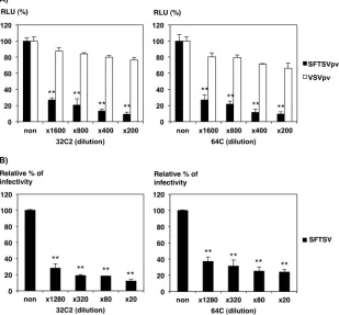
Neutralization assays.
The capacities of the sera collected
from convalescent SFTS patients to neutralize SFTSVpv and
SFTSV were compared. Sera from convalescent SFTS patients
partic-ularly neutralized both SFTSVpv and SFTSV in a dilution-dependent
manner, whereas no neutralization of VSVpv infection was observed
(
Fig. 4
). The infectivity of SFTSVpv was completely neutralized at low
dilutions of sera from convalescent SFTS patients. SFTSVpv and
SFTSV infection was also neutralized by all sera from convalescent
SFTS patients (data not shown).
Involvement of C-type lectins in SFTSVpv and SFTSV
infec-tion.
C-type lectins such as DC-SIGN were reported previously to
be involved in cell entry of various viruses (
20
). SFTSVpv was
recently reported to use DC-SIGN as a receptor for host cell entry
(
5
). The involvement of several C-type lectins, SIGN,
DC-SIGNR, and LSECtin, in SFTSVpv and SFTSV infection was
eval-uated. Jurkat and Raji cells were transfected with lentiviral vectors
encoding fCD2, DC-SIGN, DC-SIGNR, or LSECtin, and these
molecules were expressed on the cell surface (
Fig. 5A
). The
expres-sion of DC-SIGN, DC-SIGNR, and LSECtin in Jurkat or Raji cells
drastically increased SFTSVpv infection and moderately increased
RVFVpv infection, whereas no significant increase in VSVpv
in-fection was observed (
Fig. 5B
). To confirm the involvement of the
C-type lectins, the growth of SFTSV in Raji cells stably expressing
each molecule was examined. The growth of SFTSV was increased
by
⬃
100-fold within 8 days in all Raji cells expressing each
mole-cule (
Fig. 5C
).
Syncytium formation in SFTSV-infected cells exposed to low
pH.
To examine the formation of SFTSV-infected cell syncytia,
Vero cells were infected with SFTSV and cultured for 48 h. The
cells were then treated with the buffer at the indicated pH for 2
min. The infectivity of SFTSV was confirmed by an
immunofluo-rescence assay with anti-SFTSV-NP or -GP (data not shown).
Syncytia in SFTSV-infected cells were observed within 1 h after
treatment with buffers below pH 6.0 (
Fig. 6A
). Previously, low pH
was reported to alter the conformation of arenaviral GP in the
absence of cellular receptors, abolishing viral infectivity (
21
). To
examine the effect of low-pH exposure on SFTSVpv infection, the
viruses were treated with buffer at the indicated pH before
infec-FIG 4Neutralization of SFTSVpv or SFTSV infections by sera from convalescent SFTS patients. SFTSVpv (A) and SFTSV (B) were preincubated with the indicated dilutions of sera from two different convalescent SFTS patients (patients 32C2 [left] and 64C [right]). Thereafter, Huh7 cells were infected with SFTSVpv or SFTSV. Infectivities of SFTSVpv were determined by measuring luciferase activities at 24 h postinfection. Infectivities of SFTSV were determined by a focus-forming assay. The results shown are from three independent assays, with error bars representing standard deviations. **,P⬍0.01.
on November 7, 2019 by guest
http://jvi.asm.org/
[image:5.585.139.448.72.359.2]tion. The infectivity of SFTSVpv and RVFVpv was abolished by
low-pH treatment (
Fig. 6B
), indicating that the SFTSV-GP
con-formation may be altered under low-pH conditions in the absence
of host factors, such as receptor molecules.
Effect of 25HC on SFTSV infection.
It was recently reported
that 25HC broadly inhibited virus entry or replication steps (
12
,
13
). We investigated whether SFTSV entry was inhibited by 25HC
by examining the infectivity of SFTSVpv and the growth of SFTSV
in Vero and Huh7 cells in the presence of 25HC. SFTSVpv
infec-tion and the growth of SFTSV were particularly inhibited by 25HC
in a dose-dependent manner in both Vero and Huh7 cells (
Fig. 7A
and
B
). These results indicated that 25HC mainly affected SFTSV
cell entry. We further examined whether the inhibitory effects of
25HC targeted SFTSV fusion. Syncytium formation of
SFTSV-infected cells at low pH was inhibited by 25HC in a
dose-depen-dent manner (
Fig. 7C
and
D
). The expression of SFTSV-GP and
-NP was clearly observed in all cells treated with various
concen-trations of 25HC by immunofluorescence staining (
Fig. 7C
), and
FIG 5Involvement of C-type lectins in infection by SFTSVpv and SFTSV. (A) Cell surface expression levels of DC-SIGN, DC-SIGNR, and LSECtin in Jurkat and Raji cells expressed with each molecule were analyzed by using flow cytometry with anti-DC-SIGN/DC-SIGNR and anti-LSECtin antibodies. Both Jurkat and Raji cells expressed with fCD2 were used as negative controls. (B) Infectivities of SFTSVpv, RVFVpv, and VSVpv in Jurkat and Raji cells expressed with C-type lectins, DC-SIGN, DC-SIGNR, and LSECtin. Infectivities of SFTSVpv were determined by measuring luciferase activities at 24 h postinfection. The vertical axis in the graph indicates the relative fold infectivities standardized to the value of infectivities in cells expressing fCD2. (C) Growth of SFTSV in C-type lectin-expressing Raji cells. Cells expressed with DC-SIGN, DC-SIGNR, or LSECtin were infected with SFTSV at an MOI of 0.1. After incubation for the indicated number of days, the infectious dose of SFTSV in the supernatants of Vero cells was determined with a focus-forming assay using an anti-NP antibody. The vertical axis in the graph indicates the relative fold titer standardized to the value of infectivities at 0 days. The results shown are from three independent assays, with error bars representing standard deviations. *,P⬍0.05; **,P⬍0.01.
Cell Entry of SFTSV
on November 7, 2019 by guest
http://jvi.asm.org/
[image:6.585.139.452.61.516.2]SFTSV-infected cell syncytium formation was also inhibited even
by a 3-h exposure to 25HC (data not shown), suggesting that the
expression of SFTSV-GP was not altered. Finally, we examined the
effects of 25HC on the binding and internalization of SFTSV by
immunofluorescence staining of the virions. 25HC had no
signif-icant effect on binding and internalization (
Fig. 7E
). Taken
to-gether, these results suggest that 25HC acts potently after the
in-ternalization of SFTSV.
DISCUSSION
Currently, SFTSV has been identified only in China, Japan, and
South Korea. However, Heartland virus, Bhanja virus, and other
phleboviruses, which cause diseases similar to SFTS, have been
isolated previously (
22
,
23
). Understanding the life cycles of these
viruses may inform the development of important antiviral
strat-egies. In particular, understanding the initial steps of viral
infec-tion, binding, penetrainfec-tion, and fusion, is necessary for the specific
development of entry inhibitors to prevent virus spread.
Generally, VSV assembles and buds from the plasma
mem-brane. Therefore, viral GPs presented on the cell surface are
effi-ciently incorporated into the virions. The GP of SFTSV and those
of other bunyaviruses, including RVFV, are mainly localized in
intracellular compartments, such as the ER and Golgi apparatus of
the target cells, but not at the plasma membrane (
Fig. 1
) (
24
).
Although incorporation of the GP into SFTSVpv particles could
not be analyzed because the antibodies required for immunoblot
analysis are not yet available, SFTSVpv and RVFVpv were able to
infect various types of mammalian cell lines (
Fig. 2A
).
Further-more, SFTSVpv was neutralized by SFTS patient sera. These
re-sults suggest that SFTSVpv particles incorporated GP onto the
virion surface and that GP was involved in virus entry. Further
studies will be required to clarify the mechanisms by which SFTSV
GP is incorporated into VSV particles and the role of SFTSV GP
glycosylation in cell entry.
Previous studies demonstrated that HeLa cells were not
sus-ceptible to SFTSVpv and SFTSV infection (
5
,
7
). In this study,
HeLa cells were weakly susceptible to infection with these viruses
(
Fig. 2A
and
B
). Furthermore, COS7 cells were highly susceptible
to infection by SFTSVpv, consistent with data from a previous
report (
5
). COS7 cells were also moderately susceptible, in
con-trast data from to another previous report (
7
). Further studies will
be needed to clarify these discrepancies and to identify the viral
FIG 6Syncytium formation of SFTSV-infected cells. (A) Syncytium formation of SFTSV-infected Vero cells after treatment with low-pH buffer. Vero cells were infected with SFTSV at an MOI of 0.1. At 48 h postinfection, the cells were treated with citrate-phosphate buffer adjusted to the indicated pH value (pH 7.0, 6.0, or 5.0) for 2 min. Syncytium formations were determined by microscopic examination after the indicated times of incubation. (B) Relative infectivity of SFTSVpv after exposure to buffer at the indicated pH. SFTSVpv, RVFVpv, and VSVpv were exposed to buffer at the indicated pH for 2 min. After neutralization with DMEM containing 10% FCS, the remaining infectivities of the pseudotype viruses to Vero cells were measured. Infectivities of SFTSVpv were determined by measuring luciferase activities at 24 h postinfection. The results shown are from three independent assays, with error bars representing standard deviations. *,P⬍
0.05; **,P⬍0.01.
on November 7, 2019 by guest
http://jvi.asm.org/
[image:7.585.135.447.65.400.2]entry receptor(s) and host factor(s) essential for virus growth.
Although it is likely that lymphocytes are primary target cells of
SFTSV infection in humans, lymphocyte cell lines were not very
susceptible to SFTSVpv and SFTSV infection. However, further
work will be required to assess the susceptibility of primary
hu-man lymphocytes.
C-type lectins, such as DC-SIGN and DC-SIGNR, are utilized
as an initial binding receptor for various types of viruses. We
ob-served that not only DC-SIGN but also DC-SIGNR and LSECtin
were involved in the entry of SFTSV. Furthermore, although
SFTSV did not grow in Raji cells, when the expression of these
C-type lectins was induced, they became susceptible to SFTSV
infection (
Fig. 5B
). The expression of C-type lectins enhanced the
susceptibilities of Jurkat and Raji cells to SFTSVpv more than
those to RVFVpv or VSVpv. C-type lectins appear to play a critical
role in the binding of viruses to nonsusceptible cells, acting as
enhancing binding molecules. However, they may have a minor or
no role in the binding of viruses to target cells that express other
major binding and/or entry receptors. VSVpv can infect Jurkat
and Raji cells well; therefore, no enhancement of infectivity would
be observed in C-type lectin-expressing cells. Cell-cell fusion is
generally induced when viral GP is present at the plasma
mem-brane. Cell fusion occurred in cells infected with SFTSV or RVFV
despite the fact that GP was present mainly in the intracellular
compartment and not on the plasma membrane (
8
). Perhaps, the
overexpression of viral GP in infected cells breaks its retention in
FIG 7Effects of 25HC on infection by SFTSVpv and SFTSV. (A) Inhibition of SFTSVpv infection by 25HC. Vero (left) and Huh7 (right) cells were inoculated with pseudotype viruses after treatment of these cells with various concentrations of 25HC. Infectivities of SFTSVpv were determined by measuring luciferase activities at 24 h postinfection. (B) Inhibition of SFTSV growth by 25HC. Vero (left) and Huh7 (right) cells were inoculated with SFTSV and cultured with culture medium containing the indicated concentrations of 25HC. The SFTSV titer in the medium was determined by a focus-forming assay. The results shown are from three independent assays, with error bars representing standard deviations. *,P⬍0.05; **,P⬍0.01. (C) Inhibition of syncytium formation in SFTSV-infected Vero cells by 25HC. Vero cells were infected with SFTSV at an MOI of 0.1. At 24 h postinfection, cells were treated with the indicated concentrations of 25HC for 24 h. The cells were treated with citrate-phosphate buffer adjusted to the indicated pH value (pH 7.0, 6.0, or 5.0) for 2 min. Syncytium formations were observed by microscopic examination after 4 h of incubation. Expression of GP and NP at 48 h postinfection and after treatment with 25HC was examined by an immunofluorescence assay using anti-SFTSV NP polyclonal antibody and anti-SFTSV GP monoclonal antibody, respectively. (D) Syncytium counts in SFTSV-infected cells treated with 25HC. Vero cells were SFTSV-infected with SFTSV at an MOI of 0.05. At 24 h postinfection, cells were treated with the indicated concentrations of 25HC for 24 h. The cells were treated with citrate-phosphate buffer adjusted to pH 5.0 for 2 min. Syncytium counting was performed by microscopic observation after 4 h of incubation. The results shown are from three independent assays, with error bars representing standard deviations. *,P⬍0.05; **,P⬍
0.01. (E) Visualization of SFTSV binding and internalization in Vero cells treated with 25HC. Vero cells pretreated with or without 10M 25HC were infected with SFTSV at an MOI of 100. After incubation for 2 h at 4°C or further incubation for 1 h at 37°C, SFTSV was stained with anti-SFTSV NP monoclonal antibody. Nuclei were stained with DAPI. The data shown are representative of results from three independent experiments.
Cell Entry of SFTSV
on November 7, 2019 by guest
http://jvi.asm.org/
[image:8.585.114.471.70.410.2]the ER or Golgi apparatus; therefore, a small portion of the GP
leaks onto the plasma membrane at a level undetectable by an IFA.
Results of cell fusion in cells transfected with expression plasmids
of SFTSV-GP were also obtained (data not shown).
Cholesterol is one of the most abundant lipids in the plasma
membrane. The distribution of cholesterol, such as
cholesterol-rich microdomains, namely, rafts, in the plasma membrane affects
the entry, replication, and budding of many viruses. The oxysterol
25HC is a metabolite of cholesterol produced and secreted by
macrophages and has multiple effects on lipid metabolism.
Re-cently, 25HC was reported to be a potent antiviral mediator
against various kinds of viruses (
25
,
26
). In this study, the addition
of 25HC drastically inhibited SFTSV growth, particularly the
in-hibition of virus entry (
Fig. 7A
and
B
). 25HC might inhibit SFTSV
fusion (
Fig. 7C
to
E
). Previous studies demonstrated that 25HC
broadly inhibited viral membrane fusion, which was not specific
to particular structural classes of fusion peptides, suggesting the
involvement of more basic fusion processes (
13
). Cell-cell fusion
of Vero cells expressing Nipah virus F and G was inhibited by
25HC under pH-neutral conditions (
13
). The cell fusion of
SFTSV-infected cells may occur via a mechanism similar to that of
Nipah virus. If recombinant SFTSV encoding a color marker gene
could be constructed, the effects of 25HC on fusion events could
be clearly observed in real time. The treatment of mice with high
doses of 25HC decreased the infection and replication of some
viruses, and the capacity of virus growth was increased in
Ch25h-deficient mice (
13
). Further
in vivo
experiments will be needed to
verify the inhibitory effect of 25HC.
In conclusion, we analyzed the mechanisms of SFTSV entry
and fusion using native and pseudotype viruses possessing the
SFTSV envelope proteins. We demonstrated for the first time that
the production of SFTSV and membrane fusion of SFTSV-GP
were inhibited by 25HC. SFTSVpv is useful not only for analyses
of SFTSV cell entry but also for development of a neutralization
test for the diagnosis of SFTS at lower biosafety levels. 25HC could
be considered a therapeutic SFTSV inhibitor.
ACKNOWLEDGMENTS
Chinese SFTSV strain HB29 was a kind gift from Dexin Li and Mifang Liang at the National Institute for Viral Disease Control and Prevention, Chinese Center for Disease Control and Prevention. We gratefully ac-knowledge Momoko Ogata and Junko Hirai for their technical and secre-tarial assistance.
FUNDING INFORMATION
This work, including the efforts of Hideki Tani, was funded by Ministry of Education, Culture, Sports, Science, and Technology (MEXT) (24790451). This work, including the efforts of Hideki Tani, was funded by Ministry of Education, Culture, Sports, Science, and Technology (MEXT) (15K08510). This work, including the efforts of Masayuki Saijo, was funded by Ministry of Health, Labour and Welfare (MHLW) (H24-shinko-ippan-013). This work, including the efforts of Shigeru Morikawa, was funded by Ministry of Health, Labour and Welfare (MHLW) (H25-shinko-ippan-008). This work, including the efforts of Masayuki Saijo, was funded by Ministry of Health, Labour and Welfare (MHLW) (H25-shinko-shitei-009). This work, including the efforts of Hideki Tani, was funded by Ministry of Health, Labour and Welfare (MHLW) (H24-shinko-wakate-016). This work, including the efforts of Masayuki Shimo-jima, was funded by Ministry of Health, Labour and Welfare (MHLW) (H25-shinko-ippan-004). This work, including the efforts of Masayuki Saijo, was funded by Japan Agency for Medical Research and Develop-ment (AMED) (40102701).
REFERENCES
1.Yu XJ, Liang MF, Zhang SY, Liu Y, Li JD, Sun YL, Zhang L, Zhang QF, Popov VL, Li C, Qu J, Li Q, Zhang YP, Hai R, Wu W, Wang Q, Zhan FX, Wang XJ, Kan B, Wang SW, Wan KL, Jing HQ, Lu JX, Yin WW, Zhou H, Guan XH, Liu JF, Bi ZQ, Liu GH, Ren J, Wang H, Zhao Z, Song JD, He JR, Wan T, Zhang JS, Fu XP, Sun LN, Dong XP, Feng ZJ, Yang WZ, Hong T, Zhang Y, Walker DH, Wang Y, Li DX.2011. Fever with thrombocytopenia associated with a novel bunyavirus in China. N Engl J Med364:1523–1532.http://dx.doi.org/10.1056/NEJMoa1010095. 2.Takahashi T, Maeda K, Suzuki T, Ishido A, Shigeoka T, Tominaga T,
Kamei T, Honda M, Ninomiya D, Sakai T, Senba T, Kaneyuki S, FIG 7continued
on November 7, 2019 by guest
http://jvi.asm.org/
[image:9.585.41.285.64.570.2]Sakaguchi S, Satoh A, Hosokawa T, Kawabe Y, Kurihara S, Izumikawa K, Kohno S, Azuma T, Suemori K, Yasukawa M, Mizutani T, Omatsu T, Katayama Y, Miyahara M, Ijuin M, Doi K, Okuda M, Umeki K, Saito T, Fukushima K, Nakajima K, Yoshikawa T, Tani H, Fukushi S, Fu-kuma A, Ogata M, Shimojima M, Nakajima N, Nagata N, Katano H, Fukumoto H, Sato Y, Hasegawa H, Yamagishi T, Oishi K, Kurane I, Morikawa S, Saijo M.2014. The first identification and retrospective study of severe fever with thrombocytopenia syndrome in Japan. J Infect Dis209:816 – 827.http://dx.doi.org/10.1093/infdis/jit603.
3.Kim KH, Yi J, Kim G, Choi SJ, Jun KI, Kim NH, Choe PG, Kim NJ, Lee JK, Oh MD.2013. Severe fever with thrombocytopenia syndrome, South Korea, 2012. Emerg Infect Dis19:1892–1894.http://dx.doi.org/10.3201 /eid1911.130792.
4.Liu Q, He B, Huang SY, Wei F, Zhu XQ.2014. Severe fever with throm-bocytopenia syndrome, an emerging tick-borne zoonosis. Lancet Infect Dis
14:763–772.http://dx.doi.org/10.1016/S1473-3099(14)70718-2.
5.Hofmann H, Li X, Zhang X, Liu W, Kuhl A, Kaup F, Soldan SS, Gonzalez-Scarano F, Weber F, He Y, Pohlmann S.2013. Severe fever with thrombocytopenia virus glycoproteins are targeted by neutralizing antibodies and can use DC-SIGN as a receptor for pH-dependent entry into human and animal cell lines. J Virol87:4384 – 4394.http://dx.doi.org /10.1128/JVI.02628-12.
6.Lozach PY, Kuhbacher A, Meier R, Mancini R, Bitto D, Bouloy M, Helenius A.2011. DC-SIGN as a receptor for phleboviruses. Cell Host Microbe10:75– 88.http://dx.doi.org/10.1016/j.chom.2011.06.007. 7.Sun Y, Qi Y, Liu C, Gao W, Chen P, Fu L, Peng B, Wang H, Jing Z,
Zhong G, Li W.2014. Nonmuscle myosin heavy chain IIA is a critical factor contributing to the efficiency of early infection of severe fever with thrombocytopenia syndrome virus. J Virol88:237–248.http://dx.doi.org /10.1128/JVI.02141-13.
8.Filone CM, Heise M, Doms RW, Bertolotti-Ciarlet A.2006. Develop-ment and characterization of a Rift Valley fever virus cell-cell fusion assay using alphavirus replicon vectors. Virology356:155–164. http://dx.doi .org/10.1016/j.virol.2006.07.035.
9.Lozach PY, Mancini R, Bitto D, Meier R, Oestereich L, Overby AK, Pettersson RF, Helenius A.2010. Entry of bunyaviruses into mammalian cells. Cell Host Microbe 7:488 – 499. http://dx.doi.org/10.1016/j.chom .2010.05.007.
10. Tani H, Morikawa S, Matsuura Y.2011. Development and applications of VSV vectors based on cell tropism. Front Microbiol2:272.http://dx.doi .org/10.3389/fmicb.2011.00272.
11. McNab F, Mayer-Barber K, Sher A, Wack A, O’Garra A.2015. Type I interferons in infectious disease. Nat Rev Immunol15:87–103.http://dx .doi.org/10.1038/nri3787.
12. Blanc M, Hsieh WY, Robertson KA, Kropp KA, Forster T, Shui G, Lacaze P, Watterson S, Griffiths SJ, Spann NJ, Meljon A, Talbot S, Krishnan K, Covey DF, Wenk MR, Craigon M, Ruzsics Z, Haas J, Angulo A, Griffiths WJ, Glass CK, Wang Y, Ghazal P. 2013. The transcription factor STAT-1 couples macrophage synthesis of 25-hydroxycholesterol to the interferon antiviral response. Immunity38:
106 –118.http://dx.doi.org/10.1016/j.immuni.2012.11.004.
13. Liu SY, Aliyari R, Chikere K, Li G, Marsden MD, Smith JK, Pernet O, Guo H, Nusbaum R, Zack JA, Freiberg AN, Su L, Lee B, Cheng G.2013. Interferon-inducible cholesterol-25-hydroxylase broadly inhibits viral en-try by production of 25-hydroxycholesterol. Immunity38:92–105.http: //dx.doi.org/10.1016/j.immuni.2012.11.005.
14. Saijo M, Qing T, Niikura M, Maeda A, Ikegami T, Prehaud C, Kurane I, Morikawa S.2002. Recombinant nucleoprotein-based enzyme-linked immunosorbent assay for detection of immunoglobulin G antibodies to Crimean-Congo hemorrhagic fever virus. J Clin Microbiol40:1587–1591.
http://dx.doi.org/10.1128/JCM.40.5.1587-1591.2002.
15. Shimojima M, Stroher U, Ebihara H, Feldmann H, Kawaoka Y.2012. Identification of cell surface molecules involved in dystroglycan-independent Lassa virus cell entry. J Virol86:2067–2078.http://dx.doi.org /10.1128/JVI.06451-11.
16. Shimojima M, Nishimura Y, Miyazawa T, Kato K, Nakamura K, Izumiya Y, Akashi H, Tohya Y.2002. A feline CD2 homologue interacts with human red blood cells. Immunology105:360 –366.http://dx.doi.org /10.1046/j.0019-2805.2001.01371.x.
17. Tani H, Shiokawa M, Kaname Y, Kambara H, Mori Y, Abe T, Moriishi K, Matsuura Y.2010. Involvement of ceramide in the propagation of Japanese encephalitis virus. J Virol84:2798 –2807.http://dx.doi.org/10 .1128/JVI.02499-09.
18. Bukbuk DN, Fukushi S, Tani H, Yoshikawa T, Taniguchi S, Iha K, Fukuma A, Shimojima M, Morikawa S, Saijo M, Kasolo F, Baba SS.
2014. Development and validation of serological assays for viral hemor-rhagic fevers and determination of the prevalence of Rift Valley fever in Borno State, Nigeria. Trans R Soc Trop Med Hyg108:768 –773.http://dx .doi.org/10.1093/trstmh/tru163.
19. Tani H, Komoda Y, Matsuo E, Suzuki K, Hamamoto I, Yamashita T, Moriishi K, Fujiyama K, Kanto T, Hayashi N, Owsianka A, Patel AH, Whitt MA, Matsuura Y.2007. Replication-competent recombinant ve-sicular stomatitis virus encoding hepatitis C virus envelope proteins. J Virol81:8601– 8612.http://dx.doi.org/10.1128/JVI.00608-07.
20. Mason CP, Tarr AW.2015. Human lectins and their roles in viral infections. Molecules20:2229 –2271.http://dx.doi.org/10.3390/molecules20022229. 21. Tani H, Iha K, Shimojima M, Fukushi S, Taniguchi S, Yoshikawa T,
Kawaoka Y, Nakasone N, Ninomiya H, Saijo M, Morikawa S.2014. Analysis of Lujo virus cell entry using pseudotype vesicular stomatitis virus. J Virol88:7317–7330.http://dx.doi.org/10.1128/JVI.00512-14. 22. Matsuno K, Weisend C, Travassos da Rosa AP, Anzick SL, Dahlstrom
E, Porcella SF, Dorward DW, Yu XJ, Tesh RB, Ebihara H. 2013. Characterization of the Bhanja serogroup viruses (Bunyaviridae): a novel species of the genus Phlebovirus and its relationship with other emerging tick-borne phleboviruses. J Virol 87:3719 –3728. http://dx.doi.org/10 .1128/JVI.02845-12.
23. Xing Z, Schefers J, Schwabenlander M, Jiao Y, Liang M, Qi X, Li C, Goyal S, Cardona CJ, Wu X, Zhang Z, Li D, Collins J, Murtaugh MP.
2013. Novel bunyavirus in domestic and captive farmed animals, Minne-sota, USA. Emerg Infect Dis 19:1487–1489.http://dx.doi.org/10.3201 /eid1908.130165.
24. Gerrard SR, Nichol ST.2002. Characterization of the Golgi retention motif of Rift Valley fever virus G(N) glycoprotein. J Virol76:12200 – 12210.http://dx.doi.org/10.1128/JVI.76.23.12200-12210.2002. 25. Chen Y, Wang S, Yi Z, Tian H, Aliyari R, Li Y, Chen G, Liu P, Zhong
J, Chen X, Du P, Su L, Qin FX, Deng H, Cheng G.2014. Interferon-inducible cholesterol-25-hydroxylase inhibits hepatitis C virus repli-cation via distinct mechanisms. Sci Rep4:7242.http://dx.doi.org/10 .1038/srep07242.
26. Civra A, Cagno V, Donalisio M, Biasi F, Leonarduzzi G, Poli G, Lembo D. 2014. Inhibition of pathogenic non-enveloped viruses by 25-hydroxycholesterol and 27-25-hydroxycholesterol. Sci Rep4:7487.http://dx .doi.org/10.1038/srep07487.
27. Fukuma A, Fukushi S, Yoshikawa T, Tani H, Taniguchi S, Kurosu T, Egawa K, Suda Y, Singh H, Nomachi T, Gokuden M, Ando K, Kida K, Kan M, Kato N, Yoshikawa A, Kitamoto H, Sato Y, Suzuki T, Hasegawa H, Morikawa S, Shimojima M, Saijo M.2016. Severe fever with throm-bocytopenia syndrome virus antigen detection using monoclonal anti-bodies to the nucleocapsid protein. PLoS Negl Trop Dishttp://dx.doi.org /10.1371/journal.pntd.0004595.
Cell Entry of SFTSV
on November 7, 2019 by guest
http://jvi.asm.org/