Dependent Human Papillomavirus Life Cycle
Ayano Satsuka, Kavi Mehta, Laimonis Laimins
Department of Microbiology-Immunology, Feinberg School of Medicine, Northwestern University, Chicago, Illinois, USA
Amplification of human papillomaviruses (HPV) is dependent on the ATM DNA damage pathway. In cells with impaired p53 activity, DNA damage repair requires the activation of p38MAPK along with MAPKAP kinase 2 (MK2). In HPV-positive cells, phosphorylation of p38 and MK2 proteins was induced along with relocalization to the cytoplasm. Treatment with MK2 or p38 inhibitors blocked HPV genome amplification, identifying the p38/MK2 pathway as a key regulator of the HPV life cycle.
H
uman papillomaviruses (HPV) infect stratified epithelium and induce a variety of lesions (1). A subset of HPV types, referred to as high risk, are the causative agents of cervical and other anogenital malignancies as well as many oral cancers (2–4). Papillomaviruses infect basal epithelial cells of stratified epithe-lium that become exposed through microwounds and establish a latent infection, in which viral genomes are maintained in the nucleus as low-copy-number episomes. As infected basal cells di-vide and one daughter cell leaves the basal layer, the cell differen-tiation program is initiated, which results in activation of viral gene expression and replication. HPV genomes do not encode DNA polymerases or replication factors except for the DNA heli-case E1, and viral replication is dependent largely on host factors (5). It is, therefore, necessary for HPV-positive cells to retain the ability to reenter S/G2upon differentiation (6). This process is mediated through the action of the E6 and E7 proteins that mod-ulate the function of p53, Rb, and a number of other cell cycle regulators (7–9). In addition to maintaining cells active in the cell cycle, HPV genome amplification requires activation of the ATM (ataxia telengiectasia mutated) DNA damage pathway (10–14).The ATM pathway is responsible for the DNA damage response (DDR) to double-strand DNA breaks and is mediated through the action of downstream kinases, such as CHK2 (15). The ATR pathway is activated by single-strand breaks as well as replication fork collapse and functions through CHK1 (16). While these two pathways gener-ally act independently of each other, some overlap exists, such as when one pathway is deficient or compromised. A third DDR path-way has recently been described in cells with reduced or impaired p53 activity, and this pathway is mediated by the p38MAPK kinases (17– 21). The p38MAPK pathway is activated in response to a variety of stress-induced signals, including DNA damage, osmotic shock, or cytokine signaling. p38MAPK phosphorylates a number of down-stream effectors, such as c-Myc, c-Jun, and ATF2, but it specifically induces DDR through the phosphorylation of mitogen-activated protein (MAP) kinase-activated protein kinase 2 (MAPKAPK 2, or MK2) (20,22–26). Activation of MK2 leads to phosphorylation of a series of downstream targets that results in G2/M arrest and DNA repair (21). While the DNA repair portion of the p38MAPK/MK2 pathway is activated by ATM or ATR kinases, it functions indepen-dently and in parallel to the activities of CHK1 and CHK2 (19,21). Furthermore, p38MAPK/MK2 has been shown to be important for DDR in cells that have impaired p53 function, such as U2OS, HeLa, or immortalized fibroblasts with diminished p53 function (18). MK2 is relocalized to the cytoplasm upon activation, while activated CHK1
and CHK2 are retained in the nucleus (18). Keratinocytes that stably maintain high-risk HPV genomes have reduced levels of p53 through the action of E6/E6AP complexes, as well as altered levels of acetylated p53 through E6 modulation of p300 activity (8). We therefore
inves-Received18 September 2014Accepted13 November 2014
Accepted manuscript posted online19 November 2014
CitationSatsuka A, Mehta K, Laimins L. 2015. p38MAPK and MK2 pathways are
important for the differentiation-dependent human papillomavirus life cycle. J Virol 89:1919 –1924.doi:10.1128/JVI.02712-14.
Editor:M. J. Imperiale
Address correspondence to Laimonis Laimins, [email protected].
Copyright © 2015, American Society for Microbiology. All Rights Reserved.
doi:10.1128/JVI.02712-14
HFK HPV16 HPV18 HPV31 CIN612
0 48 96 0 48 96 0 48 96 0 48 96 0 48 96
p-p38
p38
MK2
β-actin p-MK2
TG
Ca p38
p-p38
MK2
β-actin
HFK 16 18 31 CIN
p-MK2
A B
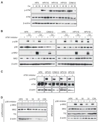
FIG 1p-38 and p-MK2 levels are increased in HPV-positive cells. (A) Western blot analysis for p-p38, p38, p-MK2, and MK2 in undifferentiated monolayer cultures of normal human foreskin keratinocytes (HFK), HPV-16, HPV-18, HPV-31, and CIN-612 cells. The HPV-16, -18, and -31 cells were generated by transfection of HFKs with recircularized HPV genomes as previously de-scribed (11,27), followed by selection for a cotransfected drug resistance marker.-Actin was used as a loading control. The following antibodies were used: p-p38(T180/Y182, D3F9; catalog number 4511), p38(D13E1; catalog number 8690), p-MK2(T334, 27B7; catalog number 3007), and MK2 (catalog number 3042) (all from Cell Signaling Technologies, San Diego, CA). Second-ary antibodies included horseradish peroxidase-linked anti-rabbit (Santa Cruz Biotechnology, Santa Cruz, CA). Levels of p-p38, p38, p-MK2, and MK2 fol-lowing differentiation in high-calcium medium for 0, 48, and 96 h were deter-mined by Western blotting. Calcium-induced differentiation was performed as described previously (27). The 0-h data represent results for undifferenti-ated cells. TG (transglutaminase) was used as a differentiation marker.-Actin was used as a loading control.
on November 7, 2019 by guest
http://jvi.asm.org/
[image:1.585.299.544.240.372.2]tigated if p38MAPK/MK2 played any role in the genome amplifica-tion of HPV-positive cells.
To determine what effect, if any, the p38/MK2 pathway plays in HPV replication, we first examined the levels and activation status of p38 and MK2 in normal and HPV-positive human keratino-cytes in undifferentiated cells grown as monolayer cultures. For this analysis, the levels of p38/MK2 proteins were examined in a series of HPV-positive human keratinocyte cell lines that were generated by transfection with recircularized genomes from HPV-16, -18, and -31, as previously described (27), as well as a biopsy sample-derived cell line that is HPV-31 positive, CIN-612. All these HPV-positive lines stably maintain episomal copies of HPV
genomes in undifferentiated monolayer cultures. As shown in Fig. 1A, the levels of total p38 were similar in both HPV-positive and normal keratinocyte cells. In contrast, the phosphorylated form of p38 (p-p38) was only detected in HPV-positive cells. Slightly increased levels of total MK2 were seen in HPV-positive cells, with minimal levels of phosphorylated MK2 (p-MK2) de-tected in undifferentiated cultures of either normal or HPV-pos-itive cells.
It was next important to investigate if the levels of p38/MK2 proteins were altered upon differentiation. For this analysis, we used calcium-induced differentiation, as this method allows for isolation of uniformly differentiated populations of cells (27).
HFK HPV31 CIN612
Ca
ATM - + - + - + - + - + - +
HFK HPV16 HPV18 - + - + - + - + - + - +
p-MK2 MK2
β-actin
p-MK2 MK2
β-actin
- + - + - + - + - +
TG TG
HFK HPV16 HPV18 HPV31 CIN612
p-ATM
ATM
0 48 96 0 48 96 0 48 96 0 48 96 0 48 96
β-actine
Ca Ca
Ca
Ca Ca Ca
undifferentiated differentiated
p-p38 Inhibitor
p38 p-MK2 MK2
β-actin
HFK 16 18 31 CIN - + - + - + - + - +
HFK 16 18 31 CIN
p38 inhibitor p38 inhibitor
A
B
C
p-ATM
HFK HPV31 CIN612
ATM - + - + - + - + - + HPV16 HPV18 Inhibitor
D
ATM
β-actin
FIG 2The ATM pathway is activated in HPV-positive cells, and addition of ATM inhibitors blocks induction of p-p38 and p-MK2. (A) Levels of total and phosphorylated p-ATM upon differentiation of HPV-positive cells as well as HFKs. Cells were seeded as monolayer cultures, followed by the addition of medium containing 1.5 mM Ca2⫹for 96 h (27), as previously described (11). Extracts were harvested at the indicated times and examined by Western blotting using the corresponding antibodies. Anti-ATM (S1981, D6H9) was obtained from Cell Signaling Technologies. (B) Levels of p-p38, p38, p-MK2, and MK2 following differentiation in high-calcium medium in the presence or absence of an ATM inhibitor. Undifferentiated cells were grown as monolayer cultures in the presence or absence of 5M ATM inhibitor KU-55933, and protein levels were determined by Western blotting. Lanes designated with Ca were induced to differentiate by the addition of high-calcium medium for 96 h with or without the presence of 5M KU-55933, and protein levels were examined by Western blotting. (C) Levels of phosphorylated ATM and total ATM following treatment with an ATM inhibitor. (D) Levels of p-MK2 in the absence or presence of a p38 inhibitor. Cells were seeded as monolayer cultures (left) or exposed to medium containing 1.5 mM Ca2⫹for 96 h (right) (11). The cells were left untreated or incubated with 10M p-p38 inhibitor SB203580 for 96 h. Extracts were harvested and examined by Western blotting using the corresponding antibodies. TG (transglutaminase) was used as a differentiation marker.-Actin was used as a loading control.
on November 7, 2019 by guest
http://jvi.asm.org/
[image:2.585.134.449.65.458.2]Amplification of viral genomes begins within 48 h and plateaus by 96 h after the calcium switch. In both normal and HPV-positive keratinocytes, the levels of phosphorylated p38 increased upon differentiation, while the total amounts were largely unchanged (Fig. 1B). Slightly higher levels of MK2 total proteins were seen in HPV-positive cells and remained unchanged upon differentia-tion. In contrast to the minimal levels p-MK2 observed in undif-ferentiated HPV-positive cells, significant induction of p-MK2 was seen upon differentiation. No activation of p-MK2 was seen in either undifferentiated or differentiated normal keratinocytes (Fig. 1B). This indicated that phosphorylation of MK2 is induced upon differentiation of HPV-positive cells.
We previously demonstrated that the ATM DNA damage pathway is constitutively activated in HPV-positive cells and that it plays a critical role in controlling amplification upon differen-tiation (11). To investigate if ATM activation is linked to p38/ MK2 activation in HPV-positive cells, the levels of ATM and its phosphorylated form (p-ATM) were examined by Western blot-ting. Consistent with previous studies, total ATM levels were rel-atively unchanged between normal and HPV-positive cells, while p-ATM was detected only in HPV-positive cells (Fig. 2A). The ATM inhibitor KU-55933 blocks phosphorylation of ATM along with its downstream targets and is specific (28). To determine if ATM was responsible for activating phosphorylation of MK2, HPV-positive cells were induced to differentiate in high-calcium medium and treated with KU-55933, and the levels of phosphor-ylated MK2 were examined by Western analysis. As seen inFig. 2B, treatment of cells with KU-55933 blocked phosphorylation of p-p38 without affecting total levels of p38. Similarly, phosphory-lation of MK2 was blocked following treatment of HPV-positive cells that had been induced to differentiate (Fig. 2B). Treatment of cells with KU-55933 also inhibited p-ATM (Fig. 2C). This dem-onstrated that phosphorylation of both p38 and MK2 is depen-dent on ATM in HPV-positive cells. Cells were also treated with an inhibitor of p38 (Fig. 2D), and MK2 phosphorylation was signif-icantly reduced, demonstrating a dependence on p-p38 activity.
Neither the p38 nor MK2 inhibitors had any effect on the expres-sion of transglutaminase, a marker of epithelial differentiation (Fig. 2C) or proliferation (data not shown).
Upon DNA damage, the p38/MK2 complex relocalizes from the nucleus to the cytoplasm to target a series of factors, some of which regulate the stability of a number of mRNAs (18,29). We therefore investigated the cellular localization of p-MK2 during the differentiation of HPV-positive cells by using immunofluores-cence with cells induced to differentiate in high-calcium medium. As shown inFig. 3, very low levels of p-MK2 were found in undif-ferentiated cells. Upon differentiation, we observed induction of MK2 phosphorylation and localization of p-MK2 to the cyto-plasm (Fig. 3). Similar localization was seen with p-p38, which formed a complex with p-MK2. One target of cytoplasmic p-MK2 kinase activity is the chaperone protein HSP-27 (30,31), and we observed via Western blotting that it became phosphorylated upon differentiation of HPV-positive cells (Fig. 4). Furthermore, the addition of MK2 inhibitors blocked phosphorylation of HSP-27 (Fig. 4). This confirmed that p-MK2 is active in differen-tiated HPV-positive cells. Many of the factors described as MK2 targets, including HSP-27, contribute to cell cycle arrest in G2/M (32) and work in parallel with factors targeted by the nuclear DNA damage kinases CHK1 and CHK2 (19).
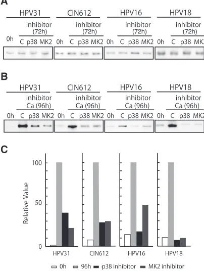
Our results indicate the p38/MK2 pathway is activated upon differentiation of HPV-positive cells but not normal cells. Next, it was important to investigate if the p38/MK2 pathway had any role in the regulation of HPV replication in undifferentiated and dif-ferentiated cells. In undifdif-ferentiated HPV-positive cells, treatment with either p38 or MK2 inhibitors showed no effect on genome copy number (Fig. 5A), indicating the p38/MK2 pathway is not involved in the stable maintenance of HPV genomes, even though ATM and p38 are activated. Upon calcium-induced differentia-tion, only about a quarter of cells amplify viral DNA, and the total amount of HPV DNA is increased (11). When HPV-positive cells were differentiated in the presence of either a p38 or MK2 inhib-itor, genome amplification was significantly suppressed (Fig. 5B FIG 3Immunofluoresence of p-p38 and p-MK2 in HPV-positive cells upon differentiation. Undifferentiated cells were stained for p-p38 or p-MK2 using appropriate antibodies. 4=,6-Diamidino-2-phenylindole (DAPI) staining was used to identify nuclei (ProLong Gold antifade reagent with DAPI; catalog number P36931; Life Technologies, Thermo Fisher Scientific, Waltham, MA). Induction and localization of p-MK2 as well as of p-p38 to the cytoplasm following Ca2⫹-induced differentiation for 72 h is shown. Normal human keratinocytes (HFK) and HPV-31-positive CIN 612 cells were examined.
on November 7, 2019 by guest
http://jvi.asm.org/
[image:3.585.126.461.67.252.2]andC). Similar effects were seen in multiple independent experi-ments. Treatment of cells with these inhibitors did not affect the phosphorylation of ATM (data not shown). These results indi-cated that the p38/MK2 pathway is activated in HPV-positive cells and plays an important role in the differentiation-specific genome amplification of HPV.
Our studies identified a novel role of the p38/MK2 pathway in the control of HPV amplification. The p38/MK2 pathway was found to be activated in HPV-positive cells upon differentiation, and it was observed to be critical for genome amplification. MK2 is a Ser/Thr kinase that is regulated through phosphorylation by p38 MAPK and ATM. The MK2 kinase has been reported to be in-volved in many cellular processes, including stress and inflamma-tory responses (33,34), nuclear export (29), gene expression reg-ulation (33,35,36), and cell proliferation (37). Recently, the p38/ MK2 pathway was identified as an alternative pathway in the DNA damage response (17–21). One of the important downstream ef-fectors of the DDR is the p53 tumor suppressor protein, whose activation can mediate cell cycle arrest to repair DNA damage or to induce apoptotic cell death (38). In p53-deficient cells, p38/ MK2 functions as a third member of the DDR pathway (21). In our studies, we determined that MK2 phosphorylation is specifi-cally induced upon differentiation of HPV-positive cells. As such, it is one of only three DNA damage factors that have been identi-fied to be induced upon differentiation. In addition to p-MK2,
␥-H2AX and p-NBS1 are two members of the ATM pathway (39) whose activation increases upon differentiation; this contrasts with ATM and CHK2, which are activated in both differentiated and undifferentiated cells (11). Since the DDR has no role in stable or transient HPV replication but only affects differentiation-de-pendent genome amplification, MK2 along with ␥-H2Ax and NBS1 are likely to be critical regulators of this process.
The MK2 pathway activates the genome amplification of HPV but not stable maintenance replication, which is consistent with
our observation that active MK2 kinases are only detected upon differentiation. MK2 has a broad range of downstream effectors and is involved in various cellular events. Cytoplasmic MK2 activ-ity is critical for checkpoint maintenance, and it acts in part by stabilizing a number of mRNAs (5), so it is possible that MK2 plays a role in regulating the stabilities of a subset of viral tran-scripts. MK2 also phosphorylates several transcriptional factors, such as SRF and CREB (40), whose function might be critical for regulating the late viral promoter. One known target of p-MK2 is the heat shock protein HSP27 (30,31), and in our studies we observed phosphorylation of HSP27 upon differentiation of HPV-positive cells that was dependent upon p-MK2 action. What role HSP27 plays in the HPV life cycle will be a focus of future studies. Finally, MK2 appears to act in parallel to CHK1 and CHK2 to activate DNA repair factors that are necessary for HPV genome amplification. Given the different cellular localizations of MK2 and CHK1/CHK2, these ki-nases seem to act coordinately to induce the full spectrum of DNA damage factors.
p-HSP27
HSP27
β-actin
HSP27
β-actin p-HSP27 TG
TG
undifferentiated
differentiated
- + - + - + - + - +
HFK 16 18 31 CIN
- + - + - + - + - +
HFK 16 18 31 CIN
MK2 inhibitor
MK2 inhibitor
FIG 4HSP27 phosphorylation is induced by p-MK2 upon differentiation. Cell lysates of HPV-16, HPV-18, HPV-31, and CIN 612 cells were induced to differentiate in high-calcium medium, and levels of p-HSP27 as well as total HSP-27 were examined by Western blotting. Cells were treated with 10M MK2 inhibitor (MK2 inhibitor III), and loss of p-HSP27 was observed. Anti-bodies used were anti-p-HSP-27 and anti-HSP27 (both from Cell Signaling Technologies). TG (transglutaminase) was used as a differentiation marker. -Actin was used as a loading control.
HPV16 HPV18 HPV31 CIN612
0h C p38 MK2 inhibitor
Ca (96h) Ca (96h) Ca (96h) Ca (96h)
0h C p38 MK2 0h C p38 MK2 0h C p38 MK2
0h C p38 MK2 0h C p38 MK2 0h C p38 MK2 0h C p38 MK2 HPV16 HPV18 HPV31 CIN612
inhibitor inhibitor inhibitor
(72h) (72h) (72h) (72h)
inhibitor inhibitor inhibitor inhibitor
A
B
0
100
0h 96h p38 inhibitor MK2 inhibitor
HPV31 CIN612 HPV16 HPV18
Relative Value
C
50
FIG 5Inhibition of p38 or MK2 blocks HPV genome amplification. (A) Southern blot analysis of undifferentiated HPV-31 cells, CIN 612 cells, HPV-16 cells, and HPV-18 cells treated with 10M p38 inhibitor (SB203580) or 10M MK2 inhibitor (MK2 inhibitor III). Cells were treated for 72 h prior to analysis. The 0-h data are results without inhibitor; C designates results after 72-h treatment with DMSO vehicle alone. p38 and MK2 indicate the inhibitor used. Viral episomes are shown. (B) Southern analysis was performed as pre-viously described (41), using the digoxigenin (DIG)-High Prime DNA labeling and detection system (Roche Diagnostics, Mannheim, Germany). The chemi-luminescent signal was visualized by using a chemichemi-luminescent image analyzer (Fc Imager; Odyssey, Lincoln, NE) to visualize DNA. Southern blot analysis results are shown for HPV-positive cells that stably maintained viral episomes treated and untreated with inhibitors of p38 or MK2 during differentiation induced by high-calcium medium for 96 h. The viral episomes are shown. (C) Quantification of the effect on amplification shown in panel B. The signal strengths were quantified by using ImageJ software (NIH, Bethesda, MD), and the results are shown in the bar chart. Each relative value was determined based on setting the signal strength of 96-h incubation with calcium as 100.
on November 7, 2019 by guest
http://jvi.asm.org/
[image:4.585.78.246.68.248.2] [image:4.585.319.521.72.341.2]ACKNOWLEDGMENTS
This work was supported by a grant to L.L. from the National Cancer Institute (CA142861).
We thank the many colleagues who provided technical assistance and were involved in manuscript preparation.
REFERENCES
1.Stubenrauch F, Laimins LA. 1999. Human papillomavirus life cycle: active and latent phases. Semin Cancer Biol9:379 –386.http://dx.doi.org /10.1006/scbi.1999.0141.
2.Durst M, Gissmann L, Ikenberg H, zur Hausen H.1983. A papilloma-virus DNA from a cervical carcinoma and its prevalence in cancer biopsy samples from different geographic regions. Proc Natl Acad Sci U S A 80:3812–3815.http://dx.doi.org/10.1073/pnas.80.12.3812.
3.zur Hausen H.2002. Papillomaviruses and cancer: from basic studies to clinical application. Nat Rev Cancer2:342–350.http://dx.doi.org/10.1038 /nrc798.
4.Leemans CR, Braakhuis BJ, Brakenhoff RH.2011. The molecular biol-ogy of head and neck cancer. Nat Rev Cancer11:9 –22.http://dx.doi.org /10.1038/nrc2982.
5.Kajitani N, Satsuka A, Kawate A, Sakai H.2012. Productive lifecycle of human papillomaviruses that depends upon squamous epithelial differ-entiation. Front Microbiol3:152.
6.Longworth MS, Laimins LA.2004. Pathogenesis of human papillomavi-ruses in differentiating epithelia. Microbiol Mol Biol Rev68:362–372. http://dx.doi.org/10.1128/MMBR.68.2.362-372.2004.
7.Scheffner M, Huibregtse JM, Vierstra RD, Howley PM. 1993. The HPV-16 E6 and E6-AP complex functions as a ubiquitin-protein ligase in the ubiquitination of p53. Cell75:495–505.http://dx.doi.org/10.1016 /0092-8674(93)90384-3.
8.Moody CA, Laimins LA.2010. Human papillomavirus oncoproteins: pathways to transformation. Nat Rev Cancer10:550 –560.http://dx.doi .org/10.1038/nrc2886.
9.Helt AM, Funk JO, Galloway DA.2002. Inactivation of both the retino-blastoma tumor suppressor and p21 by the human papillomavirus type 16 E7 oncoprotein is necessary to inhibit cell cycle arrest in human epithelial cells. J Virol76:10559 –10568.http://dx.doi.org/10.1128/JVI.76.20.10559 -10568.2002.
10. Reinson T, Toots M, Kadaja M, Pipitch R, Allik M, Ustav E, Ustav M. 2013. Engagement of the ATR-dependent DNA damage response at the human papillomavirus 18 replication centers during the initial amplifica-tion. J Virol87:951–964.http://dx.doi.org/10.1128/JVI.01943-12. 11. Moody CA, Laimins LA.2009. Human papillomaviruses activate the
ATM DNA damage pathway for viral genome amplification upon differ-entiation. PLoS Pathog5:e1000605.http://dx.doi.org/10.1371/journal.ppat .1000605.
12. Kadaja M, Isok-Paas H, Laos T, Ustav E, Ustav M.2009. Mechanism of genomic instability in cells infected with the high-risk human papilloma-viruses. PLoS Pathog5:e1000397.http://dx.doi.org/10.1371/journal.ppat .1000397.
13. Fradet-Turcotte A, Bergeron-Labrecque F, Moody CA, Lehoux M, Laimins LA, Archambault J.2011. Nuclear accumulation of the papillo-mavirus E1 helicase blocks S-phase progression and triggers an ATM-dependent DNA damage response. J Virol85:8996 –9012.http://dx.doi .org/10.1128/JVI.00542-11.
14. Wallace NA, Galloway DA.2014. Manipulation of cellular DNA damage repair machinery facilitates propagation of human papillomaviruses. Semin Cancer Biol26:30 – 42.http://dx.doi.org/10.1016/j.semcancer.2013.12.003. 15. Lavin MF.2008. Ataxia-telangiectasia: from a rare disorder to a paradigm
for cell signalling and cancer. Nat Rev Mol Cell Biol9:759 –769.http://dx .doi.org/10.1038/nrm2514.
16. Flynn RL, Zou L.2011. ATR: a master conductor of cellular responses to DNA replication stress. Trends Biochem Sci36:133–140.http://dx.doi.org /10.1016/j.tibs.2010.09.005.
17. Morandell S, Reinhardt HC, Cannell IG, Kim JS, Ruf DM, Mitra T, Couvillon AD, Jacks T, Yaffe MB. 2013. A reversible gene-targeting strategy identifies synthetic lethal interactions between MK2 and p53 in the DNA damage response in vivo. Cell Rep5:868 – 877.http://dx.doi.org /10.1016/j.celrep.2013.10.025.
18. Reinhardt HC, Hasskamp P, Schmedding I, Morandell S, van Vugt MA, Wang X, Linding R, Ong SE, Weaver D, Carr SA, Yaffe MB.2010. DNA
damage activates a spatially distinct late cytoplasmic cell-cycle checkpoint network controlled by MK2-mediated RNA stabilization. Mol Cell40:34 – 49.http://dx.doi.org/10.1016/j.molcel.2010.09.018.
19. Reinhardt HC, Yaffe MB.2009. Kinases that control the cell cycle in response to DNA damage: Chk1, Chk2, and MK2. Curr Opin Cell Biol 21:245–255.http://dx.doi.org/10.1016/j.ceb.2009.01.018.
20. Manke IA, Nguyen A, Lim D, Stewart MQ, Elia AE, Yaffe MB.2005. MAPKAP kinase-2 is a cell cycle checkpoint kinase that regulates the G2/M transition and S phase progression in response to UV irradiation. Mol Cell17:37– 48.http://dx.doi.org/10.1016/j.molcel.2004.11.021. 21. Reinhardt HC, Aslanian AS, Lees JA, Yaffe MB.2007. p53-deficient cells
rely on ATM- and ATR-mediated checkpoint signaling through the p38MAPK/MK2 pathway for survival after DNA damage. Cancer Cell 11:175–189.http://dx.doi.org/10.1016/j.ccr.2006.11.024.
22. Waas WF, Lo HH, Dalby KN.2001. The kinetic mechanism of the dual phosphorylation of the ATF2 transcription factor by p38 mitogen-activated protein (MAP) kinase alpha. Implications for signal/response profiles of MAP kinase pathways. Int J Biol Sci276:5676 –5684.http://dx .doi.org/10.1074/jbc.M008787200.
23. Marderosian M, Sharma A, Funk AP, Vartanian R, Masri J, Jo OD, Gera JF.2006. Tristetraprolin regulates cyclin D1 and c-Myc mRNA stability in response to rapamycin in an Akt-dependent manner via p38 MAPK signaling. Oncogene25:6277– 6290.http://dx.doi.org/10.1038/sj.onc.1209645. 24. Thornton TM, Rincon M.2009. Non-classical p38 MAP kinase
func-tions: cell cycle checkpoints and survival. Int J Biol Sci5:44 –51. 25. Cannell IG, Kong YW, Johnston SJ, Chen ML, Collins HM, Dobbyn
HC, Elia A, Kress TR, Dickens M, Clemens MJ, Heery DM, Gaestel M, Eilers M, Willis AE, Bushell M.2010. p38 MAPK/MK2-mediated induc-tion of miR-34c following DNA damage prevents Myc-dependent DNA replication. Proc Natl Acad Sci U S A107:5375–5380.http://dx.doi.org/10 .1073/pnas.0910015107.
26. Loesch M, Zhi HY, Hou SW, Qi XM, Li RS, Basir Z, Iftner T, Cuenda A, Chen G.2010. p38␥MAPK cooperates with c-Jun in trans-activating matrix metalloproteinase 9. J Biol Chem285:15149 –15158.http://dx.doi .org/10.1074/jbc.M110.105429.
27. Fehrmann F, Laimins LA.2005. Human papillomavirus type 31 life cycle: methods for study using tissue culture models. Methods Mol Biol292: 317–330.http://dx.doi.org/10.1385/1-59259-848-x:317.
28. Hickson I, Zhao Y, Richardson CJ, Green SJ, Martin NM, Orr AI, Reaper PM, Jackson SP, Curtin NJ, Smith GC.2004. Identification and characterization of a novel and specific inhibitor of the ataxia-telangiectasia mutated kinase ATM. Cancer Res64:9152–9159.http://dx .doi.org/10.1158/0008-5472.CAN-04-2727.
29. Engel K, Kotlyarov A, Gaestel M.1998. Leptomycin B-sensitive nuclear export of MAPKAP kinase 2 is regulated by phosphorylation. EMBO J 17:3363–3371.http://dx.doi.org/10.1093/emboj/17.12.3363.
30. Rane MJ, Coxon PY, Powell DW, Webster R, Klein JB, Pierce W, Ping P, McLeish KR.2001. p38 Kinase-dependent MAPKAPK-2 activation functions as 3-phosphoinositide-dependent kinase-2 for Akt in human neutrophils. J Biol Chem276:3517–3523.http://dx.doi.org/10.1074/jbc .M005953200.
31. Rouse J, Cohen P, Trigon S, Morange M, Alonso-Llamazares A, Zama-nillo D, Hunt T, Nebreda AR.1994. A novel kinase cascade triggered by stress and heat shock that stimulates MAPKAP kinase-2 and phosphory-lation of the small heat shock proteins. Cell78:1027–1037.http://dx.doi .org/10.1016/0092-8674(94)90277-1.
32. Gaestel M.2006. MAPKAP kinases – MKs - two’s company, three’s a crowd. Nat Rev Mol Cell Biol7:120 –130.http://dx.doi.org/10.1038/nrm1834. 33. Winzen R, Kracht M, Ritter B, Wilhelm A, Chen CY, Shyu AB, Muller M,
Gaestel M, Resch K, Holtmann H.1999. The p38 MAP kinase pathway signals for cytokine-induced mRNA stabilization via MAP kinase-activated protein kinase 2 and an AU-rich region-targeted mechanism. EMBO J18: 4969 – 4980.http://dx.doi.org/10.1093/emboj/18.18.4969.
34. Kotlyarov A, Neininger A, Schubert C, Eckert R, Birchmeier C, Volk HD, Gaestel M.1999. MAPKAP kinase 2 is essential for LPS-induced TNF-alpha biosynthesis. Nat Cell Biol1:94 –97.http://dx.doi.org/10.1038 /10061.
35. McCormick C, Ganem D.2005. The kaposin B protein of KSHV activates the p38/MK2 pathway and stabilizes cytokine mRNAs. Science307:739 – 741.http://dx.doi.org/10.1126/science.1105779.
36. Chrestensen CA, Schroeder MJ, Shabanowitz J, Hunt DF, Pelo JW, Worthington MT, Sturgill TW.2004. MAPKAP kinase 2 phosphorylates
on November 7, 2019 by guest
http://jvi.asm.org/
tristetraprolin on in vivo sites including Ser178, a site required for 14-3-3 binding. J Biol Chem279:10176 –10184.http://dx.doi.org/10.1074/jbc .M310486200.
37. Guay J, Lambert H, Gingras-Breton G, Lavoie JN, Huot J, Landry J. 1997. Regulation of actin filament dynamics by p38 map kinase-mediated phosphorylation of heat shock protein 27. J Cell Sci110:357–368. 38. Sengupta S, Harris CC.2005. p53: traffic cop at the crossroads of DNA
repair and recombination. Nat Rev Mol Cell Biol6:44 –55.http://dx.doi .org/10.1038/nrm1546.
39. Xu Y, Xu C, Price BD.2012. Mechanistic links between ATM and histone
methylation codes during DNA repair. Prog Mol Biol Transl Sci110:263– 288.http://dx.doi.org/10.1016/B978-0-12-387665-2.00010-9.
40. Heidenreich O, Neininger A, Schratt G, Zinck R, Cahill MA, Engel K, Kotlyarov A, Kraft R, Kostka S, Gaestel M, Nordheim A.1999. MAPKAP kinase 2 phosphorylates serum response factor in vitro and in vivo. J Biol Chem274:14434 –14443.http://dx.doi.org/10.1074/jbc.274.20.14434. 41. Satsuka A, Yoshida S, Kajitani N, Nakamura H, Sakai H.2010. Novel
human papillomavirus type 18 replicon and its application in screening the antiviral effects of cytokines. Cancer Sci101:536 –542.http://dx.doi .org/10.1111/j.1349-7006.2009.01411.x.