Acid Binding Region
Sandra Afione,aMichael A. DiMattia,b*Sujata Halder,bGiovanni Di Pasquale,aMavis Agbandje-McKenna,bJohn A. Chiorinia NIDCR, NIH, Molecular Physiology and Therapeutics Branch, Bethesda, Maryland, USAa; University of Florida, Department of Biochemistry and Molecular Biology, Gainesville, Florida, USAb
ABSTRACT
As a genus, the dependoviruses use a diverse group of cell surface carbohydrates for attachment and entry. Despite the fact that a majority of adeno-associated viruses (AAVs) utilize sialic acid (SIA) for binding and transduction, this virus-carbohydrate inter-action is poorly understood. Utilizing X-ray crystallography, two SIA binding regions were mapped for AAV5. The first site mapped to the depression in the center of the 3-fold axis of symmetry, while the second site was located under theHI loop close to the 5-fold axis. Mutagenesis of amino acids 569 and 585 or 587 within the 3-fold depression resulted in elimination or altera-tion in SIA-dependent transducaltera-tion, respectively. This change in SIA binding was confirmed using glycan microarrays. Mu-tagenesis of the second site identified a role in transduction that was SIA independent. Further studies of the mutants at the 3-fold site demonstrated a change in transduction activity and cell tropismin vivoas well as resistance to neutralization by a polyclonal antibody raised against the wild-type virus.
IMPORTANCE
Despite the fact that a majority of AAVs utilize sialic acid for binding and transduction, this virus-carbohydrate interaction is poorly understood. Utilizing X-ray crystallography, the sialic acid binding regions of AAV5 were identified and studied using a variety of ap-proaches. Mutagenesis of this region resulted in elimination or alteration in sialic acid-dependent transduction in cell lines. This change in sialic acid glycan binding was confirmed using glycan arrays. Further study also demonstrated a change in transduction and activity and cell tropismin vivoas well as resistance to neutralization by antibodies raised against the wild-type virus.
T
he first step in viral infection is attachment of the virus to the cell surface, and cell surface carbohydrates play an important role in this process. Due to the broad array of structural motifs possible with carbohydrates compared to proteins, many viruses and pathogens utilize carbohydrates as initial cell attachment re-ceptors. The carbohydrate moieties mediating these interactions are modified proteins or lipids in the form of glycoproteins and glycosphingolipids, respectively, or exist as glycosaminoglycan (GAG) chains attached to proteins in the form of proteoglycans.The members ofDependoparvovirus, a genus belonging to the single-stranded DNA (ssDNA)-packaging family Parvoviridae, use a diverse group of cell surface carbohydrates for attachment, entry and cellular transduction. Adeno-associated virus serotype 2 (AAV2), AAV3B, AAV6, and AAV13 bind to heparan sulfate pro-teoglycans (HSPGs) (1–7). However, these viruses differ in their affinity and specificity for HS (1). AAV1, AAV4, AAV5, and AAV6 all use different forms of sialic acid (SIA) (7–10). While both AAV4 and AAV5 require the␣2,3 form of SIA, treatment of cells with specific glycosylation inhibitors and resialylation experi-ments with neuraminidase-treated erythrocytes demonstrated that AAV4 preferentially attached to an␣2,3 SIA present on an O-linked carbohydrate core and AAV5 attached to the N-linked type (8). Analysis of AAV1 and AAV6 determined that both use either␣2,3-linked or␣2,6-linked SIA when transducing numer-ous cell types and that SIA supersedes HS in controlling AAV6 transduction (7,10). Similarly, an AAV isolate found as a contam-inant in a stock of bovine adenovirus, termed BAAV, also requires cell surface SIA for transduction and internalization, but the ter-minal SIA groups must be linked to a glycosphingolipid core of a ganglioside (11).
Carbohydrate structural motifs are not static, and their presen-tation on the cells’ surface varies with cell differentiation and mat-uration, all of which can affect viral attachment. Furthermore, their polarized surface expression or presence in the extracellular matrix or fluids, such as saliva or bronchoalveolar lavage fluid, can affect and block a virus’s attachment to a cell. For example, the protective mucins secreted by airway epithelia are heavily glyco-sylated, with an abundance of O-linked SIA. Binding and compe-tition experiments demonstrate that AAV4 will bind to and is inhibited by purified muc-1 but not its deglycosylated form (9). In contrast, AAV5 only weakly binds muc-1 and its transduction is not inhibited in competition experiments or by bronchoalveolar fluid (9,12).
Extensive mutagenesis on AAV2 localized its HS binding re-gion to a basic patch of amino acids, with R585 and R588 (AAV2
Received30 August 2014 Accepted10 November 2014
Accepted manuscript posted online19 November 2014
CitationAfione S, DiMattia MA, Halder S, Pasquale GD, Agbandje-McKenna M, Chiorini JA. 2015. Identification and mutagenesis of the adeno-associated virus 5 sialic acid binding region. J Virol 89:1660 –1672.doi:10.1128/JVI.02503-14.
Editor:M. J. Imperiale
Address correspondence to Mavis Agbandje-McKenna, mckenna@ufl.edu, or John A. Chiorini, jchiorini@dir.nidcr.nih.gov.
* Present address: Michael A. DiMattia, NIAMS, NIH, Laboratory of Structural Biology Research, Bethesda, Maryland, USA.
S.A. and M.A.D. contributed equally to this article.
Copyright © 2015, American Society for Microbiology. All Rights Reserved.
doi:10.1128/JVI.02503-14
on November 7, 2019 by guest
http://jvi.asm.org/
VP1 numbering) located close to the top of the protrusions that surround the icosahedral 3-fold symmetry axis of the capsid, shown to be the most critical for this interaction (13,14). Inter-estingly, while a mutation in this region blocks virus binding and transductionin vitro, it appears to alter but not ablate transduc-tionin vivo. Kern et al. reported that a mutation of amino acids R585 and R588 in AAV2 results in a vector with improved speci-ficity for heart tissue compared to wild-type (WT) virus, which can direct gene transfer to both the heart and liver. However, while insertion of the peptide comprising AAV2’s residues 585-RGNR-588 onto AAV5 can confer heparin binding activity to this sero-type, it does not confer sensitivity to heparin competition during transduction, suggesting that cellular transduction by AAV5 is likely controlled by its initial SIA interaction, which is not ablated by the peptide insert (14).
In this study, we have used X-ray crystallography, mutagenesis, and glycan arrays to map the SIA binding and transduction region on AAV5. X-ray data showed that SIA is able to bind at two sites on the surface of the viral particle. While mutations in both sites affected transduction activity, only one site at the center of the 3-fold axis of symmetry is responsible for the SIA-dependent transduction and binding activity of AAV5. Binding specificity of the mutants on a glycan array confirmed the importance of this site in SIA binding and their effect on transduction activity.In vivo
experiments showed that mutations of the SIA binding residues can alter AAV5 transduction activityin vivo, and antibody neu-tralization studies confirmed the importance of this site in AAV5 immunogenicity by creating escape mutants.
MATERIALS AND METHODS
Cell cultures.African green monkey kidney COS cells (The American Type Culture Collection [ATCC], Manassas, VA) and 293T cells (human embryonic kidney cells) were cultured in Dulbecco’s modified Eagle’s medium supplemented with 10% fetal bovine serum (FBS; HyClone, Lo-gan, UT), 2 mML-glutamine, 100 U of penicillin/ml, and 0.1 mg of strep-tomycin/ml (Invitrogen, Carlsbad, CA). The human tumor cell lines IGROV-1 and SF-268 were maintained in RPMI 1640 medium
(Bio-source, Camarillo, CA) supplemented with 10% FBS and 2 mML
-glu-tamine (15). Cells were maintained at 37°C under a 5% CO2humidified
atmosphere. In the transduction and binding experiments, cells trans-duced with the recombinant AAVs were placed in medium supplemented with 5% FBS (HyClone).
Crystallographic analysis of the AAV5-SIA complex.The expression, purification, and crystallographic studies of AAV5 virus-like particles (VLPs) have been previously described (16). To study the interaction of AAV5 with SIA, crystals grown in 20 mM Tris-HCl (pH 7.5), 350 mM
NaCl, 10 mM MgCl2, and 1.5% polyethylene glycol (PEG) 8000 at room
temperature (RT) using the hanging-drop vapor diffusion method were soaked with 1 mM SIA (Sigma) for⬃48 h prior to X-ray diffraction data collection (17). Crystals were soaked for 30 s in cryoprotectant solution consisting of the crystallization buffer with 10% PEG 8000 and 30% glyc-erol and flash cooled in liquid nitrogen vapor prior to X-ray diffraction data collection. A total of 199 diffraction images were collected from 4 crystals at two different synchrotron beam lines: F1 at the Cornell High Energy Synchrotron Source (CHESS, Cornell University, Ithaca, NY) at a crystal-to-detector distance of 400 mm, oscillation angle of 0.3° per image, and exposure time of 45 s per image at a wavelength () of 0.9799 Å; and X29 at the National Synchrotron Light Source (NSLS) at the Brookhaven National Laboratory with a crystal-to-detector distance of 400 mm, an oscillation angle of 0.3° per image, and an exposure time of 30 s per image at aof 1.10 Å.
The reflections were indexed and integrated with the HKL2000 suite of programs and scaled and merged with Scalepack (18). The crystals
dif-fracted X-rays to 3.5-Å resolution, and theRmergeand completeness for
this data set were 14.9% and 59.4%, respectively. The data processing statistics are given inTable 1. The crystals belong to the orthorhombic space group P212121with unit cell dimensions as follows:a⫽264.7 Å,b⫽
447.9 Å, andc⫽629.7 Å. This is isomorphous with the published AAV5
VLP structure (PDB accession no.3NTT) (19). Thus, a difference map, Fo-Fc(whereFoandFcare the observed and calculated structure factor
amplitudes, respectively), was calculated to localize the potential binding site for SIA using the CNS program (20).
The averaged Fo-Fcelectron density map, when contoured at a
thresh-old of 2.0, revealed positive electron densities at the 3-fold axis and under theHI loop, defined as sites A and B, respectively, which could be interpreted as an SIA molecule. To enable modeling and refinement of the SIA molecules, the coordinate files for SIA were obtained from the HIC-Up server (21) and the geometry restraints and dictionary files were generated using the phenix.elbow subroutine in the Phenix program (22). The SIA molecules were docked into Fo-Fcdensities using interactive
rig-id-body rotations and translations in the Coot program (23). The fit of the docked molecules was refined against the Fo-Fcdifference map using the
Real_Space refinement option in Coot. The amino acids constituting the SIA binding sites were predicted based on the list of amino acid resi-dues with interactions within 3.6 Å of the docked SIA molecules. The figures were generated using the Pymol program (24).
Construction of mutant capsid plasmids.Mutations, predicted to abrogate A- and B-site SIA interactions by switching the AAV5 residue types (that differ to AAV2 at the structurally mapped sites) to those pres-ent at the structurally equivalpres-ent positions in AAV2, were introduced into the AAV5 capsid gene using QuikChange site-directed mutagenesis (Stratagene, San Diego, CA) per the manufacturer’s instructions. For each AAV5 mutant, two complementary PCR primers were used to introduce
changes into a pAAV5 RepCap plasmid (Table 2). The A-site mutants
include M569V, Y585V, and L587T mutants. A double mutant was also made, Y585V/L587T mutant. The B-site mutants include D652F, T362M, Q359D, E350Q, P533S, P533G, and AAV5 Loop VII mutants, a VR-VII (19) substitution mutant. All mutations were confirmed by sequencing the final plasmid.
[image:2.585.299.545.86.225.2]Generation of recombinant virus.Wild-type AAV5 and capsid mu-tant vectors expressing a nuclear localized green fluorescent protein (GFP) were produced as previously described (25). Briefly, 293T cells TABLE 1Data collection, processing, and refinement statistics of AAV5-SIA
Parameter (unit) AAV5-SIAa
Wavelength (, Å) 0.9799 (CHESS)/1.10 (NSLS, BNL)
No. of films 199
Space group P212121
Unit cell parameters (Å) a⫽264.7,b⫽447.9,c⫽629.7
Resolution (Å) 50.00–3.50 (3.63–3.50)
Total no. of reflections 4,035,746
No. of unique reflections 551,907 (29,433)
Rmerge
b(%) 14.9 (23.2)
Completeness (%) 59.4 (31.9)
I/ 5.7 (2.2)
Rfactor
c/R
free
d(%) 21.6/21.8
RMSDeof bonds (Å) and angles (°) 0.009, 1.49
a
Values in parentheses are for the highest-resolution shell. bR
merge⫽(⌺|I⫺ ⬍I⬎|/⌺ ⬍I⬎)⫻100, whereIis the intensity of an individual
reflection with indices h, k, and l and⬍I⬎is the average intensity of all symmetry equivalent measurements of that reflection; the summation is over all intensities. c
Rfactor⫽(⌺|Fo|⫺|Fc|/⌺|Fo|)⫻100, whereFoandFcare the observed and calculated
structure factor amplitudes, respectively. d
Rfreeis calculated in the same way asRfactor, except that it uses 5% of the reflection
data partitioned from the refinement process. e
RMSD, root mean square deviation.
on November 7, 2019 by guest
http://jvi.asm.org/
were cotransfected with pAAV5-NLS-GFP, pAAV5 RepCap mutants, and the Ad helper plasmid 449B (26). Recombinant (rAAV-GFP) vectors were purified by CsCl gradient centrifugation. DNase-resistant genome copy numbers for the vectors were determined by quantitative real-time PCR using the TaqMan system (Applied Biosystems) with probes specific to the cytomegalovirus (CMV) promoter contained within the packaged ge-nome.
Cellular transduction following digestion of cell surface SIA. Expo-nentially growing COS, IGROV-1, and SF-268 cells were plated at a den-sity of 5⫻103cells/well in a flat-bottom 96-well plate. Twenty-four hours
after seeding, cells were incubated for 45 min with 1 mU of the
broad-spectrum neuraminidase (NA) from Vibrio cholerae(Calbiochem, La
Jolla, CA) or Glyko sialidase A (SA; recombinant fromArthrobacter ure-afaciens; Prozyme, San Leandro, CA) to remove SIA. Cells were then washed with medium and transduced with 1⫻109particles of rAAV-GFP vectors.
GFP expression, which serves as a surrogate marker for transduction, was detected 42 h later with a fluorescent cell counter (BD FACSArray Bioana-lyzer; BD Biosciences, San Jose, CA).
Cell surface vector binding assay.COS cells were seeded at 2⫻104
cells/well in a flat-bottom 96-well plate. Twenty-four hours after seeding, cells were incubated for 45 min with 1 mU of NA or SA and incubated for 45 min at 37°C. Cells were then chilled for 5 min at 4°C and incubated for 30 min at 4°C with 3⫻109rAAV-GFP particles. After this incubation,
cells were washed twice with cold medium, once with phosphate-buffered saline (PBS), and lysed in 50l buffer P1 from the Qiaprep Miniprep kit (Qiagen, Germany). Copy numbers of cell-associated vector genomes in the cell lysates were determined by quantitative real-time PCR using the TaqMan system (Applied Biosystems) with probes specific to the CMV promoter.
Antibody neutralization assay.COS cells were seeded at a density of 5⫻103cells/well in a flat-bottom 96-well plate 1 day before infection with
2⫻107rAAV-GFP vectors, which had been preincubated with serial
dilutions of a polyclonal serum, raised against AAV5, in medium for 1 h at
RT. Cells were incubated at 37°C for 1 h and then washed with medium. Twenty-four hours after infection, cells were analyzed for GFP expression by flow cytometry (BD FACSArray Bioanalyzer).
Glycan microarray analysis of AAV5 glycan interactions.The glycan binding profiles for WT AAV5 VLPs and rAAV5-GFP particles were char-acterized using glycan microarrays produced by the Consortium for Functional Glycomics (CFG) (http://www.functionalglycomics.org/static /consortium/resources/resourcecoreh.shtml). The screening was con-ducted several times on different microarrays with different biological replicates (PA Ver 2 to Ver 4.1). The microarrays contained 262 to 371 sialylated and nonsialylated glycans with different linkages and
mod-ifications (http://www.functionalglycomics.org/glycomics/publicdata
/selectedScreens.jsp). Purified AAV5 VLPs or rAAV5-GFP (at a
con-centration of 100 to 200g/ml) used to probe the array was detected
using the ADK5a anti-AAV5 capsid antibody and by goat anti-mouse secondary IgG with a fluorescein isothiocyanate (FITC) label; alterna-tively, VLPs were directly labeled with an Alexa Fluor 488 label for detection. A similar approach was used for profiling the glycan binding properties of two AAV5 SIA binding mutants, M569V and L587T mu-tants, following confirmation of reduced cell binding and infectivity phenotypes using binding and transduction assays. These mutants, produced in 293T cells, were screened on array PA v3.1 and detected with ADK5a and the Alexa Fluor 488 goat mouse secondary anti-body.
In vivostudy of transgene expression.All mouse studies were con-ducted in an AAALAC-accredited facility under the Institutional An-imal Care and Use Committee Protocol approval (NIDCR). Vector
particles (1⫻1010) of WT AAV5 or L587T mutant vectors encoding
luciferase were injected into male BALB/c mice (27) by three different routes, the submandibular salivary glands, the hind limb, or lungs (1⫻ 1011viral particles). Transduction was visualized by photon imaging of
[image:3.585.37.551.78.366.2]the whole animal using a Xenogen camera (IVIS Lumina; PerkinEl-mer) at 7 months postinfection.
TABLE 2Oligonucleotide primers used to make the different A-site and B-site AAV5 cap mutants
Site and mutation(s) Sense Sequence (5=to 3=)
A site
M569V Forward GTCGGCGGGCAGGTGGCCACCAACAAC
Reverse GTTGTTGGTGGCCACCTGCCCGCCGAC
Y585V Forward GCGACCGGCACGGTCAACCTCCAGGAAATC
Reverse GATTTCCTGGAGGTTGACCGTGCCGGTCGC
L587T Forward CCGGCACGTACAACACCCAGGAAATCGTGCCC
Reverse GGGCACGATTTCCTGGGTGTTGTACGTGCCGG
Y585V/L587T Forward CGCGACCGGCACGGTCAACACCCAGGAAATCGTGCCC
Reverse GGGCACGATTTCCTGGGTGTTGACCGTGCCGGTCGCG
B site
D652F Forward ACCAGCTTCTCGGCCGTGCCCGTCAGCAG
Reverse TGCTGACGGGCACGGCCGAGAAGCTGGTG
T362M Forward CGTCCAAGTGTTTATGGACGACGACTACCAG
Reverse TGGTAGTCGTCGTCCATAAACACTTGGACGG
Q359D Forward AACCTCACCTCCACCGTCGATGTGTTTACGGACGACGAC
Reverse TCGTCGTCCGTAAACACATCGACGGTGGAGGTGAGGTTG
E350Q Forward GCAACGGGACCCAGGGATGCCTGC
Reverse CAGGCATCCCTGGGTCCCGTTGCC
P533S Forward TTCAACAGCCAGTCGGCGAACCCGGG
Reverse CCGGGTTCGCCGACTGGCTGTTGAAG
P533G Forward ATCTTCAACAGCCAGGGGGCGAACCCGGGCAC
Reverse TGCCCGGGTTCGCCCCCTGGCTGTTGAAGATC
AAV5 loop VII Forward GGAGAACACTATGATCTTCGGGAAGCAAGGCTCAGAGAAAAC AAATGTGGACATT
GAAAAGGTCATGATCACCAGCGAGAGCGAG
Reverse CTCGCTCTCGCTGGTGATCATGACCTTTTCAATGTCCACATTTGT TTTCTCTGAG
CCTTGCTTCCCGAAGATCATAGTGTTCTCC
on November 7, 2019 by guest
http://jvi.asm.org/
RESULTS
Structural studies of AAV5-SIA interactions.SIA was observed in two separate sites in AAV5 using an averaged Fo-Fcdifference
density map contoured at a threshold of 2.0. The A site is located in the depression centered at the icosahedral 3-fold symmetry axis, and the B site is located under the HI loop, which sits above a 5-fold symmetry-related VP monomer and is also adjacent to the
glycerol molecule (GOL) observed in the previously reported crystal structure of AAV5 (Fig. 1) (19). An effort at crystallo-graphic refinement of the AAV5 VP3-SIA complex structure re-sulted in a reduction in the density signal for the SIA N-acetamido and glycerol side groups, especially for the A-site molecule, so interpretation was limited to the real-space refined model inside the Fo-Fcdensity. This observation suggests that the SIA might not FIG 1The SIA binding site on the AAV5 capsid. (A) Surface representation of the AAV5 capsid with the icosahedral reference (REF; gray), 5-fold (5F; pink), and 3-fold (3F; yellow, cyan) symmetry related VP3 monomers labeled and colored differently. The residues interacting with the SIA in the A site and B site are colored in red and blue, respectively. The SIA coordinates are shown as a stick model (C, green; O, red; N, blue). The SIA is modeled inside the Fo-Fcelectron density map
contoured at a threshold of 2.0(blue mesh). (B) Closeup of the SIA binding pocket in the A site with the ribbon diagram of the VP3 from the reference (gray) and 3-fold symmetry related monomers (cyan and yellow) within a transparent density surface. (C) The amino acid residues from the reference (gray) and one of the 3-fold related monomers (cyan) that interact with the SIA in the A site are depicted in the stick model. (D) Closeup of the SIA binding pocket in the B site with the ribbon diagram of the VP3 from the reference (gray) within a transparent density surface. The A-site binding region is shown in red on the ribbon diagram. (E) The amino acid residues from the reference (gray) and 5-fold related monomer (pink) that interact with the SIA and GOL in the B site are depicted in a stick model. In panels A, B, and D, the positions of icosahedral 2-, 3-, and 5-fold symmetry axes are depicted as a black filled oval, triangle, and pentagon, respectively. This figure was generated using the PyMol program (24).
on November 7, 2019 by guest
http://jvi.asm.org/
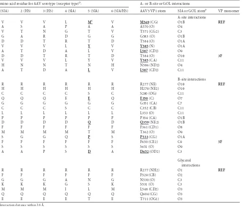
[image:4.585.137.447.69.522.2]interact with all the 60 copies of the VP in a similar manner or is flexible enough to adopt slightly different conformations at the 60 sites. These two possible scenarios would be inconsistent with the icosahedral symmetry imposed during the structure refinement and would lead to lack of ordering of the density for the SIA side groups. The amino acid residues that interact with SIA in the A site are M569, A570, T571, G583, T584, Y585, N586, and L587 (con-tact distance of 2.4 to 3.6 Å) (Table 3;Fig. 1C). The residues inter-acting with SIA in the B site are R277, H279, S280, E350, G351, C352, L353, P354, Q359, F361, T362, P553, F650, S651, and D652) (Table 3;Fig. 1E). The residues that interact with GOL in the B site are R277, F529, N530, S531, L548, Q604, and T711.
The A site but not the B site is important in SIA-dependent transduction.The biological significance of the A-site and B-site AAV5-SIA interactions was studied by making point mutations at AAV5 contact residues that differed with respect to AAV2 (a
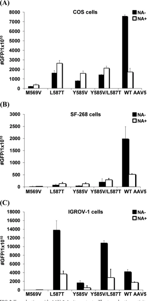
[image:5.585.45.547.76.500.2]non-SIA-binding AAV) and have side chain interactions, and their transduction activity was tested. Very little difference in vector particle yields or physical properties was observed between prep-arations of WT AAV5 and the mutants (data not shown). All mu-tants were tested on three diverse cell lines, COS, SF-268, and IGROV-1. For the A-site mutants, in the COS cells, although transduction activity for the M569V mutant was almost com-pletely abolished, L587T, Y585V, and Y585V/L587T mutants all showed approximately 7-fold-lower transduction activity than that of WT AAV5 (Fig. 2A). However, in contrast to WT AAV5, removal of cell surface SIA with NA did not further inhibit the transduction of L587T, Y585V, and Y585V/L587T mutants. Inter-estingly, all the mutants showed a slight increase in transduction following terminal SIA removal. Similar results were observed with the SF-268 cells (Fig. 2B). A different pattern of transduction was observed on the IGROV-1 cells. Although the M569V mutant
TABLE 3SIA-VP interactions in AAV5 capsida
Amino acid residue for AAV serotype (receptor type)b: A- or B-site or GOL interactions
1 (SIA) 2 (HS) 3 (HS) 4 (SIA) 5 (SIA) 6 (SIA/HS) AAV5 VP1 atom SIA or GOL atomb VP monomer
A-site interactions
V V V L Mc V M569(CG) O1B REF
A S A P A A A570 (O) O4
V T N G T V T571 (CG2) C3
G A R D G G G583 (O) O1B
D D T R T D T584 (O) O1A
V V V L Y V Y585(N) O1A
A T D A L V L587(CD2) O9
D D T R T D T584 (O) O1 3F
V V V L Y V Y585(CA) C11
H N N T N H N586 (ND2) O4
A T D A L V L587(CD2) C11
B-site interactions
R R R R R R R277 (NE) O1 REF
H H H H H H H279 (NE2) O1
C C C C S C S280 (OG) C11
Q Q Q E E Q E350(C) O7
G G G G G G G351 (CA) C7
C C C S C C C352 (CB) C11
L L L L L L L353 (O) C11
P P P P P P P354 (CA) O1B
D D D D Q D Q359(NE2) O1B
F F F F F F F361 (CD1) O8
M M M M T M T362 (O) O9
S G G Q P S P533(CG) O1A
F F F F F F F650 (CE2) C9 5F
S S S S S S S651 (O) O9
A A P S D A D652(OD2) C3
Glycerol interactions
R R R R R R R277 (NH2) O1 REF
F F F F F F F529 (CB) O2
G G G A N G N530 (O) C3
K K K G S K S531 (O) C3
M M M I L M L548 (CD2) O3
Q Q Q Q Q Q Q604 (CG) O3
E E E E T E T711 (OG1) O3
aInteraction distance within 3.6 Å.
b
Receptor type for the particular AAV serotype in parentheses.
cAmino acid residues mutated in this study are in boldface and underlined.
on November 7, 2019 by guest
http://jvi.asm.org/
displayed little transduction activity, similar to the observation in COS cells, the L587T mutation alone or in combination with Y585V improved transduction in an SIA-dependent manner compared to WT AAV5, with the WT virus transduction being lower than in COS cells (Fig. 2C). Compared with COS, L587T mutant transduction activity increased 3.5-fold on the IGROV-1 cells, while the Y585V/L587T double mutant showed an⬃3-fold
increase. Transduction activity of the Y585V mutant also in-creased on IGROV-1 cells compared to COS but was⬃2-fold lower than for WT AAV5. To test if the transduction by the two variants was SIA dependent, cells were treated with neuraminidase and the change in transduction was compared with that of un-treated cells. In contrast to the transduction activity on COS cells, L587T, Y585V, and Y585V/L587T mutants all exhibited neur-aminidase-sensitive transduction activity.
Similar to the A-site mutants, all of the B-site mutants dis-played a significant decrease in transduction activity on all cell types, suggesting that this region is also important in AAV5 trans-duction (Fig. 3). For the Q359D and P533S mutations, there was a loss in GFP expression. Neuraminidase treatment diminished the number of transduced cells, similar to WT AAV5. However, in contrast to the A-site mutants, all of the B-site mutants displayed SIA-dependent transduction activity following neuraminidase treatment on all three target cells. There was an average 5-fold decrease in transduction on all cells following pretreatment with neuraminidase, which was similar to the fold decrease observed with WT AAV5. Transductions of SF-268 and IGROV-1 were lower than on COS cells but still displayed SIA-dependent trans-duction. These results suggest that while both the A-site and B-site regions are important in transduction, mutation of the A-site re-moves neuraminidase-sensitive transduction, suggesting that this region is critical to the binding of SIA that is necessary for virus attachment and transduction.
A-site mutations change SIA-dependent cell attachment.To confirm if the change in transduction activity was associated with disruption in cell surface SIA interaction, mutant vector binding to cells was compared to that of WT AAV5 with or without pre-treatment of the target cells with neuraminidase on COS cells (Fig. 4). While the number of bound particles was smaller for the A-site mutants than for WT AAV5, very little difference in binding was observed if the cells were pretreated with neuraminidase. In agreement with the transduction data, B-site mutations D652A, T362M, and Q359D showed strong neuraminidase-sensitive binding effects. Given that WT AAV5 and the B-site mutants transduction decreased greater than 10-fold with neuraminidase treatment, this observation suggests that the A site is important in both SIA-dependent cell attachment and transduction.
Specificity of carbohydrate binding by A-site mutants on a glycan microarray.Among the A-site mutations, two phenotypes of transduction were observed, complete inhibition of transduc-tion on all cells as seen with M569V or cell type-dependent neur-aminidase-sensitive transduction with Y585V and L587T. Based on these data, we hypothesized that M569V completely abolished SIA binding while L587T and to a lesser extent Y585V altered SIA specificity. To test this hypothesis WT, M569V, and L587TrAAV-GFP vectors were used to probe glycan microarrays developed by the CFG. WT AAV5 reproducibly bound a subset of SIA-contain-ing glycans on the array (Table 4;Fig. 5): NeuAc␣2-3[6OSO3]Gal 1-4GlcNAc-Sp8 (glycan 45/46) and Neu5Ac␣2-3(6OSO3)Gal1-4(Fuc␣1-3)GlcNAc-Sp8 (glycan number 206/208), both with ␣2,3-linked terminal SIA and sulfated on the penultimate galac-tose group. Glycan 206/208 differs from 45/46 by having a fucose group on theN-acetylglucosamine of the glycan chain. Two addi-tional SIA-containing glycans bound by AAV5 on newer arrays were Neu5Ac␣2-3Gal1-4GlcNAc1-2Man␣1-3(Neu5Ac␣2-3Gal1-4GlcNAc1-2Man␣1-6)Man1-4GlcNAc1-4GlcNAc -Sp12 (glycan 141/143), and
Neu5Ac␣2-3Gal1-4GlcNAc1-FIG 2Transduction with AAV-5 A-site mutants. The transduction activity and the effect of neuraminidase were compared for mutation in the A site using COS (A), SF-268 (B), and IGROV-1 (C) cells. Transduction was measured either with (NA⫹) or without (NA⫺) pretreatment with neuraminidase to enzymatically remove terminal cell surface SIA groups. The values are means from three experiments; the error bars represent standard deviations.
on November 7, 2019 by guest
http://jvi.asm.org/
[image:6.585.41.288.68.559.2]2Man␣1-3(Neu5Ac␣2-6Gal1-4GlcNAc1-2Man␣1-6) Man1-4GlcNAc1-4GlcNAc-Sp12 (glycan 316/318). Glycans 141/143 and 316/318 contain the same glycans as 45/46 and 206/208, but without the fucosylation and sulfation, linked to core mannose on both branches. Glycans 141/143 and 316/318 differ in that the terminal SIA in one of the branches in glycan 316/318 is linked by
␣2,6 rather than␣2,3 to galactose. Binding of AAV5 to these sulfo-nated and branched glycans was also recently reported elsewhere (28).
Unlike the data for the WT AAV5, the M569V mutant showed nonspecific binding to most of the glycans on the microarray. The top hits, with acceptable percent coefficient of variation (%CV) values, did not contain terminal SIA, and most of them were sul-fated (Fig. 5;Table 4). For the L587T mutant, the binding to all the glycans was low, with all potential hits for glycans with terminal SIA having high %CV values consistent with low signal-to-noise ratios. Interestingly, the top hit for this mutant is 9NAcNeu5Aca-Sp8 (glycan number 47) with a %CV of 38 (Table 4). Significantly, SIA 9-O-acetylation is upregulated (e.g., 9-O-acetylated GD3) in human melanoma cells (29). Thus, it is possible that this or an-other modified glycan is being utilized by the L587T mutant for the infection of cancer cells such as the IGROV-1 cells.
The glycan microarray observations for the mutant viruses are consistent with the reduced binding in COS cells compared to WT AAV5 and the lack of significant difference in binding to cells without/with neuraminidase treatment (Fig. 4). The data also support the transduction data and indicate that (i) the A site is critical for SIA binding and either (ii) mutation in this region can completely abolish SIA binding or (iii) mutation in this region can retarget the capsid to different modified glycans, such as for the L587T mutant vector.
Mutation of L587 alters SIA binding for AAV5.Previous re-search had demonstrated robust transgene expression following WT AAV5 vector delivery to both lung or salivary gland epithelia (30, 31). Thus, to test if the altered SIA binding specificity by L587T also alters tropismin vivo, gene expression was monitored in mice for WT AAV5 or L587T vectors encoding luciferase. In agreement with the change inin vitrocarbohydrate binding and cell tropism, the L587T mutant also demonstrated a change in transduction activityin vivo(Fig. 6). Following vector delivery to
FIG 3Transduction with AAV-5 B-site mutants. The transduction activity and the effect of neuraminidase were compared for mutation in the B site using COS (A), SF-268 (B), and IGROV-1 (C) cells. Transduction was measured either with (NA⫹) or without (NA⫺) pretreatment with neuraminidase to enzymatically remove terminal cell surface SIA groups. The values are means from three experiments; the error bars represent standard deviations.
FIG 4Cell binding by WT and mutant vectors. The effects of neuraminidase
on vector binding were compared on COS cells with (NA⫹) or without
(NA⫺) neuraminidase pretreatment. Bound virus was measured by quantita-tive PCR using primers specific for the CMV promoter. Values are means from three experiments. Error bars represent standard deviations.
on November 7, 2019 by guest
http://jvi.asm.org/
[image:7.585.296.541.64.273.2] [image:7.585.42.309.64.576.2]the submandibular salivary glands, L587T mutant demonstrated a 3- to 4-fold increase in transduction activity compared to WT AAV5. Similarly, a direct injection of L587T mutant in the hind limb showed a 3- to 4-fold increase in luciferase expression com-pared with WT AAV5. In contrast, L587T mutant gene transfer to the lung was greatly reduced compared with WT AAV5. M569V and Q359D mutants were also evaluated in the salivary glands and showed no transduction activity (data not shown). These data suggest that alteration in glycan recognition can affect vector ac-tivityin vivo.
AAV5 SIA binding residues map to an antigenic region.We hypothesized that alteration of a critical region for transduction could alter the antigenic profile of the vector and thus its neutral-ization by serum antibodies. The antigenic profiles of selected A-site and B-A-site mutants were explored in comparison to that of WT AAV5 (Fig. 7). At low dilutions of an anti-AAV5 polyclonal se-rum, all mutants were neutralized to the same extent as WT AAV5. However, further serial dilution of this serum demonstrated dis-tinct properties for the A-site and B-site mutants. In contrast to the B-site Q359D mutant, which was neutralized to an extent sim-ilar to that of WT AAV5, both of the A-site mutants showed a sharp reduction in neutralization activity with dilutions of 100-fold or greater than the Q359D mutant or WT AAV5. Thus, in addition to being critical for SIA-dependent transduction, the A site represents an important antigenic region on AAV5, and its alteration creates an antigenic escape mutant (L587T mutant) still capable of robustin vitroandin vivotransgene expression.
DISCUSSION
To date, over 150 AAV capsid sequences have been cloned from a variety of sources, but there is very little understanding of their host cell requirements for entry and of which regions on the sur-face of the particles are involved in entry. What is known is that these viruses have evolved to utilize the glycans abundant on cell surfaces as essential primary attachment points for entry. In addi-tion, available data show that the 13 characterized serotypes can
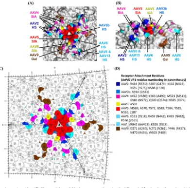
be grouped into those that bind HSPG (AAV2, AAV3B, AAV6, AAV13), SIA (AAV1, AAV4, AAV5, AAV6), GAL (AAV9), and unknown (AAV7, AAV8, AAV10, and AAV11) (32).
Efforts to manipulate the AAV capsid to target specific cell types have generated interest in understanding the AAV capsid structures and mapping their capsid surface regions used to inter-act with cellular molecules. The mapping of the HSPG binding site on AAV2, with R585 and R588 being the major determinants of the interaction, has allowed for the capsid loop containing these residues to be deleted or altered by peptide insertions for retarget-ing of tissue tropism (33). This ability to manipulate a known receptor attachment region has had a major impact on the field of AAV vectorology, and this knowledge is routinely used by several labs. We recently reported the identification of a new HSPG-de-pendent transduction region on the surface of AAV(VR-942), now AAV13, which is distinct from the HSPG binding site in AAV2 (2) but similar to that used by AAV6 to interact with this glycan (10) (Fig. 8). This suggests that distinct capsid regions on different AAV isolates can be responsible for the same activity (32).
In contrast to the HSPG binding AAVs, whose interaction sites are conferred by basic amino acid patches on the capsid, less is known about AAV SIA binding. In our targeted combined struc-tural and mutagenesis studies of the AAV5 SIA binding interac-tion, we have identified two interesting sites: A and B. Cell binding and transduction assays showed that both these sites are determi-nants of cellular transduction. However, the A site appears to be the SIA-dependent transduction determinant. This binding re-gion, configured from symmetry-related VP monomers at the ico-sahedral 3-fold axis of the capsid, is close to but distinct from amino acids affecting AAV4 SIA interaction, the AAV2, AAV6, and AAV13 HSPG binding regions, and the AAV9 galactose (GAL) footprint (Fig. 8). Rather than being exposed on the wall/ top of the 3-fold protrusions (AAV2-HS, AAV4-SIA) or outside, at the base of the protrusions (AAV13-HS, AAV6-HS, and AAV9-GAL), the AAV5 SIA binding region is recessed into the
depres-TABLE 4WT AAV5 and AAV5 M569V and L587T mutant capsid glycan specificity on glycan arraysa
AAV5 and glycan no. Glycan name RFU SD SEM %CV
Wild-type AAV5
45/46b NeuAc␣2-3[6OSO3]Gal1-4GlcNAc–Sp8 38,621 2,164 1,082 6
206/208b Neu5Ac␣2-3(6-O-Su)Gal1-4(Fuc␣1-3)GlcNAc–Sp8 34,986 4,816 2,408 14
141/143b Neu5Ac␣2-3Gal1-4GlcNAc1-2Man␣1-3(Neu5Ac␣2-3Gal1-4GlcNAc1-2Man␣1-6)Man
1-4GlcNAc1-4GlcNAc-Sp12
29,649 9,158 4,579 31
316/318b Neu5Ac␣2-3Gal1-4GlcNAc1-2Man␣1-3(Neu5Ac␣2-6Gal1-4GlcNAc1-2Man␣1-6)Man
1-4GlcNAc1-4GlcNAc-Sp12
22,531 6,079 3,040 27
M569V mutant
265 [3OSO3]Galb1-4[Fuca1-3][6OSO3]GlcNAc-Sp8 9,148 1,045 523 11
284 [3OSO3]Galb1-4[6OSO3]GlcNAcb-Sp0 7,875 565 282 7
16 b-d-Gal–Sp8 7,179 1,267 634 18
34 [3OSO3]Galb1-4[6OSO3]GlcNAcb-Sp8 7,174 604 302 8
L587T mutant
47 9NAcNeu5Aca-Sp8 1,092 411 205 38
337 GlcNAca1-4Galb1-4GlcNAcb1-3Galb1-4(Fuca1-3)GlcNAcb1-3Galb1-4(Fuca1-3)GlcNAcb-Sp0 929 682 341 73
260 Neu5Gca2-6GalNAca-Sp0 929 762 381 82
375
GalNAcb1-4GlcNAcb1-2Mana1-6(GalNAcb1-4GlcNAcb1-2Mana1-6)Manb1-4GlcNAcb1-4GlcNAc-Sp12
908 422 211 46
aThe top four glycans with the highest relative fluorescence units (RFU) for each vector are listed.
b
Numbers are based on PA Ver. 3.1/3.0.
on November 7, 2019 by guest
http://jvi.asm.org/
[image:8.585.39.554.78.294.2]sion formed by the mounds of the protrusions that surround this axis. This more protected site may serve a selective advantage to the virus as it may make it more difficult for cells to develop neu-tralizing antibodies that interfere with this interaction.
Consis-FIG 5Glycan microarray graphs. Top, WT AAV5; middle, M569V mutant; bottom, L587T mutant. The top hits for the WT AAV5 are labeled. The relative fluorescence units (RFU) are shown in theyaxis, and the glycan array number is given in thexaxis. The top 4 glycan hits (highest RFU) are indicated.
FIG 6In vivotransduction activity of WT AAV5 and A-site L587T mutant. The indicated recombinant vectors encoding luciferase were delivered into either BALB/c mouse salivary glands, muscle (tibialis anterior), or lungs. Rel-ative luciferase expression was visualized by Xenogen imaging.
on November 7, 2019 by guest
http://jvi.asm.org/
[image:9.585.41.552.57.634.2] [image:9.585.276.538.59.668.2]tently, two of the three mouse monoclonal IgG antibodies avail-able for AAV5, 3C5 and ADK5a, are nonneutralizing (34, 35) (data not shown).
The B site, which is mostly buried under the HI loop, is located close to an ordered GOL molecule, which was also observed in the WT AAV5 crystal structure (19). The GOL molecule was postu-lated to be playing a role in capsid stabilization and possibly oc-cupying a substrate-ligand interaction site by analogy to similarity with the GOL binding site observed in the structure of succinyl-CoA/3-oxoacid coenzyme A transferase (36). In this enzyme, the GOL molecule was reported to occupy the acetate cosubstrate binding. Thus, it is possible that this SIA and GOL binding site serves to bind glycans required downstream of the infection path-way, such as a receptor required for internalization. The platelet-derived growth factor receptor (PDGFR), which serves as a recep-tor for AAV5, is a sialylated glycoprotein (15). Thus, while the role for the B-site SIA interaction in cellular infection requires further investigation, it is tempting to speculate that this site may serve as the contact region for this glycoprotein receptor.
The residues comprising the A site are reminiscent of SIA bind-ing sites for other viruses, for example, influenza virus (37) and the parvovirus minute virus of mice (MVM) (38), and include mostly hydrophobic and polar residues. Indeed, several mapped SIA binding sites, including that of MVM, contain a hydrophobic aromatic residue, centered in a depression. The pockets can be located in different regions of the capsid, for example, the icosa-hedral 2-fold depression for MVM in contrast to the 3-fold de-pression used by AAV5. The three amino acids selected to confirm the role of this site in AAV5 SIA binding included two hydropho-bic residues, M569 and L587, and a polar noncharged residue, Y585, mutated to the corresponding residues in the non-SIA-binding AAV2. While mutation of residue 569 completely ablates SIA binding activity, mutations of residues 587 and 585 appear to change the binding specificity. Based on data from the glycan mi-croarray, the AAV5 M569V mutant has no affinity for the
sialy-lated glycans recognized by WT AAV5 but binds to highly sulfated glycans. On the other hand, AAV5 L587T mutant displayed sig-nificantly reduced affinity to glycans in general with a switch from ␣2,3 recognition to␣2,6 recognition and the recognition of non-sialylated glycans (Table 4).
The altered receptor recognition by the AAV5 mutants sup-ports their altered cellular transduction phenotypes. The lack of cellular transduction by the AAV5 M569V mutant is likely due to a reduced or lack of abundance of sulfated glycans on cell surfaces. The increased transduction of IGROV-1 cells by AAV5 L587T mutant is likely due to the upregulation of modified glycans on the cell surface associated with transformed cells, while the slight in-crease in transduction following neuraminidase treatment in COS cells may be due to the affinity (albeit low) for glycans without terminal SIA and recognition of GAL. As mentioned above, sev-eral AAVs in addition to AAV5, e.g., AAV1, AAV4, and AAV6, utilize SIA as their primary engagement receptor for infection. The SIA binding region for AAV1 is analogous to the GAL binding region for AAV9 in a pocket at the base of the 3-fold protrusion and different from the AAV5 A-site (L.-Y. Huang and M. Ag-bandje-McKenna, unpublished data) and is likely to be the same site for the closely related AAV6. The amino acids in AAV1 and AAV9 differ in this pocket and engage different glycans. A random mutagenesis study of the AAV4 3-fold protrusions identified six amino acids, K492, K593, M523, G581, Q583, and N585, as play-ing a role in AAV4 SIA utilization (39) (Fig. 8). These residues are located closer to the top of the protrusions but on the same face as the AAV5-SIA footprint. While there are no equivalent residues between AAV4 and AAV5 in their SIA contact region and the 3-fold region is one of the most structurally variable between the AAVs, some of the residues in the binding footprints are adjacent (Fig. 8). Significantly, AAV2 also binds its HSPG receptor using the analogous region on its capsid with different amino acids (Fig. 8). The observation suggests that the structural features, such as the depressions at the center and base of the 3-fold protrusions, in addition to amino acid type, are important for glycan recognition by the AAV capsid.
As observed in this study, changes in the primary amino acid sequence can alter the biologic activity of the particle. Changing single amino acids on the capsid at the AAV5 SIA A-site, for ex-ample, L587T, altered the glycan recognitionin vitroand thein vivotransduction activity. In addition, the ability to neutralize A-site mutant vectors with a polyclonal serum raised against WT AAV5 is significantly reduced. These findings further confirm the importance of the A site in AAV5 transduction and the role of glycan recognition in AAV5 transduction.
For other parvoviruses, SIA interactions are reported for bo-vine parvovirus (40), canine parvovirus (CPV), feline panleuko-penia virus (FPV) (41), H-1 parvovirus (H-1PV) (42), and por-cine parvovirus (43) in addition to MVM (38). The amino acids utilized for binding SIA are unknown for bovine and porcine par-vovirus, but like MVM, CPV, FPV, and H-1PV bind SIA in or close to the 2-fold depression. For MVM and H-1PV, residues in the vicinity of the SIA binding site also play a role in tissue tropism determination (38,42), and for MVM, they also dictated virulence and pathogenicity (38). However, in CPV and FPV, the SIA inter-action either alone or in the context of their receptor transferrin does not seem to be important for infection or host range (44). Thus, the available data for these viruses indicate that, unlike the AAVs, there is commonality in the use of the same capsid region to
FIG 7Neutralization of transduction by WT AAV5 and SIA binding site mutants. The indicated recombinant vector encoding GFP was incubated with serially diluted anti-AAV5 polyclonal antibody raised in rabbits prior to infec-tion of COS cells. Transducinfec-tion efficiencies relative to those of an untreated control were plotted against the reciprocal of the dilution of sera incubated with the vector. Values are means from three experiments; error bars represent standard deviations.
on November 7, 2019 by guest
http://jvi.asm.org/
[image:10.585.40.286.66.251.2]engage the same glycan. However, for autonomous viruses, at-tachment and even infection do not equate to pathogenicity.
In summary, we have identified a region used for SIA binding by AAV5, an essential interaction for successful infection. The binding site conserves the general features observed in other virus-capsid interactions. Further analysis of the SIA binding pocket in AAV5 and characterization of other SIA-dependent AAVs will be necessary to identify the interplay between the bound glycans and the receptors required for internalization. The data so far clearly point to the icosahedral 3-fold axis as being important in cell binding by multiple isolates of AAV, while versatility in capsid interactions shows that the 2-fold region of the autonomous par-vovirus capsid is also suitable for SIA receptor attachment. Since structure is more conserved than sequence, information on all receptor-interacting regions can inform vector-retargeting devel-opments. Manipulation of these regions, given knowledge of their functions, will lead to improvements in the development of the AAV gene transfer system.
ACKNOWLEDGMENTS
We thank the Protein-Carbohydrate Interaction Core H of The Consor-tium for Functional Glycomics for conducting the glycan array analysis.
This work was supported by an NIH NIDCR intramural research grant to J.A.C. and funds from the University of Florida College of Medicine, the McKnight Brain Institute, and NIH R01 GM082946 to M.A.-M.
REFERENCES
1.Rabinowitz JE, Rolling F, Li C, Conrath H, Xiao W, Xiao X, Samulski RJ.2002. Cross-packaging of a single adeno-associated virus (AAV) type 2 vector genome into multiple AAV serotypes enables transduction with broad specificity. J Virol76:791– 801.http://dx.doi.org/10.1128/JVI.76.2 .791-801.2002.
2.Schmidt M, Govindasamy L, Afione S, Kaludov N, Agbandje-McKenna M, Chiorini JA. 2008. Molecular characterization of the heparin-dependent transduction domain on the capsid of a novel adeno-associated virus isolate, AAV(VR-942). J Virol82:8911– 8916.http://dx.doi.org/10 .1128/JVI.00672-08.
3.Halbert CL, Allen JM, Miller AD.2001. Adeno-associated virus type 6 (AAV6) vectors mediate efficient transduction of airway epithelial cells in FIG 8AAV-receptor interactions. (A and B) Closeup of the available glycan receptor footprints for different AAVs colored on the surface of AAV5 capsid. The residues involved in glycan receptor binding for AAV2, AAV3b, AAV4, AAV5, AAV6, AAV9, and AAV13 are highlighted. In panel A, the capsid is viewed down the icosahedral 3-fold axis, and panel B depicts the side view of the 3-fold axis. (C) Stereographic Roadmap projection (45) of the receptor footprint viewed down the 3-fold axis. Residues are labeled by type (three-letter code) and number (AAV5 VP1 numbering). The boundary for each residue is shown in black. The amino acid residues that are exposed on the capsid exterior are visible in this image. (D) Color key for panels A to C. Panels A and B were generated using the PyMol program (24).
on November 7, 2019 by guest
http://jvi.asm.org/
[image:11.585.98.489.65.451.2]mouse lungs compared to that of AAV2 vectors. J Virol75:6615– 6624.
http://dx.doi.org/10.1128/JVI.75.14.6615-6624.2001.
4.Handa A, Muramatsu S, Qiu J, Mizukami H, Brown KE.2000. Adeno-associated virus (AAV)-3-based vectors transduce haematopoietic cells not susceptible to transduction with AAV-2-based vectors. J Gen Virol 81:2077–2084.http://vir.sgmjournals.org/content/81/8/2077.long. 5.Seiler MP, Miller AD, Zabner J, Halbert CL.2006. Adeno-associated
virus types 5 and 6 use distinct receptors for cell entry. Hum Gene Ther 17:10 –19.http://dx.doi.org/10.1089/hum.2006.17.10.
6.Summerford C, Samulski RJ.1998. Membrane-associated heparan sul-fate proteoglycan is a receptor for adeno-associated virus type 2 virions. J Virol72:1438 –1445.
7.Wu Z, Miller E, Agbandje-McKenna M, Samulski RJ.2006. Alpha2,3 and alpha2,6 N-linked sialic acids facilitate efficient binding and transduc-tion by adeno-associated virus types 1 and 6. J Virol80:9093–9103.http: //dx.doi.org/10.1128/JVI.00895-06.
8.Kaludov N, Brown KE, Walters RW, Zabner J, Chiorini JA. 2001. Adeno-associated virus serotype 4 (AAV4) and AAV5 both require sialic acid binding for hemagglutination and efficient transduction but differ in sialic acid linkage specificity. J Virol75:6884 – 6893.http://dx.doi.org/10 .1128/JVI.75.15.6884-6893.2001.
9.Walters RW, Pilewski JM, Chiorini JA, Zabner J.2002. Secreted and transmembrane mucins inhibit gene transfer with AAV4 more efficiently than AAV5. J Biol Chem277:23709 –23713.http://dx.doi.org/10.1074/jbc .M200292200.
10. Wu Z, Asokan A, Grieger JC, Govindasamy L, Agbandje-McKenna M, Samulski RJ.2006. Single amino acid changes can influence titer, heparin binding, and tissue tropism in different adeno-associated virus serotypes. J Virol80:11393–11397.http://dx.doi.org/10.1128/JVI.01288-06. 11. Schmidt M, Chiorini JA. 2006. Gangliosides are essential for bovine
adeno-associated virus entry. J Virol80:5516 –5522.http://dx.doi.org/10 .1128/JVI.02393-05.
12. Rooney CP, Denning GM, Davis BP, Flaherty DM, Chiorini JA, Zabner J.2002. Bronchoalveolar fluid is not a major hindrance to virus-mediated gene therapy in cystic fibrosis. J Virol76:10437–10443.http://dx.doi.org /10.1128/JVI.76.20.10437-10443.2002.
13. Kern A, Schmidt K, Leder C, Muller OJ, Wobus CE, Bettinger K, Von der Lieth CW, King JA, Kleinschmidt JA. 2003. Identification of a heparin-binding motif on adeno-associated virus type 2 capsids. J Virol 77:11072–11081.http://dx.doi.org/10.1128/JVI.77.20.11072-11081.2003. 14. Opie SR, Warrington KH, Jr, Agbandje-McKenna M, Zolotukhin S, Muzyczka N.2003. Identification of amino acid residues in the capsid proteins of adeno-associated virus type 2 that contribute to heparan sul-fate proteoglycan binding. J Virol 77:6995–7006.http://dx.doi.org/10 .1128/JVI.77.12.6995-7006.2003.
15. Di Pasquale G, Davidson BL, Stein CS, Martins I, Scudiero D, Monks A, Chiorini JA.2003. Identification of PDGFR as a receptor for AAV-5 transduction. Nat Med9:1306 –1312.http://dx.doi.org/10.1038/nm929. 16. DiMattia M, Govindasamy L, Levy HC, Gurda-Whitaker B, Kalina A,
Kohlbrenner E, Chiorini JA, McKenna R, Muzyczka N, Zolotukhin S, Agbandje-McKenna M.2005. Production, purification, crystallization and preliminary X-ray structural studies of adeno-associated virus sero-type 5. Acta Crystallogr F Struct Biol Cryst Commun61:917–921.http: //dx.doi.org/10.1107/S1744309105028514.
17. McPherson A.1982. Preparation and analysis of protein crystals, 1st ed. Wiley, New York, NY.
18. Otwinowski Z, Minor W.1997. Processing of X-ray diffraction data in oscillation mode. Methods Enzymol276:307–326.http://dx.doi.org/10 .1016/S0076-6879(97)76066-X.
19. Govindasamy L, DiMattia MA, Gurda BL, Halder S, McKenna R,
Chiorini JA, Muzyczka N, Zolotukhin S, Agbandje-McKenna M.2013. Structural insights into adeno-associated virus serotype 5. J Virol87: 11187–11199.http://dx.doi.org/10.1128/JVI.00867-13.
20. Brunger AT, Adams PD, Clore GM, DeLano WL, Gros P,
Grosse-Kunstleve RW, Jiang JS, Kuszewski J, Nilges M, Pannu NS, Read RJ, Rice LM, Simonson T, Warren GL.1998. Crystallography & NMR sys-tem: a new software suite for macromolecular structure determination. Acta Crystallogr D Biol Crystallogr54:905–921.
21. Kleywegt GJ.2007. Crystallographic refinement of ligand complexes. Acta Crystallogr D Biol Crystallogr63:94 –100.http://dx.doi.org/10.1107/S090 7444906022657.
22. Adams PD, Afonine PV, Bunkoczi G, Chen VB, Davis IW, Echols N, Headd JJ, Hung LW, Kapral GJ, Grosse-Kunstleve RW, McCoy AJ,
Mo-riarty NW, Oeffner R, Read RJ, Richardson DC, Richardson JS, Terwilliger TC, Zwart PH.2010. PHENIX: a comprehensive Python-based system for macromolecular structure solution. Acta Crystallogr D Biol Crystallogr66: 213–221.http://dx.doi.org/10.1107/S0907444909052925.
23. Emsley P, Lohkamp B, Scott WG, Cowtan K.2010. Features and devel-opment of Coot. Acta Crystallogr D Biol Crystallogr66:486 –501.http: //dx.doi.org/10.1107/S0907444910007493.
24. DeLano WL.2002, posting date. The PyMOL molecular graphics system. DeLano Scientific, San Carlos, CA.
25. Kaludov N, Handelman B, Chiorini JA.2002. Scalable purification of adeno-associated virus type 2, 4, or 5 using ion-exchange chromatogra-phy. Hum Gene Ther13:1235–1243.http://dx.doi.org/10.1089/10430340 2320139014.
26. Smith RH, Afione SA, Kotin RM.2002. Transposase-mediated construc-tion of an integrated adeno-associated virus type 5 helper plasmid. Bio-techniques33:204 –206, 208, 210 –211.
27. Quinn K, Quirion MR, Lo CY, Misplon JA, Epstein SL, Chiorini JA. 2011. Intranasal administration of adeno-associated virus type 12 (AAV12) leads to transduction of the nasal epithelia and can initiate trans-gene-specific immune response. Mol Ther19:1990 –1998.http://dx.doi .org/10.1038/mt.2011.146.
28. Mietzsch M, Broecker F, Reinhardt A, Seeberger PH, Heilbronn R. 2014. Differential adeno-associated virus serotype-specific interaction patterns with synthetic heparins and other glycans. J Virol88:2991–3003.
http://dx.doi.org/10.1128/JVI.03371-13.
29. Ritter G, Boosfeld E, Markstein E, Yu RK, Ren SL, Stallcup WB, Oettgen HF, Old LJ, Livingston PO.1990. Biochemical and serological characteristics of natural 9-O-acetyl GD3 from human melanoma and
bovine buttermilk and chemically O-acetylated GD3. Cancer Res50:
1403–1410.
30. Katano H, Kok MR, Cotrim AP, Yamano S, Schmidt M, Afione S, Baum BJ, Chiorini JA.2006. Enhanced transduction of mouse salivary glands with AAV5-based vectors. Gene Ther13:594 – 601.http://dx.doi.org/10 .1038/sj.gt.3302691.
31. Zabner J, Seiler M, Walters R, Kotin RM, Fulgeras W, Davidson BL, Chiorini JA.2000. Adeno-associated virus type 5 (AAV5) but not AAV2 binds to the apical surfaces of airway epithelia and facilitates gene transfer. J Virol74:3852–3858.http://dx.doi.org/10.1128/JVI.74.8.3852-3858.2000. 32. Huang LY, Halder S, Agbandje-McKenna M.2014. Parvovirus glycan
interactions. Curr Opin Virol7:108 –118.http://dx.doi.org/10.1016/j .coviro.2014.05.007.
33. Girod A, Ried M, Wobus C, Lahm H, Leike K, Kleinschmidt J, Deleage G, Hallek M.1999. Genetic capsid modifications allow efficient re-targeting of adeno-associated virus type 2. Nat Med5:1052–1056.http: //dx.doi.org/10.1038/12491.
34. Harbison CE, Weichert WS, Gurda BL, Chiorini JA,
Agbandje-McKenna M, Parrish CR.2012. Examining the cross-reactivity and neu-tralization mechanisms of a panel of mAbs against adeno-associated virus serotypes 1 and 5. J Gen Virol93:347–355.http://dx.doi.org/10.1099/vir .0.035113-0.
35. Kuck D, Kern A, Kleinschmidt JA.2007. Development of AAV serotype-specific ELISAs using novel monoclonal antibodies. J Virol Methods140: 17–24.http://dx.doi.org/10.1016/j.jviromet.2006.10.005.
36. Coker SF, Lloyd AJ, Mitchell E, Lewis GR, Coker AR, Shoolingin-Jordan PM.2010. The high-resolution structure of pig heart succinyl-CoA:3-oxoacid coenzyme A transferase. Acta Crystallogr D Biol Crystal-logr66:797– 805.http://dx.doi.org/10.1107/S0907444910018366. 37. Eisen MB, Sabesan S, Skehel JJ, Wiley DC.1997. Binding of the influenza
A virus to cell-surface receptors: structures of five hemagglutinin-sialyloligosaccharide complexes determined by X-ray crystallography. Vi-rology232:19 –31.http://dx.doi.org/10.1006/viro.1997.8526.
38. Lopez-Bueno A, Rubio MP, Bryant N, McKenna R, Agbandje-McKenna M, Almendral JM.2006. Host-selected amino acid changes at the sialic acid binding pocket of the parvovirus capsid modulate cell binding affinity and determine virulence. J Virol80:1563–1573.http://dx.doi.org/10.1128 /JVI.80.3.1563-1573.2006.
39. Shen S, Troupes AN, Pulicherla N, Asokan A.2013. Multiple roles for sialylated glycans in determining the cardiopulmonary tropism of adeno-associated virus 4. J Virol87:13206 –13213.http://dx.doi.org/10.1128/JVI .02109-13.
40. Thacker TC, Johnson FB.1998. Binding of bovine parvovirus to eryth-rocyte membrane sialylglycoproteins. J Gen Virol79(Part 9):2163–2169.
on November 7, 2019 by guest
http://jvi.asm.org/
41. Barbis DP, Chang SF, Parrish CR.1992. Mutations adjacent to the dimple of the canine parvovirus capsid structure affect sialic acid binding. Virology191: 301–308.http://dx.doi.org/10.1016/0042-6822(92)90192-R.
42. Allaume X, El-Andaloussi N, Leuchs B, Bonifati S, Kulkarni A, Marttila T, Kaufmann JK, Nettelbeck DM, Kleinschmidt J, Rommelaere J, Marchini A.2012. Retargeting of rat parvovirus H-1PV to cancer cells through genetic engineering of the viral capsid. J Virol86:3452–3465.http: //dx.doi.org/10.1128/JVI.06208-11.
43. Boisvert M, Fernandes S, Tijssen P.2010. Multiple pathways involved in
porcine parvovirus cellular entry and trafficking toward the nucleus. J Virol84:7782–7792.http://dx.doi.org/10.1128/JVI.00479-10.
44. Palermo LM, Hafenstein SL, Parrish CR.2006. Purified feline and canine transferrin receptors reveal complex interactions with the capsids of ca-nine and feline parvoviruses that correspond to their host ranges. J Virol 80:8482– 8492.http://dx.doi.org/10.1128/JVI.00683-06.
45. Xiao C, Rossmann MG.2007. Interpretation of electron density with stereographic roadmap projections. J Struct Biol158:182–187.http://dx .doi.org/10.1016/j.jsb.2006.10.013.