Nucleic Acid Interactions: Implications for Genomic RNA Packaging
Meng Sun,aIwen F. Grigsby,bRobert J. Gorelick,cLouis M. Mansky,bKarin Musier-Forsytha
‹Department of Chemistry and Biochemistry, Center for Retroviral Research, and Center for RNA Biology, The Ohio State University, Columbus, Ohio, USAa; Institute for
Molecular Virology, Departments of Diagnostic and Biological Sciences and Microbiology, School of Dentistry and Medical School, University of Minnesota, Minneapolis, Minnesota, USAb; AIDS and Cancer Virus Program, Leidos Biomedical Research, Inc., Frederick National Laboratory for Cancer Research, Frederick, Maryland, USAc
Retroviral RNA encapsidation involves a recognition event between genomic RNA (gRNA) and one or more domains in Gag. In
HIV-1, the nucleocapsid (NC) domain is involved in gRNA packaging and displays robust nucleic acid (NA) binding and
chaper-one functions. In comparison, NC of human T-cell leukemia virus type 1 (HTLV-1), a deltaretrovirus, displays weaker NA
bind-ing and chaperone activity. Mutation of conserved charged residues in the deltaretrovirus bovine leukemia virus (BLV) matrix
(MA) and NC domains affects virus replication and gRNA packaging efficiency. Based on these observations, we hypothesized
that the MA domain may generally contribute to NA binding and genome encapsidation in deltaretroviruses. Here, we examined
the interaction between HTLV-2 and HIV-1 MA proteins and various NAs
in vitro
. HTLV-2 MA displays higher NA binding
af-finity and better chaperone activity than HIV-1 MA. HTLV-2 MA also binds NAs with higher afaf-finity than HTLV-2 NC and
dis-plays more robust chaperone function. Mutation of two basic residues in HTLV-2 MA
␣
-helix II, previously implicated in BLV
gRNA packaging, reduces NA binding affinity. HTLV-2 MA binds with high affinity and specificity to RNA derived from the
pu-tative packaging signal of HTLV-2 relative to nonspecific NA. Furthermore, an HIV-1 MA triple mutant designed to mimic the
basic character of HTLV-2 MA
␣
-helix II dramatically improves binding affinity and chaperone activity of HIV-1 MA
in vitro
and restores RNA packaging to a
⌬
NC HIV-1 variant in cell-based assays. Taken together, these results are consistent with a role
for deltaretrovirus MA proteins in viral RNA packaging.
W
hen retroviruses assemble in infected cells, two copies of
full-length genomic RNA (gRNA) are selected for
packag-ing. Although gRNA constitutes only a very small portion of total
RNA in the cytoplasm, it is selectively packaged into virions (
1
).
The specific packaging process is believed to involve recognition
of gRNA packaging signals by the Gag polyprotein (
1–4
). In
HIV-1 assembly, the nucleocapsid (NC) domain of Gag is the
dominant nucleic acid (NA) binding region and is essential for
specific incorporation of gRNA. HIV-1 NC is also a robust
chap-erone protein, wherein it remodels NAs to their most
thermody-namically stable state through duplex destabilization, aggregation,
and rapid binding kinetics (
5–8
). The solution structures of HIV-1
NC bound to stem-loop 2 (SL2) and SL3 derived from the psi (
⌿
)
packaging signal have been studied by nuclear magnetic resonance
(NMR) spectroscopy. In these structures, the two zinc finger
mo-tifs of NC specifically bind to guanosines in the G-rich RNA
te-traloops (
9
,
10
).
The matrix (MA) domain of Gag also has several established
functions in retroviral replication (
11
). HIV-1 MA is required for
targeting of Gag to the plasma membrane of infected cells via its
myristoyl moiety (
12
,
13
). Basic residues of HIV-1 MA also
con-tribute to membrane binding (
14–16
). In the absence of NC and
protease activity, the HIV-1 MA domain has been shown to bind
RNA and facilitate immature virus particle formation (
17
,
18
). A
number of additional studies have supported the NA binding
properties of HIV-1 MA (
19–21
), yet how this capability
contrib-utes to virus replication has not been elucidated. A more recent
study showed that HIV-1 MA-RNA interactions can be
outcom-peted by phosphatidylinositol-4,5-bisphosphate [PI(4,5)P
2]-con-taining liposomes but not other liposomes (
22
). It has been
pro-posed that RNA binding to MA negatively regulates membrane
binding both by preventing nonspecific interactions between
ba-sic residues and acidic lipids and by suppressing
myristate-depen-dent hydrophobic interactions (
23
).
An early study suggested that the MA domain of bovine
leuke-mia virus (BLV), a deltaretrovirus, plays a more significant role in
specific RNA binding than NC (
24
). BLV MA was reported to
form a specific complex with the dimeric 5
=
end of the gRNA
sequence but not with other RNAs
in vitro
, whereas BLV NCp12
bound randomly and nonspecifically (
24
). More recent studies
showed that both the BLV MA and NC domains are involved in
gRNA packaging and that conserved charged residues in MA are
critical for this function. When two conserved residues, K41 and
H45, were individually mutated to alanine, RNA packaging
effi-ciency was significantly reduced (by 66 and 92%, respectively). In
contrast, mutation of these same residues does not affect Gag
membrane localization (
25
). The MA domain of the
deltaretrovi-rus human T-cell leukemia videltaretrovi-rus type 2 (HTLV-2) contains 11
basic residues scattered throughout the primary sequence, which
form a cluster of exposed positive charges at the surface of the
protein; 8 of the 11 residues are also present in HTLV-1 MA (
26
,
27
). Surprisingly, replacement of the basic residues of HTLV-1
MA with Leu/Ile did not affect intracellular targeting of Gag, even
though most of the mutations completely abolished viral
infectiv-ity and dramatically reduced viral particle production (
27
). Based
Received1 August 2013 Accepted4 November 2013
Published ahead of print13 November 2013
Address correspondence to Karin Musier-Forsyth, [email protected].
Copyright © 2014, American Society for Microbiology. All Rights Reserved.
doi:10.1128/JVI.02151-13
on November 7, 2019 by guest
http://jvi.asm.org/
on these data, it has been suggested that, as for BLV MA, HTLV-1
MA may also play a role in gRNA packaging (
25
).
Interestingly, HTLV-1 NC displays reduced NA binding
affin-ity and chaperone function relative to those of HIV-1 NC yet has
robust duplex-destabilizing capabilities (
28
,
29
). Our laboratory
has previously explored the mechanistic basis for the poor NA
binding and chaperone properties of HTLV-1 NC, and our studies
show that removal of HTLV-1 NC’s anionic C-terminal domain
(CTD) improves the chaperone function to a level comparable to
those of other retroviral NCs (
30
). An intramolecular N-terminal
domain (NTD)-CTD interaction reduces the kinetics of
associa-tion with NAs in the unbound state, whereas an NTD-CTD
inter-action between neighboring molecules reduces the NC-NA
disso-ciation in the bound state. These properties inhibit both NA
aggregation and rapid protein dissociation from single-stranded
DNA (ssDNA), which are required for chaperone function (
6
).
The amino acid sequences of HTLV-1 and HTLV-2 NCs are 72%
identical and have similar isoelectric points, close to neutral.
Therefore, HTLV-2 NC is likely to possess structural and
bio-chemical properties similar to those of HTLV-1 NC.
Sequence alignment of HTLV-1, HTLV-2, BLV, and HIV-1
MA proteins shows high homology among the three
deltaretrovi-ruses, especially HTLV-1 and HTLV-2, which share 58% identity.
These data suggest that there is likely to be conserved function
among deltaretroviral MA proteins. In contrast, HIV-1 and
HTLV-2 MA proteins share only
⬃
10% sequence identity.
Nev-ertheless, they adopt quite similar secondary structures, with
N-terminal basic residues exposed in similar positions on one side of
␣
-helix II, as shown by NMR spectroscopy (
26
). The functional
similarity between these two MA proteins is unknown.
The gRNA packaging signal of BLV is a bipartite RNA motif
consisting of a primary (SL1 and SL2) region and a secondary
region containing a single stem-loop (
31
). It has been shown that
replacement of the BLV packaging signal with a similar region
from either HTLV-1 or HTLV-2 leads to only a partial BLV
rep-lication defect (
32
,
33
). These data support at least some level of
conserved function in deltaretroviral RNA packaging signals.
Based on the available data, we hypothesized that in
deltaret-roviruses, MA plays an equally important role in gRNA
recogni-tion and packaging as that of NC. To test this hypothesis, we
com-pared the capabilities of HTLV-2 MA and HIV-1 MA to bind and
aggregate NAs and to chaperone the annealing of complementary
structures. We chose HTLV-2 MA as a representative
deltaretro-virus for these studies due to the availability of a high-resolution
NMR structure (
26
). Comparisons between HTLV-2 MA and NC
were also made. In addition, to probe the NA binding specificity,
HTLV-2 MA
␣
-helix II variants were prepared and compared to
the wild-type (WT) protein (
Fig. 1
). Finally, an HIV-1 MA variant
designed to mimic HTLV-2 MA (
Fig. 1
) was prepared and tested
in vitro
as well as in cell-based studies. Taken together, our results
support an important role for HTLV-2 MA in NA binding and
chaperone activities and support the conclusion that MA may
generally function in deltaretrovirus gRNA packaging. The results
highlight retrovirus-specific differences in protein-RNA
interac-tions that play critical roles in the retrovirus life cycle.
MATERIALS AND METHODS
Plasmid construction.Plasmids containing the genes encoding histidine-tagged HTLV-2 MA in a pET-11a vector and histidine-histidine-tagged HIV-1 MA in a pET-16b vector were constructed by using standard methods. The
R47A/K51A HTLV-2 MA and E40R/E42L/N47K HIV-1 MA (TRI-MUT) variants were constructed by using the QuikChange mutagenesis kit from Stratagene (La Jolla, CA). BL21-CodonPlus(DE3)-RP competent cells (Stratagene, La Jolla, CA) were transformed with plasmids encoding the WT and mutant MA proteins, and mutations were confirmed by sequenc-ing of the entire gene. The WT/⌬NC HIV-1 proviral plasmid used in this study was the delNC construct (34), a gift from David Ott, AIDS and Cancer Virus Program. The MAE40R/E42L/N47K/⌬NC HIV-1 proviral plas-mid was generated by mutating nucleotides (nt) 907 to 909 from GAG to CGC, nt 913 to 914 from GA to TT, and nt 930 from G to T (nt positions refer to those from HIV-1 pNL4-3 [GenBank accession no.AF324493]).
Protein preparation.All MA proteins were purified according to a previously reported protocol (26), except that Talon metal affinity resin (Clontech Laboratories, Inc., Mountain View, CA) was used and the pu-rified proteins were dialyzed into a solution containing 50 mM Tris-HCl (pH 8.0), 200 mM NaCl, 5 mM 2-mercaptoethanol (-ME), and 1 mM dithiothreitol (DTT). The concentrations of purified proteins were deter-mined by using the Bradford assay (35).
The HTLV-2 NC protein was prepared essentially as described previ-ously (28,36,37). NC was stored in a lyophilized form at⫺80°C. Prior to use, NC was resuspended in NC storage buffer containing 20 mM HEPES (pH 7.5), 5 mM-ME, and 0.1 mM Tris(2-carboxyethyl)phosphine hy-drochloride (TCEP-HCl) (pH 7.5). The concentration of NC was deter-mined by measuring its absorbance at 280 nm and using the extinction coefficient 11,740 M⫺1cm⫺1.
Circular dichroism spectroscopy.Circular dichroism (CD) spectra were measured at room temperature by using an Aviv 202 CD spectrom-eter (Aviv Biomedical, Lakewood, NJ) with a 0.1-cm-path-length cuvette. Prior to analysis, proteins were dialyzed into 10 mM sodium phosphate (pH 7.5) and diluted to a concentration of 0.2 mg/ml. Spectra were accu-mulated over three scans.
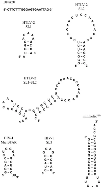
Nucleic acid preparation.NA oligonucleotides used in this work are shown inFig. 2. The 6-carboxyfluorescein (FAM)-labeled 20-nt ssDNA oligonucleotide (5=-FAM DNA20) was obtained from TriLink Biotech-nologies (San Diego, CA). The following high-performance liquid chro-matography (HPLC)-purified FAM- and fluorescein (Fl)-labeled HTLV-2 RNA oligonucleotides and unlabeled HTLV-2 SL1 RNA were purchased from Dharmacon RNA Technologies (Lafayette, CO): 5=-Fl-U UAUGGGACAAAUCCA-3=(5=-Fl-SL1) and 5=-Fl-UUGGGCUUUCCC CAACUCCAAUACCCAAAGCCC-3=(5=-Fl-SL2) (note that the two U’s in italics are not encoded by HTLV-2). RNAs derived from HIV-1 (3= -FAM-MicroTAR and 3=-FAM-SL3) and 3=-FAM-minihelixLys,3, derived from the acceptor-T⌿C stem-loop of human tRNALys,3, were also pur-chased from Dharmacon RNA Technologies (Lafayette, CO).
HIV-1trans-activation response element (TAR) DNA (38) was ob-tained from Integrated DNA Technologies (Coralville, IA), purified on a 12% (wt/vol) denaturing polyacrylamide gel, and stored at⫺20°C.
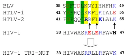
Unla-FIG 1Structure-based sequence alignment of MA␣-helix II from HTLV-2, HTLV-1, and HIV-1 (26) along with BLV MA. Residues are boxed as follows: green, identical in HTLV-1 and -2 and BLV; yellow, conserved in HTLV-1 and -2 and BLV. Basic residues are shown in blue, and acidic residues are shown in red. Residues in boldface type indicate residues changed to generate HTLV-2 and HIV-1 MA variants. Arrows show the changes made to generate the HIV-1 MA TRI-MUT variant to mimic HTLV-2 MA.
on November 7, 2019 by guest
http://jvi.asm.org/
[image:2.585.314.525.66.159.2]beled HTLV-2-derived SL2 and SL1-SL2, human tRNALys,3 (39), and HIV-1 TAR RNA (38) werein vitrotranscribed with T7 RNA polymerase as described previously (40). For gel shift annealing assays, TAR RNA was internally radiolabeled with [␣-32P]GTP duringin vitrotranscription with T7 RNA polymerase by using standard protocols, followed by gel purification. All RNA oligonucleotides were dissolved in diethyl pyrocar-bonate-treated water and stored at⫺20°C.
The concentrations of RNA and DNA oligonucleotides were deter-mined by measuring the absorbance at 260 nm with the following extinc-tion coefficients: 6.04⫻105M⫺1cm⫺1for human tRNALys,3(76-mer), 5.648⫻105M⫺1cm⫺1for TAR DNA (59-mer), 5.337⫻105M⫺1cm⫺1 for TAR RNA (59-mer), 4.586⫻105M⫺1cm⫺1for SL1-SL2 (50-mer), 3.499⫻105M⫺1cm⫺1for 3=-FAM-minihelixLys,3(37-mer), 3.242⫻105 M⫺1cm⫺1for 5=-Fl-SL2 (33-mer), 2.139⫻105M⫺1cm⫺1for 5=-FAM DNA20, 1.893⫻105M⫺1cm⫺1for 3=-FAM-MicroTAR (18-mer), 1.88⫻ 105M⫺1cm⫺1for 5=-Fl-SL1 (16-mer), and 1.506⫻105M⫺1cm⫺1for 3=-FAM-SL3 (16-mer).
Prior to use, all oligonucleotides except 5=-FAM DNA20 were re-folded in a solution containing 25 mM HEPES (pH 7.5) and 100 mM NaCl by heating at 80°C for 2 min and cooling to 60°C for 2 min, followed by the addition of MgCl2to a final concentration of 10 mM and placement on ice.
Fluorescence anisotropy measurements.Equilibrium dissociation constants were determined by measuring the fluorescence anisotropy
(FA) of 20 nM fluorescently labeled NA as a function of increasing con-centrations of unlabeled proteins. The labeled NAs were incubated with various amounts of the unlabeled proteins for 30 min at room tempera-ture in a solution containing 20 mM HEPES (pH 7.5) and 50 mM NaCl. Anisotropy measurements were carried out on a SpectraMax M5 micro-plate reader (Molecular Devices, Sunnyvale, CA). The excitation and emission wavelengths were 485 and 525 nm, respectively. All measure-ments were performed at least three times, and the data were averaged. The titration curves were fit to the following equation, which assumes a 1:1 binding stoichiometry (41–43):A⫽Amin⫹{(Y⫹S⫹Kd)⫺[(Y⫹
S⫹Kd)2⫺(4YS)]1/2} · (A
max⫺Amin)/(2Y), whereAis the measured anisotropy at a particular total concentration of the protein (S) and the labeled NAs (Y),Aminis the minimum anisotropy,Amaxis the final max-imum anisotropy, andKdis the dissociation constant. For FA competition assays, fluorescently labeled NAs were prebound to proteins of interest at saturating concentrations (5M for HTLV-2 MA, 5M for HTLV-2 NC, and 7M for HIV-1 MA) for 10 min under the same conditions as those described above for direct FA measurements, followed by incubation with increasing amounts of unlabeled SL1, SL2, SL1-SL2, tRNA, or inositol hexaphosphate (IP6) for 30 min prior to taking FA readings.
Sedimentation assays.Sedimentation assays were carried out essen-tially as described previously (44). Briefly, refolded32P-labeled TAR RNA (15 nM) was combined with TAR DNA (90 nM) in a solution containing 20 mM HEPES (pH 7.5), 20 mM NaCl, and 5 mM DTT. Upon addition of proteins to a final concentration of 1, 5, or 10M, reaction mixtures (30 l) were incubated at 37°C for 30 min. Solutions were then centrifuged at 12,000 rpm in a microcentrifuge for 20 min. The supernatant (2l) was collected and analyzed by scintillation counting. The percent radioactivity remaining in the supernatant, relative to the RNA-only sample (set to 100%), was plotted as a function of the protein concentration.
Annealing assays.TAR DNA/RNA annealing assays were performed essentially as described previously (44). Briefly, refolded32P-labeled TAR RNA was combined with unlabeled complementary TAR DNA in a solu-tion containing 20 mM HEPES (pH 7.5), 20 mM NaCl, and 5 mM DTT at 37°C. Reactions were initiated by adding 90 nM DNA and MA or NC proteins (5M) to 15 nM RNA, followed by incubation in the reaction buffer for the indicated times. Reactions were quenched by placing solu-tions on ice, followed by the addition of SDS to a 1% (vol/vol) final con-centration. Samples were extracted twice with a 4:1 mixture of phenol-chloroform and loaded onto SDS–12% polyacrylamide gels (375 mM Tris-HCl [pH 8.8], 0.1% [wt/vol] SDS, and a 19:1 mixture of acrylamide-bisacrylamide [wt/vol]) run at 25°C in Tris-glycine (25 mM Tris, 250 mM glycine [pH 8.3]) running buffer. Gels were visualized by using a Typhoon Trio Imager and quantified with Bio-Rad Quantity One software.
Determination of RNA packaging efficiency.HIV-1 proviral plasmid DNA (10g) was transfected into 2 million 293T cells by using a calcium phosphate-mediated method in 10-cm plates (45). Cells and supernatant were collected at 48 h posttransfection for preparation of RNAs and ly-sates. For quantification of viral RNA, virus-like particles (VLPs) were harvested by filtering the supernatant through a 0.2-m syringe filter prior to ultracentrifugation in a Beckman 50.2 Ti rotor at 25,000 rpm for 2 h at 4°C. Viral RNA was extracted from VLP pellets with a High Pure Viral RNA kit (Roche Applied Science, Indianapolis, IN). Total cellular RNAs were prepared by using an RNeasy kit and QIAshredder (Qiagen, Inc., Valencia, CA). Total viral and cellular RNAs were then treated with DNase by using a DNA-free kit (Ambion, Inc., Austin, TX) to remove any contaminating genomic DNA. Reverse transcription (RT) and quantita-tive PCR (qPCR) were performed individually for both viral and cellular RNAs with the Transcriptor High Fidelity cDNA synthesis kit (Roche) and the SYBR green qPCR reagent kit (Invitrogen Corp., Carlsbad, CA), respectively. Primers used for quantitative PCR of HIV-1gagRNA were
5=-ACATCAAGCAGCCATGCAAAT and 5=-ATGTCACTTCCCCTTGG
TTCTCT. A serial dilution of an HIV-1 vector (10⫺6to 10 ng) containing the target sequence was prepared as a standard for quantitative PCR anal-ysis. The quantity of Gag can be determined from viral and cellular
sam-FIG 2Oligonucleotide constructs used in this work.
on November 7, 2019 by guest
http://jvi.asm.org/
[image:3.585.60.264.67.446.2]ples based on the defined standard. Experiments were performed with a MyiQ single-color real-time PCR detection system (Bio-Rad Laborato-ries, Hercules, CA), and data were analyzed with MyiQ software (version 1.0; Bio-Rad). For quantification of VLP production, the supernatant was filtered through a 0.2-m syringe filter prior to ultracentrifugation in a Beckman 50.2 Ti rotor at 25,000 rpm for 2 h at 4°C. Transfected cells were also harvested and washed with Dulbecco’s phosphate-buffered saline (Invitrogen). VLPs and cell pellets were resuspended individually in ra-dioimmunoprecipitation assay (RIPA) buffer (150 mM NaCl, 1.0% Igepal CA-630, 0.5% sodium deoxycholate, 0.1% SDS, 50 mM Tris-HCl [pH 8.0], 5 mM EDTA). Lysates were electrophoresed on 12.5% SDS-poly-acrylamide gels and transferred onto nitrocellulose membranes (Bio-Rad). HIV-1 Gag was detected with a primary rabbit anti-HIV-1 p24 antiserum (Advanced Biotechnologies, Inc., Columbia, MD) at a 1:1,500 dilution, followed by horseradish peroxidase-conjugated goat anti-rabbit IgG (Thermo Fisher Scientific, Inc., Rockford, IL) at a 1:10,000 dilution. Band intensities were quantified with the ChemiDoc XRS system (Bio-Rad). The RNA packaging efficiency was determined by using methods described previously (25).
RESULTS
Nucleic acid binding of HTLV-2 MA and NC.
Previous studies
have shown that HIV-1 NC is an excellent RNA binding and
chap-erone protein and plays an essential role in gRNA packaging, while
HIV-1 MA is not required for specific RNA packaging (
46
). To test
whether HTLV-2 NC and MA proteins play roles similar to those
of their HIV-1 counterparts, FA assays were used to determine
equilibrium dissociation constants (
K
d) for HTLV-2 NC and MA
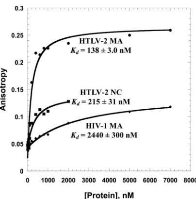
binding to a nonspecific 20-mer ssDNA (5
=
-FAM DNA20) and to
HTLV-2 SL1 and SL2 sequences derived from the putative gRNA
packaging signal (
Fig. 2
) (
32
). Representative binding curves for
5
=
-FAM DNA20 are shown in
Fig. 3
. The data were fit to a binding
model that assumes a 1:1 binding stoichiometry, and apparent
K
dvalues of 215 nM and 138 nM were determined for the HTLV-2
NC and MA proteins, respectively. The
K
dvalues for HTLV-2
genome-derived SL1 and SL2 sequences are summarized in
Table
1
. Under the relatively low-ionic-strength conditions of 50 mM
NaCl, the binding of NC to ssDNA and SL2 was
⬃
2-fold and
⬃
6-fold weaker than the binding of MA, respectively. Under these
conditions, neither HTLV-2 MA or NC has a strong preference for
binding to SL1 or SL2 relative to ssDNA, although MA binds with
a
⬃
2-fold-higher affinity to SL2 than to ssDNA.
Although MA does not display a strong preference for binding
to SL2 at low ionic strength, the binding to SL2 is less sensitive to
increasing salt concentrations than the binding to ssDNA (
Table
1
). At 100 mM NaCl, SL2 binding was 5-fold stronger than ssDNA
binding, whereas at 150 mM NaCl, binding was
⬃
13-fold
stron-ger. Thus, under physiological conditions, HTLV-2 MA may play
a role in selective binding to gRNA.
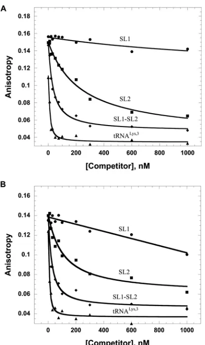
FA competition assays were performed to further test the
spec-ificity of HTLV-2 MA binding. In these experiments, MA was
prebound to fluorescently labeled minihelix
Lys,3, derived from the
acceptor-T
⌿
C sequence of human tRNA
Lys,3, or HIV-1 SL3 (
Fig.
2
). The complexes were titrated with unlabeled HTLV-2-derived
SL1, SL2, or SL1-SL2 or human tRNA
Lys,3. As shown in
Fig. 4
, SL1
was unable to compete effectively for MA binding to the prebound
RNAs. In contrast, SL2 and SL1-SL2 readily competed off the
non-specific RNAs. Surprisingly, tRNA
Lys,3was even more effective
than SL1-SL2 at competing for binding, and the same result was
obtained with another tRNA (tRNA
Ala) tested (data not shown).
Nucleic acid chaperone activity of HTLV-2 MA and NC.
An-nealing of TAR DNA hairpin to a complementary TAR RNA
hair-pin, which mimics the annealing step of minus-strand transfer in
reverse transcription, was performed as a model assay to study the
chaperone function of the HTLV-2 NC and MA proteins (
38
). A
comparison of the annealing of TAR RNA/DNA hairpins in the
presence of saturating concentrations of NC and MA is shown in
Fig. 5
. These data suggest that HTLV-2 MA exhibits much better
chaperone activity than NC. The effective annealing rates and final
percentages of RNA annealed in the presence of MA were
signifi-cantly higher (
⬃
15-fold and
⬃
12-fold, respectively) than those in
the presence of NC. In contrast to HIV-1, in which NC is highly
basic, HTLV-2 NC has an acidic C-terminal domain (overall pI
⬃
7.0), similar to HTLV-1 NC, which also lacks strong NA binding
and chaperone activities (
28
). However, HTLV-2 MA is a
rela-tively basic protein (overall pI
⬃
9.6), which may explain its
rela-tively robust NA binding and chaperone functions.
[image:4.585.62.264.62.267.2]Role of basic amino acid residues in
␣
-helix II of HTLV-2
MA.
The sequence alignment reveals high homology between
del-taretroviral MA proteins, including the presence of charged
resi-dues in
␣
-helix II (
Fig. 1
), which is predicted to have an overall
positive charge. In contrast, HIV-1 MA possesses a helix II that is
neutral overall. A previous study showed that mutation of BLV
TABLE 1Binding parameters of HTLV-2 proteinsa
HTLV-2 protein (NaCl concn [mM])
MeanKd(nM)⫾SD
ssDNA SL1 SL2
NC (50) 215⫾31 614⫾120 419⫾170
MA (50) 138⫾3.0 341⫾170 74⫾18
MA (100) 945⫾200 — 186⫾80
MA (150) 6,200⫾1,600 — 493⫾97
R47A/K51A MA (50) 864⫾320 — 633⫾200
R47A/K51A MA (100) NB — NB
R47A/K51A MA (150) NB — NB
aShown are apparent equilibrium dissociation constants (K
d) obtained from FA
measurements performed at various ionic strengths. NB indicates that no binding was detected with up to 7M protein. A dash indicates that the value was not determined. FIG 3Representative FA binding assays wherein 20 nM 5=-FAM DNA20 was
titrated with HTLV-2 MA, HTLV-2 NC, and HIV-1 MA. The curves are single exponential fits of the data.
on November 7, 2019 by guest
http://jvi.asm.org/
[image:4.585.299.544.595.697.2]MA K41 and H45 to Ala significantly reduced the gRNA packaging
efficiency (
25
). To test whether these conserved residues influence
RNA binding of HTLV-2 MA
in vitro
, the two corresponding basic
residues, R47 and K51, were mutated to alanine. Binding to
non-specific 5
=
-FAM DNA20 and HTLV-2 SL2 RNA was tested by
using FA.
Table 1
shows that when assays were conducted with 50
mM NaCl, the simultaneous mutation of two residues to generate
the R47A/K51A MA variant led to
⬃
6-fold and
⬃
8-fold
reduc-tions in ssDNA and SL2 binding, respectively. The effect on SL2
binding was even more dramatic under conditions of higher ionic
strength (
Table 1
). At
ⱖ
100 mM NaCl, binding to ssDNA and SL2
was undetectable, whereas the WT protein still bound SL2 with a
relatively high affinity (186 nM and 493 nM at 100 mM and 150
mM NaCl, respectively). WT MA binding to ssDNA was much
more salt sensitive, with apparent
K
dvalues of 945 nM and 6,200
nM measured at 100 mM and 150 mM NaCl, respectively. Thus,
R47 and K51 may interact with SL2 through primarily
nonelec-trostatic forces.
Comparison of HTLV-2 and HIV-1 MA proteins.
Superposi-tion of the HTLV-2 and HIV-1 MA structures reveals a similar
three-dimensional fold despite limited primary sequence identity
(
26
). To gain further insights into functional differences between
these proteins, we conducted FA assays to compare their binding
to 5
=
-FAM DNA20. This fairly random ssDNA sequence was used
to minimize sequence-specific binding effects. The
K
dvalue
ob-tained for HTLV-2 MA (138 nM) is
⬃
17-fold lower than that
measured for HIV-1 MA (2,440 nM), showing that HTLV-2 MA
binds NAs with a higher affinity than HIV-1 MA (
Fig. 3
).
Further-more, SL2 binding of HIV-1 MA was 6-fold weaker and more
sensitive to increasing salt concentrations than HTLV-2 MA (
Ta-bles 1
and
2
).
Figure 5
compares the annealing of TAR DNA/RNA hairpins
in the presence of saturating concentrations of HTLV-2 MA and
HIV-1 MA. HTLV-2 MA facilitates the annealing reaction more
effectively than HIV-1 MA, with an
⬃
2-fold-higher
k
obs. The
abil-ity to aggregate NAs is another critical component of a chaperone
protein, and a sedimentation assay was used to measure this
prop-erty (data not shown). The higher percentage of aggregated RNA
observed at all protein concentrations tested shows that HTLV-2
MA is a much more effective NA-aggregating agent than HIV-1
MA. HTLV-2 MA achieved 80% aggregation at all concentrations
tested (1 to 10
M), whereas HIV-1 MA aggregated only
⬃
36% of
the NAs at the lowest concentration tested (1
M) and aggregated
even smaller amounts at 5
M (25%) and 10
M (6%). Although
it is not known why less aggregation was observed with increasing
FIG 4FA competition assays wherein 20 nM fluorescently labeled HIV-1 SL3 (A) or minihelixLys,3(B) was preincubated with 5M HTLV-2 MA, followed by titration with unlabeled HTLV-2-derived SL1, SL2, and SL1-SL2 RNAs or tRNALys,3.
[image:5.585.61.269.63.412.2]FIG 5Annealing time courses of 15 nM TAR RNA and 90 nM TAR DNA at 37°C in the presence of 5M proteins.
TABLE 2Binding parameters of WT HIV-1 MA and variantsa
HIV-1 MA protein (NaCl concn [mM])
MeanKd(nM)⫾SD
ssDNA MicroTAR SL2
WT (50) 2,440⫾300 1,360⫾280 471⫾150
WT (100) — — 1,320⫾140
WT (150) — — 5,190⫾700
E40R (50) 154⫾6.0 254⫾76 —
E40R/E42L (50) 461⫾190 271⫾170 —
E40R/E42L/N47K (50) 230⫾31 185⫾16 —
aShown are apparent equilibrium dissociation constants (K
d) obtained from FA
measurements performed at various ionic strengths. A dash indicates that the value was not determined.
on November 7, 2019 by guest
http://jvi.asm.org/
[image:5.585.298.545.78.170.2] [image:5.585.61.266.496.700.2]HIV-1 MA concentrations, it may be due to the increase in the salt
concentration as more protein was added.
Electrostatic potential surfaces of HTLV-2 and HIV-1 MA
proteins obtained by using Swiss-PdbViewer reveal a major
difference between the two proteins in the N terminus. In
par-ticular, four basic residues (R47, R48, K51, and K55) located in
HTLV-2 MA helix II form a large patch of positive charge (data
not shown). In contrast, HIV-1 MA lacks a cluster of basic
residues on helix II. Instead, two basic (R39 and R43) and two
acidic (E40 and E42) residues form a neutral surface on one
face of the N-terminal domain. In order to test whether the
basic character of helix II specifically affects the NA binding
properties of HIV-1 MA, an E40R/E42L/N47K chimeric triple
mutant (TRI-MUT) was designed to mimic the more basic
HTLV-2 MA helix II domain. The HIV-1 MA TRI-MUT
vari-ant was overexpressed, purified, and shown to be well folded by
CD spectroscopy (data not shown). Binding of this HIV-1 MA
variant to both nonspecific DNA and HIV-1-derived RNA was
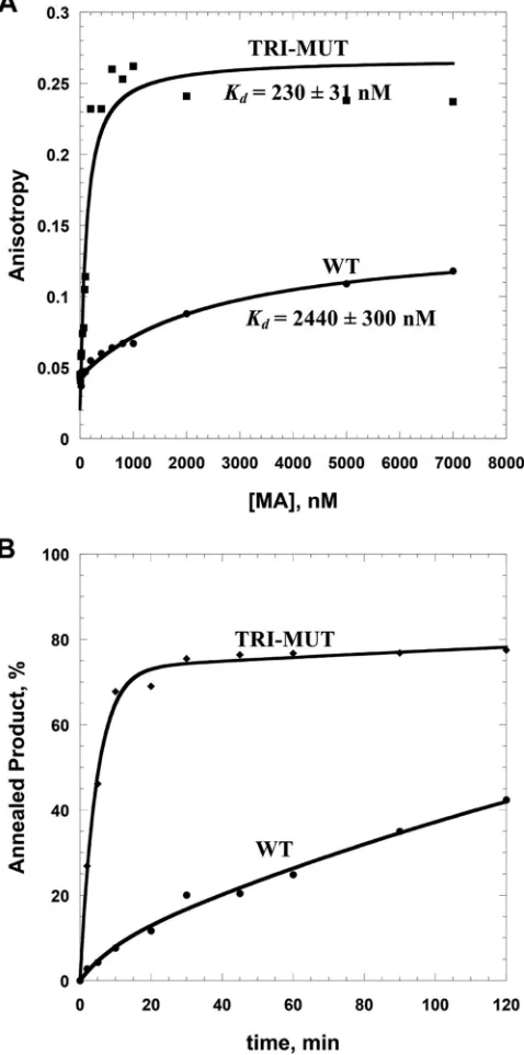
investigated next. FA binding assays showed that the TRI-MUT
variant showed a
⬃
10-fold-higher ssDNA binding affinity than
WT HIV-1 MA (
Fig. 6A
and
Table 2
) as well as significantly
improved chaperone function (
Fig. 6B
). Binding to an HIV-1
TAR RNA-derived sequence (MicroTAR) was also
⬃
7-fold
tighter for the TRI-MUT variant. The binding and chaperone
properties of the TRI-MUT variant are similar to those of
HTLV-2 MA (
Fig. 3
,
5
, and
6
). Surprisingly, even the double
mutant variant (E40R/E42L) and a single point mutant (E40R)
demonstrated significantly improved binding properties
rela-tive to those of WT HIV-1 MA when measured using both
ssDNA and MicroTAR RNA (
Table 2
). Taken together, the data
support a critical function for the conserved basic residues
lo-cated in helix II of HTLV-2 MA on NA binding and chaperone
properties.
IP6 competes for MA-nucleic acid binding.
Since a primary
function of all retroviral MA domains involves membrane
binding to phospholipids, we tested the NA binding of HTLV-2
MA, HTLV-2 NC, and HIV-1 MA in the presence of increasing
concentrations of IP6, which has been shown to have an effect
on HIV-1 Gag assembly
in vitro
(
47
) (
Fig. 7
). Proteins were
prebound to HTLV-2 SL2 at saturating concentrations. As
ex-pected, addition of IP6 did not compete for HTLV-2 NC-SL2
interactions, consistent with the fact that NC does not
partici-pate in membrane binding. In contrast, both HTLV-2 and
HIV-1 MAs were displaced from Fl-SL2 by IP6. The final
an-isotropy value of
⬃
0.05 at 20
M IP6 suggests that Fl-SL2 is
completely displaced. These results suggest that IP6 interacts
with both HTLV-2 and HIV-1 MA proteins but not with
HTLV-2 NC in the presence of RNA.
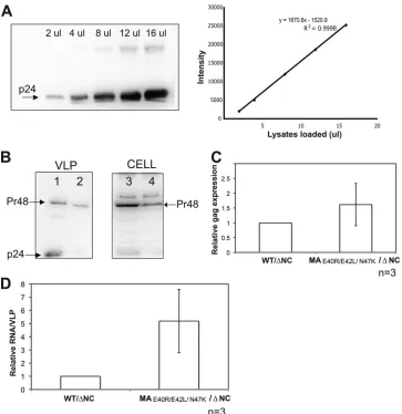
Cell-based assays to probe the role of MA helix II in HIV-1
RNA packaging.
Although NC is the key player in HIV-1 gRNA
packaging (
4
,
48–51
), MA has also been shown to participate in
BLV gRNA packaging (
25
). Furthermore, data provided in this
study and elsewhere (
17
,
19–21
,
34
) suggest that HIV-1 MA
inter-acts with gRNA. We have shown that the HIV-1 MA TRI-MUT
variant exhibits higher NA binding affinity
in vitro
than WT
HIV-1 MA. To test the effect of the triple mutation on RNA
pack-aging in HIV-1, we transfected WT/
⌬
NC and MA
E40R/E42L/N47K/
⌬
NC HIV-1 clones individually into 293T cells and harvested
VLPs. The amount of Gag polypeptide (Pr48) was detected and
quantified from VLPs as well as from virus-producing cells
(
Fig. 8A
and
B
). Total RNAs from VLPs and virus-producing cells
were used in a two-step, real-time RT-PCR analysis to quantify the
expression level of the HIV-1
gag
gene (
Fig. 8C
). The viral RNA
packaging efficiency was determined as described previously (
25
).
The results shown in
Fig. 8D
indicate that HIV-1 RNA packaging
is about 5 times more efficient for MA
E40R/E42L/N47K/
⌬
NC virus
than for WT/
⌬
NC virus (
n
⫽
3). (The packaging efficiency of the
WT virus with NC, as determined in independent experiments,
was
⬃
17-fold higher than that of WT/
⌬
NC virus [data not
FIG 6Comparison of WT HIV-1 MA and TRI-MUT NA binding and chap-erone functions. (A) Representative FA binding assays using 20 nM 5=-FAM DNA20 in the presence of increasing amounts of protein. Lines are fits to the data (see equation in the text). (B) Annealing time courses of 15 nM TAR RNA and 90 nM TAR DNA in the presence of 5M protein.
on November 7, 2019 by guest
http://jvi.asm.org/
[image:6.585.302.541.72.553.2]shown].) These cell-based data support the
in vitro
results and
show that the introduction of basic charges into helix II of HIV-1
MA enhances the RNA binding and packaging ability of HIV-1
MA in the absence of NC.
DISCUSSION
In this work, the NA binding and chaperone properties of two
different MA proteins, HTLV-2 and HIV-1, were compared
in
vitro
. FA binding studies demonstrated that in the deltaretrovirus
HTLV-2, MA binds NAs with higher affinity than NC, which is in
contrast to HIV-1. Furthermore, salt-dependent binding assays
support specific binding of HTLV-2 MA to the SL2 stem-loop
structure derived from the putative HTLV-2 RNA packaging
sig-nal and suggest that conserved basic residues in helix II contribute
to binding. Competition binding studies also supported
preferen-tial binding to SL2 or the combined SL1-SL2 over SL1 alone, but
surprisingly, tRNA was found to be even more effective than
SL1-SL2 at competing for HTLV-2 MA binding. This may be due to
nonspecific binding interactions of MA with longer nucleic acid
sequences. A recent study of BLV MA showed high-affinity (10 to
20 nM) binding to RNAs derived from the BLV genome,
support-ing a conservation of function among deltaretroviral MA proteins
(
52
). In contrast, previous work has shown that HIV-1 MA is not
required for specific gRNA binding (
46
).
The chaperone activities of HTLV-2 MA and NC were tested by
using gel shift annealing assays and compared with those of HIV-1
proteins. Our data suggest that HTLV-2 MA facilitates TAR RNA/
DNA annealing more effectively than NC, which is also in contrast
to HIV-1, where NC is a potent chaperone protein. The different
behaviors of the two retroviral genera may be due to the different
distributions of local electrostatic potential in the respective Gag
proteins. HIV-1 NC is well known for its highly basic character (pI
9.86), whereas HTLV-2 NC is neutral overall (pI 7.68); in contrast,
HTLV-2 MA (pI 9.51) exhibits a more basic character than HIV-1
MA (pI 9.02). A basic cluster is located in the N-terminal domain
of HTLV-2 MA, forming a highly positively charged surface that is
absent from HIV-1 MA. We have previously shown that HTLV-1
NC, which shares high sequence identity with HTLV-2 NC, lacks
NA-aggregating capabilities and displays relatively poor
chaper-one activity despite the fact that it is a strong duplex destabilizer
compared to other retroviral NCs (
28
). Combined with previous
cell-based studies, which demonstrated that basic residue variants
of BLV MA are defective in RNA packaging (
25
), these data
sug-gest that deltaretroviral MA proteins are major players in the
ini-tial stage of gRNA selection and packaging, whereas NC plays a
more minor role at this stage of the life cycle. Further functional
analyses will be needed to map the MA-genome interactions at the
molecular level.
The basic residues of MA also play a role in directing
mem-brane targeting of Gag. In HIV-1, a highly basic region spanning
residues in the N terminus forms an interface with acidic
phos-pholipids and, along with the N-terminal myristoyl group,
facili-tates membrane binding of Gag to PI(4,5)P
2-containing
lipo-somes (
23
,
53
,
54
). Interestingly, Gag targeting and membrane
binding mediated by HTLV-1 MA do not appear to require
PI(4,5)P
2(
55
). Instead, HTLV-1 Gag binding to liposomes is
driven largely by electrostatic interactions instead of specific
in-teractions with PI(4,5)P
2. In contrast to HIV-1, HTLV-1 Gag
membrane binding
in vitro
is not suppressed by RNA. These data
suggested that HIV-1 and HTLV-1 use different mechanisms to
regulate membrane targeting (
55
). Previous studies have also
in-dicated that HTLV-1 and HIV-1 Gag proteins target different
plasma membrane microdomains in Jurkat T cells (
56
). Our
petition assays with IP6 demonstrate that HTLV-2 MA is
com-peted off NAs even more effectively than HIV-1 MA and suggest
that the MA-IP6-interacting surface likely overlaps the MA-NA
binding site. Similar results were recently reported for BLV MA
(
52
). IP6 possesses more negative-charge density than the head
group of PI(4,5)P
2. Therefore, these results are in agreement with
previous studies showing that electrostatic interactions are the
major driving force for HTLV-1 Gag-liposome binding (
55
),
al-though it is still unclear which specific phospholipid is required
for HTLV-1 and -2 membrane targeting. More detailed liposome
binding studies are needed to determine the specific factor that
contributes to deltaretroviral MA membrane targeting and
sub-cellular localization.
The chaperone activity of retroviral NC proteins plays a
critical role in facilitating NA remodeling events throughout
reverse transcription. With a 228-nt gRNA R region, which is
predicted to fold into a complex secondary structure (
57
),
HTLV-1 would be expected to require a robust chaperone to
facilitate the minus-strand transfer steps of reverse
transcrip-tion. Surprisingly, HTLV-1 and -2 NC proteins display
rela-tively poor NA binding and overall chaperone activity;
how-ever, once bound, HTLV-1 NC is a strong duplex destabilizer
(
28
,
29
). We therefore suggest that in deltaretroviruses, MA is
involved in key early steps involving genome recognition and
packaging; however, once the local concentration of Gag is
high enough, MA preferentially binds to the membrane, and
NC domain binding to NAs occurs, allowing NC to carry out
the chaperone function required for reverse transcription
(
Fig. 9
).
The fact that we can increase RNA packaging efficiency (and
perhaps specificity as well) of a
⌬
NC HIV-1 virus by increasing
the basic character of the helix II domain of HIV-1 MA is
remarkable. This result is consistent with previous studies
FIG 7FA competition assays wherein 20 nM 5=-Fl-SL2 was preincubated with HTLV-2 MA, HIV-1 MA, or HTLV-2 NC at saturating protein concentrations (5M for HTLV-2 MA, 5M for HTLV-2 NC, and 7M for HIV-1 MA), followed by titration with IP6. The inset shows an expanded view of the low-IP6-concentration region of the titration.
on November 7, 2019 by guest
http://jvi.asm.org/
[image:7.585.43.285.63.256.2]showing that HIV-1 MA and NC have redundant roles in virus
assembly (
17
,
34
). Mutations introduced into NA binding
re-gions of either NC or MA do not severely affect RNA
incorpo-ration; however, mutation of RNA binding areas of both
do-mains results in particles without gRNA packaged (
17
,
34
).
Although the cluster of basic amino acids located in helix II of
BLV MA is known to contribute to gRNA incorporation (
25
),
additional studies are needed to confirm that the basic
charac-ter of MA in other retroviruses, such as HTLV-1 and -2, is
involved in genome packaging.
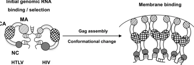
In summary, based on previous studies and the new results
reported here, we propose a model (
Fig. 9
) in which initial gRNA
binding and selection involve a folded conformation of Gag with
both MA and NC domains binding to RNA. In HIV-1, NC plays a
major role at this stage and binds the genome specifically. We have
recently shown that HIV-1 Gag binds to psi-specific RNAs
differ-ently than to non-psi RNAs; our data suggest that psi binding
involves primarily the NC domain, whereas non-psi binding
in-volves both the NC and MA domains (
58
). In contrast, we propose
that in deltaretroviruses, MA plays a larger role in recognizing
gRNA at the initial stage. When Gag assembles at the plasma
membrane, it triggers a conformational change (
59
,
60
), wherein
phosphorylated phosphatidylinositides or other membrane
mi-crodomains bind to the MA domain while NC remains bound to
RNA. Furthermore, once a high local concentration is achieved,
the slow NA dissociation kinetics of HTLV NC (
28
) allow it to
remain bound to the RNA. Taken together, these studies suggest
that deltaretroviruses use distinct protein-RNA interactions for
gRNA packaging. The implications of these findings for specific
viral RNA recognition by HTLV MA and MA’s role in the context
FIG 8HIV-1 gRNA packaging efficiency of the HIV-1 MA TRI-MUT variant in an NC deletion background. (A) Serial dilution of WT HIV-1 p24 for protein quantification. (Left) Immunoblot displaying a serial dilution (2l to 16l lysate per loading) of WT HIV-1 p24. (Right) Band intensities (arbitrary units) (y
axis) plotted against the volume of lysates loaded (xaxis) to evaluate the linearity and accuracy of protein quantification. The linear equation and anR2value are indicated. (B) Immunoblots were probed with antisera against HIV-1 p24. The amount of protein was determined based on the linear standard shown in panel A. Lanes 1 and 2 show VLP lysates collected from WT/⌬NC (lane 1) and MAE40R/E42L/N47K/⌬NC (lane 2). Lanes 3 and 4 show lysates from producing cells expressing the WT/⌬NC clone (lane 3) and the MAE40R/E42L/N47K/⌬NC clone (lane 4). (C) Relative expression level ofgagdetected by two-step quantitative RT-PCR. The expression level ofgagfrom MAE40R/E42L/N47K/⌬NC is normalized to that from WT/⌬NC. Error bars represent standard deviations from three independent experiments (n⫽3). (D) Relative fold change of RNA packaging efficiency. The RNA packaging efficiency (yaxis) was calculated as previously described (25). The RNA packaging efficiency of the MAE40R/E42L/N47K/⌬NC mutant was normalized to that of WT/⌬NC. Error bars represent standard deviations from three individual experiments (n⫽3).
on November 7, 2019 by guest
http://jvi.asm.org/
[image:8.585.111.474.64.438.2]of Gag remain to be explored. Additional studies to map HTLV
MA-RNA interactions are under way to gain further insights into
MA’s diverse roles in the viral life cycle.
ACKNOWLEDGMENTS
This work was supported by the Intramural Research Program of the NIH, National Cancer Institute, Center for Cancer Research, and by NIH grants GM065056 (to K.M.-F.) and GM098500 (to L.M.M.). This project has been funded in whole or in part with federal funds from the National Cancer Institute, National Institutes of Health, under contract HHSN261200800001E with Leidos Biomedical Research, Inc. (R.J.G.).
The content of this publication does not necessarily reflect the views or policies of the Department of Health and Human Services, nor does mtion of trade names, commercial products, or organizamtions imply en-dorsement by the U.S. Government.
We thank members of the AIDS and Cancer Virus Program, Frederick National Laboratory for Cancer Research, David E. Ott for providing the delNC HIV-1 proviral plasmid, and Donald G. Johnson and Catherine V. Hixson for assistance with preparation of the HTLV-2 NCp15 protein.
REFERENCES
1.Berkowitz R, Fisher J, Goff SP.1996. RNA packaging. Curr. Top. Microbiol. Immunol.214:177–218.http://dx.doi.org/10.1007/978-3-642-80145-7_6. 2.Jewell NA, Mansky LM.2000. In the beginning: genome recognition, RNA
encapsidation and the initiation of complex retrovirus assembly. J. Gen. Virol.
81:1889 –1899.http://vir.sgmjournals.org/content/81/8/1889.long. 3.Rein A.1994. Retroviral RNA packaging: a review. Arch. Virol. Suppl.
9:513–522.
4.Berkowitz RD, Ohagen A, Hoglund S, Goff SP.1995. Retroviral nucleo-capsid domains mediate the specific recognition of genomic viral RNAs by chimeric Gag polyproteins during RNA packaging in vivo. J. Virol.69:
6445– 6456.
5.Levin JG, Guo J, Rouzina I, Musier-Forsyth K. 2005. Nucleic acid chaperone activity of HIV-1 nucleocapsid protein: critical role in reverse transcription and molecular mechanism. Prog. Nucleic Acid Res. Mol. Biol.80:217–286.http://dx.doi.org/10.1016/S0079-6603(05)80006-6. 6.Cruceanu M, Gorelick RJ, Musier-Forsyth K, Rouzina I, Williams MC.
2006. Rapid kinetics of protein-nucleic acid interaction is a major com-ponent of HIV-1 nucleocapsid protein’s nucleic acid chaperone function. J. Mol. Biol.363:867– 877.http://dx.doi.org/10.1016/j.jmb.2006.08.070. 7.Narayanan N, Gorelick RJ, DeStefano JJ.2006. Structure/function
map-ping of amino acids in the N-terminal zinc finger of the human immuno-deficiency virus type 1 nucleocapsid protein: residues responsible for nu-cleic acid helix destabilizing activity. Biochemistry45:12617–12628.http: //dx.doi.org/10.1021/bi060925c.
8.Darlix JL, Garrido JL, Morellet N, Mely Y, de Rocquigny H. 2007. Properties, functions, and drug targeting of the multifunctional nucleo-capsid protein of the human immunodeficiency virus. Adv. Pharmacol.
55:299 –346.http://dx.doi.org/10.1016/S1054-3589(07)55009-X. 9.Amarasinghe GK, De Guzman RN, Turner RB, Chancellor KJ, Wu ZR,
Summers MF.2000. NMR structure of the HIV-1 nucleocapsid protein bound to stem-loop SL2 of the psi-RNA packaging signal. Implications for genome recognition. J. Mol. Biol.301:491–511.http://dx.doi.org/10.1006 /jmbi.2000.3979.
10. De Guzman RN, Wu ZR, Stalling CC, Pappalardo L, Borer PN, Sum-mers MF.1998. Structure of the HIV-1 nucleocapsid protein bound to the SL3 psi-RNA recognition element. Science279:384 –388.http://dx.doi .org/10.1126/science.279.5349.384.
11. Parent LJ, Gudleski N.2011. Beyond plasma membrane targeting: role of the MA domain of Gag in retroviral genome encapsidation. J. Mol. Biol.
410:553–564.http://dx.doi.org/10.1016/j.jmb.2011.04.072.
12. Bryant M, Ratner L. 1990. Myristoylation-dependent replication and assembly of human immunodeficiency virus 1. Proc. Natl. Acad. Sci. U. S. A.87:523–527.http://dx.doi.org/10.1073/pnas.87.2.523.
13. Spearman P, Wang JJ, Vander Heyden N, Ratner L.1994. Identification of human immunodeficiency virus type 1 Gag protein domains essential to membrane binding and particle assembly. J. Virol.68:3232–3242. 14. Chukkapalli V, Hogue IB, Boyko V, Hu WS, Ono A.2008. Interaction between
the human immunodeficiency virus type 1 Gag matrix domain and phosphati-dylinositol-(4,5)-bisphosphate is essential for efficient gag membrane binding. J. Virol.82:2405–2417.http://dx.doi.org/10.1128/JVI.01614-07.
15. Perez-Caballero D, Hatziioannou T, Martin-Serrano J, Bieniasz PD.
2004. Human immunodeficiency virus type 1 matrix inhibits and confers cooperativity on gag precursor-membrane interactions. J. Virol.78:9560 – 9563.http://dx.doi.org/10.1128/JVI.78.17.9560-9563.2004.
16. Zhou W, Parent LJ, Wills JW, Resh MD. 1994. Identification of a membrane-binding domain within the amino-terminal region of human immunodeficiency virus type 1 Gag protein which interacts with acidic phospholipids. J. Virol.68:2556 –2569.
17. Ott DE, Coren LV, Gagliardi TD.2005. Redundant roles for nucleocap-sid and matrix RNA-binding sequences in human immunodeficiency vi-rus type 1 assembly. J. Virol.79:13839 –13847.http://dx.doi.org/10.1128 /JVI.79.22.13839-13847.2005.
18. Ott DE, Coren LV, Shatzer T.2009. The nucleocapsid region of human immunodeficiency virus type 1 Gag assists in the coordination of assembly and Gag processing: role for RNA-Gag binding in the early stages of as-sembly. J. Virol.83:7718 –7727.http://dx.doi.org/10.1128/JVI.00099-09. 19. Hearps AC, Wagstaff KM, Piller SC, Jans DA.2008. The N-terminal
basic domain of the HIV-1 matrix protein does not contain a conventional nuclear localization sequence but is required for DNA binding and protein self-association. Biochemistry 47:2199 –2210. http://dx.doi.org/10.1021 /bi701360j.
20. Lochrie MA, Waugh S, Pratt DG, Jr, Clever J, Parslow TG, Polisky B.
1997. In vitro selection of RNAs that bind to the human immunodefi-ciency virus type-1 gag polyprotein. Nucleic Acids Res.25:2902–2910.
http://dx.doi.org/10.1093/nar/25.14.2902.
21. Purohit P, Dupont S, Stevenson M, Green MR.2001. Sequence-specific interaction between HIV-1 matrix protein and viral genomic RNA re-vealed by in vitro genetic selection. RNA7:576 –584.http://dx.doi.org/10 .1017/S1355838201002023.
22. Alfadhli A, Still A, Barklis E.2009. Analysis of human immunodeficiency virus type 1 matrix binding to membranes and nucleic acids. J. Virol.
83:12196 –12203.http://dx.doi.org/10.1128/JVI.01197-09.
FIG 9Proposed mechanism of the Gag-genome interaction. Larger circles indicate a major role in NA binding, and smaller circles indicate a more minor role. In initial genome selection (left), Gag binds to gRNA with both NC and MA domains. In HIV, the NC domain has specific binding interactions with the psi packaging signal, with only minor contributions from MA. In HTLV, the MA domain contributes significantly more to initial binding and specific interactions with gRNA than NC. Upon reaching the plasma membrane, the conformation of Gag changes, as the MA domain preferentially binds to membrane lipids. In HTLV, the high local concentration of Gag allows the NC domain to remain bound to the genome despite its relatively weak affinity.
on November 7, 2019 by guest
http://jvi.asm.org/
[image:9.585.138.448.68.172.2]23. Chukkapalli V, Oh SJ, Ono A.2010. Opposing mechanisms involving RNA and lipids regulate HIV-1 Gag membrane binding through the highly basic region of the matrix domain. Proc. Natl. Acad. Sci. U. S. A.
107:1600 –1605.http://dx.doi.org/10.1073/pnas.0908661107.
24. Katoh I, Kyushiki H, Sakamoto Y, Ikawa Y, Yoshinaka Y.1991. Bovine leukemia virus matrix-associated protein MA(p15): further processing and formation of a specific complex with the dimer of the 5=-terminal genomic RNA fragment. J. Virol.65:6845– 6855.
25. Wang H, Norris KM, Mansky LM.2003. Involvement of the matrix and nucleocapsid domains of the bovine leukemia virus Gag polyprotein pre-cursor in viral RNA packaging. J. Virol.77:9431–9438.http://dx.doi.org /10.1128/JVI.77.17.9431-9438.2003.
26. Christensen AM, Massiah MA, Turner BG, Sundquist WI, Summers MF.1996. Three-dimensional structure of the HTLV-II matrix protein and comparative analysis of matrix proteins from the different classes of pathogenic human retroviruses. J. Mol. Biol.264:1117–1131.http://dx .doi.org/10.1006/jmbi.1996.0700.
27. Le Blanc I, Rosenberg AR, Dokhelar MC.1999. Multiple functions for the basic amino acids of the human T-cell leukemia virus type 1 matrix protein in viral transmission. J. Virol.73:1860 –1867.
28. Stewart-Maynard KM, Cruceanu M, Wang F, Vo MN, Gorelick RJ, Wil-liams MC, Rouzina I, Musier-Forsyth K.2008. Retroviral nucleocapsid proteins display nonequivalent levels of nucleic acid chaperone activity. J. Virol.82:10129 –10142.http://dx.doi.org/10.1128/JVI.01169-08.
29. Darugar Q, Kim H, Gorelick RJ, Landes C.2008. Human T-cell lym-photropic virus type 1 nucleocapsid protein-induced structural changes in transactivation response DNA hairpin measured by single-molecule fluo-rescence resonance energy transfer. J. Virol.82:12164 –12171.http://dx .doi.org/10.1128/JVI.01158-08.
30. Qualley DF, Stewart-Maynard KM, Wang F, Mitra M, Gorelick RJ, Rouzina I, Williams MC, Musier-Forsyth K.2010. C-terminal domain modulates the nucleic acid chaperone activity of human T-cell leukemia virus type 1 nucleocapsid protein via an electrostatic mechanism. J. Biol. Chem.285:295–307.http://dx.doi.org/10.1074/jbc.M109.051334. 31. Mansky LM, Krueger AE, Temin HM.1995. The bovine leukemia virus
encapsidation signal is discontinuous and extends into the 5=end of the gag gene. J. Virol.69:3282–3289.
32. Mansky LM, Wisniewski RM.1998. The bovine leukemia virus encapsi-dation signal is composed of RNA secondary structures. J. Virol.72:3196 – 3204.
33. Mansky LM, Gajary LC.2002. The primary nucleotide sequence of the bovine leukemia virus RNA packaging signal can influence efficient RNA packaging and virus replication. Virology301:272–280.http://dx.doi.org /10.1006/viro.2002.1578.
34. Ott DE, Coren LV, Chertova EN, Gagliardi TD, Nagashima K, Sowder RC, II, Poon DT, Gorelick RJ.2003. Elimination of protease activity restores efficient virion production to a human immunodeficiency virus type 1 nucleocapsid deletion mutant. J. Virol.77:5547–5556.http://dx.doi .org/10.1128/JVI.77.10.5547-5556.2003.
35. Bradford MM.1976. A rapid and sensitive method for the quantitation of microgram quantities of protein utilizing the principle of protein-dye binding. Anal. Biochem. 72:248 –254. http://dx.doi.org/10.1016/0003 -2697(76)90527-3.
36. Carteau S, Gorelick RJ, Bushman FD.1999. Coupled integration of human immunodeficiency virus type 1 cDNA ends by purified integrase in vitro: stimulation by the viral nucleocapsid protein. J. Virol.73:6670 – 6679. 37. Guo J, Wu T, Anderson J, Kane BF, Johnson DG, Gorelick RJ,
Hen-derson LE, Levin JG.2000. Zinc finger structures in the human immu-nodeficiency virus type 1 nucleocapsid protein facilitate efficient minus-and plus-strminus-and transfer. J. Virol.74:8980 – 8988.http://dx.doi.org/10 .1128/JVI.74.19.8980-8988.2000.
38. Vo MN, Barany G, Rouzina I, Musier-Forsyth K.2009. HIV-1 nucleo-capsid protein switches the pathway of transactivation response element RNA/DNA annealing from loop-loop “kissing” to “zipper.” J. Mol. Biol.
386:789 – 801.http://dx.doi.org/10.1016/j.jmb.2008.12.070.
39. Stello T, Hong M, Musier-Forsyth K.1999. Efficient aminoacylation of tRNA(Lys,3) by human lysyl-tRNA synthetase is dependent on covalent continuity between the acceptor stem and the anticodon domain. Nucleic Acids Res.27:4823– 4829.http://dx.doi.org/10.1093/nar/27.24.4823. 40. Milligan JF, Uhlenbeck OC.1989. Synthesis of small RNAs using T7 RNA
polymerase. Methods Enzymol. 180:51– 62. http://dx.doi.org/10.1016 /0076-6879(89)80091-6.
41. Lundblad JR, Laurance M, Goodman RH.1996. Fluorescence polariza-tion analysis of protein-DNA and protein-protein interacpolariza-tions. Mol. En-docrinol.10:607– 612.http://dx.doi.org/10.1210/me.10.6.607.
42. Reid SL, Parry D, Liu HH, Connolly BA.2001. Binding and recognition of GATATC target sequences by the EcoRV restriction endonuclease: a study using fluorescent oligonucleotides and fluorescence polarization. Biochemistry40:2484 –2494.http://dx.doi.org/10.1021/bi001956p. 43. Muller B, Restle T, Reinstein J, Goody RS.1991. Interaction of
fluores-cently labeled dideoxynucleotides with HIV-1 reverse transcriptase. Bio-chemistry30:3709 –3715.http://dx.doi.org/10.1021/bi00229a017. 44. Vo MN, Barany G, Rouzina I, Musier-Forsyth K.2006. Mechanistic
studies of mini-TAR RNA/DNA annealing in the absence and presence of HIV-1 nucleocapsid protein. J. Mol. Biol.363:244 –261.http://dx.doi.org /10.1016/j.jmb.2006.08.039.
45. Graham FL, van der Eb AJ. 1973. A new technique for the assay of infectivity of human adenovirus 5 DNA. Virology52:456 – 467.http://dx .doi.org/10.1016/0042-6822(73)90341-3.
46. Poon DT, Li G, Aldovini A.1998. Nucleocapsid and matrix protein contributions to selective human immunodeficiency virus type 1 genomic RNA packaging. J. Virol.72:1983–1993.
47. Datta SA, Zhao Z, Clark PK, Tarasov S, Alexandratos JN, Campbell SJ, Kvaratskhelia M, Lebowitz J, Rein A.2007. Interactions between HIV-1 Gag molecules in solution: an inositol phosphate-mediated switch. J. Mol. Biol.365:799 – 811.http://dx.doi.org/10.1016/j.jmb.2006.10.072. 48. Cimarelli A, Sandin S, Hoglund S, Luban J.2000. Basic residues in
human immunodeficiency virus type 1 nucleocapsid promote virion as-sembly via interaction with RNA. J. Virol.74:3046 –3057.http://dx.doi .org/10.1128/JVI.74.7.3046-3057.2000.
49. Gorelick RJ, Chabot DJ, Rein A, Henderson LE, Arthur LO.1993. The two zinc fingers in the human immunodeficiency virus type 1 nucleocap-sid protein are not functionally equivalent. J. Virol.67:4027– 4036. 50. Poon DT, Wu J, Aldovini A. 1996. Charged amino acid residues of
human immunodeficiency virus type 1 nucleocapsid p7 protein involved in RNA packaging and infectivity. J. Virol.70:6607– 6616.
51. Kafaie J, Song R, Abrahamyan L, Mouland AJ, Laughrea M. 2008. Mapping of nucleocapsid residues important for HIV-1 genomic RNA dimerization and packaging. Virology375:592– 610.http://dx.doi.org/10 .1016/j.virol.2008.02.001.
52. Qualley DF, Lackey CM, Paterson JP.2013. Inositol phosphates compete with nucleic acids for binding to bovine leukemia virus matrix protein: implications for deltaretroviral assembly. Proteins81:1377–1385.http: //dx.doi.org/10.1002/prot.24281.
53. Ono A, Orenstein JM, Freed EO.2000. Role of the Gag matrix domain in targeting human immunodeficiency virus type 1 assembly. J. Virol.74:
2855–2866.http://dx.doi.org/10.1128/JVI.74.6.2855-2866.2000. 54. Hill CP, Worthylake D, Bancroft DP, Christensen AM, Sundquist WI.
1996. Crystal structures of the trimeric human immunodeficiency virus type 1 matrix protein: implications for membrane association and assembly. Proc. Natl. Acad. Sci. U. S. A.93:3099 –3104.http://dx.doi.org/10.1073/pnas.93.7 .3099.
55. Inlora J, Chukkapalli V, Derse D, Ono A.2011. Gag localization and virus-like particle release mediated by the matrix domain of human T-lymphotropic virus type 1 Gag are less dependent on phosphatidylinositol-(4,5)-bisphosphate than those mediated by the matrix domain of HIV-1 Gag. J. Virol.85:3802–3810.http://dx.doi.org/10.1128/JVI.02383-10.
56. Mazurov D, Heidecker G, Derse D.2006. HTLV-1 Gag protein associates with CD82 tetraspanin microdomains at the plasma membrane. Virology
346:194 –204.http://dx.doi.org/10.1016/j.virol.2005.10.033.
57. Askjaer P, Kjems J.1998. Mapping of multiple RNA binding sites of human T-cell lymphotropic virus type I Rex protein within 5=- and 3=-Rex response elements. J. Biol. Chem.273:11463–11471.http://dx.doi.org/10 .1074/jbc.273.19.11463.
58. Webb JA, Jones CP, Parent LJ, Rouzina I, Musier-Forsyth K. 2013. Distinct binding interactions of HIV-1 Gag to Psi and non-Psi RNAs: implications for viral genomic RNA packaging. RNA19:1078 –1088.http: //dx.doi.org/10.1261/rna.038869.113.
59. Datta SA, Curtis JE, Ratcliff W, Clark PK, Crist RM, Lebowitz J, Krueger S, Rein A.2007. Conformation of the HIV-1 Gag protein in solution. J. Mol. Biol.365:812– 824.http://dx.doi.org/10.1016/j.jmb.2006.10.073.
60. Rein A, Datta SA, Jones CP, Musier-Forsyth K.2011. Diverse interac-tions of retroviral Gag proteins with RNAs. Trends Biochem. Sci.36:373– 380.http://dx.doi.org/10.1016/j.tibs.2011.04.001.
on November 7, 2019 by guest
http://jvi.asm.org/