Thesis by
Colwyn Boyd Trevarthen
In Partial Fulfillment of the Requirements For the Degree of
Doctor of Philosophy
The unity of perception and its di visibili ty "Jere examined by a method of double learning. Polarized light and polarizing filters i'lere used to present monkeys i'lith ti'lO contradictory visual tasks simultaneously, one visible to each eye. Subjects i'lere trained after surgical division of the visual pathways at the optic chiasm, and after the cerebral cortices 'iJere separated by cutting the corpus callosum. The distribution of learning betvleen the two halves of the brain gave information about the location of visual learning, and about the relationship between visual attention and the intention to respond vii th a par-ticular limb. Two subjects learned conflicting tasks si-multaneously. In many tests, however, there remained
I am pleased to acknowledge the advice and
assistance I have had in the course of this study from
my Professor, Dr. R. 1:l. Sperry. I am also indebted to Dr. H. L. Arora vrho performed the surgery. My i'mrk was supported by funds from a National Science Foundation
1. INTRODUCTION II. J'.'lETHODS .
General methods Apparatus
Training
Visual stimuli
Surgery, recovery and postmortems Subjects
III. RESULTS . . Introduction
Section I Performance of normal subjects
Summary and conclusions for Section I Section II
Tests for double visual learning in split-brain subjects
Summary and conclusions for Section II Section III
Habits of limb-use in split-brain monkeys when learning visual tasks
Summary and conclusions for Section III IV. DISCUSSION AND CONCLUSIONS
CHAPTER I INTRODUCTION
In the mid 19th century an English physician named David Ferrier became interested in the effects of stimula-tion and ablastimula-tion of the surface of the brain. He applied small electric shocks from an induction coil to points on the brain of a lightly anesthetized monkey and he noted responses in various parts of the body (1). The movements 'l'Tere unlike the reflex twitches or spasms which could even be obtained from an isolated spinal cord, and more elabor-ate than the movements following cortical stimulation which had been reported a few years before in the now classical paper of Fritsch and Hitzig (2); they were more like "bits of motor acts." For the first time a function of higher order, a fragment of voluntary behavior, had been located in one part of the cerebral cortex.
injury to chosen parts of the cerebral cortex failed again and again. At the end of the century the higher functions of intelligence, of "mind" in the sense of Charles Sherr-ington, remained as mysterious and elusive as ever
(4,5).
Modern neurophysiology has probed deep into the brain and found signs of complex partnership between
brain-stem and cortex. The latter can no longer be regarded as an autonomous seat of intelligence in which perceptions are formed and "There skillful acts become directed accord-ing to learned associations. I1assed nerve cells in the deep parts of the cerebral hemispheres and in the core of the brain-stem receive information from all parts of the central nervous system including the cortex. They have been shown to regulate the activity of cortical neurones, and to modulate the passage of information centralwards in sensory pathways
(6,7).
But in general the electrophysiological results, either the recorded patterns of activity in single or massed neurones, or the effects of artificial electrical
stimulation, have not been easily equated to normal func-tioning. The experiments which test for losses resulting from lesions to nervous tissue are at a disadvantage be-cause of the bilateral symmetry of the brain. For almost every part lost or damaged there is a duplicate, mirror -image part ready to stand in its place. Valuable informa-tion has been obtained by carefully doubling the lesions so that both lateral halves of a chosen system are equally involved. But there are often doubts about the extent of removal. An ever-present difficulty concerns the capacity of the brain to compensate for injuries. This has continually
confounded surgical analysis of intelligence.
Studies of split-brain subjects made in the past 5 or
6
years have shown that perceptual learning of complex discriminations in vision or touch may be kept in one half of the brain containing a cortical projection area while the other half remains in ignorance. But it has also beenfound that the brain stem contains a coordinated mechanism of response which is able to couple with a perceptual
recognition process initiated in either cerebral hemisphere. Of the commissural fibers which connect the b'lo
cerebral hemispheres of higher mammals, the greater number by far are aggregated in a massive bridge called the corpus
callosum (10). It has been estimated that there are 10
6
nerve fibers in the corpus callosum of man (11). But, surgical section or congenital lack of this bridge has
been found to result in remarkably slight defect in visual, somesthetic or motor coordinations of animal or human sub-jects.
The most extensive studies of human subjects have been made by Akeleitis and Smith on cases which had received partial or complete section of the corpus callosum in the hopes of preventing spread of epileptic seizures from one half of the brain to the other. They have made tests of motor functions, of visual and tactile recognition of
stylus-maze learning (12-23). These and other studies have yielded results which are almost entirely negative (24) .
The only dependable indications of postoperative effects of callosum section have been occasional exaggeration of motor disabilities which were already present before surgery, and some exceptional reports of inability to recognize letters by their feel in the subordinate hand (25), or their
appear-ance in the subordinate half-field (26,27). It is possible that such effects have always been due to injury to one hemisphere, rather than to dissociation of the subordinate from the dominant hemisphere. In an extensive recent review of experimental and clinical data concerning the fUnctions of the corpus callosum Bremer, Brihaye and Andre-Balisaux
(24) have weighed the reliable evidence and concluded that the callosum has no important role in perceptual integration and determination of motor skills, even in man where the dominance of one hemisphere would seem to make the integra-tion via this commissure particularly imperative.
In the course of an investigation of the role of the corpus callosum in the interocular transfer of learning in the cat, Myers achieved the combined separation of sensory input pathways and interhemispheric commissures which results in a split-brain preparation (28-31).
Optic fibers from a portion of each retina cross to the other side of the brain and form the optic chiasm.
the roof of the mouth and through this hole he made a midline incision cutting all the crossed fibers (31). ~fuen cats with both chiasm and callosum cut were subjected to psychological tests of learning and retention through one eye at a time, it was found that all but the most difficult tasks would transfer substantially from one eye to the other, provided the posterior segment of the corpus callosum was left in-tact (32). This transfer of learning was immediately abolished when the posterior 1/3 of the callosum \-laS cut
(31,33).
Further experiments have rigorously tested the isolation of learning which follows combined chiasm and callosum section. Sperry, Stamm and Miner compared succes-sive, completed learning of a single task by the two eyes and found no sign of savings in the second learning
(34).
Their cats did not even show a capacity to attend to the critical visual cues more efficiently when using the second trained eye. They simply learned the problem over again, did so in the same general way so that the two learning curves for each subject were remarkably alike. All the results supported the conclusion that tvlO separate and rather equal visual learning mechanisms were produced in the brain of each operated cat.of a nev, engram in, or through mediation of, the untrained
half of the brain (35,36). vn1en the visual cortex of the
trained hemisphere was removed, some process of
reduplica-tion of the engram in the other hemisphere allovfed
reten-tion by the untrained eye, and the same retenreten-tion of a
transferred learning vms found after the corpus callosum
vms cut beb'Teen the hemispheres (37).
After chiasm and callosum section, cats appear to
move with normal coordination and exhibit no confusion or
other outward sign of the separated brain processes. The
only easily detected loss is the expected slight lateral
visual field defect due to loss of the crossing fibers (31).
The indications are that the visual processes themselves
are isolated somewhere in the parts of the two hemispheres
which communicate through the posterior corpus callosum,
and that processes determining motor functions, "nth which
they are to be linked, remain distributed in both halves
of the brain.
The cats of the above tests "lere trained with one
or other eye covered by a rubber face-mask. Tney were
trained to push open hinged doors, on "Thich the visual
cues "lere mounted, to obtain a morsel of food (29,31,38).
The for'l'Tard pushes with forehead or nose "rere roughly
symmetric with respect to the axis of the body, and
pre-sumably both halves of the brain were involved. An
to originate in one-half of the brain was reported for visual learning by Schrier and Sperry
(39).
They demon-strated that the chiasm-callosum sectioned cat could work with either paw when vision was restricted to one eye by a face-mask. Alternate forced periods of work with left and right paw allowed learning of both combinations with a given eye to proceed side by side. No superiority of learning by particular eye-hand pairs was observed. From this we may conclude that, while visual learning by one eye is contained ,dthin one hemisphere after chiasm and callosum section, the motor system necessary for controlled movements of the forelimbs is represented in, or connected with, both hemispheres.StUdies of the sensory processes and their elabora-tion, of perception and recall of meaning, have employed the split-brain in attempts to locate essential regions where learning and recall occur in each hemisphere. Ex-tensive ablations may be performed in one half-brain
with-out incapacitating the animal.
region in each hemisphere which is necessary and sufficient
for tactile discrimination learning and retention by the
forelimb of the other side of the body (41).
This limitation of the essential sensory analysis
to a small piece of cortex receiving projection fibers has
not been possible, however, in the case of visual learning.
Sperry, Myers and Schrier (42) found that monocular deficits
in visual coordination and retention followed removal of
nonvisual cortex on one side of the chiasm-sectioned cats.
The losses were more pronounced after subsequent sectioning
of the corpus callosum. Two-, and three-stage removals of
cortex revealed that both parietal and frontal regions
were involved in visual processes. Gradual recovery of
simpler visual functions occurred over many weeks of
ob-servation and some of the tasks which had been trained
be-fore surgery recovered; particularly simpler tasks which
were learned more quickly by normal subjects. The cats
were described as suffering impairment of the visual
per-ceptual process itself rather than its motor expression.
The eye which is connected to the isolated visual
projection area on one side of the split-brain may still
be used alone for reflex placing reactions (42) and for
withdrawal reflexes conditioned to a flashing light (43).
These eye-limb coordinations have been found to survive
even when the eye is used in combination with the limb
the other, intact cerebral hemisphere. Some kind of
inter-hemispheric coupling at sub-callosal levels may account for
these results.
This possibility has been tested by making reciprocal
ablations in the two hemispheres. Myers, Sperry and Miner
have found that eye-paw coordinations survived removal of
the frontal cortex with all of somatic areas I and II from
one hemisphere, plus removal of the entire temporal, occipital
and posterior parietal areas from the other hemisphere in
cats with total section of the corpus callosum, optic chiasm,
anterior and hippocampal commissures (41).
When interpreting this experiment it is necessary
to remember that bilateral anterior ablations lead to
paral-ysis and paresthesia of the limbs, and that animals with
bi-lateral posterior ablations which include all of the occipital
area make only the simplest visual discriminations, such as
of brightness differences or of the movement of striped
pat-terns. Further data on interhemispheric integrations,
ob-tained wi th monkeys, is di scu s sed be low.
Experiments with split-brain monkeys have, to a
large extent, replicated the various results obtained vdth
cats
(
9
).
However, there are differences, particularlyconcerning the elaboration of the response, and there are
indications that callosal communication is more efficient
Dissociation of visual learning in the two hemispheres
following chiasm and callosum section has been reported in
brief communications from two laboratories. Sperry has
described efficient interhemispheric communication of
visual learning. involving brightness, size, color, 3-D
/
shape and flat-pattern discriminations, after section of
the chiasm, anterior commissure and anterior half of the
corpus callosum, This transfer was abolished by subsequent
cutting of the remaining part of the corpus callosum
(44).
Downer has found absence of interhemispheric communication
for pattern and color discriminations after section of the
chiasm and corpus callosum
(45).
In both cases the subjectswere required to choose between small, visually distinct
ob-jects and to displace one of them by hand in order to obtain
a food reward.
Do\~er claims that color discriminations were laid
down on both sides \'lhen learned through one eye after chiasm
section, as long as the callosum remained intact. Pattern
discriminations on the other hand were said to be more often
than not restricted to the side receiving sensory information
by direct thalamic projection
(45).
However, in other studiescats have been found to transfer pattern discrimination
learn-ing across the corpus callosum (32), and the writer has
ob-served efficient transfer of pattern discrimination learning
in a monkey \'uth approximately four square millimeters of
Sperry trained opposing discriminations concurrently to the two eyes of split-brain monkeys and found no signs
of interference. He observed that even temperamental ef-fects (sulking) with a particular problem could be coupled exclusively to one eye and the half-brain functioning with
i t (44).
In a study of intermanualsomesthetic transfer in
split-brain monkeys, Glickstein and Sperry reaffirmed an
earlier observation (44) that, though transfer was fre-quently blocked by callosum section, it could occur; and
they discovered, furthermore, that transfer in one direction could be aided by unilateral damage to one somatic cortical area
(46).
This latter effect arose presumably becauselearning was forced to occur predominantly on the undamaged side, even when the ipsilateral hand (the one most affected by surgery) was in use. Glickstein and Sperry have drawn attention to the fact that general motor habits, including exploratory comparison of the stimuli, transferred to the
untrained limb after callosum section, even in those cases
"'hen learning of the correct choice between critical tactile
cues failed to be transferred. Transfer of somesthetic learning may be explained as due to the presence of un-crossed somesthetic sensory fibers which allo,"T both limbs to have representation in each hemisphere.
An apparent conflict of interpretation has appeared
processes of visual perception and regulation of limb
move-ments in split-brain monkeys. Downer has reported a very
strong tendency for spontaneous choice of the contralateral
hand when vision is restricted to one eye
(47).
In mostof his cases, 90-100% of voluntary moves made over tests of
300 to 700 trials in length ",ere made \d th the right hand
"Then ·the left eye was in use, and vice versa. Changes of
limb use followed rapidly upon the change of vision from one eye to the other. In the few cases where an ipsilateral
combination of eye and hand were used for a few trials, a
preference for use of the same hand had already been clear
when both eyes were open or before callosum section.
Ipsi-lateral hand movements were described as "clumsy and a",kward
with much pawing and groping," and the impression was gained
that the movements following an initial orientation to the
response situation were "as if blind."
Tests with artifiCial restraint of one limb at a time revealed that ipsilateral combinations could become
effective in performing the tasks on visual cues
(47).
After long training the movements became "surer, swifter and nruch less fumbling," in all cases, but the proficiency always fell behind that for the other eye. This slo\,1
im-provement was found to be similar to that seen in recovery
of function following ablation of the precentral (motor)
Sperry has reported that the monkey ~dll use either hand to perform visual discriminations, even though the visual input is restricted to one side. One case has been described in which, after removal of the arm area from the right side of the cerebrum and recovery of function of the left limb, the left eye and hand could be used together for good performance
(4
8
).
Perhaps the discrepancy shown by the two reports is a reflection of differences in technique. Specifically, attention may be dravm to the very different methods em-ployed for restricting vision. Downer approximated the
two trimmed eye-lids together with surgical thread and allowed them to remain continuously closed together for periods of some weeks. His subjects were therefore forced
to use monocular viSion with a particular eye continuously for this length of time. Sperry's experiments were per
-formed with restriction of vision only at the time of
viewing of the stimuli. The subjects Nere free to use both eyes, except during each trial when they voluntarily placed the head behind a Single eye-hole and peered out at the other"dse inviSible response situation and visual cues (9).
is "conditional" on the stimulation of the other. The correct choice of an object to be moved by hand must be made in these tests on the basis of both feel and appear-ance of the object
(48,9).
Monkeys, first trained with the right eye and left hand, learned the same task v,i th left eye and right hand without any sign of transfer of learning. However, once this second training was completed both ipsilateral
eye-and-hand combinations performed with high level of retention and only a little hesitancy during the first several trials.
When the somesthetic area was removed from one hemi-sphere, it was found that visuo-somesthetic discriminations were retained for the unaffected limb working in combination with either eye. The ipsilateral combination of eye and limb, requiring linking of function between the hemispheres, reached criterion within 50 trials. Six weeks after the ablation, as soon as the hand contralateral to the lesion Nas recovered sufficiently for tests to be performed with it, retention was found to be complete ",lith either eye, but only after a preliminary fluctuating performance for the first
3
days with the ipsilateral combination(48).
the present results in which there is modification by visual processes contained in one hemisphere of processes thought
to be confined to the other hemisphere by callosum section.
The degree to which the cerebral hemispheres may
interact or cooperate in learning after the corpus callosum
has been sectioned is still uncertain. Most of the tests
of learning have indicated that sensory processes are
isolated. Contradictory discriminations have been used to
test the independence of the two halves of a split-brain
during learning. Myers trained cats conflicting visual
pattern discrimination tasks in seriatim (29-31), and Sperry
trained monkeys to learn such tasks concurrently; with 5
or 10 trials to one eye, then as many to the other eye, and
so on, alternately (44,48). The same has been done with
the two pa\'1S on reverse tactile discrimation problems (40,46).
In no case was evidence of confusion or frustration observable
in the behavior of the subjects.
Myers has shown that normal transfer through the
corpus callosum may be suppressed by presentation of
con-flicting stimuli to the two hemispheres. Subjects trained
with the chiasm sectioned, but with the callosum intact
showed transfer of the general nature of a problem through
the callosum, and suppression of callosal communication
when two problems, different in detail and contradictory
in general feature s, were trained \fl th alternation bet\,leen
information through the corpus callosum is continued, but is
without effect at times when there is competition from
in-formation arriving directly through the geniculo-striate
projection.
Tne present study was designed to obtain further
in-formation about the distribution of visual learning and,
at the same time, to investigate the mechanism of eye-hand
coordination in split-brain monkeys. The independence of
visual learning in the two hemispheres after chiasm and
callosum were sectioned was tested by presenting h-ro
con-tradictory tasks simultaneously, one to each eye. These
tasks required perception of differences beh-reen paired
visual stimuli and both tasks converged upon a single
response situation in which either hand could be used for
obtaining a reward.
The studies of Sperry and Myers have sho~m that
con-tradictory discriminations may be learned and retained in
the two halves of the split-brain regardless of the form
of the response. But contradictory discriminations may be
learned and retained by normal monkeys, provided there is
some opportunity for the subject to switch his set for the
interpretation of the stimuli one way or the other. It
seems possible that in the case of the tests with
split-brain animals the alternations of monocular training or
testing periods, however brief, might provide cues which
discriminations to be kept apart. On the other hand, if
the perception processes for the two eyes are completely
separated in the split-brain, it might be possible for
two conflicting engrams to be acquired sinrultaneously.
It was planned to test this possibility directly;
to follow the course of learning in both halves of the
split-brain by measuring the retention by each eye as
soon as a dependable criterion of learning with both eyes
at once had been attained. A variety of visual tasks were
chosen in an attempt to test the further possibility that
some tasks would be more subject to interhemispheric
mix-ing than others.
It was immediately noted that any disbalance of
learning between the two halves of the brain could be
correlated with the development of a habit for use of one
particular limb. Thenceforth, careful record was kept of
the use of the two hands in performance of responses and,
in addition, tests were made with forced modification of
habits which had become established freely.
Cases of sinrultaneous learning of two contradictory
problems "Jere observed, but gradually it became clear that
the method could yield information about factors of learning
which might lead one eye to ascendency over the other.
Finally, observations were being made in an attempt to
underly the learning, and to determine the way in which
the visual choice arises in conjunction with an intention
CHAPTER II
METHODS
General Methods
Monkeys were trained to push by hand on one of two
small plastic screens placed side by side, and to make
their choice of movement on the basis of simple
differ-ences between visual stimuli projected onto the screens.
Most subjects were trained after "split-brain"
surgery. Nerve fibers which cross over from each eye to
the other side of the brain were cut at the chiasm, and
direct communication between visual areas of the cerebral
cortices were eliminated by cutting of the corpus callosum
and other commissures of the forebrain. Visual structures
of the brain stem below the cerebral hemispheres were also
separated in cases where additional surgery was performed
to midbrain commissures.
The two parallel visual systems of the split-brain
subjects were presented with different visual tasks
simul-taneously as a test of their independence during learning.
This was accomplished by use of polarized visual stimuli
viewed by the subject through polarizing filters.
While overlapping polarizers allow only a minute
amount of light to pass when their polarization planes are
crossed at right angles, filters of like orientation
polarized at right angles to one another, were superimposed
upon the pair of plastic screens, and individual eye-windows
of polarizing material, also oriented at right angles, caused
each pair of stimuli to be visible to a different eye. In
this way the split-brain subjects were presented with two
visual discrimination tasks converging upon a single response.
In most of the experiments to be described, the two
visual tasks presented at one time were mutually
contradic-tory. In consequence, any common brain process associated
with the recognition of the stimuli, and receiving component
information from both retinas, would be annulled. When
working on a trial the subject looked out with both eyes
upon a simplified visual field conSisting of a black surface
in front of the eyes, a shallow metal shelf just beloiV' the
level of the eyes, and above the shelf, set in the black
surface, two squares of \llhite plastic illuminated from
be-hind. To this field seen equally by both eyes were added
the differential pairs of polarized cues (see fig.
4).
When a particular task was presented for the first
time, both eyes of the subject were left free to receive
their respective stimuli, and both hands were free for
responses. Training was continued in this way until a
criterion of statistically significant learning had been
achievedj then the distribution of visual learning processes
betlV'een the two halves of the brain could be found by
ADJUSTABLE
HEAD
RE
S
TRAINTS
SUBJECT
IN
RESTRAINED
WORKING
POSITION,
EYES
BEHIND
POLARIZING WINDOWS
AUTOMATIC
REW
A
RD
DISPEN
S
ER
+'
CONTROL
BOX
FIG.
I
-TRAINING
BOX
AND
APPARATUS
FOR
PROJECTING
POLARIZED
STIMULI
to have poor retention of the task to which it had already
been exposed during binocular training, this eye was further trained alone until learning was complete. Finally, the effect of forced use of combinations of eye and hand other than those chosen voluntarily by the subject was studied, all combinations being eventually trained to criterion.
The performance during training ",as recorded as sequences of movements, each made by a particular hand to a particular response screen and scored as correct or in-correct.
Notes 1'rere made of the eagerness with "rhich the response was made, the hesitancy of the subject in choosing a response, any ineffective gestures made by either hand to the screens, and the reaction by the subject to success or failure when this reaction was particularly emotional. A one-\<lay glass "r.i.ndow in the side of the training box made it possible to observe all movements of the subject closely.
Apparatus
The aim of providing simultaneous, but different, stimulation of the eyes required that the subjects "rere to be trained in a box which limited their access to the visual
FIG. 2 - FRONT OF TRAINING BOX TO SHOW EYE-HCLES
. AND HEAD-RESTRAINTS.
A- FRaIl IN FRONT C- FRONTI£ SIDE
When looking at the stimul~, as in figure 1, the
subject was placed with each eye 5 mm. behind a small
spectacle-like window (see figs. 2,3). In this way the
paths of vision of the two eyes were separated. Stimuli
mounted as standard 2" x 2" slides were back-projected onto
two white plastic screens at 5-6" directly in front of the
eyes, and easily visible to both of them. Without moving from position, the subject could reach through a horizontal
space to push one or the other screen, and, if the correct
side had been chosen, a switch behind the screen caused
delivery of a peanut onto a shelf in front of the screens.
A small partition beti'Teen the screens prevented double
pushing by a hand aimed at both screens.
Simultaneous presentation of different stimuli to the eyes was made possible by polarization of the light
between the projectors and the screens. Polarizing material
mounted in thin plastic sheet in front of the projectors polarized the stimulus figures before they were focussed on
the screens. The latter were made of an opal plastic, 1/16"
thick, chosen for minimum depolarization of the light
pat-*
tern. A second pair of polarizing filters, one in front
of each eye, allowed control of vision. If the planes of polarization of a projector-filter and of an eye-filter
were parallel, a slight reduction in intensity of the
pro-jected image occurred, but it remained plainly visible.
PROJECTORS
INTERCHANGEABLE -.:m:~~m
POLAR IZING "" FILTERS
HINGED SCREENS
EYE FILTERS
TO L. HEMISPHERE TO R. HEMISPHERE
FIG. 3 - PLAN OF PROJECTION APPARATUS.
h a V HORIZONTAL 8 VERTICAL POLARIZING FILTERS
STIMULI PROJECTED AS IN FIGURE 4
FIG. 4 - STIMULI OF TASK C AS SEEN BY SLEJECT.
TRIAL IN WHICH LEFT SIDE REWARDED.
ABOVE - OVERLAPPING STIMULI SEEN WITHOUT POLARIZATION.
BELOW - WITH POLARIZING FILTERS.
If, however, the filters were a crossed pair, all but a very small part of the projected light was blocked. In this case a figure could then be made invisible to the eye by a low level of unpolarized background illumination. Thus the relative orientation of projector-filters and eye-filters could determine which eye would see the
stimuli from a particular projector (figa 1 and
3).
Two sets of a pair of stimulus figures werepro-jected with equal brightness of total illumination, so as to overlap on the screens with one figure of each pair on each screen. The two sets were mutually contradictory. If, for instance, one set from one projector presented a cross on the left and a circle on the r~ght, the other, from the second projector, showed the circle on the left and the cross on the right. Each set was viSible to only one eye, left or right, depending upon the plane of polariza-tion of the projector-filter relative to the filter in front of the eyes. When the projector-filters were interchanged in accordance with a standard schedule of alternation for side of rewarded response, the stimulus pairs were simul-taneously reversed for each eye (figs. 3 and
4).
A note on leakage of light through crossed polarizing filters
The filters used were made of Polaroid HN 32
mounted between 1/3211 thick laminated plastic. These, when
through the filters since there is higher transmittance in
the blue-violet region. A bright white light appears purple
when viewed through the crossed polaroids.
ifuen a pattern of light of the contrast of brightness
used in these experiments is projected through such a filter
onto a screen of the white plastic material used (Plexiglas
#w-2067),
and this is viewed in a darkened room through asheet of polaroid oriented at right angles to the pattern
of light emitted from the screen, a faint bluish ghost of
the pattern is visible. This becomes invisible when even
a low level of unpolarized overlapping illumination is
added. \fuen a second pattern is projected, the threshold
of visibility of the ghost is further reduced.
Thus, when two patterns of light are vertically
polarized and the other horizontally polarized, are both
projected at comparable levels of illumination so as to
overlap on the plastic screens, only one is visible through
a filter oriented parallel to it.
Training
Preliminary training
Familiarization of each subject with the restrained
head position began with the rear head-barrier removed and
the side barriers \'lidely separated (fig. 2). Food placed
on the shelf in front of the eyes enticed the subject to look
out and also to reach up through the arm space while looking.
bet",een depression of one of the screens and deli very of
rew'ard VlaS soon discovered. Automatic cut-off of the
re-ward after each push caused the subjects to learn rapidly
that only one push at a time would be effective.
The warning tone (cf. fig. 1) ,'las soon introduced as a cue to effective response, and then the visual stimuli
of the first problem ",ere projected onto the screens.
Binocular training of the contra-dictory visual tasks
In each daily training session, 50 to 100 trials
'1'1'ere presented as rapidly as the animal 1-lOuld work 'I'ri thout
becoming excited or satiated with the revlard. Each group
of ten successive trials include 5 rewarded on the left
side, and 5 rewarded on the right.
The criterion of learning
A criterion of correctness, better than the 0.025%
level of probability by chance, 1-laS set by training until twenty successive trials (two groups) included 2 or fewer
errors. Experience sh01tIS that a monkey has learned and 1'rill normally retain a task trained to this level of
correctness. No overtraining beyond this level preceded
the monocular tests.
Monocular tests for retention, and monocular training in absence of retention
Upon attainment of the above criterion, with both
f-1onocular attention was forced by placing a small blackened
metal flap over one vnndow immediately in front of one eye.
In general, the eye of the opposite side of the body to the
limb chosen for response was tested first; then, after 10 or
20 trials, the other eye was tested in a similar \"ay.
In all cases, training with one eye alone required
presentation of only one pair of stimuli in each trial.
Under these circumstances only one projector was employed
in each trial.
Although restriction of vision to either eye would
sometimes cause a drop of performance to belov, criterion,
at least one eye rapidly became as effective alone as when
both eyes \"ere open. This most-retentive' eye vias regarded
as the one to which attention had been directed during
binocular learning. In some cases the second eye performed
\'/'i th equal efficiency with the reverse task. Usually its
performance was immediately inferior, and in this event
training was continued until the criterion of correct
performance had again been attained. Subsequent tests
depended upon the course of performance as will be described
in the presentation of the results.
Control of training
Trials ",ere presented 1tith irregular alternation
of the side rewarded according to the principle laid d01'Vn
by Gellerman for blO-choice discrimination training (50).
correlation of performance ,,;i th re1'rard except by attending
to and learning the visual stimuli.
Spurious visual cues were eliminated by interchange
of the projectors from time to time, exchange of slides
bearing the stimulus figures, and like methods of control.
Auditory cues of change of stimuli or reward were
prevented by use of silent mercury sNitches for shift of
re'l'lard, or for interchange of the projectors where this '\'I'as used for reversal of the cues in monocular training.
The reversal of polarization planes of the
projector-filters ''las accomplished with almost no sound, and false,
reversed moves Nere used to check this out as a source
of learned information. In no case Nas evidence obtained
of attention to this cue.
Control s1utches, operated by the experimenter in
the inter-trial interval, enabled each trial to be set up
'l'ri th proper distribution of stimulus figures and appropriate
connection of the re'l'lard circuit. Upon depression of a
start s\utch by the experimenter, both projectors were
turned on and the reward circuit, controlling the automatic
peanut vendor, was completed, Simultaneously, a lO'l'l-pitched
warning tone alerted the subject to the possibility of
making a response. Any move made by the subject 1'lhich
caused depression of either switch behind the two response
screens immediately resulted in the disconnection of the
whole circuit and further pushes were 1U thout effect until
FIG. 5 - THE VISUAL STIMULI. 1/3 -SCALE.
Vi sual Stimuli
The stimuli were projected with the aid of a
standard 300 watt 2ft x 2ft slide projector placed as shown
in figure 1.
In the following account of results each
discrimina-tion task is referred to by one of the letters A through O.
The tasks are shown, with stimuli in proportion to the size
they were actually projected, in figure 5 .
•
Task A.--Two, half-inch diameter, transparent, plastic
push-buttons mounted in a transparent plastiC surround, and
illuminated from behind. Both buttons and surround were
covered with sheet polarizing filter. I'ihen viewed through
one or other eye window of the training box, the buttons
appeared as one black and the other white in a grey surround
half-way in brightness between the two. These relative
brightnesses ~Tere determined by the orientation of the
polarizing material relative to the eye filters.
Task B.--Relatively complex and distinct colored
patterns. A blue triangle, bordered ~Qth a yellow line,
and containing red, purple, green and dark blue spots was
coupled with a stack of green, orange, red and blue
hori-zontal bars.
Task C.--A black cross and a black circle of equal
Task D.--Uni~orm di~ferences in illumination. The
level of light on one response screen was reduced by
inser-tion of a Kodak Wratten Neutral Density Filter No.
9
6
overhalf the projected field of light. A filter of density
1.00, with 10% transmittance, was used.
Task E.--Scattered small figures forming overall
patterns differing in composition. In one, irregular curved
lines were added to the common pattern of irregular,
dif-ferent-sized stars. The patterns were approximated in
overall brightness.
Task F.--Black outline stars with equal area; one
with
5
points, the other with6.
Task G.--Orthogonal, concentric patterns o~ fine
black lines. Concentric circles and radiating lines.
Task H.--Uniform blue and orange illuminations.
The hue was determined with Kodak 1:i'ratten Filters No. 44A
and No. 23A, respectively. Brightnesses were balanced for
human vision by addition of Kodak N.D. No.
96,
with 80%transmittance, to No. 23A.
Task I.--A black circular spot and a black triangle
of equal area.
Task J.--Horizontal and vertical pairs of
rec-tangular lines.
Task K.--Two line drawings representing two Necker
three-dimensional orientation. Each figure was made
un-ambiguous by breaks in certain of the lines which favored
the appearance of one or other of the two possible
inter-pretations.
Task L.--Photographs of grey cylinders, illuminated
from one side and tilted in two diagonal directions, with
top or bottom nearest to the subject.
Task M.--Yellow and green uniform illuminations.
Kodak Wratten Filters No.9 (plus N.D. No.
96
with 40%transmittance) and No.
57.
Task N.--Green and violet uniform illuminations.
Kodak Wratten Filters No. 11 and No. 32 (plus N.D. No.
96,
transmittance 63%).
Task O.--Yellow and blue uniform illuminations.
Kodak 1Ilratten Filters No.8 (plus N.D. No.
96,
transmit-tance 16%) and No. 46.
The dominant wavelengths of the color filters,
when used in conjunction with an incandescent tungsten
Task
H
M
N
0
*
SurgeryDominant T,lJavelength
Filter Color
rrr
44A Blue 492
23A Orange 606
9 Yellow 583
57 Green 531
11 Yellow-Green 553
32 Violet 531
8 Yellow 581
46 Indigo Blue 475
Surgery, Recovery and Postmortems
Barbiturate anesthesia was administered after ether
induction, by both intrapleural and intramuscular injections.
Aseptic technique was used throughout, and close
visual control of the operation was obtained with the aid
of a wide angle binocular microscope with coaxial illumina-tion. The head of the subject 'tTaS held firmly tied in a
moulded plastic frame fitting to the contours of the lower jaw.
An elliptical bone segment, approximately 4 cm x 3 cm
was removed from the skull, extending rather more do~m the
left side than to the right of the midline. A longitudinal
incision was made in the dura to the left of the sagittal
sinus, and deflected towards the falx cerebri. For
ex-posure of the commissures, the left cerebral hemisphere
was gently retracted a few mm, and cutting was carried out
with small knives and fine glass suction tubes with sharp
tips. The field of operation was kept clear of blood and
cerebrospinal fluid by suction. A specially constructed
speculum allowed separation of the two halves of the cerebrum
and visualization of all phases of the surgery, including
cutting of the optic chiasm at the base of the brain and
separation of the superior colliculi. In conclusion, the
dura was sutured loosely with surgical silk, the bone flap
was replaced and held in place, without pressure on the
cerebrum, by ,'lire sutures, then muscular layers and scalp
were approximated and sutured together.
Antibiotics were administered upon completion of
surgery .
Post surgical recovery and abnormalities consequent to surgery
As a rule, abnormal consequences of surgery, such
as weakness, lack of responsiveness, transient minor
paral-yses or seizures, were over by the end of the first week
after the operation. The following points require special
Subjects CHC, BRS and IGR all showed slight weakness
of the right side and a tendency to turn head and eyes to
the right, and to circle to the right when walking in the
first postoperative week. CHC and IGR suffered Jacksonian
type seizures of the right leg, arm and face on the 4th and
6th days respectively. The next day, in each case, the
seizures were absent and thereafter there was steady
re-covery with attainment of apparently normal use of all limbs
and good vision within the following week.
Subject HDN was first operated upon in the usual way
for section of callosum and chiasm. After signs of
inter-ocular transfer of pattern and color discrimination
learn-ing, a second operation was performed
7
months later. Asmall portion of the extreme posterior edge of the corpus
callosum approximately 5 sq. rom in area was found intact.
Corpus callosum and optic chiasm were reseparated, and
further surgery was performed to divide the posterior
commissure and separate the superior colliculi.
After operation to the midbrain structures,
sub-jects JNY and HDN showed characteristic signs which have
since become recognized as characteristic of this extended
split-brain surgery. In this condition the eyes appear
wide open, slightly protuberant and with some\'1hat dilated
pupils. There is generally a trembling of the eyes which
takes the form of an oscillatory see-saw nystagmus, one
This movement varies in intensity and appears most
pro-nounced when the subjects are staring vacantly inth
ap-prehension, and least pronounced when they are
concen-trating visual attention on some visual task. The tremor
has a frequency of about 5 oscillations per second; there
are associated trembling movements of the eye-brows and
head when the nystagmus is most severe.
These effects persist long after surgery; subject
JNY has protruding eyes with see-saw movements 16 months
after surgery.
Both JNY and HDN showed weakness and a certain
ineptness in coordination when free in a large cage after
the midbrain surgery, but when in familiar situations they
were capable of competent and apparently normal behavior.
Subject HDN was considerably less alert and learned
poorly after the second operation, though tasks learned
before this surgery were retained perfectly. It was noted
3
weeks after the operation that HDN walked vaguely andgently about a large exercise cage. Visual fixation
ap-peared poor; peanuts could be seen and picked up but with
rather more than usual concentration.
The subject miscalculated the position of perches
in the cage when forced to move quickly, and stumbled.
Nervous threatening gestures were made to the experimenter
and the head twitched, usually cocked to the left meanwhile.
call was made when the monkey was lert alone. In the
training box this subject showed less ale.rtness but more
compulsive activity than berore the operation, and made
submissive or agressive races and gestures orten accompanied
by grunting noises to the mirror rormed by the one-way-vision
glass rront. Frequently the hair or the shoulders and arms
was erected.
At the time or sacririce, 8 months arter the second
opera tion it vvas no ted that the right pupi 1 (6 mID di ame ter )
was somewhat more dilated than the lert (4 mm).
Post-morten examination or the extent or surgery
Tnree subjects have been sacririced and examined ror
surgical erfects. All were perrused with 10% rormalin
im-mediately upon death following administration or a lethal
dose of barbiturate. The brains \"ere carerully removed,
examined grossly, then blocked for histological pre paration
and microscopic examination when there was doubt concerning
the extent or the surgery. The results were as follows:
CHC.--The corpus callosum, anterior and hippocampal
commissures and optic chiasm were found to be completely
severed. The massa intermedia was round separated in the
anterior haIr but rully intact in the posterior half. No
attempt had been made to separate the two halves or the
thalamus at surgery. The right fornix had been injured
was near the midline but veered slightly to the right at the posterior aspect.
No injuries \-,ere noted on the surface of the brain.
IGR.--The corpus callosum, anterior and hippocampal
commissures and optic chiasm , ... ere found to be perfectly
sectioned. The chiasm was divided exactly in the midline.
Three tiny lesions, made by the drill during removal of
the bone plate, were noted in the cortex; but these were
obviously of inconsequential dimenSions.
HDN.--The corpus callosum, anterior hippocampal
habenular and posterior commissures and optic chiasm were
sectioned, and the superior and inferior colliculi , ... ere
separated by an incision extending down to the third
ventricle, and posteriorly to a point just anterior to
the trigeminal decussation.
A large lesion (fig.
6),
apparently caused byretraction of the left hemisphere and postoperative
in-fection of the brain, had produced an excavation in
this hemisphere extending from just anterior to the
pre-central motor area for the right foot and dot.,rn into the
cingulate gyrus, completely removing the cortex of the
supplementary motor area of il}'oolsey, et al. (51). The
corpus callosum was absorbed, and a hydrocephalic
con-dition had distended all cerebral ventricles. Sections
through the thalamus revealed that nuclei had suffered
It has been noted above that HDN had certain
ab-normal signs after this operation, which were not seen by
other cases ~tlth similar surgery. These may largely be
accounted for by the losses of cortical tissue from the
medial face of the left hemisphere. The effects produced
by experimental ablation of one cingulate gyrus have been
reported by Showers and Crosby (52). They noted the
following effects:
Deviation of the head, neck, eyes and tongue
tm·;ards the side of the lesion.
Lm<Tered body temperature and piloerection over
the face, neck, trunk and upper extremities.
Dilation of the pupil of the eye contralateral
to the lesion.
The animals remained alternately sleepy and
hyper-kinetic for about
4
days. They remained more active thanothers in the colony and were more vocal, more aggressive
and less fearful.
These observations bear many pOints of
corres-pondence with the postoperative behavior of HDN described
The Subjects
The seven monkeys used are referred to in the text by three-letter abbreviation of their names. Their
dif-ferent characteristics are summarized in Table I.
Name HLN
ELZ
CHC IGR BRS HDN
JNY
TABLE I
SEecies Sex HeiE;ht* Lbs. Personalit;'l
R m 14 Young and lively. Cooperative. C f 12 Tame and gentle. Had been
house-pet.
R m 8 Young and excitable.
R m 20 Robust. Serious. Became aggressive.
R m 17 Eager and excitable.
R m
15
Large but timid. Changed by surgery (cf. p. 40).R m 11 Young, quiet and shy. Cooperative.
R = Macaca (Rhesus) mulatta;
C = Macaca (Cynamolgus) irus.
m
=
male f=
female [image:50.530.58.491.220.653.2]RESULTS
Introduction
Two factors were measured as quantitative indices
of behavior. The number of correct moves, or of errors
made in each group of 10 trials, indicated h01tl accurately
the subject could choose bet1tleen the visual stimuli. The
distribution of correct moves and incorrect moves between the hands, and the relative amount of use made of each hand, averaged over a number of trials, could be obtained as information about the brain mechanisms of visuo-motor coordination.
In the follm'fing, the changes of visual choice
will be recorded as numbers of trials correct in each
group of ten, and given in the form of learning curves, or as numbers of errors made in each group of 10 trials and tabulated. Each measure was taken from the initiation
of a particular training, at the beginning of a new task or new condition of vision, until criterion had been reached. In tables of errors, those errors made during the 20 criterial trials will be omitted. They number 0,
1 or 2 in accord with the definition of criterion used (cf. p.
30 ).
Motor performance is reported as numbers of correct and incorrect moves made by each hand in each
from these numbers. Figures show the distribution of
errors and correct moves between left and right hands,
and the changes vlhich occur during learning or retention
testing.
Section I
Performance of normal subjects
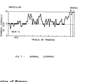
Fig.
7
shows the course of learning by a normal,naive subject, MLN, under conditions in which both eyes
were presented the same stimuli in each trial. Task C,
requiring discriminati on between a black cross and a
black circle, was used (cf. fig.
5).
For the first few days of training the subject
was nervous and could not be worked for more than 10-50
trials in anyone session. During this time many
con-secuti ve pushes ,'lere made by the left hand to the left
response screen; a "position preference" which enabled
escape from choice between the visual cues. "\mile a
position habit persists, each group of 10 trials includes
5 in which reward is obtained.*
After 200 trials there 'I'laS a period of learning
which, however, did not last. The learning curve
fluc-tuates uncertainly and there is occasional reappearance
of a position preference. Criterion was attained after
a total of 750 trials by a sudden reappearance of learning.
.... u w
a: a:
BINOCULAR MONOC .
8~ 5~,~,,"'-ll
....
a:
~~ ::<
:>
MLN-C
z O~~~--~--~ ____ L -_ _ ~ _ _ - L _ _ ~ _ _ ~U
100
TRIALS OF TRAINING
FIG.7 - NORMAL LEARNING.
Explanation of FigureS.
The followinG conventions were used in the learning curves of figures 7 to
14:-Each subject is represented by a three-letter symbol as in the text, and this is followed by a letter indicating the task presented. e.g. MIn'-C means, "performance by subject MIn' on Task Cn•
Each point represents one group of ten trials •
R
L
-In figure
lett rect
• random choice} 5 trials correct in each group of 10.
II: binocular performance with contradictory st1Jllu.ll. c monocular performance •
.. right eye.
= lett eye.
=
performance with a position preference} see p. 47. [image:53.536.114.470.166.441.2] [image:53.536.69.472.175.688.2]Subsequent tests revealed that both eyes had per-fect retention of the task when used individually.
Although all moves during learning were made with the left hand, the right hand could be used with either eye for perfect performance; '<Then a barrier ,<Tas placed ac~oss the left half of the arm-slot preventing use of the left hand, the follm<Ting two trials .<Tere prolonged, with a few frustrated moves by the left hand and signs of nervousness and confusion. However, by the third trial no attempt was made to use the left hand, and thenceforth either hand could be used without difficulty.
The reaction to contradictory overlapping pairs of stimuli by normal subjects varies with past experience. Naive animals were no. more disturbed by the contradictory cues than by the unfamiliarity of the whole situation. But, when experienced subjects were presented
contradic-tory stimuli, after they had learned to use similar stimuli for directing their responses, there were signs of frustration.
The above subject, r-1LN, was immediately bei'Tildered by overlapping polarized pairs of stimuli presented after learning and made a few nervous responses, then stopped '<Tork.
stimuli to the two eyes. Thus the correct button in each
trial now appeared black, as before, to the right eye,
but white to the left eye (cf. fig.
5).
The initial binocular learning was normal and
there w"as perfect retention by both eyes as in the case
of ~~N. The curve for performance with contradictory
ones (fig. 8) shows that learning could occur, but that
only periodically, during long exposure to the situation,
could choices be made accurately.
The fluctuations of performance do not show
cor-respondence lrith the daily training sessions. A few
inter-spersed monocular tests of
6
trials each indicate that goodperformance occurred Ivhen the left eye ''las ina ttenti ve.
The right eye shO"\'lS good retention throughout. Apparently
temporary escape from the conflict could be obtained by
inattention to the eye which suffered reversal of cues.
At first the subject was greatly disturbed by
the frustrating stimulation and became obviously tense
and nervous each period of superior performance. The
temperamental displays included jumping, making of faces
to the reflecting glass in the front of the training box,
biting at wrists and ankles, and sulking ~dth back turned
to the eye -,'lindo,'ls and screens. v.Jhen she ''las sulking an
offered peanut ''lould be pointedly rejected.
In both the above cases, and in other tests of
.... u .... 0:: O::z 0
....
u
....
O::z ....
-ID ~ :> z
0' " " 100 .... 500 1,000 1,500 o TRIALS OF TRAINING
l:! 0::
'
O~ 0'" O::z ....
-ID ~
consistent, except occasionally at times of confusion or excitement. After reaching cautiously with either hand when first introduced to the training box, and when food inducements were laid in front of the response screen, the subject came to prefer a hand for response which there-after ,'las always used for pushing. Peanuts Vlere picked up by either hand from the metal shelf to \'lhich they were delivered.
Summary and conclusions for Section I
Visual discrimination learning by a normal subject proceeded
va
th an initial period in Nhich random responses may be regulated by position preferences. These evenrecurred after there ~I(as a period of improved score indica-tive of learning. The final learning of the visual cues Nas rapid and a steady high level of choice \I(as maintained thereafter.
Both eyes exhibited perfect retention of the visual task immediately learning ,-las completed.
Although a particular limb \I(as chosen for learning Nhen both hands \-Tere free to work, the previously unused hand could be brought to work for perfect performance im-mediately the preferred hand Nas restrained. There ~"as a brief confusion as hands Nere exchanged for the first time.
to each eye. Such stimuli are discouraging to, and
avoided by a subject previously trmned to choose
be-tween the same cues without contradiction.
A normal subject may learn to pay attention to
one eye and so resolve a conflict of visual stimulation
introduced after normal training, but this restriction
of attention is maintained poorly and involves
consider-able emotional strain. Periodic improvement of
perfor-mance due to resolution of conflict alternates with
periods of collapse in which random choices supervene.
Section II
Tests for double visual learning in split-brain subjects
Differences between subjects with the forebrain
*
commissures cut, and those with additional surgery to
**
the commissures of the roof of the midbrain make it
convenient to consider these b'1o groups of subjects
separately.
A. Subjects ~'1i th chiasm and forebrain commissures
cut. --In Figures 9 and 10 are shmm the results of four
complete experiments with three subjects,CHC, IGR and BRS.
*
The corpus callosum, anterior commissure, hippocampal commissure were cut, but the habenular commissure was left intact.BINOCULAR R.EYE LEYE
>-u
Afl
wa:: a::z
ow
u>-a:: z
w-ID
:;
::::l CHC -A
z a
100 100
TRIALS OF TRAINING
FIG.9 - DOUBLE LEARNING, SUBJECT CHC.
-5
O~--~I~O~O--~----~--~~--~----~--~--OL---~IO~O=---LJ
0
Z 100 o 100
W
t-~ 10 R L 10 r---__ -iR.,."L ___ -,
-t-U
W
Il: Il:
0 5
u
~
.
....Il:
W
CD BRS-
BRS-::;;
:::>
0
z
-D
-c o
'----'-'---TRIALS OF TRAINING
FIG. 10 - LEARNING OF SUBJECTS IGR AND BRS WITH TASKS B, CaD.
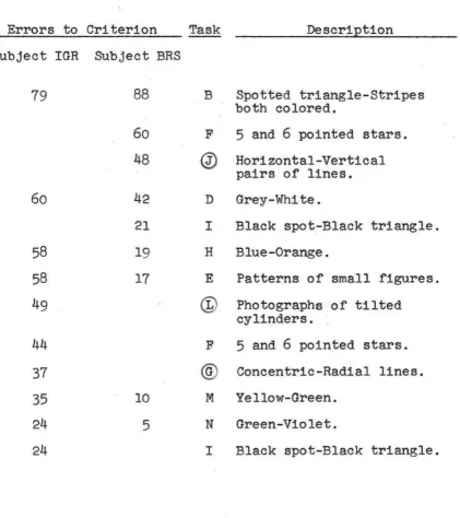
Table II summarizes the error scores to criterion
of learning for IGR and BRS over a series of
14
tasks,and presents them in the order in which they were given
to the animals.
This data will be considered, first, as information
concerning the presence or absence of conflict during
binoc-ular training; and second, for such evidence of independent
learning or interaction of learning as may be obtained from
the monocular retention tests.
The learnin