Differential Inhibitory Receptor
Expression on T Cells Delineates
Functional Capacities in Chronic Viral
Infection
Jeffrey E. Teigler,
a,bGennadiy Zelinskyy,
cMichael A. Eller,
a,bDiane L. Bolton,
a,bMary Marovich,
a,b,dAlexander D. Gordon,
a,bAljawharah Alrubayyi,
a,bGalit Alter,
eMerlin L. Robb,
a,bJeffrey N. Martin,
fSteven G. Deeks,
gNelson L. Michael,
aUlf Dittmer,
cHendrik Streeck
a,b,hU.S. Military HIV Research Program, Walter Reed Army Institute of Research, Silver Spring, Maryland, USAa; Henry M. Jackson Foundation for the Advancement of Military Medicine, Inc., Bethesda, Maryland, USAb; Institute for Virology, University Hospital Essen, University of Duisburg-Essen, Essen, Germanyc; NIAID, National Institutes of Health, Bethesda, Maryland, USAd; Massachusetts General Hospital, Boston, Massachusetts, USAe; Department of Epidemiology and Biostatistics, University of California, San Francisco, California, USAf; UCSF School of Medicine, University of California, San Francisco, California, USAg; Institute for HIV Research, University Hospital Essen, University Duisburg-Essen, Essen, Germanyh
ABSTRACT
Inhibitory receptors have been extensively described for their
impor-tance in regulating immune responses in chronic infections and cancers. Blocking
the function of inhibitory receptors such as PD-1, CTLA-4, 2B4, Tim-3, and LAG-3 has
shown promise for augmenting CD8 T cell activity and boosting pathogen-specific
immunity. However, the prevalence of inhibitory receptors on CD4 T cells and their
relative influence on CD4 T cell functionality in chronic HIV infection remains poorly
described. We therefore determined and compared inhibitory receptor expression
patterns of 2B4, CTLA-4, LAG-3, PD-1, and Tim-3 on virus-specific CD4 and CD8 T
cells in relation to their functional T cell profile. In chronic HIV infection, inhibitory
receptor distribution differed markedly between cytokine-producing T cell subsets
with, gamma interferon (IFN-
␥
)- and tumor necrosis factor alpha (TNF-
␣
)-producing
cells displaying the highest and lowest prevalence of inhibitory receptors,
respec-tively. Blockade of inhibitory receptors differentially affected cytokine production by
cells in response to staphylococcal enterotoxin B stimulation. CTLA-4 blockade
in-creased IFN-
␥
and CD40L production, while PD-1 blockade strongly augmented
IFN-
␥
, interleukin-2 (IL-2), and TNF-
␣
production. In a Friend retrovirus infection
model, CTLA-4 blockade in particular was able to improve control of viral replication.
Together, these results show that inhibitory receptor distribution on HIV-specific CD4
T cells varies markedly with respect to the functional subset of CD4 T cells being
an-alyzed. Furthermore, the differential effects of receptor blockade suggest novel
methods of immune response modulation, which could be important in the context
of HIV vaccination or therapeutic strategies.
IMPORTANCE
Inhibitory receptors are important for limiting damage by the
im-mune system during acute infections. In chronic infections, however, their
expres-sion limits immune system responsiveness. Studies have shown that blocking
inhibi-tory receptors augments CD8 T cell functionality in HIV infection, but their influence
on CD4 T cells remains unclear. We assessed the expression of inhibitory receptors
on HIV-specific CD4 T cells and their relationship with T cell functionality. We
uncov-ered differences in inhibitory receptor expression depending on the CD4 T cell
func-tion. We also found differences in functionality of CD4 T cells following blocking of
different inhibitory receptors, and we confirmed our results in a Friend virus
retrovi-ral model of infection in mice. Our results show that inhibitory receptor expression
Received28 July 2017Accepted24 August
2017
Accepted manuscript posted online13
September 2017
CitationTeigler JE, Zelinskyy G, Eller MA,
Bolton DL, Marovich M, Gordon AD, Alrubayyi A, Alter G, Robb ML, Martin JN, Deeks SG, Michael NL, Dittmer U, Streeck H. 2017. Differential inhibitory receptor expression on T cells delineates functional capacities in chronic viral infection. J Virol 91:e01263-17.https://doi .org/10.1128/JVI.01263-17.
EditorFrank Kirchhoff, Ulm University Medical
Center
Copyright© 2017 American Society for
Microbiology.All Rights Reserved. Address correspondence to Hendrik Streeck, hendrik.streeck@uk-essen.de.
crossm
on November 7, 2019 by guest
http://jvi.asm.org/
on CD4 T cells is linked to CD4 T cell functionality and could be sculpted by
block-ade of specific inhibitory receptors. These data reveal exciting possibilities for the
development of novel treatments and immunotherapeutics.
KEYWORDS
CD4 T cells, CTLA-4, HIV, PD-1, inhibitory receptors
A
ntiviral T cells play a pivotal role in the clearance of acute viral infections. In some
cases T cells fail to eliminate infection but then control viral replication to lower,
nonpathogenic levels. However, under continuous viral replication and immune
acti-vation encountered in certain chronic viral infections, such as human
immunodefi-ciency virus (HIV) and hepatitis C virus, T cells progressively lose the ability to mount
antiviral effector activities, such as cytokine secretion, proliferation, and cytotoxicity,
necessary to control viral replication (1–3). This progressive loss of function, termed
exhaustion, in turn contributes to the lack of viral clearance and may even lead to
diminished viral suppression in chronic stages of disease (2, 4, 5). An important feature
of exhausted T cells is the upregulation of inhibitory receptors such as CD244 (2B4),
cytotoxic T-lymphocyte-associated protein 4 (CTLA-4), lymphocyte-activation gene-3
(LAG-3), programmed death-1 (PD-1), and T cell immunoglobulin and mucin
domain-containing-3 (Tim-3) (5–8). Inhibitory receptors exert their effect through various
mech-anisms, resulting in activation or attenuation of signaling cascades, interference with T
cell ligation of activating coreceptor, or interference with ligation of major
histocom-patibility complex (MHC)-peptide complexes (9–15). Notably, these receptors are also
commonly found on activated T cells during acute infection in order to limit
immuno-pathology due to an exaggerated immune response or excessive and persistent
inflammation (16, 17), yet their continued expression on T cells in chronic infection
becomes detrimental by limiting the ability to control viral replication (2, 4, 18–25).
Thus, inhibitory receptors and their ligands play crucial roles in shaping the immune
response to pathogens, providing the immune system with a mechanism to fine-tune
adaptive immune responses and ensuring pathogen control without excessive
immune-mediated damage. The identity and prevalence of inhibitory receptor
distri-bution in chronic viral infection is therefore of critical importance to understanding the
corresponding potency of virus-specific cellular immunity.
As inhibitory receptors function to limit T cell responses during chronic infections as
well as several cancers, their blockade is being actively investigated as a means to
restore T cell functionality and achieve therapeutic cures (26–28). Studies in the murine
chronic lymphocytic choriomeningitis virus (LCMV), Friend virus (FV), and tumor models
have shown a partial or full restoration of cytotoxic CD8 T lymphocyte activity through
blockade of CTLA-4, PD-1, LAG-3, or Tim-3 (29). Studies of human chronic infections
such as hepatitis B virus (HBV), hepatitis C virus (HCV), and HIV have demonstrated that
ex vivo
blockade of receptors alone or in combination can rescue cytotoxic CD8 T
lymphocyte proliferation, cytokine production, or cytolytic activity (25, 30–42).
Impor-tantly, these studies also showed that inhibitory receptor functions are nonredundant,
as made apparent by studies showing both rescue of different effector functions
dependent on the inhibitory receptor blocked and additional expression of individual
inhibitory receptors progressively shutting down effector functions (10, 34, 36, 43–45).
The utility of inhibitory receptor blockade has been further demonstrated in clinical
trials, where blocking reagents against CTLA-4, PD-1, and LAG-3 improved survival
times and reduced tumor burdens for multiple cancers and lowered viral loads in virus
infections (46–50).
While most studies have focused on the expression, influence, and blockade of
inhibitory receptors on cytotoxic CD8 T lymphocytes, less is known about the influence
of inhibitory receptors on CD4 T cell function. Tim-3 has been shown to be important
for the generation of gamma interferon (IFN-
␥
)-secreting CD4 T cells in the setting of
acute
Mycobacterium tuberculosis
and HCV infection. Furthermore, PD-1 and LAG-3
expression on HIV-specific CD4 T cells has been shown to be important for regulating
cytokine secretion (37, 51–56). Despite the known role of inhibitory receptors in the
on November 7, 2019 by guest
http://jvi.asm.org/
restraint of T cell responses in chronic infections, the relative contribution of different
inhibitory receptors to CD4 T cell function impairment in chronic HIV infection is poorly
understood. As we and others have shown, a robust CD4 T cell response to HIV is
influential in controlling infection (57–60). Indeed, factors which modulate CD4 T cell
functions in HIV infection, such as the ability of CD4 T cells to produce cytokines
supporting CD8 T cell and B cell function and HIV-specific CD4 T cells’ ability to directly
kill infected cells, are important for disease status (61). The relative prevalence of
inhibitory receptors on CD4 T cells and their ability to influence and sculpt HIV-specific
CD4 T cell responses therefore would likely have great importance for understanding
both the elicitation and control of these crucial antiviral functions.
We therefore assessed the inhibitory receptor profile of functional subsets of
HIV-specific CD4 and CD8 T cells from HIV-infected donors able to control viral infection
to various degrees. In addition, we studied changes in the functional profiles of T cells
after blockade of inhibitory receptors and confirmed these findings in a mouse model
of retroviral infection. These results are important for understanding HIV pathology and
have important implications for the design of immunotherapeutic interventions.
RESULTS
Marked differences in inhibitory receptor expression between CD4 and CD8 T
cells in HIV-infected progressors and controllers.
We first determined the expression
levels of the inhibitory receptors 2B4, CTLA-4, LAG-3, PD-1, and Tim-3 on CD4 and CD8
T cells from treatment-naive, chronically HIV-infected individuals with detectable viral
loads of
⬎
2,000 RNA copies/ml (termed HIV progressors; average
⫽
54,248
⫾
15,890
HIV RNA copies/ml) and individuals spontaneously able to control HIV replication
(
⬍
2000 HIV RNA copies/ml) in the absence of antiretroviral medication (termed HIV
controllers; average
⫽
566
⫾
120 HIV RNA copies/ml). Interestingly, expression levels of
inhibitory receptors differed markedly between CD4 and CD8 T cells. The most common
inhibitory receptors expressed on CD4 T cells for all individuals were PD-1 and CTLA-4,
expressed on
⬃
20% and 6% of cells, respectively (Fig. 1A). While PD-1 was expressed
at similar levels on both CD4 and CD8 T cells, CTLA-4 expression was marginally present
on CD8 relative to CD4 T cells. The most commonly expressed receptor on CD8 T cells
was 2B4 (
⬃
75% of cells), while less than 10% of CD4 T cells expressed this receptor. The
FIG 1Inhibitory receptor distribution on CD4 T cells is diminished in HIV controllers. Levels of 2B4, CTLA-4, LAG-3, PD-1, and Tim-3 were assessed on total CD4 and CD8 T cells in HIV progressors (P; viral load of⬎2,000 HIV RNA copies/ml;n⫽29) and controllers (C; viral load of⬍2,000 HIV RNA copies/ml;n⫽23). (A) Levels were compared for individual receptor prevalence on CD4 (circles) and CD8 (squares) T cells, with progressors in black and controllers in red. Summary data are shown as means⫾standard errors of the means (SEM). Bars indicate aPvalue of⬍0.05 by Wilcoxon rank-sum tests, with compared groups being at the ends of the bar. (B) Prevalence of multiple and single receptor-expressing subsets on total CD4 and CD8 T cells. Colors of slices indicate the presence of 2 (blue), 1 (red), or 0 (gray) inhibitory receptors. Pies were compared by SPICE permutation tests with 10,000 permutations.
on November 7, 2019 by guest
http://jvi.asm.org/
[image:3.585.44.543.75.280.2]largest difference between HIV controllers and progressors was LAG-3 and CTLA-4
expression, which were both elevated on CD4 and CD8 T cells in controllers (LAG-3,
0.065% versus 0.021% [P
⫽
0.0002] and 0.047% versus 0.004% [P
⫽
0.0003],
respec-tively; CTLA-4, 7.43% versus 6.04% [P
⫽
0.03] and 0.67% versus 0.37% [P
⫽
0.005],
respectively). Only PD-1 showed significant upregulation on CD4 T cells in HIV
pro-gressors compared to controllers (21.61% versus 16.29%;
P
⫽
0.047). CD8 T cells were
unique in their larger percentage of 2B4
⫹cells as well as the predominance of 2B4
⫹PD-1
⫹cells, both of which were not observed to the same extent on CD4 T cells.
To evaluate levels of redundancy in the inhibitory receptor expression on T cells, we
compared coexpression profiles of inhibitory receptors on total CD4 and CD8 T cells
using SPICE, version 5.1 (62). While inhibitory receptor distribution on CD8 T cells was
similar in progressors and controllers (P
⫽
0.33), we observed significantly lower
inhibitory receptor-expressing subsets in CD4 T cells from HIV controllers (P
⫽
0.026)
(Fig. 1B). This observation is interesting, as the only significant difference in individual
populations between progressors and controllers were higher levels of CTLA-4
single-receptor-expressing cells in controllers relative to progressors (data not shown). This
point suggests that an aggregate total of several receptor-negative populations that
did not reach monovariate statistical significance contribute to the overall difference in
inhibitory receptor distribution between progressors and controllers. Together these
results indicate that CD4 T cells in both HIV progressors and controllers express
different amounts and combinations of the inhibitory receptors measured relative to
CD8 T cells.
Inhibitory receptor distribution differs on CD4 T cell subsets in association with
their functional profile but are largely similar between HIV progressors and
controllers.
Given the various functions of CD4 T cells in chronic viral infection, we next
sought to determine whether inhibitory receptor expression differs on the basis of
functionality of HIV-specific CD4 T cells. PBMC from HIV progressors and controllers
were stimulated with HIV-1 Gag PTE peptide pools, and production of IFN-
␥
, CD40L,
tumor necrosis factor alpha (TNF-
␣
), and interleukin-2 (IL-2) was determined by
intra-cellular cytokine staining. While Gag-specific CD4 T cell response does not represent the
totality of the HIV-specific CD4 T cell responses, we previously determined that it is a
good representation of the overall HIV-specific CD4 T cell response (63). Inhibitory
receptor expression on these cytokine-producing cells was assessed and compared
between groups. Interestingly, we observed marked differences in the inhibitory
re-ceptor expression distribution between CD4 T cells based on their functionality
(Fig. 2A). A general pattern was observed where HIV-specific IFN-
␥
-producing CD4 T
cells displayed the greatest expression of several inhibitory receptors relative to other
subsets, with progressively lower levels observed on CD40L-, TNF-
␣
-, and
IL-2-producing CD4 T cells, respectively. This pattern was most evident for CTLA-4 and 2B4
and was observed in HIV progressors as well as HIV controllers. Notably, HIV-specific
IL-2-producing CD4 T cells bearing Tim-3 and LAG-3 were negligible in both
progres-sors and controllers. In contrast to other inhibitory receptors, the expression patterns of
PD-1 did not fit this general pattern. While IFN-
␥
-producing cells had the highest levels
of PD-1 in progressors and controllers (76.1% and 71.3%, respectively), IL-2-producing
cells also displayed elevated levels of PD-1 relative to CD40L- and TNF-
␣
-producing
cells in progressors (59.4% versus 54.0% and 45.2%;
P
values of 0.20 and 0.063) and
controllers (58.8% versus 48.3% and 36.5%, respectively;
P
values of 0.5 and 0.0002).
These data show that the inhibitory receptor expression levels on CD4 T cells in both
HIV progressors and controllers directly relate to functional capacity.
We next analyzed the distribution of single and multiple inhibitory
receptor-expressing populations on HIV-specific CD4 T cell subsets in both progressors and
controllers using SPICE analysis. Similar to our monovariate analyses, in Gag-stimulated
cells, we found that the overall expression of inhibitory receptors was highest on
IFN-
␥
-secreting CD4 T cells followed by CD40L-, TNF-
␣
-, and IL-2-secreting CD4 T cells
in both progressors (P
⫽
0.0004,
P
⬍
0.0001, and
P
⫽
0.0004, respectively; SPICE
permutation tests, with 10,000 permutations) and controllers (P
⫽
0.02,
P
⬍
0.0001, and
on November 7, 2019 by guest
http://jvi.asm.org/
P
⫽
0.01, respectively) (Fig. 2B). HIV-specific IFN-
␥
-secreting CD4 T cells were unique in
their high levels of CTLA-4 and PD-1 coexpression, which significantly differed between
progressors and controllers (P
⫽
0.023; SPICE permutation tests, with 10,000
permu-tations) (Fig. 2B and 3). The inhibitory receptor levels were similar between CD40L- and
IL-2-producing CD4 T cells, while TNF-
␣
-producing cells displayed the smallest amount
of these receptors. Notably, as cytokine production was assessed in a monovariate
fashion, it is unclear whether these cytokine-producing subsets represent distinct
subpopulations of CD4 T cells or whether they represent cells at different stages of
progressive exhaustion.
Differential impact of inhibitory receptor blockade of HIV-specific CD4 T cell
subsets.
It is widely believed that inhibitory receptors serve to downregulate certain T
cell functions, and their actions may synergize to enact successively greater levels of T
cell inhibition, with various influences on different T cell functionalities. To investigate
the role of inhibitory receptors on the functionality of HIV-specific CD4 and CD8 T cells,
we performed
in vitro
blockade of the most commonly expressed inhibitory receptors,
2B4, CTLA-4, PD-1, and Tim-3, after staphylococcal enterotoxin B (SEB) stimulation in a
subset of HIV progressors. Cytokine production by proliferating cells then was
mea-FIG 2Bulk inhibitory receptor distribution varies on different cytokine-producing CD4 T cell subsets but is similar between HIV progressors and controllers. (A) PBMC from HIV progressors (n⫽29) and controllers (n⫽23) were stimulated with HIV-1 Gag PTE peptide pools and assessed for prevalence of single inhibitory receptors on cytokine-producing subsets: IFN-␥(gray), CD40L (red), TNF-␣(blue), and IL-2 (green). Individual receptor prevalence on cytokine-producing subsets in HIV progressors (top) and HIV controllers (bottom) is shown. Summary data are shown as means⫾SEM. Bars indicate aPvalue of⬍0.05 by Wilcoxon signed-rank tests, with compared groups being at the ends of the bar. (B) Prevalence of single- and multiple-receptor subsets on cytokine-producing CD4 T cells displayed using SPICE. Colors of slices indicate the presence of 3 (green), 2 (blue), 1 (red), or 0 (gray) inhibitory receptors. The prevalence of subsets expressing single and multiple inhibitory receptors in HIV progressors (top;n⫽29) and controllers (bottom;n⫽23) is shown. Bars indicate aPvalue of⬍0.05 by SPICE permutation tests with 10,000 permutations, with compared groups being at the ends of the bar.
on November 7, 2019 by guest
http://jvi.asm.org/
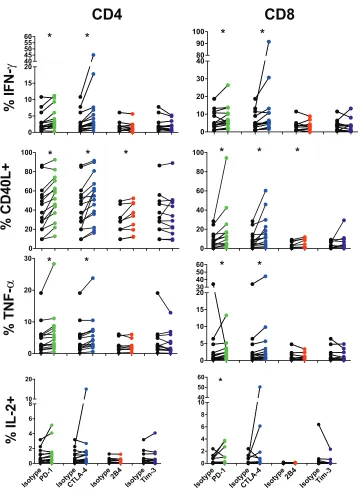
[image:5.585.43.541.72.436.2]sured 5 days following stimulation and compared to an isotype-matched antibody
control. LAG-3 was omitted from this analysis due to the exceedingly low level of
expression observed on both bulk CD4 and CD8 T cells in our studies. In agreement
with previous studies (39, 52), we observed significant differences in the functional
responses of CD8 T cells after receptor blockade with PD-L1, which resulted in increased
production of IFN-
␥
(7.4% versus 5.6), CD40L (17.6% versus 8.3%), and IL-2 (0.88%
versus 0.45%) (Fig. 4). Blockade of CTLA-4 resulted in increases in IFN-
␥
(13.2% versus
5.6%), CD40L (16.4% versus 8.3%), and TNF-
␣
(5.1% versus 3.8%) production by CD8 T
cells relative to isotype controls.
CD4 T cells showed an overall similar pattern of increased production of IFN-
␥
(4.2%
versus 3.0%), CD40L (52.3% versus 41.4%), and TNF-
␣
(6.3% versus 4.8%) after PD-1
receptor blockade (Fig. 4). Additionally, blockade of CTLA-4 resulted in increased
production relative to an isotype control of IFN-
␥
(7.0% versus 3.0%), CD40L (53.0%
versus 41.4%), and TNF-
␣
(6.0% versus 4.8%). Therefore, both PD-1 and CTLA-4 are able
to repress cytokine production by CD4 and CD8 T cells in chronic HIV infection, and
their blockade is able to partially rescue cytokine production. Interestingly, while
blockade of 2B4 had no discernible effect on production of IFN-
␥
, TNF-
␣
, or IL-2, its
blockade resulted in increased levels of CD40L production for both CD4 (31.8% versus
27.0%) and CD8 (4.8% versus 3.7%) T cells. Interestingly, single blockade of Tim-3 had
no apparent effect on cytokine elicitation by CD4 T cells.
We next sought to explore the hierarchy of inhibitory receptor responses by
combining blockade of 2B4, CTLA-4, and PD-1 on T cells in a subset of HIV progressors
in response to SEB. Interestingly, combinatorial blockade was unable to augment
increases in IFN-
␥
production by either CD4 or CD8 T cells beyond that achieved by
Progressors
Controllers
Tim-3 PD-1 LAG-3 CTLA-4 2B4 0.0 10.0 20.0 30.0 40.0 -+ -+ + -+ + -+ -+ -+ -+ + + -+ + -+ + -+ -+ -+ -+ -+ -+ -+ -+ -+ -0.0 10.0 20.0 30.0 40.0 50.0 IFN-γCD40L TNF-a IL-2 IFN-γ CD40L TNF-a IL-2 Tim-3 PD-1 LAG-3 CTLA-4 2B4 -+ -+ + -+ + -+ -+ -+ -+ + + -+ + -+ + -+ -+ -+ -+ -+ -+ -+ -+ -+ -Frequency (%) F requency (%)
FIG 3Expression of single- and multiple-inhibitory-receptor subsets in cytokine-producing CD4 T cells from HIV progressors (n⫽29; top) and controllers (n⫽23; bottom) following Gag PTE stimulation. Bars indicate aPvalue of⬍0.05 by Wilcoxon signed-rank tests, with compared groups being at the ends of the bar.
on November 7, 2019 by guest
http://jvi.asm.org/
[image:6.585.44.475.75.397.2]single-receptor blockade (Fig. 5). Together, these data suggest that PD-1 and CTLA-4
share common roles in reducing cytokine production by IFN-
␥
-, IL-2-, or TNF-
␣
-producing CD4 T cell subsets in chronic HIV, but that CD40L responses are also
modulated through 2B4. However, given the subtle degree to which combinations of
receptor blockade influenced cytokine production, further exploration of other
signa-tures, such as transcriptional profiles following blockade, would improve our
under-standing of inhibitory receptor hierarchies.
Blockade of CTLA-4 similarly augments T cell responses in murine retroviral
infection.
The importance of inhibitory receptor functionality to sculpt antiviral
im-0 10 20 30 40 80 90
100
*
*
0 5 10 15 20 40 45 50 55
60
*
*
% I
FN-γ
0 20 40 60 80
100
*
*
*
% CD40L
+
0 10 20
30
*
*
%
TN
F-α
IsotypePD-1 IsotypeCTLA-4 Isotype2B4 IsotypeTim-3
0 2 4 6 8 10 20
% I
L-2+
CD4
CD8
IsotypePD-1 IsotypeCTLA-4 Isotype2B4 IsotypeTim-3
0 2 4 6 8 10 40 50 60
*
0 5 10 15 20 30 40 5060
*
*
0 20 40 60 80
100
*
*
*
FIG 4Inhibitory receptors differentially control cytokine production by T cells in HIV infection. PBMC from HIV progressors (n⫽9 to 15) were stained with CFSE and blocked with an isotype control (black) or for PD-1 (green), CTLA-4 (blue), 2B4 (red), or Tim-3 (purple) and then stimulated with SEB. Five days following stimulation, cells were washed and stimulated with SEB (5g/ml), and the prevalence of cytokine-producing CFSE-low CD4 (left) and CD8 (right) cells was assessed. Asterisks indicate aPvalue of⬍0.05 by Wilcoxon signed-rank tests.
on November 7, 2019 by guest
http://jvi.asm.org/
[image:7.585.43.403.67.558.2]mune responses has been demonstrated in several settings for CD8 T cells. We sought
to expand and confirm our observations implicating receptor blockade as a means of
supporting CD4 T cell responses in a murine model of retroviral infection. Mice infected
with Friend virus (FV) were subjected to inhibitory receptor blockade using antibodies
blocking CTLA-4, LAG-3, PD-L1, or Tim-3 or isotype controls. Mice were sacrificed 14
days following infection, and levels of infectious virus as well as cytokine-producing
CD4 and CD8 T cells were assessed (Fig. 6A). Relative expression of inhibitory receptors
on effector CD4
⫹CD62L
⫺T cells before and after FV infection was first assessed by
mean fluorescence intensity (MFI) in both naive and FV-infected mice. Levels of every
inhibitory receptor were found to be increased at day 14 following FV infection (Fig.
6B). In the context of inhibitory receptor-blocking antibodies, only CTLA-4 blockade
significantly influenced FV viral loads in the bone marrow, the main site of FV
replica-tion, relative to untreated animals (149.7 versus 26.9 infectious cells/1
⫻
10
6,
respec-tively;
P
⫽
0.01 by Student’s
t
tests) (Fig. 6C). CTLA-4 or LAG-3 inhibition resulted in
increased numbers of bone marrow CD4 T cells able to produce IFN-
␥
(19.3% versus
11.3% and 27.4% versus 11.3%, respectively) or TNF-
␣
(8.6% versus 4.7% and 9.1%
versus 4.7%, respectively) relative to untreated animals (Fig. 6D). A similar
augmenta-tion of IFN-
␥
was observed for CD8 T cells in animals blocked with CTLA-4 or LAG-3
antibodies (43.3% versus 32.5% and 35.1% versus 23.5%, respectively). Together, these
data highlight the role of CTLA-4 in influencing antiretroviral CD4 and CD8 T cell
responses. However, as different viruses and infections covering different time scales
were involved in comparing FV to HIV infection, further exploration is warranted.
DISCUSSION
Inhibitory receptors play a critical role in regulating immune responses and
pro-tecting against immunopathology during acute infections. Despite their crucial role in
protecting against hyperactivation of immune cells early in infection, their continued
expression in chronic infection is associated with T cell exhaustion and viral persistence.
In this study, we found that inhibitory receptor distribution in chronic HIV infection
differed significantly between functionally distinct cytokine-producing CD4 T cells and
FIG 5PBMC from HIV-positive donors (n⫽10) were stained with CFSE and blocked with an isotype control or for PD-1, CTLA-4, 2B4, or a combination of these receptors and then stimulated with SEB. Five days following stimulation, cells were washed and restimulated with SEB (5g/ml), and the prevalence of cytokine-producing CFSE-low CD4 and CD8 T cells was assessed. Data are shown as means⫾SEM. Bars indicate aPvalue of⬍0.05 by Wilcoxon signed-rank tests, with compared groups being at the ends of the bar.on November 7, 2019 by guest
http://jvi.asm.org/
[image:8.585.56.358.70.298.2]were more elevated on CD4 T cells from progressors than controllers. Treatment with
inhibitory receptor blockade yielded generally similar augmentation of cytokine
pro-duction between CD4 T cells and CD8 T cells, and these results were confirmed in a
murine model of retroviral infection. Together these results show that CD4 T cells in
chronic HIV infection express inhibitory receptors that can be used to modulate
immune responses and that the expression profiles differ across functionally distinct
cell subsets. Moreover, blockade of the inhibitory receptors PD-1, CTLA-4, and 2B4 show
differential abilities to rescue cytokine production upon stimulation.
The ability to control HIV infection is constrained in part by the cumulative
acqui-sition of inhibitory receptor expression on both CD4 and CD8 T cells (38, 39, 52, 64, 65).
In chronic HIV infection, CD8 T cells have been shown to have increased levels of
inhibitory receptors, such as PD-1, 2B4, LAG-3, and Tim-3, relative to HIV-uninfected
individuals as well as relative to CD8 T cells specific for other chronic infections, such
as Epstein-Barr virus or cytomegalovirus (40). Levels of these inhibitory receptors have
been suggested to correlate with HIV loads and their accumulation results in an
exhausted phenotype, which is at least in part reversible upon receptor blockade (52,
66, 67). Our data show that the CD4 T cell compartment expresses lower overall levels
of inhibitory receptors than CD8 T cells, mirroring prior studies (51, 54).
FIG 6CTLA-4 or LAG-3 blockade reduces viral burden and augments CD4 and CD8 T cell responses in murine retroviral infection. Mice (n⫽5 to 8/group) were infected with Friend’s leukemia virus (FLV) in the presence of control or inhibitory receptor-blocking antibodies. (A) Experimental layout of infection of mice with Friend’s leukemia virus (FLV) in the presence of inhibitory receptor-blocking antibodies. (B) Mean fluorescence intensity (MFI) of inhibitory receptors on CD4⫹CD62L⫺effector T cells in spleen in both naive (n⫽5) and FV-infected (n⫽8; 10 days postinfection) mice. Symbols represent levels in naive (open) and
FV-infected (closed) mice. (C) Cells from bone marrow were harvested 14 days following infection, and numbers of cells containing infectious virus were assessed. Data are shown as means⫾SEM. Bars represent aPvalue of⬍0.05 by Student’sttests, with compared groups being at the ends of the bar. (D) Cells from bone marrow were harvested 14 days following infection, and cells were restimulated with CD3/CD28 antibodies. Levels of cytokine-producing cells were assessed. Levels in naive mice are included for reference. Data are shown as means⫾SEM. Bars represent aPvalue of⬍0.05 by Student’sttests, with compared groups being at the ends of the bar.
on November 7, 2019 by guest
http://jvi.asm.org/
[image:9.585.45.542.69.424.2]While there indeed appear to be shared mechanisms of CD4 and CD8 T cell
exhaustion, there are also unique features of CD4 T cell exhaustion signatures (68). For
instance, while CD8 T cell dysfunction in chronic viral diseases is strongly linked to a
distinct and predictable exhaustion program, alterations of pathogen-specific CD4 T
cell functions result from a broader combination of skewed lineage differentiation and
by factors unique to T helper cells. In addition, MHC class II-T cell receptor (TCR)
functional avidity is generally lower in HIV infection than MHC class I-TCR interaction,
which can further alter apparent CD4 T cell exhaustion (69). In the context of HIV
infection, infection and depletion of HIV-specific CD4 T cells also may play a role in
observed CD4 T cell dysfunction (70). Therefore, while we have observed differences in
exhaustion between CD4 T cell subsets in HIV infection, without assessment of
tran-scriptional programs associated with known exhaustion pathways, such as Blimp-1, it is
difficult to specifically assign a mechanism to the observed influence of inhibitory
receptors on impaired CD4 T cell functionality we observed. It will therefore be of
interest to further explore the genetic signatures of exhaustion between CD4 and CD8
T cells during HIV infection to assess whether CD4 T cells display an exhausted genetic
phenotype similar to that of CD8 T cells and whether other inhibitory receptors are
involved. In particular, understanding differences in exhaustion signatures using
te-tramer technology will provide a much cleaner assessment than cytokine secretion.
A hallmark of the exhausted phenotype of CD8 and CD4 T cells in chronic infection
is the progressive loss of cellular functions, such as the ability to produce different
cytokines as well as the ability to proliferate in response to antigen. Indeed, in chronic
simian immunodeficiency virus (SIV) infection, as CD8 T cell exhaustion progresses,
SIV-specific CD8 T cells have been shown first to lose the ability to produce IL-2 in
response to antigen, then TNF-
␣
, and finally IFN-
␥
(71–74). Furthermore, the ability to
produce several cytokines upon stimulation with antigen has been observed to be
sensitive to the amount of PD-1 present on a given cell, with previous studies showing
inhibition of IL-2 production requiring little expression of PD-1 while MIP-1

production
is only inhibited in the presence of high levels of PD-1 expression (43). Our studies
suggest a similar phenomenon occurs with HIV-specific CD4 T cells in chronic HIV
infection. IFN-
␥
-producing CD4 T cells displayed a significantly higher prevalence of
inhibitory receptor expression than CD4 T cells that produced CD40L and IL-2. A
possible explanation for these differences is that the ability to produce certain
cyto-kines is lost as inhibitory receptors accumulate on a cell type in a manner similar to
what has been observed for CD8 T cells. Alternatively, inhibitory receptor expression
patterns may delineate distinct functional subsets rather than identify CD4 T cells at
various stages of an exhaustion pathway. For example, PD-1 also serves as a marker for
T follicular helper (T
FH) cells, and its role in T
FHfunction remains uncertain but has been
proposed to regulate B cell function (75). It is important to note that differential
distribution of memory phenotypes within these subsets could account for some of the
observed differences in inhibitory receptor expression. However, in these studies we
focus on the ability of the HIV-specific CD4 T cell compartment to execute a given
function, regardless of the cells which make up the population providing that function.
It would be of interest to further examine the subsets which comprise these functional
compartments and their relative influence on observed inhibitory receptor prevalence
(63). Lastly, as cytokine production was assessed in a monovariate fashion in these
studies, it is unclear whether the larger amounts of inhibitory receptors present on
IFN-
␥
-producing CD4 T cells is the result of progressive loss of functions as inhibitory
receptors accumulate, similar to CD8 T cells, or whether the inhibitory receptors
delineate different populations of cytokine-producing cells.
The ability of CD4 T cells to maintain help in chronic retroviral infection has
numerous benefits that aid in the control of viral infection. Our results indicate that
differential blockade of inhibitory receptors can augment CD4 T cell responses to
varying degrees, and while the distribution pattern of these receptors remains largely
unaltered between HIV progressors and controllers on CD8 T cells, their bulk expression
is altered between progressors and controllers on CD4 T cells. Furthermore, in both HIV
on November 7, 2019 by guest
http://jvi.asm.org/
controllers and progressors, a substantial proportion of cytokine-producing CD4 T cells
expresses CTLA-4, PD-1, or both. While it is possible that this phenomenon captures a
portion of activated CD4 T cells as well, these receptors likely play a central role in
restraining CD4 T cell responses in HIV infection. This central role of CTLA-4 was also
observed in a separate retroviral system, Friend virus (FV) infection in mice. This result
indicates that inhibitory receptor expression on CD4 T cells in chronic infection follows
generalizable rules and that while certain receptors such as LAG-3 or PD-1 are uniquely
expressed during FV and HIV infection, respectively, CTLA-4 expression is similarly
regulated in both diseases. This possibility is strengthened by previous reports showing
the ability of CTLA-4 blockade to augment CD4 T cell responses against other chronic
viral infections, such as Epstein-Barr virus, hepatitis B virus, and hepatitis C virus. It is
interesting that PD-1 has also been shown to be a prominent marker of CD4 T cells
hypothesized to be the main reservoir population in chronic HIV infection (76).
There-fore, coexpression of other inhibitory receptors and their relative contribution to the
establishment or maintenance of HIV reservoir-harboring cells would be of interest.
Further exploration of the hierarchy of inhibitory receptor usage in these systems
would build a better understanding of the process by which CD4 T cells become
impaired in chronic infection as well as their potential contribution to the HIV reservoir.
Taken together, we demonstrated that CD4 T cells display marked differences in
their inhibitory receptor profiles in chronic viral infection dependent on their
function-ality. Cytokine-producing cellular subsets displayed inhibitory receptor distributions
reminiscent of cytokine-production hierarchies previously observed for CD8 T cells,
suggesting similarities in T cell exhaustion between CD4 and CD8 T cells, particularly
during chronic HIV infection. Notably, we observed an influence of PD-1 and CTLA-4 on
HIV Gag-specific CD4 T cell function and confirmed the importance of CTLA-4 in a
murine retroviral infection model. These studies suggest that CD4 T cell functionality
during chronic viral infection is able to be sculpted by blockade or engagement of
different inhibitory receptors, providing exciting avenues for the continued
develop-ment of HIV immunotherapeutic interventions.
MATERIALS AND METHODS
Ethics statement.Cryopreserved peripheral blood mononuclear cells (PBMC) from treatment-naive chronically HIV-1-infected individuals were used. Subjects were enrolled either at the United States Military HIV Research Program (MHRP) RV149 cohort or the University of California, San Francisco (UCSF), SCOPE cohort. All study subjects were adults and gave written consent, and IRB approval was obtained by the Walter Reed National Military Medical Center, the Walter Reed Army Institute of Research, the Naval Medical Research Center, and the University of California, San Francisco.
Animal experiments were performed in strict accordance with the German regulations of the Society for Laboratory Animal Science (GV-SOLAS) and the European Health Law of the Federation of Laboratory Animal Science Associations (FELASA). The protocol was approved by the North Rhine-Westphalia State Agency for Nature, Environment, and Consumer Protection (LANUV) (permit number G 1518/15). All efforts were made to minimize suffering.
Mice.Inbred C57BL/6 (B6) mice were maintained under pathogen-free conditions. C57BL/6 mice (H-2b/b, Fv1b/b, Fv2r/r) are resistant to FV-induced leukemia. All mice were females of 8 to 12 weeks of age
at the beginning of the experiments.
Human cell stimulation for cytokine production.PBMCs were thawed in 37°C RPMI 1640 supple-mented with 10% fetal calf serum,L-glutamine, and penicillin-streptomycin (R10) and rested overnight. PBMCs were then diluted to 1⫻106/ml and stimulated with HIV-1 Gag PTE peptide pools (1g/ml; NIH
AIDS Reagent Bank) in the presence of CD28/49d costimulatory molecules (BD), brefeldin A (Sigma-Aldrich), and monensin (BD) and incubated for 6 h at 37°C, 5% CO2.
Alternatively, PBMC were stained with carboxyfluorescein succinimidyl ester (CFSE) (0.5 M in phosphate-buffered saline [PBS], 37°C, 5 min) and blocked with 10g/ml mouse IgG1 isotype control (MG1-45; BioLegend), anti-human 2B4/CD244 (eBioPP35; eBioscience), anti-human CD152/CTLA-4 (L3D10; BioLegend), anti-human CD274/PD-L1 (29E.2A3; BioLegend) and anti-human CD273/PD-L2 (MIH18; BioLegend), or anti-human CD366/Tim-3 (F38-2E2; BioLegend) 1 h prior to stimulation with staphylococcal enterotoxin B (SEB; 5g/ml). Cells were incubated at 37°C, 5% CO2, and then washed with
R10 prior to restimulation with SEB and BFA-monensin treatment as described above.
Virus and viral infection.The FV stock used in these experiments was FV complex containing B-tropic Friend murine leukemia helper virus (F-MuLV) and polycythemia-inducing spleen focus-forming virus (77). Virus stock was prepared as a 10% spleen cell homogenate from BALB/c mice infected 14 days previously with 3,000 spleen focus-forming units of noncloned virus stock. Experimental mice were injected intravenously with 20,000 spleen focus-forming units of lactate dehydrogenase-elevating virus (LDV)-free FV complex.
on November 7, 2019 by guest
http://jvi.asm.org/
In vivoreceptor blockade.For blockade of inhibitory pathways in acute FV-infected mice, 200g anti-mouse CTLA-4 (9H10), 100g anti-mouse LAG-3 (C9B7W), 200g rat anti-mouse PD-L1 A (10F.9G2), or 100g anti-mouse Tim-3 antibody (RMT3-23) (BioXCell) was administered intraperitoneally every second day for a total of four times. T cell responses and viral loads were analyzed 1 day posttreatment.
Infectious center assays.Infectious center assays were performed as described previously (78).
Multicolor flow cytometry.Following peptide stimulation, cells were washed with PBS–2% fetal calf serum (FCS) and stained with an amine-reactive viability dye (Live/Dead aqua; Life Technologies) for 30 min at room temperature (RT). Cells then were washed and blocked with PBS–10% normal mouse serum (Life Technologies) for 15 min at 4°C. Following blocking, cells were stained with anti-human anti-CD14 brilliant violet 510 (BV510) (M5E2; BioLegend), anti-CD19 BV510 (HIB-19; BioLegend), anti-LAG-3/CD223 phycoerythrin-cyanine 7 (PE-Cy7) (3DS223H; eBioscience), anti-2B4/CD244 PE (C1.7; BioLegend), anti-PD-1/CD279 BV605 (EH12.2H7; BioLegend), anti-CTLA-4/CD152 PE-CF594 (BNI3; BD), and anti-Tim-3/CD366 BV421 (F38-2E2; BioLegend) for 30 min at 4°C. Cells then were washed and fixed in Fix/Perm buffer A (Life Technologies) for 15 min at RT. Intracellular staining was done in Fix/Perm Buffer B at RT with anti-human anti-CD3 Alexa Fluor 700 (UCHT1; BD), anti-CD4 fluorescein (RPA-T4; BioLegend), anti-CD8 allophyco-cyanin (APC)-Cy7 (RPA-T8; BioLegend), and one of the following cytokine-specific antibodies: anti-CD40L/ CD154 (24-31; BioLegend), anti-interferon gamma (IFN-␥) (B27; BioLegend), anti-IL-2 (MQ1-17H12; Bio-Legend), or anti-TNF-␣ (MAb11; BioLegend), conjugated to APC. Cells were washed twice and resuspended in PBS for flow cytometry. Data were collected on a four-laser LSR II flow cytometer using FACSDiva software (BD) and subsequently analyzed using FlowJo (version 9.410; TreeStar).
Mouse cell surface and intracellular staining by flow cytometry.Surface and intracellular staining were performed as described previously (79). For surface staining, we used antibodies specific against mouse anti-CD3 APC-Cy7 (17A2; BioLegend), anti-CD4 BV605 (RM4-5; BioLegend), anti-CD8 Alexa Fluor 700 (53-6.7; eBioscience), CD11b BV650 (M17/4; BioLegend), CD43 PE or PerCP (peridinin chlorophyll protein; 1B11; BioLegend), CD62L PE-Cy7 (MEL-14; eBioscience), and CD69 PE (H1.2F3; BioLegend), and for intracellular staining we used a cross-reactive antibody specific against human granzyme B, AF700 (GB11; ThermoFisher) (80). Intracellular staining for IFN-␥fluorescein isothiocyanate (XMG1.2; eBiosci-ence), TNF-␣BV510 (MP6-XT22; BioLegend), and IL-2 eF450 (JES6-5H4; eBioscience) was performed as described previously (81). Data were acquired on a four LSR II flow cytometer (Becton Dickinson) from 350,000 to 500,000 lymphocyte-gated events per sample. Analyses were done using FACSDiva software (Becton Dickinson) and FlowJo software v10 (TreeStar).
Statistical analyses. Monovariate statistical analysis was performed using GraphPad Prism, v6 (GraphPad Software, Inc.). For comparison between flow cytometry groups, nonparametric Mann-Whitney U tests or Wilcoxon signed-rank tests were used for data sets which were nonpaired or paired, respectively. Data are presented as means and standard errors of the means (SEM). Flow cytometry analysis and presentation of distributions was performed using SPICE, version 5-1.2, downloaded from http://exon.niaid.nih.gov/spice(62). Comparison of distributions was performed using a Student’sttest and a partial permutation test.
ACKNOWLEDGMENTS
This work was funded by the National Institutes of Health (NIH; R01 AI091450-01 and
R01 AI094602-01) and a cooperative agreement (W81XWH-11-2-0174) between the
Henry M. Jackson Foundation for the Advancement of Military Medicine, Inc., and the
U.S. Department of Defense (DOD). This work was further supported by DFG TRR60 and
DFG STR1069/2-1. The SCOPE cohort is supported by P30 AI027763.
The following were obtained through the NIH AIDS Reagent Program, Division of
AIDS, NIAID, NIH: HIV-1 PTE Gag and Env peptides. Human recombinant IL-2 was
obtained from Maurice Gately, Hoffmann-La Roche, Inc.
Material has been reviewed by the Walter Reed Army Institute of Research. There is
no objection to its presentation and/or publication. The opinions or assertions
con-tained here are the private views of the author and are not to be construed as official
or as reflecting true views of the Department of the Army or the Department of
Defense.
REFERENCES
1. Antoine P, Olislagers V, Huygens A, Lecomte S, Liesnard C, Donner C, Marchant A. 2012. Functional exhaustion of CD4⫹T lymphocytes during primary cytomegalovirus infection. J Immunol 189:2665–2672.https:// doi.org/10.4049/jimmunol.1101165.
2. Wherry EJ, Ha S, Kaech SM, Haining WN, Sarkar S, Kalia V, Subramaniam S, Blattman JN, Barber DL, Ahmed R. 2007. Resource molecular signature of CD8⫹ T cell exhaustion during chronic viral infection. Immunity 27:670 – 684.https://doi.org/10.1016/j.immuni.2007.09.006.
3. Wherry EJ, Ahmed R. 2004. Memory CD8 T-cell differentiation during
viral infection. J Virol 78:5535–5545. https://doi.org/10.1128/JVI.78.11 .5535-5545.2004.
4. Blackburn SD, Shin H, Haining WN, Zou T, Workman CJ, Polley A, Betts MR, Freeman GJ, Vignali DAA, Wherry EJ. 2009. Coregulation of CD8⫹T cell exhaustion by multiple inhibitory receptors during chronic viral infection. Nat Immunol 10:29 –37.https://doi.org/10.1038/ni.1679. 5. West EE, Youngblood B, Tan WG, Jin H, Araki K, Alexe G, Konieczny BT,
Calpe S, Freeman GJ, Terhorst C, Haining WN, Ahmed R. 2011. Tight regulation of memory CD8⫹ T cells limits their effectiveness during
on November 7, 2019 by guest
http://jvi.asm.org/
sustained high viral load. Immunity 35:285–298.https://doi.org/10.1016/ j.immuni.2011.05.017.
6. Odorizzi PM, Wherry EJ. 2012. Inhibitory receptors on lymphocytes: insights from infections. J Immunol 188:2957–2965.https://doi.org/10 .4049/jimmunol.1100038.
7. Jin H-T, Anderson AC, Tan WG, West EE, Ha S-J, Araki K, Freeman GJ, Kuchroo VK, Ahmed R. 2010. Cooperation of Tim-3 and PD-1 in CD8 T-cell exhaustion during chronic viral infection. Proc Natl Acad Sci U S A 107:14733–14738.https://doi.org/10.1073/pnas.1009731107.
8. Joller N, Hafler JP, Brynedal B, Spoerl S, Levin SD, Sharpe AH, Kuchroo VK. 2011. Cutting edge: TIGIT has T cell-intrinsic inhibitory functions. J Immunol 186:1338 –1342.https://doi.org/10.4049/jimmunol.1003081. 9. Yokosuka T, Takamatsu M, Kobayashi-Imanishi W, Hashimoto-Tane A,
Azuma M, Saito T. 2012. Programmed cell death 1 forms negative costimulatory microclusters that directly inhibit T cell receptor signaling by recruiting phosphatase SHP2. J Exp Med 209:1201–1217.https://doi .org/10.1084/jem.20112741.
10. Brooks DG, Ha S-J, Elsaesser H, Sharpe AH, Freeman GJ, Oldstone MBA. 2008. IL-10 and PD-L1 operate through distinct pathways to suppress T-cell activity during persistent viral infection. Proc Natl Acad Sci U S A 105:20428 –20433.https://doi.org/10.1073/pnas.0811139106.
11. Eissmann P, Beauchamp L, Wooters J, Tilton JC, Long EO, Watzl C. 2005. Molecular basis for positive and negative signaling by the natural killer cell receptor 2B4 (CD244). Blood 105:4722– 4730. https://doi.org/10 .1182/blood-2004-09-3796.
12. Sage PT, Paterson AM, Lovitch SB, Sharpe AH. 2014. The coinhibitory receptor CTLA-4 controls B cell responses by modulating T follicular helper, T follicular regulatory, and T regulatory cells. Immunity 41: 1026 –1039.https://doi.org/10.1016/j.immuni.2014.12.005.
13. Schneider H, Martin M, Agarraberes FA, Yin L, Rapoport I, Kirchhausen T, Rudd CE. 1999. Cytolytic T lymphocyte-associated antigen-4 and the TCR/CD3 complex, but not CD28, interact with clathrin adaptor com-plexes AP-1 and AP-2. J Immunol 163:1868 –1879.
14. Wang CJ, Heuts F, Ovcinnikovs V, Wardzinski L, Bowers C, Schmidt EM, Kogimtzis A, Kenefeck R, Sansom DM, Walker LSK. 2015. CTLA-4 controls follicular helper T-cell differentiation by regulating the strength of CD28 engagement. Proc Natl Acad Sci U S A 112:524 –529.https://doi.org/10 .1073/pnas.1414576112.
15. Way SS, Havenar-Daughton C, Kolumam GA, Orgun NN, Murali-Krishna K. 2007. IL-12 and type-I IFN synergize for IFN-gamma production by CD4 T cells, whereas neither are required for IFN-gamma production by CD8 T cells after Listeria monocytogenes infection. J Immunol 178: 4498 – 4505.https://doi.org/10.4049/jimmunol.178.7.4498.
16. Zelinskyy G, Myers L, Dietze KK, Roggendorf M, Liu J, Lu M, Anke R, Teichgräber V, Hasenkrug KJ. 2011. Virus-specific CD8⫹T cells upregu-late programmed death-1 expression during acute friend retrovirus infection but are highly cytotoxic and control virus replication. J Immu-nol 187:3730 –3737.https://doi.org/10.4049/jimmunol.1101612. 17. Akhmetzyanova I, Drabczyk M, Neff CP, Gibbert K, Palmer E, Dittmer U,
Zelinskyy G. 2015. PD-L1 expression on retrovirus-infected cells mediates immune escape from CD8⫹T cell killing. PLoS Pathog 11:e1005224. https://doi.org/10.1371/journal.ppat.1005224.
18. Frebel H, Nindl V, Schuepbach RA, Braunschweiler T, Richter K, Vogel J, Wagner CA, Loffing-cueni D, Kurrer M, Ludewig B, Oxenius A. 2012. Programmed death 1 protects from fatal circulatory failure during sys-temic virus infection of mice. J Exp Med 209:2485–2499.https://doi.org/ 10.1084/jem.20121015.
19. Ito T, Ueno T, Clarkson MR, Yuan X, Jurewicz MM, Yagita H, Azuma M, Sharpe AH, Auchincloss H, Sayegh MH, Najafian N. 2005. Analysis of the role of negative T cell costimulatory. J Immunol 174:6648 – 6656.https:// doi.org/10.4049/jimmunol.174.11.6648.
20. Koehn BH, Ford ML, Ferrer IR, Gangappa S, Kirk AD, Larsen CP, Koehn BH, Ford ML, Ferrer IR, Borom K, Gangappa S, Kirk AD, Larsen CP. 2008. PD-1-dependent mechanisms maintain peripheral tolerance of donor-reactive CD8⫹T cells to transplanted tissue. J Immunol 181:5313–5322. https://doi.org/10.4049/jimmunol.181.8.5313.
21. Okazaki T, Okazaki I, Wang J, Sugiura D, Nakaki F, Yoshida T, Kato Y, Fagarasan S, Muramatsu M, Eto T, Hioki K, Honjo T. 2011. PD-1 and LAG-3 inhibitory co-receptors act synergistically to prevent autoimmunity in mice. J Exp Med 208:395– 407.https://doi.org/10.1084/jem.20100466. 22. Venner JM, Famulski KS, Badr D. 2014. Molecular landscape of T
cell-mediated rejection in human kidney transplants: prominence of CTLA4 and PD ligands. Am J Transplant 14:2565–2576.https://doi.org/10.1111/ ajt.12946.
23. Linsley BPS, Greene JL, Tan P, Bradshaw J, Ledbetter JA, Anasetti C, Damle NK. 1992. Coexpression and functional cooperation of CTLA-4 and CD28 on activated T lymphocytes. J Exp Med 176:1595–1604. https://doi.org/10.1084/jem.176.6.1595.
24. Linsley PS, Bradshaw J, Greene J, Peach R, Bennett KL, Mittler RS. 1996. Intracellular trafficking of CTLA-4 and focal localization towards sites of TCR engagement. Immunity 4:535–543.https://doi.org/10.1016/S1074 -7613(00)80480-X.
25. Zhang Z, Zhang J-Y, Wherry EJ, Jin B, Xu B, Zou Z-S, Zhang S-Y, Li B-S, Wang H-F, Wu H, Lau GKK, Fu Y-X, Wang F-S. 2008. Dynamic pro-grammed death 1 expression by virus-specific CD8 T cells correlates with the outcome of acute hepatitis B. Gastroenterology 134:1938 –1949. https://doi.org/10.1053/j.gastro.2008.03.037.
26. Hamanishi J, Mandai M, Iwasaki M, Okazaki T, Tanaka Y, Yamaguchi K, Higuchi T, Yagi H, Takakura K, Minato N, Honjo T, Fujii S. 2007. Pro-grammed cell death 1 ligand 1 and tumor-infiltrating CD8⫹T lympho-cytes are prognostic factors of human ovarian cancer. Proc Natl Acad Sci U S A 104:3360 –3365.https://doi.org/10.1073/pnas.0611533104. 27. Matsuzaki J, Gnjatic S, Mhawech-fauceglia P, Beck A, Miller A, Tsuji T.
2010. Tumor-infiltrating NY-ESO-1-specific CD8⫹T cells are negatively regulated by LAG-3 and PD-1 in human ovarian cancer. Proc Natl Acad Sci U S A 107:7875–7880.https://doi.org/10.1073/pnas.1003345107. 28. Nomi T, Sho M, Akahori T, Hamada K, Kubo A, Kanehiro H, Nakamura S,
Enomoto K, Yagita H, Azuma M, Nakajima Y. 2007. Clinical significance and therapeutic potential of the programmed death-1 ligand/ programmed death-1 pathway in human pancreatic cancer. Clin Cancer Res 13:2151–2158.https://doi.org/10.1158/1078-0432.CCR-06-2746. 29. Dietze KK, Zelinskyy G, Liu J, Kretzmer F, Schimmer S, Dittmer U. 2013.
Combining regulatory t cell depletion and inhibitory receptor blockade improves reactivation of exhausted virus-specific CD8⫹T cells and efficiently reduces chronic retroviral loads. PLoS Pathog 9:e1003798. https://doi.org/10.1371/journal.ppat.1003798.
30. McMahan RH, Golden-Mason L, Nishimura MI, McMahon BJ, Kemper M, Allen TM, Gretch DR, Rosen HR. 2010. Tim-3 expression on PD-1⫹ HCV-specific human CTLs is associated with viral persistence, and its blockade restores hepatocyte-directed in vitro cytotoxicity. J Clin Inves-tig 120:4546 – 4557.https://doi.org/10.1172/JCI43127.
31. Schurich A, Khanna P, Lopes AR, Han KJ, Peppa D, Micco L, Nebbia G, Kennedy PTF, Geretti A-M, Dusheiko G, Maini MK. 2011. Role of the coinhibitory receptor cytotoxic T lymphocyte antigen-4 on apoptosis-prone CD8 T cells in persistent hepatitis B virus infection. Hepatology 53:1494 –1503.https://doi.org/10.1002/hep.24249.
32. Kroy DC, Ciuffreda D, Cooperrider JH, Tomlinson M, Hauck GD, Aneja J, Berger C, Wolski D, Carrington M, Wherry EJ, Chung RT, Tanabe KK, Elias N, Freeman GJ, de Kruyff RH, Misdraji J, Kim AY, Lauer GM. 2014. Liver envi-ronment and HCV replication affect human T-cell phenotype and expres-sion of inhibitory receptors. Gastroenterology 146:550 –561.https://doi.org/ 10.1053/j.gastro.2013.10.022.
33. Nebbia G, Peppa D, Schurich A, Khanna P, Singh HD, Cheng Y, Rosen-berg W, Dusheiko G, Gilson R, Chinaleong J, Kennedy P, Maini MK. 2012. Upregulation of the Tim-3/Galectin-9 pathway of T cell exhaustion in chronic hepatitis B virus infection. PLoS One 7:e47648.https://doi.org/ 10.1371/journal.pone.0047648.
34. Bengsch B, Seigel B, Ruhl M, Timm J, Kuntz M, Blum ME, Pircher H, Thimme R. 2010. Exhausted HCV-specific CD8⫹ T cells is linked to antigen recognition and T cell differentiation. PLoS Pathog 6:e1000947. https://doi.org/10.1371/journal.ppat.1000947.
35. Raziorrouh B, Schraut W, Gerlach T, Nowack D, Gruner NH, Ulsenheimer A, Zachoval R, Wachtler M, Spannagl M, Haas J, Diepolder HM, Jung M-C. 2010. The immunoregulatory role of CD244 in chronic hepatitis B infec-tion and its inhibitory potential on virus-specific CD8 1 T-cell funcinfec-tion. Hepatology 52:1934 –1947.https://doi.org/10.1002/hep.23936. 36. Nakamoto N, Cho H, Shaked A, Olthoff K, Valiga ME, Gostick E, Price DA,
Freeman GJ, Wherry EJ, Chang K-M. 2009. Synergistic reversal of intra-hepatic HCV-specific CD8 T cell exhaustion by combined PD-1/CTLA-4 blockade. PLoS Pathog 5:e1000313.https://doi.org/10.1371/journal.ppat .1000313.
37. Golden-Mason L, Palmer BE, Kassam N, Townshend-Bulson L, Livingston S, McMahon BJ, Castelblanco N, Kuchroo V, Gretch DR, Rosen HR. 2009. Negative immune regulator Tim-3 is overexpressed on T cells in hepatitis C virus infection and its blockade rescues. J Virol 83:9122–9130.https:// doi.org/10.1128/JVI.00639-09.
38. Jones RB, Ndhlovu LC, Barbour JD, Sheth PM, Jha AR, Long BR, Wong JC, Satkunarajah M, Schweneker M, Chapman JM, Gyenes G, Vali B, Hyrcza
on November 7, 2019 by guest
http://jvi.asm.org/
MD, Yue FY, Kovacs C, Sassi A, Loutfy M, Halpenny R, Persad D, Spotts G, Hecht FM, Chun T, Mccune JM, Kaul R, Rini JM, Nixon DF, Ostrowski MA. 2008. Tim-3 expression defines a novel population of dysfunctional T cells with highly elevated frequencies in progressive HIV-1 infection. J Exp Med 205:2763–2779.https://doi.org/10.1084/jem.20081398. 39. Day CL, Kaufmann DE, Kiepiela P, Brown JA, Moodley ES, Reddy S,
Mackey EW, Miller JD, Leslie AJ, DePierres C, Mncube Z, Duraiswamy J, Zhu B, Eichbaum Q, Altfeld M, Wherry EJ, Coovadia HM, Goulder PJR, Klenerman P, Ahmed R, Freeman GJ, Walker BD. 2006. PD-1 expression on HIV-specific T cells is associated with T-cell exhaustion and disease progression. Nature 443:350 –354.https://doi.org/10.1038/nature05115. 40. Chew GM, Fujita T, Webb GM, Burwitz BJ, Wu HL, Reed JS, Hammond KB, Clayton KL, Ishii N, Abdel-Mohsen M, Liegler T, Mitchell BI, Hecht FM, Korman AJ, Deeks SG, Sacha JB, Ndhlovu LC. 2016. TIGIT marks ex-hausted T cells, correlates with disease progression, and serves as a target for immune restoration in HIV and SIV infection. PLoS Pathog 12:e1005349.https://doi.org/10.1371/journal.ppat.1005349.
41. Raziorrouh B, Heeg M, Kurktschiev P, Schraut W, Zachoval R, Wendtner C, Wächtler M, Spannagl M, Denk G, Ulsenheimer A, Bengsch B, Pircher H, Diepolder HM, Grüner NH, Jung M-C. 2014. Inhibitory phenotype of HBV-specific CD4⫹T-cells is characterized by high PD-1 expression but absent coregulation of multiple inhibitory molecules. PLoS One 9:e105703.https://doi.org/10.1371/journal.pone.0105703.
42. West EE, Jin H, Rasheed A, Penaloza-Macmaster P, Ha S, Tan WG, Youngblood B, Freeman GJ, Smith KA, Ahmed R. 2013. PD-L1 blockade synergizes with IL-2 therapy in reinvigorating exhausted T cells. J Clin Investig 123:2604 –2615.https://doi.org/10.1172/JCI67008.
43. Wei F, Zhong S, Ma Z, Kong H, Medvec A, Freeman GJ, Krogsgaard M, Riley JL. 2013. Strength of PD-1 signaling differentially affects T-cell effector functions. Proc Natl Acad Sci U S A 110:2–11.https://doi.org/10 .1073/pnas.1305394110.
44. Parry RV, Chemnitz JM, Frauwirth KA, Lanfranco AR, Braunstein I, Sumire V, Linsley PS, Thompson CB, Riley L, Kobayashi SV, Riley JL. 2005. CTLA-4 and PD-1 receptors inhibit T-cell activation by distinct mechanisms CTLA-4 and PD-1 receptors inhibit T-cell activation by distinct mecha-nisms. Mol Cell Biol 25:9543–9553.https://doi.org/10.1128/MCB.25.21 .9543-9553.2005.
45. Viganò S, Banga R, Bellanger F, Pellaton C, Farina A, Comte D, Harari A, Perreau M. 2014. CD160-associated CD8 T-cell functional impairment is independent of PD-1 expression. PLoS Pathog 10:e1004380.https://doi .org/10.1371/journal.ppat.1004380.
46. Gardiner D, Lalezari J, Lawitz E, DiMicco M, Ghalib R, Reddy KR, Chang K-M, Sulkowski M, O’Marro S, Anderson J, He B, Kansra V, McPhee F, Wind-Rotolo M, Grasela D, Selby M, Korman AJ, Lowy I. 2013. Assessment of BMS-936558, a fully human monoclonal antibody to programmed death-1 (PD-1), in patients with chronic hepatitis C virus infection. PLoS One 8:e63818.https://doi.org/10.1371/journal.pone.0063818.
47. Brignone C, Escudier B, Grygar C, Marcu M, Triebel F. 2009. Cancer therapy: clinical A phase I pharmacokinetic and biological correlative study of IMP321, a novel MHC class II agonist, in patients with advanced renal cell carcinoma. Clin Cancer Res 15:6225– 6232.https://doi.org/10 .1158/1078-0432.CCR-09-0068.
48. Lee PP, Yee C, Savage PA, Fong L, Brockstedt D, Weber JS, Johnson D, Swetter S, Thompson J, Greenberg PD, Roederer M, Davis MM. 1999. Characterization of circulating T cells specific for tumor-associated antigens in melanoma patients. Nat Med 5:677– 685.https://doi.org/10.1038/9525. 49. Callahan MK, Wolchok JD. 2013. At the bedside: CTLA-4- and
PD-1-blocking antibodies in cancer immunotherapy. J Leukoc Biol 94:41–53. https://doi.org/10.1189/jlb.1212631.
50. Brahmer JR, Tykodi SS, Chow LQM, Hwu W-J, Topalian SL, Hwu P, Drake CG, Camacho LH, Kauh J, Odunsi K, Pitot HC, Hamid O, Bhatia S, Martins R, Eaton K, Chen S, Salay TM, Alaparthy S, Grosso JF, Korman AJ, Parker SM, Agrawal S, Goldberg SM, Pardoll DM, Gupta A, Wigginton JM. 2012. Safety and activity of anti-PD-L1 antibody in patients with advanced cancer. N Engl J Med 366:2455–2465.https://doi.org/10.1056/NEJMoa1200694.
51. Porichis F, Hart MG, Zupkosky J, Barblu L, Kwon DS, Mcmullen A, Brennan T, Ahmed R, Freeman GJ, Kavanagh DG, Kaufmann DE. 2014. Differential impact of PD-1 and/or interleukin-10 blockade on HIV-1. J Virol 88:2508 –2518.https://doi.org/10.1128/JVI.02034-13.
52. Petrovas C, Casazza JP, Brenchley JM, Price DA, Gostick E, Adams WC, Precopio ML, Schacker T, Roederer M, Douek DC, Koup RA. 2006. PD-1 is a regulator of virus-specific CD8⫹T cell survival in HIV infection. J Exp Med 203:2281–2292.https://doi.org/10.1084/jem.20061496.
53. Yang B, Wang X, Jiang J, Cheng X. 2013. Involvement of CD244 in
regulating CD4⫹T cell immunity in patients with active tuberculosis. PLoS One 8:e63261.https://doi.org/10.1371/journal.pone.0063261. 54. Porichis F, Kwon DS, Zupkosky J, Tighe DP, Mcmullen A, Brockman MA,
Pavlik DF, Rodriguez-Garcia M, Pereyra F, Freeman GJ, Kavanagh DG, Kaufmann DE. 2011. Responsiveness of HIV-specific CD4 T cells to PD-1 blockade responsiveness of HIV-specific CD4 T cells to PD-1 blockade. Blood 118:965–974.https://doi.org/10.1182/blood-2010-12-328070. 55. Brooks DG, Teyton L, Oldstone MBA, McGavern DB. 2005. Intrinsic
func-tional dysregulation of CD4 T cells occurs rapidly following persistent viral infection intrinsic functional dysregulation of CD4 T cells occurs rapidly following persistent viral infection. J Virol 79:10514 –10527. https://doi.org/10.1128/JVI.79.16.10514-10527.2005.
56. Oxenius A, Zinkernagel RM, Hengartner H. 1998. Comparison of activa-tion versus inducactiva-tion of unresponsiveness of virus-specific CD4⫹and CD8⫹T cells upon acute versus persistent viral infection. Immunity 9:449 – 457.https://doi.org/10.1016/S1074-7613(00)80628-7.
57. Aubert RD, Kamphorst AO, Sarkar S, Vezys V, Ha S-J, Barker DL, Ye L, Sharpe AH, Freeman GJ, Ahmed R. 2011. Antigen-specific CD4 T-cell help rescues exhausted CD8 T cells during chronic viral infection. Proc Natl Acad Sci U S A 108:21182–21187. https://doi.org/10.1073/pnas .1118450109.
58. Chevalier MF, Jülg B, Pyo A, Flanders M, Ranasinghe S, Soghoian DZ, Kwon DS, Rychert J, Lian J, Muller MI, Cutler S, McAndrew E, Jessen H, Pereyra F, Rosenberg ES, Altfeld M, Walker BD, Streeck H. 2011. HIV-1-specific interleukin-21⫹CD4⫹T cell responses contribute to durable viral control through the modulation of HIV-specific CD8⫹T cell func-tion. J Virol 85:733–741.https://doi.org/10.1128/JVI.02030-10. 59. Ranasinghe S, Flanders M, Cutler S, Soghoian DZ, Ghebremichael M,
Davis I, Lindqvist M, Pereyra F, Walker BD, Heckerman D, Streeck H. 2012. HIV-specific CD4 T cell responses to different viral proteins have discor-dant associations with viral load and clinical outcome. J Virol 86: 277–283.https://doi.org/10.1128/JVI.05577-11.
60. Soghoian DZ, Streeck H. 2010. Cytolitic CD4(⫹) T cells in viral immunity. Expert Rev Vaccines 9:1453–1463.https://doi.org/10.1586/erv.10.132. 61. Swain SL, McKinstry KK, Strutt TM. 2012. Expanding roles for CD4⫹T
cells in immunity to viruses. Nat Rev Immunol 12:136 –148.
62. Roederer M, Nozzi J, Nason M. 2012. SPICE: exploration and analysis of post-cytometric complex multivariate datasets. Cytom A 79:167–174. 63. Corneau A, Cosma A, Even S, Katlama C, Le Grand R, Frachet V, Blanc C,
Autran B. 2017. Comprehensive mass cytometry analysis of cell cycle, activation, and coinhibitory receptors expression in CD4 T cells from healthy and HIV-infected individuals. Cytometry B Clin Cytom 92:21–32. https://doi.org/10.1002/cyto.b.21502.
64. Kaufmann DE, Kavanagh DG, Pereyra F, Zaunders JJ, Mackey EW, Miura T, Palmer S, Brockman M, Rathod A, Piechocka-Trocha A, Baker B, Zhu B, Le Gall S, Waring MT, Ahern R, Moss K, Kelleher AD, Coffin JM, Freeman GJ, Rosenberg ES, Walker BD. 2007. Upregulation of CTLA-4 by HIV-specific CD4⫹T cells correlates with disease progression and defines a reversible immune dysfunction. Nat Immunol 8:1246 –1254.https://doi .org/10.1038/ni1515.
65. Velu V, Kannanganat S, Ibegbu C, Chennareddi L, Villinger F, Freeman GJ, Ahmed R, Amara RR. 2007. Elevated expression levels of inhibitory receptor programmed death 1 on simian immunodeficiency virus-specific CD8 T cells during chronic infection but not after vaccination. J Virol 81:5819 –5828.https://doi.org/10.1128/JVI.00024-07.
66. Shankar P, Russo M, Harnisch B, Patterson M, Skolnik P, Lieberman J. 2000. Impaired function of circulating HIV-specific CD8(⫹) T cells in chronic human immunodeficiency virus infection. Blood 96:3094 –3101. 67. Goepfert PA, Bansal A, Edwards BH, Ritter GD, Tellez I, McPherson SA, Sabbaj S, Mulligan MJ. 2000. A significant number of human immuno-deficiency virus epitope-specific cytotoxic T lymphocytes detected by tetramer binding do not produce gamma interferon. J Virol 74: 10249 –10255.https://doi.org/10.1128/JVI.74.21.10249-10255.2000. 68. Crawford A, Angelosanto JM, Kao C, Doering TA, Odorizzi PM, Barnett BE,
Wherry EJ. 2014. Resource molecular and transcriptional basis of CD4⫹ T cell dysfunction during chronic infection. Immunity 40:289 –302. https://doi.org/10.1016/j.immuni.2014.01.005.
69. Ranasinghe S, Cutler S, Davis I, Lu R, Soghoian DZ, Qi Y, Sidney J, Kranias G, Flanders MD, Lindqvist M, Kuhl B, Alter G, Deeks SG, Walker BD, Gao X, Sette A, Carrington M, Streeck H. 2013. Association of HLA-DRB1-restricted CD4⫹T cell responses with HIV immune control. Nat Med 19:930 –933.https://doi.org/10.1038/nm.3229.
70. Douek DC, Brenchley JM, Betts MR, Ambrozak DR, Hill BJ, Okamoto Y, Casazza JP, Kuruppu J, Kunstman K, Wolinsky S, Grossman Z, Dybul M,
on November 7, 2019 by guest
http://jvi.asm.org/
Oxenius A, Price DA, Connors M, Koup RA. 2002. HIV preferentially infects HIV-specific CD4⫹ T cells. Nature 417:95–98.https://doi.org/10.1038/ 417095a.
71. Kuroda MJ, Schmitz JE, Charini WA, Nickerson CE, Lifton MA, Lord CI, Forman MA, Letvin NL. 1999. Emergence of CTL coincides with clearance of virus during primary simian immunodeficiency virus infection in rhesus monkeys. J Immunol 162:5127–5133.
72. Hel Z, Nacsa J, Kelsall B, Tsai WP, Letvin N, Parks RW, Tryniszewska E, Picker L, Lewis MG, Edghill-Smith Y, Moniuszko M, Pal R, Stevceva L, Altman JD, Allen TM, Watkins D, Torres JV, Berzofsky JA, Belyakov IM, Strober W, Franchini G. 2001. Impairment of Gag-specific CD8⫹T-cell
function in mucosal and systemic compartments of simian immunode-ficiency virus mac251- and simian-human immunodeimmunode-ficiency virus KU2-infected macaques. J Virol 75:11483–11495.https://doi.org/10.1128/JVI .75.23.11483-11495.2001.
73. Vogel TU, Allen TM, Altman JD. 2001. Functional impairment of simian immunodeficiency virus-specific CD8⫹T cells during the chronic phase of infection. J Virol 75:2458 –2461.https://doi.org/10.1128/JVI.75.5.2458 -2461.2001.
74. Xiong Y, Luscher MA, Altman JD, Hulsey M, Robinson HL, Ostrowski M, Barber BH, MacDonald KS. 2001. Simian immunodeficiency virus (SIV) infection of a rhesus macaque induces SIV-specific CD8(⫹) T cells with a defect in effector function that is reversible on extended interleukin-2 incubation. J Virol 75:3028 –3033.https://doi.org/10.1128/JVI.75.6.3028 -3033.2001.
75. Pissani F, Streeck H. 2014. Emerging concepts on T follicular helper cell
dynamics in HIV infection. Trends Immunol 35:278 –286.https://doi.org/ 10.1016/j.it.2014.02.010.
76. Perreau M, Savoye A-L, De Crignis E, Corpataux J-M, Cubas R, Haddad EK, De Leval L, Graziosi C, Pantaleo G. 2013. Follicular helper T cells serve as the major CD4 T cell compartment for HIV-1 infection, replication, and production. J Exp Med 210:143–156. https://doi.org/10.1084/jem .20121932.
77. Lilly F, Steeves R. 1973. B-tropic Friend virus: a host-range pseudotype of spleen focus-forming virus (SFFV). Virology 55:363–370.https://doi.org/ 10.1016/0042-6822(73)90176-1.
78. Dittmer U, Brooks DM, Hasenkrug KJ. 1998. Characterization of a live-attenuated retroviral vaccine demonstrates protection via immune mechanisms. J Virol 72:6554 – 6558.
79. Dietze KK, Zelinskyy G, Gibbert K, Schimmer S, Francois S, Myers L. 2011. Transient depletion of regulatory T cells in transgenic mice reactivates virus-specific CD8⫹T cells and reduces chronic retroviral set points. Proc Natl Acad Sci USA 108:2420 –2425. https://doi.org/10.1073/pnas .1015148108.
80. Zelinskyy G, Kraft ARM, Schimmer S, Arndt T, Dittmer U. 2006. Kinetics of CD8⫹effector T cell responses and induced CD4⫹regulatory T cell re-sponses during friend retrovirus infection. Eur J Immunol 36:2658 –2670. https://doi.org/10.1002/eji.200636059.
81. He H, Messer RJ, Sakaguchi S, Yang G, Robertson SJ, Hasenkrug KJ. 2004. Reduction of retrovirus-induced immunosuppression by in vivo modu-lation of T cells during acute infection. J Virol 78:11641–11647.https:// doi.org/10.1128/JVI.78.21.11641-11647.2004.
on November 7, 2019 by guest
http://jvi.asm.org/