Virus Protease Modulates the Efficiency of Virus Assembly
Laura A. VanBlargan,a,bKaitlin A. Davis,a,cKimberly A. Dowd,aDavid L. Akey,dJanet L. Smith,d,eTheodore C. Piersona
Viral Pathogenesis Section, Laboratory of Viral Diseases, National Institute of Allergy and Infectious Diseases, National Institutes of Health, Bethesda, Maryland, USAa; Biological Sciences Graduate Program, University of Maryland, College Park, Maryland, USAb; Department of Biology, Johns Hopkins University, Baltimore, Maryland, USAc; Life Sciences Institutedand Department of Biological Chemistry,eUniversity of Michigan, Ann Arbor, Michigan, USA
ABSTRACT
The molecular mechanisms that define the specificity of flavivirus RNA encapsulation are poorly understood. Virions composed of the structural proteins of one flavivirus and the genomic RNA of a heterologous strain can be assembled and have been devel-oped as live attenuated vaccine candidates for several flaviviruses. In this study, we discovered that not all combinations of flavi-virus components are possible. While a West Nile flavi-virus (WNV) subgenomic RNA could readily be packaged by structural pro-teins of the DENV2 strain 16681, production of infectious virions with DENV2 strain New Guinea C (NGC) structural propro-teins was not possible, despite the very high amino acid identity between these viruses. Mutagenesis studies identified a single residue
(position 101) of the DENV capsid (C) protein as the determinant for heterologous virus production. C101 is located at the P1=
position of the NS2B/3 protease cleavage site at the carboxy terminus of the C protein. WNV NS2B/3 cleavage of the DENV struc-tural polyprotein was possible when a threonine (Thr101 in strain 16681) but not a serine (Ser101 in strain NGC) occupied the
P1=position, a finding not predicted byin vitroprotease specificity studies. Critically, both serine and threonine were tolerated
at the P1=position of WNV capsid. More extensive mutagenesis revealed the importance of flanking residues within the
polypro-tein in defining the cleavage specificity of the WNV protease. A more detailed understanding of the context dependence of viral protease specificity may aid the development of new protease inhibitors and provide insight into associated patterns of drug re-sistance.
IMPORTANCE
West Nile virus (WNV) and dengue virus (DENV) are mosquito-borne flaviviruses that cause considerable morbidity and mor-tality in humans. No specific antiflavivirus therapeutics are available for treatment of infection. Proteolytic processing of the flavivirus polyprotein is an essential step in the replication cycle and is an attractive target for antiviral development. The design of protease inhibitors has been informed by insights into the molecular details of the interactions of proteases and their sub-strates. In this article, studies of the processing of WNV and DENV capsid proteins by the WNV protease identified an unex-pected contribution of the sequence surrounding critical residues within the cleavage site on protease specificity. This demon-stration of context-dependent protease cleavage has implications for the design of chimeric flaviviruses, new therapeutics, and the interpretation of flavivirus protease substrate specificity studies.
W
est Nile virus (WNV) and the four serotypes of dengue virus (DENV1 to -4) are mosquito-borne viruses of theFlavivirusgenus that significantly impact public health (1,2). Despite a clear need, neither vaccines nor therapeutics for WNV or DENV have been licensed for use in humans. The flavivirus genome is an⬃ 11-kb, single-stranded, positive-sense RNA that encodes a single open reading frame flanked by 5=and 3=untranslated regions. The viral genome is translated on endoplasmic reticulum (ER)-de-rived membranes into a single polyprotein that undergoes co- and posttranslational cleavage by the viral protease NS2B/3 and host proteases into 10 functionally distinct proteins, including the structural proteins capsid (C), premembrane (prM), and enve-lope (E) that form the virus particle. During assembly, mem-brane-anchored prM and E glycoproteins are incorporated into virions as they bud into the ER lumen. The C protein associates with the viral genome in the cytoplasm to form an unstructured nucleocapsid that is incorporated into the budding particle via unknown mechanisms (3). The carboxy terminus (C terminus) of the C protein includes a signal sequence, flanked by protease cleavage sites, that directs the translocation of prM into the ER lumen and tethers C to the cytosolic face of the ER membrane.
Cleavage at both sites is essential for virion morphogenesis and occurs in a sequential manner (4–6). NS2B/3 first cleaves the C protein on the cytosolic side, resulting in its release from the mem-brane and increased exposure of the cleavage site on the luminal side of the ER membrane that is recognized by a cellular signalase. Signalase cleavage remains inefficient until NS2B/3-mediated cleavage has occurred, and uncoupling the coordinated process-ing of the C protein decreases the nucleocapsid incorporation into virions (7,8).
Received13 May 2015Accepted2 June 2015 Accepted manuscript posted online10 June 2015
CitationVanBlargan LA, Davis KA, Dowd KA, Akey DL, Smith JL, Pierson TC. 2015. Context-dependent cleavage of the capsid protein by the West Nile virus protease modulates the efficiency of virus assembly. J Virol 89:8632–8642. doi:10.1128/JVI.01253-15.
Editor:R. W. Doms
Address correspondence to Theodore C. Pierson, [email protected].
Copyright © 2015, American Society for Microbiology. All Rights Reserved.
doi:10.1128/JVI.01253-15
on November 7, 2019 by guest
http://jvi.asm.org/
The flavivirus protease is a complex of the viral proteins NS2B and NS3, the latter of which contains the proteolytic domain (9– 11). NS2B is an essential cofactor that aids in NS3 folding and substrate recognition and contains a hydrophobic domain that tethers the protease to the ER membrane (12,13). NS2B/3 cleaves at least five positions within the membrane-associated viral poly-protein in addition to the C poly-protein (9,14,15). Due to its essential role in flavivirus replication, the proteolytic activity of NS2B/3 is an attractive target for the development of antiviral drugs (re-viewed by Lin and Shi [16]). The feasibility of designing flavivirus protease inhibitors is suggested by the successful development of protease inhibitors for hepatitis C virus (HCV), a related hepaci-virus in theFlaviviridaefamily. Numerous HCV protease inhibi-tors have been approved for treatment of HCV by the FDA (re-viewed in reference 17). However, while compounds with inhibitory activity against recombinant flavivirus proteases with activity in cell culture have been described (16,18–23), no com-pounds within vivoactivity have been described.
The design of peptidic or peptidomimetic inhibitors that bind the active site of viral proteases is informed by a detailed under-standing of the specificity of substrate recognition. Alignments of flavivirus NS2B/3 cleavage sites reveal a highly conserved dibasic motif at the P1 and P2 positions (upstream of the cleavage site) and a small-side-chain amino acid (G, A, S, or T) at the P1= posi-tion (downstream of the cleavage site), according to the Schechter and Berger nomenclature (24). This is reflected in the results of substrate specificity profiling studies performed with recombi-nant proteases of several flaviviruses, including WNV and DENV (25–33). However, while considerable insight has been gained from studies with recombinant proteases or model substrates, it is not known how the conditions used in these assays influence cleavage specificity or if they recapitulate the activity of NS2B/3 in infected cells.
In this study, we have identified novel features of WNV pro-tease substrate specificity using a cell culture-based assay. In ef-forts to produce virus particles bytranscomplementation, we dis-covered that the packaging of a WNV subgenomic RNA by DENV structural proteins was strikingly strain dependent. This pheno-type was mapped to the substrate specificity of the WNV NS2B/3 protease. WNV NS2B/3 cleavage of the DENV2 C protein was substantially reduced by conservative amino acid substitutions at the P1=position of the cleavage site; cleavage was possible with a glycine or serine but not threonine at this site. In contrast, cleavage of the WNV C protein was efficient with any of the three residues at the P1=position. This fine specificity was not predicted by stud-ies with recombinant NS2B/3 protease and short peptide sub-strates (33). We found that the sensitivity of WNV NS2B/3 to changes at the P1=position could be altered when residues at the P6 to P2 and P2=to P4=positions were mutated in various com-binations. These findings suggest the context in which the cleavage site is presented has a marked impact on protease recognition and has implications for studies of flavivirus protease specificity and inhibition.
MATERIALS AND METHODS
Cell lines.All cell lines were maintained at 37°C and 7% CO2. HEK-293T
cells were passaged in Dulbecco’s modified Eagle medium (DMEM) con-taining Glutamax (Invitrogen, Carlsbad, CA), supplemented with 7% fe-tal bovine serum (FBS) (HyClone, Logan, UT) and 100 U/ml penicillin-streptomycin (PS) (Invitrogen, Carlsbad, CA). Raji cells expressing the
C-type lectin DC-SIGNR were passaged in RPMI 1640 medium contain-ing Glutamax (Invitrogen, Carlsbad, CA), supplemented with 7% FBS and 100 U/ml PS.
Plasmids.Plasmids encoding subgenomic replicons of the WNV lin-eage II strain 956 (herein referred to as WNVrep) and DENV2 strain 16681 (DENVrep) have been described previously (34,35); both were engineered to express a cassette encoding green fluorescent protein (GFP) and containing a gene conferring resistance to Zeocin. Plasmids that ex-press the CprME structural genes of WNV strain NY99, DENV1 strain Western Pacific-74 (WP), and DENV2 strain 16681 have been described previously (34,35). Additionally, the CprME structural genes of DENV2 strains New Guinea C (NGC) (GenBank accession no.AAA42941) and IQT2913 (GenBank accession no.AF100468) were cloned into the expres-sion vector pcDNA6.2 (Invitrogen, Carlsbad, CA). Single amino acid mu-tations were introduced into CprME expression constructs using the Quikchange site-directed mutagenesis kit (Stratagene, La Jolla, CA) ac-cording to the manufacturer’s instructions. Overlap extension PCR was used to introduce larger numbers of amino acid changes into the NGC CprME C gene, to construct DENV2 NGC/16681 CprME chimeras and to add a V5 tag (GKPIPNPLLGLDST) to the amino terminus of the C pro-tein in NGC and WNV CprME plasmids. All plasmid propagation and cloning procedures were performed using Escherichia coli Stbl2 cells grown at 30°C (Invitrogen, Carlsbad, CA).
Production of RVPs.Reporter virus particles (RVPs) were produced by the genetic complementation of a DNA-launched, subgenomic repli-con with a CprME expression plasmid, as described previously (34,36). Briefly, preplated HEK-293T cells were cotransfected with replicon and CprME expression plasmids in a 1:3 ratio by mass using Lipofectamine LTX or Lipofectamine 3000 (Invitrogen, Carlsbad, CA) in accordance with the manufacturer’s instructions. Transfected cells were incubated in a low-glucose formulation of DMEM containing 25 mM HEPES (Invit-rogen, Carlsbad, CA), 7% FBS, and 100 U/ml PS. Transfected cells were incubated at 30°C, and supernatant was harvested at various times post-transfection, filtered using a 0.22-m-pore syringe filter, and stored at
⫺80°C.
Measuring the infectious titer of RVPs.The infectious titer of RVPs was assayed using Raji cells expressing DC-SIGNR, as described previ-ously (34,36). Briefly, serial 2-fold dilutions of RVP-containing superna-tant were added to cells, followed by incubation at 37°C for 2 (WNVrep) or 3 (DENVrep) days. Infection was scored by flow cytometry as the percentage of cells expressing GFP. To calculate infectious titers, only values on the linear portion of virus infectivity titrations were used to solve the following equation: IU/sample volume⫽(% GFP⫹cells)⫻(no. of cells)⫻(dilution factor), where IU represents infectious units.
Quantification of viral RNA.The amount of viral RNA in RVP-con-taining supernatants was measured by quantitative reverse transcription-PCR (qRT-transcription-PCR). Supernatant was treated with recombinant DNase I (Roche Diagnostics, Indianapolis, IN) followed by lysis and RNA isolation (EZ1 virus minikit v2.0; Qiagen, Valencia, CA). RNA content was quan-tified with the Superscript III one-step RT-PCR system with PlatinumTaq
DNA polymerase (Invitrogen, Carlsbad, CA) and primers specific for the 3=untranslated region of the WNV lineage II replicon (37).
Measuring the efficiency of capsid cleavage.To measure the effi-ciency of C protein cleavage by the WNV or DENV protease, V5-CprME expression constructs were transfected into HEK-293T cells as described above. Two days posttransfection, cells were disrupted using radioimmu-noprecipitation assay (RIPA) lysis and extraction buffer (Thermo Scien-tific, Waltham, MA) supplemented with protease inhibitors (cOmplete mini-protease inhibitor cocktail tablets; Roche Diagnostics, Indianapolis, IN) according to the manufacturers’ instructions. The total protein con-centration in cell lysates was measured using the Thermo Scientific Pierce bicinchoninic acid (BCA) protein assay kit (Thermo Scientific, Waltham, MA) and used to normalize loading of samples for analysis by SDS-PAGE and Western blotting. Immunoblots were performed using an anti-V5 mouse monoclonal antibody (Invitrogen, Carlsbad, CA) at 1g/ml,
on November 7, 2019 by guest
http://jvi.asm.org/
lowed by IRDye 800CW goat anti-mouse IgG (1/2500 dilution) (LiCor, Lincoln, Nebraska). Blots were imaged using a LiCor Odyssey infrared imaging system (LiCor, Lincoln, Nebraska). C protein cleavage was de-tected by the presence of a band corresponding to the V5-tagged C protein (⬃13 kDa), while uncleaved C protein was detected by the presence of a band corresponding to CprM (⬃33 kDa). Cleavage efficiency was quan-tified by comparing the amount of cleaved C (intensity of 13-kDa band) to total C (13-kDa band intensity plus 33-kDa band intensity). Cleavage efficiency is presented as the percentage of cleaved over total C protein⫾ 1 standard error of the mean.
Statistical analyses.Statistical analyses were performed using Prism software version 6.0f for Mac OS X (GraphPad Software, San Diego, CA). Log10IU per milliliter infectious titer values and cleavage efficiency values
were compared by one-way analysis of variance (ANOVA) followed by Tukey’s multiple-comparison test.
Surface electrostatic potential calculation.Surface electrostatic po-tential calculations were done with APBS (38) and pdb2pqr (39,40) using default parameters (protein dielectric, 2.0; solvent dielectric, 78.0). Im-ages were rendered in PyMOL (41).
RESULTS
DENV2 strain-dependent complementation of a WNV
sub-genomic replicon.Pioneering studies by Alexander Khromykh
and colleagues demonstrated that subgenomic flavivirus replicon RNAs lacking the structural genes could be encapsidated by CprME proteins expressed intrans(42). This genetic complemen-tation results in the production of pseudoinfectious virus-like particles capable of only a single round of infection, referred to herein as reporter virus particles (RVPs). RVPs have been used extensively to study flavivirus biology (43–48) and virus-antibody interactions (35,36,49–51) and as tools to screen antiviral com-pounds (19,52,53). The mechanism of flavivirus RNA packaging is not well understood and appears to be relatively nonselective. Replicon RNAs of one flavivirus can be packaged by structural genes derived from another flavivirus (34,54–56). Additionally, several flavivirus vaccine candidates are chimeric viruses in which the structural proteins of one virus are inserted into the genomic background of a heterologous virus type (reviewed by Ishikawa et al. [57]). To study mechanisms of flavivirus neutralization, our laboratory has produced RVPs by complementation of a WNV replicon (WNVrep) with structural genes derived from WNV, DENV, and the tick-borne flavivirus Langat (34,35,44). In par-ticular, high-titer RVP stocks have been generated by complemen-tation of WNVrep with the structural genes of DENV2 strain 16681 (34,58).
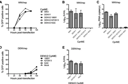
To expand the DENV2 strains available for study using this approach, we generated CprME structural gene constructs for the strains New Guinea C (NGC) and IQT2913. DENV2 NGC is the strain from which the DENV2 component of the NIAID tetrava-lent vaccine candidate was derived (59). IQT2913 is an isolate from Iquitos, Peru (60).Figure 1Ashows RVP titers generated by complementation of the WNV replicon with CprME from DENV2 NGC and IQT2913, as well as from flaviviruses previously shown to result in infectious RVPs (DENV2 16681, DENV1 West-ern Pacific-74 [WP], and the NY99 strain of WNV). Complemen-tation with CprME from WNV, DENV1, and DENV2 16681 produced comparable RVP titers that peaked⬃72 to 96 h post-transfection, similar to previous observations (34). Mean titers for WNV, DENV1, and DENV2 16681 RVPs collected at 72 h post-transfection ranged from 1⫻106to 4⫻106IU/ml (n⫽3) (Fig. 1B). Conversely, complementation of the WNV replicon with CprME from DENV2 strains NGC and IQT2913 resulted in no
detectable RVP production (n⫽3) (Fig. 1AandB). These results were surprising, as DENV2 16681 shares 98% and 97% amino acid identity with strains NGC and IQT2913, differing by 17 and 24 amino acid residues within CprME, respectively. Analysis of the RNA content of DENV2 NGC and IQT2913 RVP preparations revealed ⬎1,000-fold lower RNA content than DENV2 16681 RVP-containing supernatants produced in parallel (n⫽3) (Fig. 1C). Thus, the inability to complement the WNV replicon with structural genes from DENV2 strains NGC or IQT2913 reflected a defect prior to the release of virus particles into the supernatant.
We next performed complementation experiments with a DENV2 subgenomic replicon of similar design (DENVrep). RVPs produced using this replicon resulted in comparable titers for all three DENV2 strains (16681, NGC, and IQT2913), with mean titers ranging from 1⫻105to 5⫻105IU/ml at 120 h
posttrans-fection (n⫽3) (Fig. 1DandE). As noted previously (34), RVP production using the DENV replicon was delayed (Fig. 1Aversus
D) and less efficient (Fig. 1BversusE) compared to that in exper-iments with WNVrep.
The C protein governs DENV2 RVP production following
WNV replicon complementation.To determine the molecular
basis of the DENV2 strain-specific RVP production, we created chimeras of DENV2 16681 and DENV2 NGC structural proteins (Fig. 2A). Each chimera was evaluated in complementation exper-iments with the WNV replicon. RVP production studies revealed that all CprME chimeras containing the C gene of 16681, regard-less of the origin of the prM or E genes, resulted in robust RVP titers (mean titers of⬎105IU/ml [n⫽3]). In contrast, all CprME chimeras containing the NGC C gene resulted in undetectable RVP production (n⫽3) (Fig. 2B). These data reveal a critical role for the C protein in modulating DENV2 RVP production using a heterologous viral RNA.
The C proteins of DENV2 16681 and NGC differ at only two amino acid residues (residues 9 and 101). We created NGC vari-ants encoding a single substitution at these positions to corre-spond to the amino acid found in 16681 and evaluated them in RVP production experiments with WNVrep. Complementation with wild-type (WT) NGC and the NGC R9K variant resulted in undetectable RVP titers (n⫽3). In contrast, the T101S mutation in the NGC background resulted in RVP production similar to titers obtained with 16681 (⬎105IU/ml [n⫽3];P⫽0.36) (Fig.
2C). A similar pattern was observed following the introduction of mutation T101S into the DENV2 IQT2913 C protein (data not shown). Conversely, the reciprocal S101T mutation in the 16681 C protein rendered this construct incapable of RVP production following complementation of the WNV replicon (Fig. 2D). To-gether, these results identify C protein residue 101 as the determi-nant of compatibility between the structural genes of DENV2 and a WNV replicon RNA during virus particle production.
WNV NS2B/3 cannot cleave the DENV2 C protein when a
threonine occupies the P1=position.During polyprotein
pro-cessing, NS2B/3 cleaves the C protein immediately after residue 100, removing the C-terminal signal sequence and liberating it from its membrane anchor. C protein residue 101 identified above is located immediately downstream of the scissile bond in the P1= position of the cleavage site (Fig. 2E) and therefore has the poten-tial to impact cleavage by the WNV protease. To investigate this hypothesis, we developed an assay to measure C protein cleavage in cells harboring a replicating flavivirus subgenomic RNA. A V5 tag was inserted at the amino terminus of the C protein in DENV2
on November 7, 2019 by guest
http://jvi.asm.org/
NGC CprME expression constructs (V5-CprME), enabling quan-titation of NS2B/3 cleavage efficiency in RVP-producing cells by Western blotting. Using this system, capsid cleavage resulted in the production of a V5-tagged C protein that is considerably smaller than its uncleaved C-prM precursor (13 kDa and 33 kDa, respectively). Cleavage studies in HEK293T cells cotransfected with the WNV replicon and WT NGC CprME genes resulted in no detectable cleaved C protein in cell lysates. In contrast, partial cleavage of NGC T101S (the 16681 P1=residue) and NGC T101G (the WNV P1=residue) was observed in experiments performed in parallel (31%⫾16% and 63%⫾12%, respectively [n⫽4]) (Fig. 3AandB). These results correlated well with the ability of these structural gene constructs to produce infectious RVPs by comple-mentation (Fig. 3CversusFig. 1BandE). RVP production with the WT NGC V5-CprME construct resulted in undetectable titers, whereas NGC constructs with T101S or T101G mutations at the P1=site enabled efficient RVP release (⬎105IU/ml [n⫽2]). By
comparison, cleavage experiments using the DENV replicon
re-sulted in efficient cleavage of the C protein regardless of whether the amino acid at residue 101 was a T, S, or G (⬎90% cleaved C [n⫽4]) (Fig. 3AandB). RVP titers in the corresponding super-natants were similar for all three structural gene constructs (⬃3⫻ 103IU/ml [n⫽2]). These results demonstrated that even
chemi-cally conservative amino acid variation at the P1=position modu-lates cleavage of the DENV C protein and RVP production in cells replicating a WNV replicon.
WNV NS2B/3 efficiently cleaves the WNV C protein when a
threonine occupies the P1=position.Because a T residue in the
P1=position of the DENV2 NGC C protein cleavage site prevents processing by the WNV NS2B/3 protease, we hypothesized that introduction of a T into the P1=position (residue 106) of the WNV C protein cleavage site would have a similar phenotype. Surpris-ingly, complementation of the WNV replicon with a WNV G106T variant resulted in efficient RVP production (3⫻106IU/ml [n⫽
3]) (Fig. 4A), with only a 0.5-log reduction in titer compared to WT WNV (P⫽0.015 [n⫽3]). Likewise, a WNV G106S variant
FIG 1DENV2 strain-dependent complementation of a WNV subgenomic replicon. (A to C) RVPs were produced by complementation of a GFP-expressing WNV replicon (WNVrep) and a CprME plasmid containing the structural genes of WNV (NY99), DENV1 (WP), or DENV2 (strains 16681, NGC, and IQT2913). (A) RVPs were harvested at the times indicated posttransfection and used to infect Raji DC-SIGNR cells. Infection was scored by GFP expression 2 days postinfection using flow cytometry. Error bars represent the range of duplicate infections. The results shown are representative of three independent experiments. (B) The infectious titer of RVPs collected 72 h posttransfection was determined. Error bars represent the standard error of the mean from three independent experiments. (C) RVPs collected 72 h posttransfection were analyzed for viral RNA content by qRT-PCR. Error bars represent the standard error of the mean from three independent experiments. (D and E) RVPs were produced by complementation of a GFP-expressing DENV replicon (DENVrep) with DENV2 CprME (strains 16681, NGC, and IQT2913). (D) RVP samples collected at the indicated times posttransfection were analyzed for infectivity of Raji DC-SIGNR cells 3 days postinfection. Error bars represent the range of duplicate infections. The results shown are representative of three independent experiments. (E) The infectious titer of RVPs collected at 120 h posttransfection was determined. Error bars represent the standard error of the mean from three independent experiments.
on November 7, 2019 by guest
http://jvi.asm.org/
[image:4.585.43.542.65.395.2]also supported RVP titers similar to WT WNV (P⫽0.18 [n⫽3]). Biochemical studies revealed that the levels of WNV protease cleavage efficiency of all three constructs were similar (Fig. 4Band
C) (n⫽3;P⬎0.05 for all comparisons). Altogether, these data demonstrate that a threonine in the P1=position of the C protein cleavage site impacts WNV NS2B/3 protease efficiency in a strik-ingly context-dependent manner.
The specificity of WNV NS2B/3 at the P1=position is
influ-enced by surrounding residues.We next investigated how the
sequence surrounding the protease cleavage site affected WNV protease specificity at the P1=position. We replaced residues ad-jacent to the incompatible threonine at the P1=position of NGC capsid with those corresponding to the WNV capsid (Fig. 5A). Cleavage of each NGC C protein variant was measured by Western blotting of cells expressing the WNV replicon; the efficiently cleaved NGC T101G variant was used as a positive control in these studies. Efficient cleavage was observed when positions P4 to P2= (55%⫾9%) and P6 to P4=(86%⫾6%) were mutated (Fig. 5B
andC). Mutations at residues flanking just one side of the cleavage site were poorly cleaved (14%⫾3% and 13%⫾7% for mutants P2=-10=and P10-P2, respectively). Notably, the cleaved C protein of mutant P2=-P10=migrated more slowly during gel electropho-resis than C protein expressed from other NGC constructs (Fig. 5B). In this construct, the order of cleavage may be uncoupled by mutations within the signal sequence, which have the potential to
impact the display of the capsid-signal sequence junction and re-sult in a cleaved C protein that retains a C-terminal signal se-quence (8). As detailed above, the efficiency of C protein cleavage directly impacted the release of infectious virus particles. The rel-atively efficiently cleaved capsid variants supported virus produc-tion, including NGC T101G (9⫻105IU/ml), mutant P4=-P2=
(7⫻104IU/ml [13-fold reduction from T101G]), and mutant P6-P4=(9⫻104IU/ml [10-fold reduction from T101G]), while
low-to-undetectable titers for mutants P2=-10=and P10-P2 (⬍102 IU/ml) were achieved (Fig. 5D). These results suggest that amino acids directly surrounding the cleavage site influence the ability of WNV NS2B/3 to cleave a substrate when a T occupies the P1= position.
Our data suggested that the ability of WNV NS2B/3 to cleave DENV C proteins with a threonine at the P1=position can be influenced by residues between P4 and P2=. Therefore, we con-structed NGC C protein variants with single amino acid substitu-tions (at the P4, P3, P2, or P2=position) to determine if individual substitutions influence WNV NS2B/3 specificity. Unlike the effi-cient C protein cleavage detected for NGC mutant P4-P2= encod-ing four mutations (Fig. 5C), minimal cleavage of variants with a single mutation at either P4, P3, P2, or P2=was observed (n⫽3,
P⬎0.25 for WT NGC versus each mutant) (Fig. 5EandF). No-tably, among these constructs, the C protein of the P2=mutant was most efficiently cleaved (10%⫾3%) (Fig. 5F) and supported RVP
FIG 2Genetic basis of DENV2 strain-dependent complementation of the WNV subgenomic replicon. (A) Schematics of DENV2 16681/NGC CprME chimeras, with genes from 16681 in gray and genes from NGC in a stippled pattern. Locations of amino acid sequence variation between the two strains are indicated by “⫹” marks. (B to D) RVPs were produced by WNVrep complementation with the DENV2 CprME construct indicated on thexaxis. The infectious RVP titer was determined at 72 h posttransfection; error bars represent the standard error of the mean from three independent experiments. Infectious RVP titers are shown for DENV2 16681, NGC, and the 16681/NGC CprME chimeras shown in panel A (B), DENV2 16681, NGC, and NGC containing point mutations R9K or T101S in the C protein (C), and DENV2 16681 and 16681 C protein mutant S101T (D). (E) Alignment of sequences surrounding the NS2B/3 cleavage site in the C protein for DENV2 strains 16681, NGC, and IQT2913.
on November 7, 2019 by guest
http://jvi.asm.org/
[image:5.585.40.543.64.357.2]production (5⫻104IU/ml [a 51-fold reduction from T101G]) (Fig. 5G) to levels similar to that observed when multiple residues were changed (mutants P6-P4=and P4-P2=) (Fig. 5D).
The difference in cleavage efficiencies of the P6-P4=and P4-P2= variants (Fig. 5D) suggests a role for residues farther from the scissile bond studied above. Of interest, the P5 and P3=residues differ in charge between WNV and DENV. We constructed addi-tional NGC C protein variants with single amino acid substitu-tions at the P6, P5, P3=, and P4=positions and evaluated the effi-ciency of C protein cleavage following complementation with the WNV replicon. Complementation with mutants P6 and P4= re-sulted in inefficient cleavage (4%⫾1% and 9%⫾5%, respec-tively) (Fig. 5HandI) and low but detectable RVP titers (7⫻103 IU/ml and 3⫻104IU/ml; 59- and 15-fold reductions from T101G
titers for mutants P6 and P4=, respectively) (Fig. 5J). For mutants P5 and P3=, neither cleaved C protein nor infectious RVPs were detectable (Fig. 5HtoJ).
Altogether, these results indicate that the interactions that me-diate WNV NS2B/3 specificity for the P1=site involve multiple surrounding residues. Mutations at the P6, P2=, and P4=positions individually had a minor effect on C protein cleavage compared to multiple mutations at the P6 to P4=and P4 to P2=positions.
DISCUSSION
Little is known about the specificity of the flavivirus NS2B/3 pro-tease in the context of viral RNA replication or authentic viral polyprotein substrates (61). Most studies to define protease cleav-age site specificity employ short (3- to 8-amino-acid) synthetic
FIG 3Cleavage of the C protein of DENV2 NGC T101 mutants by WNV and DENV NS2B/3. An amino-terminal V5 tag was added to the C proteins of WT NGC, NGC T101S, and NGC T101G CprME constructs. The V5-tagged CprME constructs were used in complementation experiments with WNVrep and DENVrep. Cell lysates were harvested at 48 h posttransfection and analyzed by SDS-PAGE and Western blotting with an anti-V5 MAb. (A) A representative blot is shown. The lower band corresponds to cleaved C (⬃13 kDa), while the upper band corresponds to uncleaved CprM (⬃33 kDa). (B) Mean cleavage efficiency of the C protein from four independent experiments. Error bars represent the standard error of the mean. (C) Mean infectious titer of RVPs harvested prior to cell lysate collection (48 h posttransfection). Error bars represent the range from two independent experiments.
FIG 4Cleavage of the C protein of WNV G106 mutants by WNV NS2B/3. RVPs were produced by complementation of WNVrep with CprME derived from wild-type WNV (WT) or from WNV containing mutations at C protein residue G106 (the P1=position of the WNV C protein cleavage site). (A) Shown is the mean infectious RVP titer of supernatant collected 72 h posttransfection; error bars represent the standard error of the mean for three independent experiments. (B and C) An amino-terminal V5 tag was added to the C protein in the WNV CprME constructs (WT, G106S, and G106T). Following complementation of WNVrep with the WNV V5-tagged CprME constructs, cell lysates were collected at 48 h posttransfection and analyzed by SDS-PAGE and Western blotting with an anti-V5 MAb. (B) A representative blot is shown. The minor mobility shift for the band corresponding to the C protein of G106T was not observed in all independent repeats. (C) Mean cleavage efficiency of the WNV C protein; error bars represent the standard error of the mean from three independent experiments.
on November 7, 2019 by guest
http://jvi.asm.org/
[image:6.585.115.474.69.286.2]peptide substrates and covalently linked and truncated forms of NS2B and NS3 (25–33). Furthermore, these experiments are per-formed in the absence of membranes, at basic pH (⬃pH 9), and in the presence of detergents and high concentrations of glycerol.
These biochemical approaches have identified a requirement for dibasic residues at P1 and P2 and a small side chain at the P1= position of the cleavage site, although preferences for particular residues within that motif exist, and distinct specificities have been
FIG 5Effect of mutations surrounding C protein residue T101 on WNV NS2B/3 cleavage of the NGC C protein. (A) Sequence alignment of the C protein cleavage site of WNV, DENV2 NGC, and DENV2 NGC mutants. Highlighted in gray is the P1=residue; highlighted in black are the substitutions from the WNV cleavage site introduced into each variant. The numbering at the top corresponds to the location in the DENV2 NGC C protein amino acid sequence. (B to J) V5-tagged CprME constructs of DENV2 NGC containing a mutation at the indicated C protein cleavage site were used in complementation experiments with WNVrep. Cell lysates were collected and analyzed by SDS-PAGE and subjected to Western blotting with an anti-V5 MAb. For the NGC variants from panel A, shown is a representative blot (B), with the mean efficiency of C protein cleavage (C) and mean infectious RVP titer (D) from four independent experiments. Error bars represent the standard error of the mean. For mutants P4, P3, P2, and P2=, shown is a representative blot (E), with the mean efficiency of C protein cleavage (F) and mean infectious RVP titer (G) from three independent experiments. Error bars represent the standard error of the mean. For mutants P6, P5, P3=, and P4=, shown is a representative blot (H), with the mean efficiency of C protein cleavage (I) and mean infectious RVP titer (J) from three independent experiments. Error bars represent the standard error of the mean.
on November 7, 2019 by guest
http://jvi.asm.org/
[image:7.585.39.547.66.578.2]noted between viruses. WNV NS2B/3 has been shown to prefer-entially cleave sites with a K at P2, whereas DENV NS2B/3 most efficiently cleaves sites with an R at this position. In contrast, WNV and DENV NS2B/3 proteases both preferentially cleave sites with an R over a K at the P1 position (25,33,62). These studies have indicated that WNV NS2B/3 has a strict preference for G at both the P1=and P2=positions, while DENV NS2B/3 is less selec-tive and can cleave substrates with almost any amino acid at P1= and P2=(33). Another study, however, observed a narrower spec-ificity of the DENV protease at the P1=position (restricted to small, polar amino acids with a strong preference for S) and at the P2=position (a weak preference for acidic residues) (62).
How substrate residues outside the active site recognition site impact cleavage efficiency has not been well explored for flavivirus proteases. Allosteric and subsite cooperativities have the potential to influence protease substrate specificity (reviewed by Ng et al. [63]). Allosteric cooperativity occurs when the binding of sub-strate residues to a protease outside the active site affects proteo-lytic efficiency. Subsite cooperativity posits that individual subsite binding may be positively or negatively influenced by binding of surrounding subsites (reviewed in reference63). Both processes have been shown to contribute to HIV-1 protease recognition (64–67). As discussed above, prior studies of the WNV NS2B/3 employed short peptide substrates that do not extend significantly beyond the active site, potentially limiting their utility for identi-fying all of the interactions that impact protease specificity.
To investigate the effect of cleavage site context on WNV NS2B/3 substrate specificity, we studied WNV NS2B/3 processing of DENV and WNV C proteins. WNV NS2B/3 cleaved the DENV2 C protein when a G, but not a T, occupied the P1=position. In contrast, when the P1=residue of the WNV C protein cleavage site was mutated from a G to a T, we observed efficient WNV NS2B/3 cleavage, indicating that a T at P1=is permissive for cleavage only in the context of certain substrates. This context-dependent cleav-age specificity could be a result of either allosteric or subsite coop-erativity (or both), as the WNV and DENV2 C proteins have se-quence differences at residues both within and outside the protease binding site.
To study the effect of subsite cooperativity on WNV protease specificity, we introduced mutations in a DENV2 CprME con-struct at residues surrounding the C protein cleavage site. While keeping a T constant at the P1=position (which results in un-cleaved C protein in the context of the WT DENV2 NGC se-quence), surrounding residues were mutated to match the WNV C protein cleavage site. We found that WNV NS2B/3 was able to efficiently cleave the DENV2 C protein containing a T at the P1= position when the P4, P3, P2, and P2=positions were mutated simultaneously. When the P6, P5, P3=, and P4=positions were additionally mutated, cleavage efficiency was further enhanced. Thus, the sequence context within the active site binding site of the substrate greatly impacts WNV NS2B/3 specificity for the P1= res-idue. However, when the P6, P5, P4, P3, P2, P2=, P3=, and P4= mutations were introduced separately, their effect on the effi-ciency of DENV2 C protein cleavage by the WNV protease was greatly diminished. Thus, the interactions that mediate the con-text-dependent specificity of WNV NS2B/3 at the P1=position are likely due to the influence of multiple substrate residues.
Notably, onein vitrostudy observed strict specificity of WNV NS2B/3 for a G at P1=(33). This finding is not consistent with the observation that, while most WNV NS2B/3 cleavage sites do
con-tain a G at P1=, the P1=position of the cleavage site at the NS3/ NS4A junction is occupied by an S. It also contradicts our finding that the WNV and DENV2 C proteins were cleaved by the WNV protease when an S occupied the P1=position. Several differences in experimental design between the studies could account for this discrepancy, including assay conditions (biochemical versus cell-based assay), NS2B/3 source (recombinant, truncated NS2B/3 versus full-length NS2B/3 from the polyprotein), and the sub-strate used to probe specificity (9-mer peptides versus a CprME polyprotein).
Structural basis for WNV NS2B/3 substrate specificity.
Effi-cient peptide cleavage requires accurate placement of the scissile peptide bond in the active site. A view of substrate binding is available in crystal structures for inhibitor-bound WNV NS2B/3 (13,68,69) and DENV NS2B/3 (70). The pocket in NS2B/3 occu-pied by the substrate P1=side chain (the S1=subsite) is lined by the side chains of NS3 residue 36 (DENV I36, WNV A36) and the catalytic histidine (NS3 H51) (13). In structures of both WNV and DENV NS2B/3, the P1=side chain of a protein inhibitor binds identically to the small and relatively hydrophobic S1=subsite (13,
70). In contrast, the S2=subsite for the substrate P2=side chain is a large, open pocket on the enzyme surface between the side chains of NS3 residues 34 and 131. Here the structures differ (NS3 Y34 in WNV versus T34 in DENV; NS3 P131 in WNV versus K131 in DENV). The structures are consistent with the data presented herein that substrate sequences flanking the cleavage site play a role in specificity (70). Our data are also consistent with other observations that NS3 positions 131 and 132 contribute to speci-ficity of the P2=position, as mutation of WNV NS3 P131 and NS3 T132 to match the DENV sequence (P131K plus T132P) shifted the specificity of WNV NS2B/3 to that of DENV NS2B/3 (71). Interactions between protease and substrate on the non-prime site of the scissile bond involve the S2 and S3 pockets of the active site and are occupied by the P2 and P3 positions of the substrate, respectively. Like the situation for the substrate P1=site, the sub-strate P2 and P3 side chains bind WNV and DENV NS3 identically in the S2 and S3 subsites (13,69). Substrate-enzyme interactions well beyond the scissile bond and/or cooperativity of subsites are clearly critical.
The subsite cooperativity observed in our study may be a result of restrictions on substrate size by WNV NS2B/3, where the WNV NS2B/3 preference for the smaller substrate residues at P1=(G⬎ T) and P2 (K⬎R) can be overcome by the introduction of smaller residues at neighboring positions. We have shown that WNV NS2B/3 cleavage of substrates with a T in the P1=position is de-pendent on the surrounding residues. Notably, when the P4, P3, P2, and P2=positions were mutated to the smaller but chemically similar amino acids found in the WNV C protein cleavage site, WNV NS2B/3 could cleave the DENV2 NGC C protein substrate with a P1=threonine.
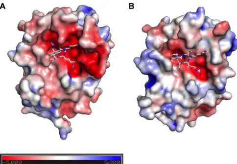
Our data together with the available structures show that proper positioning of the peptide substrate is influenced by NS3 interaction with flanking residues at a distance of up to six N-ter-minal positions and four C-terN-ter-minal positions of the target pep-tide bond. Although partial activity can be restored with a single substitution at the P1=site, efficient cleavage of natural substrate requires multiple substrate-enzyme interactions. Consistent with our data, WNV and DENV NS2B/3 structures have strikingly dif-ferent electrostatic surface potentials near the active site (Fig. 6). WNV NS2B/3 has a more negatively charged surface in the region
on November 7, 2019 by guest
http://jvi.asm.org/
surrounding the substrate entrance, while DENV NS2B/3 has a more electropositive surface. The more negative surface of WNV NS2B/3 outside the active site may account for the increased cleav-age efficiency we observed when the P6, P5, P3=, and P4=positions were mutated in addition to the P4 to P2=positions in NGC mu-tant P6 to P4=, as the P5 and P3=positions now contained posi-tively charged lysine residues. While individual mutations at P5 and P3=did not have an effect on WNV NS2B/3 cleavage of the NGC C protein, cooperative effects from multiple residues sur-rounding the cleavage site could mediate cleavage.
Our results indicate that subsite cooperativity involving mul-tiple residues within the P6 to P2 and P2=to P4=positions influ-ences the specificity of the WNV protease at the P1=position. This finding has implications for the design of experiments to probe NS2B/3 substrate specificity, as the substrate selected for specific-ity profiling may drastically affect the results of the screen. Fur-thermore, molecular details of substrate specificity can be inte-grated with structural information to facilitate rational inhibitor design.
ACKNOWLEDGMENTS
We thank Heather D. Hickman, Leslie Goo, and Richard J. Kuhn for critical reading of the manuscript.
This work was supported by the Intramural Research Program of the National Institute of Allergy and Infectious Diseases, NIH, to T.C.P. and NIH grant P01 AI-055672 to J.L.S. L.A.V. was additionally supported by supported by NIH Institutional Training grant 2T32AI051967-06A1 awarded to the University of Maryland.
REFERENCES
1.Messina JP, Brady OJ, Pigott DM, Golding N, Kraemer MU, Scott TW, Wint GR, Smith DL, Hay SI.2015. The many projected futures of dengue. Nat Rev Microbiol13:230 –239.http://dx.doi.org/10.1038 /nrmicro3430.
2.Suthar MS, Diamond MS, Gale M, Jr.2013. West Nile virus infection and immunity. Nat Rev Microbiol 11:115–128. http://dx.doi.org/10.1038 /nrmicro2950.
3.Kuhn RJ, Zhang W, Rossmann MG, Pletnev SV, Corver J, Lenches E, Jones CT, Mukhopadhyay S, Chipman PR, Strauss EG, Baker TS, Strauss JH.2002. Structure of dengue virus: implications for flavivirus
organization, maturation, and fusion. Cell108:717–725.http://dx.doi.org /10.1016/S0092-8674(02)00660-8.
4.Lobigs M.1993. Flavivirus premembrane protein cleavage and spike het-erodimer secretion require the function of the viral proteinase NS3. Proc Natl Acad Sci U S A90:6218 – 6222.http://dx.doi.org/10.1073/pnas.90.13 .6218.
5.Amberg SM, Nestorowicz A, McCourt DW, Rice CM.1994. NS2B-3 proteinase-mediated processing in the yellow fever virus structural region: in vitro and in vivo studies. J Virol68:3794 –3802.
6.Amberg SM, Rice CM.1999. Mutagenesis of the NS2B-NS3-mediated cleavage site in the flavivirus capsid protein demonstrates a requirement for coordinated processing. J Virol73:8083– 8094.
7.Stocks CE, Lobigs M.1998. Signal peptidase cleavage at the flavivirus C-prM junction: dependence on the viral NS2B-3 protease for efficient processing requires determinants in C, the signal peptide, and prM. J Virol 72:2141–2149.
8.Lobigs M, Lee E, Ng ML, Pavy M, Lobigs P.2010. A flavivirus signal peptide balances the catalytic activity of two proteases and thereby facilitates virus morphogenesis. Virology401:80 – 89.http://dx.doi.org/10.1016/j.virol.2010 .02.008.
9.Chambers TJ, Grakoui A, Rice CM.1991. Processing of the yellow fever virus nonstructural polyprotein: a catalytically active NS3 proteinase do-main and NS2B are required for cleavages at dibasic sites. J Virol65:6042– 6050.
10. Chambers TJ, Weir RC, Grakoui A, McCourt DW, Bazan JF, Fletterick RJ, Rice CM.1990. Evidence that the N-terminal domain of nonstructural protein NS3 from yellow fever virus is a serine protease responsible for site-specific cleavages in the viral polyprotein. Proc Natl Acad Sci U S A 87:8898 – 8902.http://dx.doi.org/10.1073/pnas.87.22.8898.
11. Falgout B, Pethel M, Zhang YM, Lai CJ. 1991. Both nonstructural proteins NS2B and NS3 are required for the proteolytic processing of dengue virus nonstructural proteins. J Virol65:2467–2475.
12. Clum S, Ebner KE, Padmanabhan R.1997. Cotranslational membrane insertion of the serine proteinase precursor NS2B-NS3(Pro) of dengue virus type 2 is required for efficient in vitro processing and is mediated through the hydrophobic regions of NS2B. J Biol Chem272:30715–30723. http://dx.doi.org/10.1074/jbc.272.49.30715.
13. Aleshin AE, Shiryaev SA, Strongin AY, Liddington RC.2007. Structural evidence for regulation and specificity of flaviviral proteases and evolution of theFlaviviridaefold. Protein Sci16:795– 806.http://dx.doi.org/10.1110 /ps.072753207.
14. Lin C, Amberg SM, Chambers TJ, Rice CM.1993. Cleavage at a novel site in the NS4A region by the yellow fever virus NS2B-3 proteinase is a pre-requisite for processing at the downstream 4A/4B signalase site. J Virol 67:2327–2335.
15. Zhang L, Mohan PM, Padmanabhan R.1992. Processing and localiza-tion of dengue virus type 2 polyprotein precursor NS3-NS4A-NS4B-NS5. J Virol66:7549 –7554.
16. Lim SP, Shi PY.2013. West Nile virus drug discovery. Viruses5:2977– 3006.http://dx.doi.org/10.3390/v5122977.
17. Kohli A, Shaffer A, Sherman A, Kottilil S.2014. Treatment of hepatitis C: a systematic review. JAMA 312:631– 640.http://dx.doi.org/10.1001 /jama.2014.7085.
18. Nitsche C, Holloway S, Schirmeister T, Klein CD.30 September 2014. Biochemistry and medicinal chemistry of the dengue virus protease. Chem Revhttp://dx.doi.org/10.1021/cr500233q.
19. Mueller NH, Pattabiraman N, Ansarah-Sobrinho C, Viswanathan P, Pierson TC, Padmanabhan R.2008. Identification and biochemical char-acterization of small-molecule inhibitors of West Nile virus serine pro-tease by a high-throughput screen. Antimicrob Agents Chemother52: 3385–3393.http://dx.doi.org/10.1128/AAC.01508-07.
20. Yang CC, Hsieh YC, Lee SJ, Wu SH, Liao CL, Tsao CH, Chao YS, Chern JH, Wu CP, Yueh A.2011. Novel dengue virus-specific NS2B/NS3 pro-tease inhibitor, BP2109, discovered by a high-throughput screening as-say. Antimicrob Agents Chemother 55:229 –238. http://dx.doi.org/10 .1128/AAC.00855-10.
21. Tomlinson SM, Watowich SJ. 2012. Use of parallel validation high-throughput screens to reduce false positives and identify novel dengue NS2B-NS3 protease inhibitors. Antiviral Res93:245–252.http://dx.doi .org/10.1016/j.antiviral.2011.12.003.
22. Stoermer MJ, Chappell KJ, Liebscher S, Jensen CM, Gan CH, Gupta PK, Xu WJ, Young PR, Fairlie DP.2008. Potent cationic inhibitors of West Nile virus NS2B/NS3 protease with serum stability, cell permeability FIG 6Electrostatic surface potential of the WNV and DENV proteases. The
surfaces of inhibitor-bound WNV NS2B/3 (PDB 3E90) (69) (A) and DENV NS2B/3 (PDB 3U1I) (70) (B) are colored according to the electrostatic surface potential from the negative (red,⫺5 kT) to the positive (blue,⫹5 kT).
on November 7, 2019 by guest
http://jvi.asm.org/
[image:9.585.41.283.65.232.2]and antiviral activity. J Med Chem51:5714 –5721.http://dx.doi.org/10 .1021/jm800503y.
23. Shiryaev SA, Ratnikov BI, Chekanov AV, Sikora S, Rozanov DV, Godzik A, Wang J, Smith JW, Huang Z, Lindberg I, Samuel MA, Diamond MS, Strongin AY.2006. Cleavage targets and theD -arginine-based inhibitors of the West Nile virus NS3 processing proteinase. Biochem J393:503–511.http://dx.doi.org/10.1042/BJ20051374. 24. Schechter I, Berger A.1967. On the size of the active site in proteases. I.
Papain. Biochem Biophys Res Commun27:157–162.http://dx.doi.org/10 .1016/S0006-291X(67)80055-X.
25. Niyomrattanakit P, Yahorava S, Mutule I, Mutulis F, Petrovska R, Prusis P, Katzenmeier G, Wikberg JE.2006. Probing the substrate spec-ificity of the dengue virus type 2 NS3 serine protease by using internally quenched fluorescent peptides. Biochem J397:203–211.http://dx.doi.org /10.1042/BJ20051767.
26. Chappell KJ, Stoermer MJ, Fairlie DP, Young PR.2006. Insights to substrate binding and processing by West Nile virus NS3 protease through combined modeling, protease mutagenesis, and kinetic studies. J Biol Chem281:38448 –38458.http://dx.doi.org/10.1074/jbc.M607641200. 27. Mueller NH, Yon C, Ganesh VK, Padmanabhan R.2007.
Characteriza-tion of the West Nile virus protease substrate specificity and inhibitors. Int J Biochem Cell Biol39:606 – 614.http://dx.doi.org/10.1016/j.biocel.2006 .10.025.
28. Kondo MY, Oliveira LC, Okamoto DN, de Araujo MR, Duarte dos Santos CN, Juliano MA, Juliano L, Gouvea IE.2011. Yellow fever virus NS2B/NS3 protease: hydrolytic properties and substrate specificity. Biochem Biophys Res Commun407:640 – 644.http://dx.doi.org/10.1016 /j.bbrc.2011.03.054.
29. Ang MJ, Li Z, Lim HA, Ng FM, Then SW, Wee JL, Joy J, Hill J, Chia CS. 2014. A P2 and P3 substrate specificity comparison between the Murray Valley encephalitis and West Nile virus NS2B/NS3 protease using C-ter-minal agmatine dipeptides. Peptides52:49 –52.http://dx.doi.org/10.1016 /j.peptides.2013.12.002.
30. Marcon L, Kozak D, Battersby BJ, Chappell KJ, Fairlie DP, Young P, Trau M.2008. A dual-purpose synthetic colloidal platform for protease mapping: substrate profiling for dengue and West Nile virus proteases. Anal Biochem376:151–153.http://dx.doi.org/10.1016/j.ab.2008.01.034. 31. Yusof R, Clum S, Wetzel M, Murthy HM, Padmanabhan R. 2000.
Purified NS2B/NS3 serine protease of dengue virus type 2 exhibits cofac-tor NS2B dependence for cleavage of substrates with dibasic amino acids in vitro. J Biol Chem275:9963–9969.http://dx.doi.org/10.1074/jbc.275.14 .9963.
32. Nall TA, Chappell KJ, Stoermer MJ, Fang NX, Tyndall JD, Young PR, Fairlie DP.2004. Enzymatic characterization and homology model of a catalytically active recombinant West Nile virus NS3 protease. J Biol Chem 279:48535– 48542.http://dx.doi.org/10.1074/jbc.M406810200. 33. Shiryaev SA, Kozlov IA, Ratnikov BI, Smith JW, Lebl M, Strongin AY.
2007. Cleavage preference distinguishes the two-component NS2B-NS3 serine proteinases of dengue and West Nile viruses. Biochem J401:743– 752.http://dx.doi.org/10.1042/BJ20061136.
34. Ansarah-Sobrinho C, Nelson S, Jost CA, Whitehead SS, Pierson TC. 2008. Temperature-dependent production of pseudoinfectious dengue reporter virus particles by complementation. Virology381:67–74.http: //dx.doi.org/10.1016/j.virol.2008.08.021.
35. Pierson TC, Sanchez MD, Puffer BA, Ahmed AA, Geiss BJ, Valentine LE, Altamura LA, Diamond MS, Doms RW.2006. A rapid and quanti-tative assay for measuring antibody-mediated neutralization of West Nile virus infection. Virology 346:53– 65. http://dx.doi.org/10.1016/j.virol .2005.10.030.
36. VanBlargan LA, Mukherjee S, Dowd KA, Durbin AP, Whitehead SS, Pierson TC.2013. The type-specific neutralizing antibody response elic-ited by a dengue vaccine candidate is focused on two amino acids of the envelope protein. PLoS Pathog 9:e1003761. http://dx.doi.org/10.1371 /journal.ppat.1003761.
37. Geiss BJ, Pierson TC, Diamond MS.2005. Actively replicating West Nile virus is resistant to cytoplasmic delivery of siRNA. Virol J2:53.http://dx .doi.org/10.1186/1743-422X-2-53.
38. Baker NA, Sept D, Joseph S, Holst MJ, McCammon JA.2001. Electro-statics of nanosystems: application to microtubules and the ribosome. Proc Natl Acad Sci U S A98:10037–10041.http://dx.doi.org/10.1073/pnas .181342398.
39. Dolinsky TJ, Czodrowski P, Li H, Nielsen JE, Jensen JH, Klebe G, Baker NA.2007. PDB2PQR: expanding and upgrading automated preparation
of biomolecular structures for molecular simulations. Nucleic Acids Res 35:W522–W525.http://dx.doi.org/10.1093/nar/gkm276.
40. Dolinsky TJ, Nielsen JE, McCammon JA, Baker NA.2004. PDB2PQR: an automated pipeline for the setup of Poisson-Boltzmann electrostatics calculations. Nucleic Acids Res 32:W665–W667. http://dx.doi.org/10 .1093/nar/gkh381.
41. Schrodinger LLC.2010. The PyMOL molecular graphics system, version 1.3r1.https://www.pymol.org.
42. Khromykh AA, Varnavski AN, Westaway EG.1998. Encapsidation of the flavivirus Kunjin replicon RNA by using a complementation system pro-viding Kunjin virus structural proteins in trans. J Virol72:5967–5977. 43. Davis CW, Mattei LM, Nguyen HY, Ansarah-Sobrinho C, Doms RW,
Pierson TC.2006. The location of asparagine-linked glycans on West Nile virions controls their interactions with CD209 (dendritic cell-specific ICAM-3 grabbing nonintegrin). J Biol Chem281:37183–37194.http://dx .doi.org/10.1074/jbc.M605429200.
44. Nelson S, Poddar S, Lin TY, Pierson TC.2009. Protonation of individual histidine residues is not required for the pH-dependent entry of West Nile virus: evaluation of the “histidine switch” hypothesis. J Virol83:12631– 12635.http://dx.doi.org/10.1128/JVI.01072-09.
45. de Wispelaere M, Yang PL.2012. Mutagenesis of the DI/DIII linker in dengue virus envelope protein impairs viral particle assembly. J Virol86: 7072–7083.http://dx.doi.org/10.1128/JVI.00224-12.
46. Zheng A, Yuan F, Kleinfelter LM, Kielian M. 2014. A toggle switch controls the low pH-triggered rearrangement and maturation of the den-gue virus envelope proteins. Nat Commun5:3877.http://dx.doi.org/10 .1038/ncomms4877.
47. Christian EA, Kahle KM, Mattia K, Puffer BA, Pfaff JM, Miller A, Paes C, Davidson E, Doranz BJ.2013. Atomic-level functional model of den-gue virus envelope protein infectivity. Proc Natl Acad Sci U S A110: 18662–18667.http://dx.doi.org/10.1073/pnas.1310962110.
48. Hsieh SC, Wu YC, Zou G, Nerurkar VR, Shi PY, Wang WK.2014. Highly conserved residues in the helical domain of dengue virus type 1 precursor membrane protein are involved in assembly, precursor mem-brane (prM) protein cleavage, and entry. J Biol Chem289:33149 –33160. http://dx.doi.org/10.1074/jbc.M114.610428.
49. Pierson TC, Xu Q, Nelson S, Oliphant T, Nybakken GE, Fremont DH, Diamond MS.2007. The stoichiometry of antibody-mediated neutraliza-tion and enhancement of West Nile virus infecneutraliza-tion. Cell Host Microbe 1:135–145.http://dx.doi.org/10.1016/j.chom.2007.03.002.
50. Nelson S, Jost CA, Xu Q, Ess J, Martin JE, Oliphant T, Whitehead SS, Durbin AP, Graham BS, Diamond MS, Pierson TC.2008. Maturation of West Nile virus modulates sensitivity to antibody-mediated neutraliza-tion. PLoS Pathog 4:e1000060. http://dx.doi.org/10.1371/journal.ppat .1000060.
51. Mattia K, Puffer BA, Williams KL, Gonzalez R, Murray M, Sluzas E, Pagano D, Ajith S, Bower M, Berdougo E, Harris E, Doranz BJ.2011. Dengue reporter virus particles for measuring neutralizing antibodies against each of the four dengue serotypes. PLoS One6:e27252.http://dx .doi.org/10.1371/journal.pone.0027252.
52. de Wispelaere M, LaCroix AJ, Yang PL. 2013. The small molecules AZD0530 and dasatinib inhibit dengue virus RNA replication via Fyn kinase. J Virol87:7367–7381.http://dx.doi.org/10.1128/JVI.00632-13. 53. Scaturro P, Trist IM, Paul D, Kumar A, Acosta EG, Byrd CM, Jordan
R, Brancale A, Bartenschlager R.2014. Characterization of the mode of action of a potent dengue virus capsid inhibitor. J Virol88:11540 –11555. http://dx.doi.org/10.1128/JVI.01745-14.
54. Harvey TJ, Liu WJ, Wang XJ, Linedale R, Jacobs M, Davidson A, Le TT, Anraku I, Suhrbier A, Shi PY, Khromykh AA.2004. Tetracycline-inducible packaging cell line for production of flavivirus replicon particles. J Virol78: 531–538.http://dx.doi.org/10.1128/JVI.78.1.531-538.2004.
55. Whitby K, Pierson TC, Geiss B, Lane K, Engle M, Zhou Y, Doms RW, Diamond MS.2005. Castanospermine, a potent inhibitor of dengue virus infection in vitro and in vivo. J Virol79:8698 – 8706.http://dx.doi.org/10 .1128/JVI.79.14.8698-8706.2005.
56. Davis CW, Nguyen HY, Hanna SL, Sanchez MD, Doms RW, Pierson TC.2006. West Nile virus discriminates between SIGN and DC-SIGNR for cellular attachment and infection. J Virol80:1290 –1301.http: //dx.doi.org/10.1128/JVI.80.3.1290-1301.2006.
57. Ishikawa T, Yamanaka A, Konishi E.2014. A review of successful flavi-virus vaccines and the problems with those flaviflavi-viruses for which vaccines are not yet available. Vaccine32:1326 –1337.http://dx.doi.org/10.1016/j .vaccine.2014.01.040.
on November 7, 2019 by guest
http://jvi.asm.org/
58. Mukherjee S, Dowd KA, Manhart CJ, Ledgerwood JE, Durbin AP, Whitehead SS, Pierson TC.2014. Mechanism and significance of cell type-dependent neutralization of flaviviruses. J Virol88:7210 –7220.http: //dx.doi.org/10.1128/JVI.03690-13.
59. Whitehead SS, Hanley KA, Blaney JE, Jr, Gilmore LE, Elkins WR, Murphy BR.2003. Substitution of the structural genes of dengue virus type 4 with those of type 2 results in chimeric vaccine candidates which are attenuated for mosquitoes, mice, and rhesus monkeys. Vaccine21:4307– 4316.http://dx.doi.org/10.1016/S0264-410X(03)00488-2.
60. Armstrong PM, Rico-Hesse R.2003. Efficiency of dengue serotype 2 virus strains to infect and disseminate inAedes aegypti. Am J Trop Med Hyg 68:539 –544.
61. Condotta SA, Martin MM, Boutin M, Jean F.2010. Detection and in-cell selectivity profiling of the full-length West Nile virus NS2B/NS3 serine protease using membrane-anchored fluorescent substrates. Biol Chem 391:549 –559.http://dx.doi.org/10.1515/BC.2010.051.
62. Li J, Lim SP, Beer D, Patel V, Wen D, Tumanut C, Tully DC, Williams JA, Jiricek J, Priestle JP, Harris JL, Vasudevan SG. 2005. Functional profiling of recombinant NS3 proteases from all four serotypes of dengue virus using tetrapeptide and octapeptide substrate libraries. J Biol Chem 280:28766 –28774.http://dx.doi.org/10.1074/jbc.M500588200. 63. Ng NM, Pike RN, Boyd SE. 2009. Subsite cooperativity in protease
specificity. Biol Chem390:401– 407.http://dx.doi.org/10.1515/BC.2009 .065.
64. Tozser J, Bagossi P, Weber IT, Louis JM, Copeland TD, Oroszlan S. 1997. Studies on the symmetry and sequence context dependence of the HIV-1 proteinase specificity. J Biol Chem272:16807–16814.http://dx.doi .org/10.1074/jbc.272.27.16807.
65. Tozser J, Weber IT, Gustchina A, Blaha I, Copeland TD, Louis JM, Oroszlan S.1992. Kinetic and modeling studies of S3-S3=subsites of HIV proteinases. Bio-chemistry31:4793–4800.http://dx.doi.org/10.1021/bi00135a008.
66. Ridky TW, Cameron CE, Cameron J, Leis J, Copeland T, Wlodawer A, Weber IT, Harrison RW.1996. Human immunodeficiency virus, type 1 protease substrate specificity is limited by interactions between substrate amino acids bound in adjacent enzyme subsites. J Biol Chem271:4709 – 4717.http://dx.doi.org/10.1074/jbc.271.9.4709.
67. Pettit SC, Simsic J, Loeb DD, Everitt L, Hutchison CA, III, Swanstrom R.1991. Analysis of retroviral protease cleavage sites reveals two types of cleavage sites and the structural requirements of the P1 amino acid. J Biol Chem266:14539 –14547.
68. Erbel P, Schiering N, D’Arcy A, Renatus M, Kroemer M, Lim SP, Yin Z, Keller TH, Vasudevan SG, Hommel U.2006. Structural basis for the activation of flaviviral NS3 proteases from dengue and West Nile virus. Nat Struct Mol Biol13:372–373.http://dx.doi.org/10.1038/nsmb1073. 69. Robin G, Chappell K, Stoermer MJ, Hu SH, Young PR, Fairlie DP,
Martin JL.2009. Structure of West Nile virus NS3 protease: ligand stabi-lization of the catalytic conformation. J Mol Biol385:1568 –1577.http: //dx.doi.org/10.1016/j.jmb.2008.11.026.
70. Noble CG, Seh CC, Chao AT, Shi PY.2012. Ligand-bound structures of the dengue virus protease reveal the active conformation. J Virol86:438 – 446.http://dx.doi.org/10.1128/JVI.06225-11.
71. Shiryaev SA, Ratnikov BI, Aleshin AE, Kozlov IA, Nelson NA, Lebl M, Smith JW, Liddington RC, Strongin AY.2007. Switching the substrate specificity of the two-component NS2B-NS3 flavivirus proteinase by structure-based mutagenesis. J Virol81:4501– 4509.http://dx.doi.org/10 .1128/JVI.02719-06.