Mapping an Adeno-associated Virus 9-Specific Neutralizing
Epitope To Develop Next-Generation Gene Delivery Vectors
April R. Giles,aLakshmanan Govindasamy,aSuryanarayan Somanathan,aJames M. Wilsona
aGene Therapy Program, Department of Medicine, University of Pennsylvania, Philadelphia, Pennsylvania, USA
ABSTRACT Recent clinical trials have demonstrated the potential of adeno-associated virus (AAV)-based vectors for treating rare diseases. However, significant barriers remain for the translation of these vectors into widely available therapies. In particular, exposure to the AAV capsid can generate an immune response of neutral-izing antibodies. One approach to overcome this response is to map the AAV-specific neutralizing epitopes and rationally design an AAV capsid able to evade neutralization. To accomplish this, we isolated a monoclonal antibody against AAV9 following immunization of BALB/c mice and hybridoma screening. This antibody, PAV9.1, is specific for intact AAV9 capsids and has a high neutralizing titer of ⬎1: 160,000. We used cryo-electron microscopy to reconstruct PAV9.1 in complex with AAV9. We then mapped its epitope to the 3-fold axis of symmetry on the capsid, specifically to residues 496-NNN-498 and 588-QAQAQT-592. Capsid mutagenesis demonstrated that even a single amino acid substitution within this epitope mark-edly reduced binding and neutralization by PAV9.1. In addition, in vivo studies showed that mutations in the PAV9.1 epitope conferred a “liver-detargeting” pheno-type to the mutant vectors, unlike AAV9, indicating that the residues involved in PAV9.1 interactions are also responsible for AAV9 tropism. However, we observed minimal changes in binding and neutralizing titer when we tested these mutant vectors for evasion of polyclonal sera from mice, macaques, or humans previously exposed to AAV. Taken together, these studies demonstrate the complexity of incor-porating mapped neutralizing epitopes and previously identified functional motifs into the design of novel capsids able to evade immune response.
IMPORTANCE Gene therapy utilizing viral vectors has experienced recent success, cul-minating in U.S. Food and Drug Administration approval of the first adeno-associated vi-rus vector gene therapy product in the United States: Luxturna for inherited retinal dys-trophy. However, application of this approach to other tissues faces significant barriers. One challenge is the immune response to viral infection or vector administration, pre-cluding patients from receiving an initial or readministered dose of vector, respectively. Here, we mapped the epitope of a novel neutralizing antibody generated in response to this viral vector to design a next-generation capsid to evade immune responses. Epitope-based mutations in the capsid interfered with the binding and neutralizing abil-ity of the antibody but not when tested against polyclonal samples from various sources. Our results suggest that targeted mutation of a greater breadth of neutralizing epitopes will be required to evade the repertoire of neutralizing antibodies responsible for blocking viral vector transduction.
KEYWORDS adeno-associated virus, gene therapy, neutralizing antibody, structural biology, vector engineering
A
deno-associated virus (AAV) is a small nonenveloped virus in the genus Dependo-virus(1). Due to its apparent lack of pathogenesis and low immunogenicity, AAV is undergoing extensive evaluation in the clinic as a viral vector for treating a wideReceived11 June 2018Accepted31 July 2018
Accepted manuscript posted online8 August 2018
CitationGiles AR, Govindasamy L, Somanathan S, Wilson JM. 2018. Mapping an adeno-associated virus 9-specific neutralizing epitope to develop next-generation gene delivery vectors. J Virol 92:e01011-18.https://doi.org/10 .1128/JVI.01011-18.
EditorRozanne M. Sandri-Goldin, University of California, Irvine
Copyright© 2018 American Society for Microbiology.All Rights Reserved. Address correspondence to James M. Wilson, [email protected].
GENE DELIVERY
crossm
on November 6, 2019 by guest
http://jvi.asm.org/
variety of indications. Recently, the U.S. Food and Drug Administration approved the first AAV gene therapy, the AAV2-based Luxturna for the retinal disease Leber congen-ital amaurosis (2–4). The AAV 4.6-kb single-stranded DNA genome contains two viral genes: rep and cap. These genes can be removed and replaced with a cassette expressing a therapeutic transgene along with the necessaryrepandcapgenes intrans (5). The capgene encodes three proteins, VP1, VP2, and VP3, with 60 VP monomers comprising a complete icosahedral AAV capsid at a 1:1:10 ratio of VP1:VP2:VP3 (5, 6). VP1 and VP2 proteins each contain the entire VP3 amino acid sequence, which provides the structure of the capsid. The VP1 and VP2 unique regions perform other functions such as endosomal escape (7). VP3 contains nine flexible loops called hypervariable regions (HVRs) that are responsible for the majority of sequence and structural variation between AAV serotypes, which are organized into larger groups called clades based on shared sequences; these variations confer unique properties to these serotypes, such as receptor binding, tissue tropism, and antigenicity (8–12).
As AAV is naturally occurring, most humans are exposed to it prior to adulthood and, therefore, have preexisting immunity (13). An estimated 80% of individuals are sero-positive for antibodies against at least one AAV serotype (14). Of these antibodies, those that are neutralizing antibodies (NAbs) are capable of blocking subsequent infection with AAV. A large fraction of AAV-seropositive individuals have NAbs against the virus; for example, an estimated 50% of the population has NAbs against AAV2 (14). However, these NAbs can also prevent successful gene transfer when using AAV vectors in a therapeutic setting. NAb titers as low as 1:10 significantly reduce transduction in mice and nonhuman primates following intravenous delivery, and patients with pre-existing NAbs who received intravenous AAV in the clinic historically have lower levels of gene expression compared to NAb-negative patients (15–17). For this reason, individuals with preexisting antibodies against AAV are currently excluded from most clinical trials utilizing AAV vectors administered intravenously, significantly reducing the number of patients eligible to receive a potentially life-saving therapy (3, 18, 19). Although transgene expression following AAV vector delivery can last for up to 10 years in a number of large animal models (20–22), whether this longevity will translate into adult humans is unknown. If expression is lost over time, potentially due to cellular replication and subsequent vector genome dilution in some tissues, a second admin-istration of AAV vector may be required to maintain therapeutic efficacy. The efficiency of this readministration is predicted to be significantly reduced by NAbs generated by the initial delivery of AAV, particularly following intravenous administration (23, 24). Therefore, vectors that can evade NAbs generated in response to AAV infection and/or AAV-based gene therapy would be paramount to widespread success of AAV gene therapy.
NAbs function primarily by binding to the exposed surface of the AAV capsid and blocking processes essential for cellular transduction. One approach to overcoming vector neutralization is to identify or engineer AAV capsids that structurally vary from their parent AAV capsid at the epitopes utilized by NAbs to block successful transduc-tion. Several groups have attempted to generate these vectors through discovery of novel AAV serotypes, reconstruction of predicted historical capsids, or AAVcapgene library shuffling and selection (25–27). Another strategy combines monoclonal anti-body (mAb) selection following hybridoma screening and cryo-electron microscopy (cryo-EM) reconstruction of these mAbs in complex with AAV itself in order to directly observe mAb binding to the capsid surface. This allows the generation of a represen-tative map of the repertoire of immunogenic epitopes of a given AAV serotype. This map, in combination with capsid motifs such as receptor binding sites, can inform the rational design of novel capsids that can evade NAbs while maintaining wild-type (WT) transduction properties. Several studies have leveraged this mapping approach to identify both neutralizing and nonneutralizing epitopes of a number of AAV serotypes, including AAV1, AAV2, AAV5, and AAV8. These epitopes were located at the 3-fold spikes (formed primarily by HVRs IV, V, and VIII), as well as the plateau between the
on November 6, 2019 by guest
http://jvi.asm.org/
5-fold pore and the 2-fold depression (the 2/5-fold wall) and around the 5-fold pore, suggesting a similarity in mAb binding on the surface of different AAV capsids (28–30). In this study, we sought to identify neutralizing epitopes on AAV9, which has not yet been evaluated by this epitope mapping approach. Importantly, AAV9 is currently being administered intravenously in the clinic for a number of cardiac, musculoskeletal, and central nervous system indications (31–33), most notably for spinal muscular atrophy (3). Here, we report the highest resolution AAV-Ab complex reconstructed to date: a 4.2-Å structure of AAV9 in complex with the potent NAb PAV9.1. Through the use of serotype swapping, alanine replacement, and additional point mutations, we validated the epitope of PAV9.1 and demonstrated the ability of the resulting mutants to significantly interfere with PAV9.1 binding and neutralizing. However, this impact on both the binding and neutralizing ability of PAV9.1 was markedly reduced or not observed when we tested mutants against a panel of polyclonal samples from a variety of sources. This result suggests that, although this epitope may play a role in the neutralization of AAV transduction in some circumstances, the targeted mutation of a greater breadth of neutralizing epitopes will be required to engineer a novel capsid able evade the repertoire of NAbs responsible for blocking AAV transduction.
RESULTS
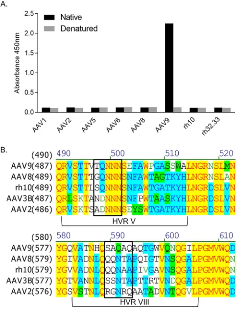
The NAb PAV9.1 is potent and specific for AAV9.We first aimed to identify a novel, potent anti-AAV9 NAb for epitope mapping. We screened a panel of 30 hybrid-oma clones for AAV reactivity by an enzyme-linked immunosorbent assay (ELISA) against a number of serotypes and for AAV9 neutralization by an NAb assay. We selected the mAb PAV9.1 from this panel due to its specificity for AAV9 (Fig. 1A). PAV9.1 FIG 1Characterization of the PAV9.1 mAb and mutagenesis strategy based on the PAV9.1 epitope. (A) PAV9.1 recognition of various AAV serotypes based on capture ELISA with native or denatured capsid protein. (B) Alignment of the AAV VP1 amino acid sequence. Residues of interest to the epitope of PAV9.1 are within the black box.
Mapping an AAV9-Specific Neutralizing Epitope Journal of Virology
on November 6, 2019 by guest
http://jvi.asm.org/
[image:3.585.85.327.72.389.2]recognized only intact capsid by ELISA (Fig. 1A) and did not recognize AAV by Western blotting (data not shown), suggesting that PAV9.1 identifies a conformational epitope on the capsid surface. This is in contrast to the remaining clones, which more broadly bound the panel of AAVs included in the screen and also recognized AAV by Western blotting (data not shown). In an NAb assay, purified PAV9.1 mAb showed an effective NAb titer of 1:163,840, indicating that this novel anti-AAV9 antibody is a potent neutralizer of AAV9. Again, this was in contrast to the other clones screened by NAb assay, none of which were able to neutralize AAV transduction.
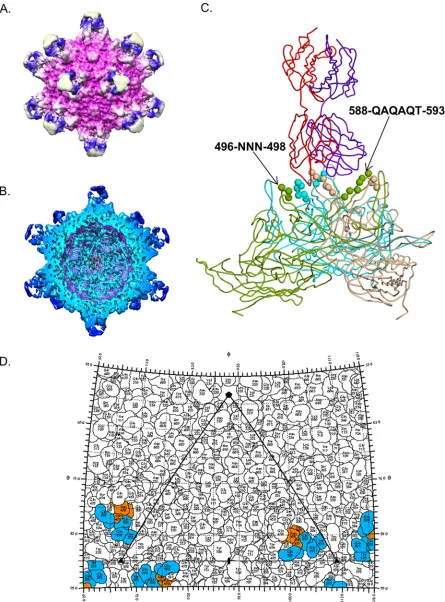
Cryo-EM reconstruction of AAV9 in complex with PAV9.1.Following complexing of AAV9 with PAV9.1 antigen-binding fragments (Fab), we captured 1,100 images, boxed 3,022 particles, and generated a 4.2-Å reconstruction of the complex using AUTO3DEM. We observed Fab electron density extending from the 3-fold axis com-prised by HVRs IV, V, and VIII and decorating the interior face of the 3-fold protrusions with the Fab electron density centered perpendicularly (Fig. 2A and B). This region was mainly comprised of charged residues, which favor strong electrostatic interactions between 3-fold related VP monomers, as well as with receptors and mAbs. A single Fab molecule was bound and extended across two of the three protrusions at each 3-fold axis, blocking binding of additional Fab molecules at these sites due to steric hindrance (Fig. 2C). The region of the PAV9.1 Fab complementary-determining regions (CDRs) in contact with the 3-fold protrusions had an average density of 2.5 sigma levels, which is comparable to densities reported for other AAV-Fab reconstructions (34). We ob-served a PAV9.1 Fab constant region density at approximately 0.8 sigma levels, or approximately one-third of the density observed for the contact region of the PAV9.1 CDRs, corresponding to a single Fab occupancy per 3-fold axis. The PAV9.1 Fab CDRs directly interacted with residues 496-NNN-498 (HVR V) and 588-QAQAQT-593 (HVR VIII) (Fig. 2C and D). PAV9.1 binding additionally occluded residues G455 and Q456 (HVR IV); T494, Q495, and E500 (HVR V); and N583, H584, S586, and A587 (HVR VIII), which do not participate in electrostatic interactions with PAV9.1 but may provide structural stability to this region of the capsid following Fab binding (Table 1). The CDRs of the heavy chain interacted with HVR V, whereas the CDRs of the light chain interacted with HVR VIII of the same VP3 monomer (Fig. 2C).
Based on the PAV9.1 footprint (Fig. 2D, Table 1), we selected two sets of five residues for focused mutagenesis for epitope validation and escape mutant design: 586-SAQAQ-590 and 494-TQNNN-498. We chose residues 586-SAQAQ-586-SAQAQ-590 because this site contains a high degree of sequence and structural diversity (Fig. 1B). The selected motif contains residues identified by the cryo-EM reconstruction to be directly interacting with PAV9.1, as well as residues identified as occluded, allowing for the interrogation of the junction between bound and occluded residues. These residues have also been implicated in neutralizing epitopes for AAV1, AAV2, and AAV8, allowing for the comparison of the AAV9 epitope residues to those previously published (35). Finally, restricting HVR VIII mutagenesis to these five residues increased the likelihood that the capsid would tolerate larger mutations, since this motif has more limited interactions with regions contributing to capsid structural integrity. Despite PAV9.1 being specific for AAV9, the HVR V motif 496-NNN-498 identified as interacting with PAV9.1 is highly conserved between serotypes (Fig. 1B). However, unpublished phage display work (data not shown) suggested the involvement of an asparagine-rich motif in the epitope of PAV9.1; thus, we selected this motif for mutagenesis. We added residues 494-TQ-495 to again interrogate the junction between bound and occluded residues and because they were previously implicated in AAV-Ab interactions (35).
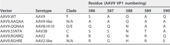
Epitope-based mutations markedly reduce AAV9-PAV9.1 binding.We first gen-erated 586-SAQAQ-590 serotype swap mutants using site-directed mutagenesis. Based on the knowledge that PAV9.1 specifically recognizes AAV9 and that the amino acid sequence and structural conformation at this location vary widely between AAV serotypes, we chose full swaps with the corresponding residues from representative serotypes from clade B (AAV2), clade C (AAV3B), and clade D/E (AAV8/rh10) (Table 2).
on November 6, 2019 by guest
http://jvi.asm.org/
FIG 2Cryo-EM reconstruction of AAV9 in complex with PAV9.1 Fab. (A) Depiction of the molecular surface of AAV9 capsid (fuchsia) bound with PAV9.1 Fab (blue at the protrusion of the 3-fold axis reconstructed at a 4.2-Å resolution. We boxed 3,022 particles and used Auto3dEM for electron microscopy reconstruction. (B) Depiction of a cross-section of the AAV9-PAV9.1 complex. (C) Pseudo-atomic model of AAV9-PAV9.1 trimer built into electron density as obtained from cryo-EM reconstruction. VP3 monomers are shown in green, wheat, and cyan. Spheres represent PAV9.1 Fab bound residues. We have illustrated a single PAV9.1 Fab with the heavy chain in indigo and the light chain in red. (D) Two-dimensional “roadmap” representation of residues involved in PAV9.1 binding, with HVR V contact residues shown in orange and HVR VIII contact residues shown in blue.
Mapping an AAV9-Specific Neutralizing Epitope Journal of Virology
on November 6, 2019 by guest
http://jvi.asm.org/
[image:5.585.42.487.71.673.2]In doing so, we expected to maximize the likelihood of efficient capsid assembly while also maximizing the natural variation at this location. We generated two additional mutants, AAV9.AAQAA (more convergent than AAV9.QQNAA) and AAV9.RGHRE (more divergent than AAV9.RGNRQ), to determine (i) the minimum mutation required to disrupt PAV9.1 interactions and (ii) the maximum disruption that we could introduce. AAV9.AAQAA, AAV9.QQNAA, and AAV9.SSNTA mutants produced vectors of equivalent titer to AAV9.WT; however, titers of AAV9.RGNRQ and AAV9.RGHRE were reduced two to 3-fold relative to AAV9.WT (data not shown). We determined the binding of PAV9.1 mAb to each mutant capsid compared to AAV9.WT by capture ELISA (Fig. 3A). The EC50, or the concentration of PAV9.1 mAb required to reach half-maximal binding, of PAV9.1 for each swap mutant was markedly increased (indicative of reduced capsid binding) relative to the EC50 for AAV9.WT. This result validated the epitope mapping results, indicating that residues 586-SAQAQ-590 are involved in AAV9-PAV9.1 interactions. The EC50increases ranged from 45-fold (AAV9.AAQAA) to almost 300-fold (AAV9.RGHRE) (Table 3); the increase in EC50 directly correlated with the degree of sequence and structural divergence from AAV9 at this location. The one exception was AAV9.RGNRQ, which shares Q590 with AAV9, potentially contributing to stronger PAV9.1 binding than that expected by sequence analysis.
As the S586A and Q590A mutations in AAV9.AAQAA were sufficient to disrupt PAV9.1 binding of AAV9, we next determined the minimal change required to induce this disruption. We introduced a point mutation at one of these positions either by alanine replacement or a more conservative replacement (S¡T or Q¡N). Mutations to either alanine or threonine at S586 did not significantly reduce PAV9.1 binding, whereas a single mutation to either alanine or asparagine at Q590 was sufficient to disrupt capsid recognition by PAV9.1 (Fig. 3C). This result indicates that Q590 is critical for PAV9.1 recognition of the AAV9 capsid.
[image:6.585.41.374.83.173.2]We next interrogated the 494-TQNNN-498 motif of HVR V for its inclusion in the PAV9.1 epitope using the same mutagenic strategy: mutating sets of residues to evolutionarily conserved amino acids or alanine alone. Since 496-NNN-498 is conserved across all serotypes tested, we used only alanine replacement for this stretch of residues; for 494-TQ-495, we mutated to AA, as well as GQ and TD, in order to represent the naturally occurring diversity at this site. Despite the specificity of PAV9.1 for AAV9 and the diversity at this location, AAV9.GQNNN, AAV9.TDNNN, and AAV9.AANNN did TABLE 1PAV9.1 Fab epitope residuesa
HVR Position(s)
Contact residues
V 496-NNN-498
VIII 588-QAQAQT-593
Occluded residues
IV G455, Q456
V T494, Q495, E500
VIII N583, H584, S586, A587
aWe used cryo-reconstruction to generate a list of residues that are either directly bound or occluded by
PAV9.1 Fab binding to AAV9.
TABLE 2Mutagenesis strategy of PAV9.1 HVR VIII epitope residues
Vector Serotype Clade
Residue (AAV9 VP1 numbering)
586 587 588 589 590
AAV9.WT AAV9 F S A Q A Q
AAV9.AAQAA AAV9-like N/A A A Q A A
AAV9.QQNAA AAV8/rh10 D/E Q Q N A A
AAV9.SSNTA AAV3B C S S N T A
AAV9.RGNRQ AAV2 B R G N R Q
AAV9.RGHRE AAV2-like N/A R G H R E
on November 6, 2019 by guest
http://jvi.asm.org/
[image:6.585.41.370.652.740.2]not increase the EC50of PAV9.1 for AAV (Fig. 3B). This confirms the conclusion from the cryo-reconstruction map that the 494-TQ-495 site does not participate in the PAV9.1 epitope. However, the AAV9.TQAAA mutation increased the PAV9.1 EC50 15-fold, indicating that despite the fact that 496-NNN-498 is a conserved motif, it still plays an important role in the AAV9-specific binding of PAV9.1. Finally, we generated combina-tion mutants from HVR V and minimal HVR VIII mutacombina-tions (AAV9.TQAAA/SAQAN and AAV9.TQAAA/SAQAA); the PAV9.1 EC50values for these combination mutants show that the effects of changing motifs in the PAV9.1 epitope are additive (Fig. 3D and E).
FIG 3Effect of epitope mutations on the EC50of PAV9.1 mAb for AAV9. We used capsid capture ELISA against AAV9 to analyze and generate binding curves for PAV9.1. Panels A to E illustrate 586-590 swap mutants (A), 494-498 mutants (B), 586-590 point mutants (C), AAV9.TQAAA and AAV9.SAQAN single and combination mutants (D), AAV9.TQAAA and AAV9.SAQAA single and combination mutants (E). We normalized absorbance to the maximum absorbance for each capsid. We determined the line of best fit and EC50using the dose response function in Prism.
Mapping an AAV9-Specific Neutralizing Epitope Journal of Virology
on November 6, 2019 by guest
http://jvi.asm.org/
[image:7.585.42.428.66.562.2]Epitope-based mutations modulate AAV9 transduction.To evaluate the ability of the novel AAV9 mutants to evade NAbs while maintaining the properties of AAV9.WT, we first assessed the in vitro and in vivo transduction. The majority of mutations resulting in a reduction in PAV9.1 binding also reduced the transduction efficiency in HEK293 cells, with the notable exception of AAV9.RGNRQ, which improved vector transduction by 2.3-fold (Fig. 4A). This improvement could have been due to the introduction of R586 and R589 (R585 and R588 by AAV2 VP1 numbering), two residues responsible for heparin recognition by AAV2, which performs significantly better than AAV9 in vitroin most cell lines likely due to the inclusion of these heparin-binding motifs (36). However, AAV9.RGHRE, which shares R586 and R589 with AAV9.RGNRQ, did not display AAV2-like transduction efficiency, suggesting the involvement of other factors. AAV9.AAQAA demonstrated the greatest reduction in transduction efficiency, indicating that S586 and/or Q590 are essential residues for AAV9 transductionin vitro. Epitope-based mutations ablate PAV9.1 neutralization.We next examined the effects of the mutations on the neutralizing titer of PAV9.1. Mutant AAV9.AANNN, which does not affect PAV9.1 binding, did not affect the neutralizing titer (Fig. 4B and I). However, all mutant vectors that increased the PAV9.1 EC50 reduced the effective neutralizing titer of PAV9.1. AAV9.RGHRE, which most dramatically increased the EC50 by almost 300-fold, reduced the NAb titer of PAV9.1 by at least 2,048-fold (from 1:163,840 to ⬍1:80, the lowest dilution tested) (Fig. 4C to K). Mutant vectors that increased the EC50more modestly, such as AAV9.SAQAN, reduced the effective NAb titer of PAV9.1 to a lesser degree (Fig. 4L). Overall, we observed a strong correlation between reduction in PAV9.1 binding, as measured by EC50, and reduction in the effective NAb titer (Fig. 5). A notable exception was again AAV9.RGNRQ, which reduced the NAb titer by only 8-fold (the second lowest reduction) despite being the fourth most effective mutant at reducing PAV9.1 binding.
The PAV9.1 epitope is important for AAV9 liver tropism.To evaluate the viability of these mutants as AAV9-like gene therapy vectors, we injected C57BL/6 mice intra-venously with 1e11 genome copies (GC)/mouse of AAV9.WT.CMV.LacZ or the AAV9 mutant vectors that reduced PAV9.1 activity (n⫽3 per group). Genome copy analysis of day 14 tissue samples indicated a reduction in liver transduction for all mutants. AAV9.QQNAA performed most similarly to AAV9.WT with 17-fold fewer GC/g DNA, whereas AAV9.RGHRE transduced liver the least efficiently with 1,110-fold fewer GC/g DNA (Fig. 6A). However, in other organs such as heart and brain, the majority of mutants maintained near AAV9.WT levels of transduction, with the exception of the AAV2-like mutants, AAV9.RGNRQ and AAV9.RGHRE. Although these differences in tissue GCs were not statistically significant, the observed trends suggest that these residues are important for AAV9 liver tropism but play a smaller role in the transduction of other tissues, as most mutants displayed a “liver-detargeting” phenotype. These results were further reflected in the expression of-galactosidase (-Gal) in liver and heart; liver
-Gal activity was highest in animals receiving AAV9.WT, whereas heart-Gal activity was similar between AAV9.WT and most mutants (with the exception of the AAV2-like mutants) (Fig. 6B and C).
[image:8.585.42.371.83.185.2]We repeated these experiments at a 10-fold higher dose (1e12 GC/mouse) for a representative subset of AAV9 mutant vectors. Although transduction differences did TABLE 3Summary of AAV9 capsid mutant characteristics followingin vitroevaluation
WT or mutant Fold NAb titer reduction Fold EC50increase % WT transduction
WT 1 1 100
AAQAA 16 45 27
QQNAA 128 124 53
SSNTA 512 264 58
RGNRQ 8 96 233
RGHRE 2048 294 60
TQAAA 16 15 50
SAQAN 16 40 76
SAQAA 4 20 54
on November 6, 2019 by guest
http://jvi.asm.org/
FIG 4Characterizing the impact of PAV9.1 epitope mutations on in vitro vector transduction and effective PAV9.1 mAb neutralizing titer. (A) Transduction efficiency of PAV9.1 capsid mutants relative to AAV9.WT in HEK293 cells. We determined significance by using a two-sided one-samplettest and compared the percent transduction of each mutant to the transduction of AAV9.WT (defined as 100%). Pvalues are indicated by asterisks (*,P⬍0.05;***,P⬍0.001). (B to K) Determination of the neutralizing titer of PAV9.1 when transducing
(Continued on next page)
Mapping an AAV9-Specific Neutralizing Epitope Journal of Virology
on November 6, 2019 by guest
http://jvi.asm.org/
[image:9.585.45.480.65.697.2]not reach significance at this dose, the tissue tropism trends were consistent with those observed at the lower dose, particularly for heart and muscle samples (Fig. 6D). Again, these results were reflected in-Gal activity in histological sections of liver, heart, and muscle (Fig. 6E to G).
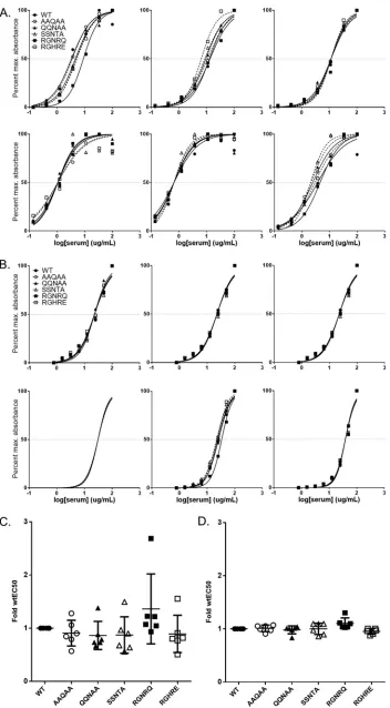
Epitope-based mutations in AAV9 do not significantly affect binding or neu-tralization by polyclonal plasma or sera. We next assessed the ability of PAV9.1 epitope-based mutant vectors to evade the binding of and neutralization by polyclonal plasma or sera. We first utilized plasma from C57BL/6 mice previously injected intra-venously with AAV9.WT (7.5e8 or 7.5e9 GC/mouse,n⫽6 per group). We determined the dilution of plasma required to reach half-maximal binding. Binding of plasma from low-dose mice to mutant vectors was almost indistinguishable from binding to AAV9.WT (Fig. 7A to C). In contrast, we observed significant differences in the EC50of plasma from high-dose mice for a subset of mutants, most notably AAV9.RGNRQ, relative to the EC50for AAV9.WT (Fig. 7B to D). Despite an average 2-fold increase in the EC50of high-dose mouse plasma for AAV9.RGNRQ, we did not observe a reduction of the effective NAb titer of the plasma in this mutant (data not shown).
To determine whether this trend in EC50increase was true for nonhuman primate samples, we obtained sera from a panel of six macaques that received AAV9 vector or a novel vector closely related to AAV9 with the same VP3 sequence (2 amino acid difference in the nonstructural VP1 unique region). We confirmed that the macaques had NAb titers against AAV9 of⬍1:5 (defined as NAb negative) prior to administration. Although we did observe some variation in the EC50s of each animal’s serum for mutant vectors compared to the EC50for AAV9.WT, no clear trend of increased or decreased binding emerged based on mutant identity (Fig. 8A and C). When testing sera from macaques with preexisting NAb titers against AAV9 (attributed to a prior AAV infection), we observed little to no variation in the EC50of the sera for the panel of AAV9 mutants (Fig. 8B and D). This was in stark contrast to the variations seen in the EC50of injected sera, suggesting fundamental differences between the relevant anti-AAV epitope rep-ertoire of serum generated in response to AAV infection and AAV vector administration. In addition, the increase in the EC50 of injected nonhuman primate sera for AAV9.RGNRQ did not decrease the effective NAb titer of the sera for AAV9.RGNRQ (data not shown).
Finally, we assessed NAb-positive serum samples from four normal human donors for binding to AAV9.WT and mutant vectors. As was the case for the uninjected
FIG 4Legend (Continued)
HEK293 cells with AAV9.WT.CMV.LacZ (B), AAV9.AAQAA (C), AAV9.QQNAA (D), AAV9.SSNTA (E), AAV9.RGNRQ (F), AAV9.RGHRE (G), AAV9.TQAAA (H), AAV9.AANNN (I), AAV9.SAQAN (J), or AAV9.SAQAA (K) in the presence of various dilutions of PAV9.1. We defined the neutralizing titer as the dilution prior to the dilution at which we could achieve transduction levels of 50% or greater than the vector without mAb (levels measured in relative light units). All data are reported as means⫾the standard deviations (SD).
FIG 5Correlation between PAV9.1 EC50and neutralizing titer for a panel of AAV9 mutants. We calculated the fold reduction in PAV9.1 neutralizing titer against each mutant relative to PAV9.1 neutralizing titer against AAV9.WT. We plotted the data on a log scale against the fold increase in PAV9.1 EC50for each mutant relative to PAV9.1 EC50for AAV9.WT on a linear scale (semi-log plot). We used GraphPad Prism to determine the semi-log line of best fit (R2⫽0.8474).
on November 6, 2019 by guest
http://jvi.asm.org/
[image:10.585.96.316.69.193.2]NAb-positive nonhuman primate serum samples, all four NAb-positive normal human donor samples demonstrated minimal variation in EC50for AAV9 mutant versus WT vectors (Fig. 9). As expected, the lack of changes in EC50 for the mutant vectors translated to a lack of reduction in NAb titer of sera toward AAV9 mutant vectors (data not shown).
FIG 6In vivoanalysis of AAV9 PAV9.1 mutant vectors. C57BL/6 mice received intravenous injections of either 1e11 GC per mouse (A to C) or 1e12 GC per mouse (D to F) AAV9.CMV.LacZ (WT or mutant;n⫽3). We sacrificed mice on day 14 and harvested tissues for biodistribution analysis (A and D) using TaqMan qPCR. Values are reported as means⫾the SD. We also harvested liver (B and E), heart (C and F), and muscle (G) for-Gal histochemistry to determine enzyme activity. Representative 10⫻images are shown. Scale bars, 200m.
Mapping an AAV9-Specific Neutralizing Epitope Journal of Virology
on November 6, 2019 by guest
http://jvi.asm.org/
[image:11.585.42.429.69.616.2]FIG 7Effect of epitope mutations on EC50of injected mouse plasma for AAV9. Using capsid capture ELISA, we analyzed the day 56 plasma of mice that received intravenous injections of either 7.5e8 GC/mouse (A) or 7.5e9 GC/mouse (B)
(Continued on next page)
on November 6, 2019 by guest
http://jvi.asm.org/
[image:12.585.39.420.66.706.2]DISCUSSION
Here, we report the cryo-EM reconstruction of AAV9 in complex with the highly potent and specific mAb PAV9.1. The epitope determined for PAV9.1 largely overlaps with the epitopic regions of other AAV NAbs isolated from mouse hybridomas, namely, ADK8 (AAV8; 586-LQQQNT-591), E4E (AAV1; 492-TKTDNNN-498), 5H7 (AAV1; 496-NNNS-499, 588-STDPATGD-595), and C37 (AAV2; 492-SADNNNS-498, 585-RGNRQ-589) (28, 29, 34). Thus, despite the large degree of sequence and structural variations among the serotypes in HVR V and VIII, this finding suggests that the 3-fold protrusions may be a significant site of AAV9 neutralization as it is for other serotypes. Previous findings regarding the repertoire of NAbs directed against other AAV capsids may therefore be applicable to AAV9. Although the various mapped neutralizing epitopes show overlap, the binding angles and orientations of the NAbs vary significantly. When bound to AAV9, PAV9.1 extends into the center of the 3-fold axis of symmetry, sterically limiting the occupancy to 20 Fab particles; in contrast, mAbs raised against other serotypes bind on the top or face outward from the 3-fold axis, allowing higher occupancy. Studies have identified both HVR V and VIII as shared antigenic regions across serotypes, including AAV2 (in complex with C37B, 11 Å), AAV8 (in complex with ADK8, 18.7 Å), and AAV1 (in complex with 5H7, 23 Å), which bears the most similarity to the binding footprint of PAV9.1 for AAV9 (28, 29, 34). Therefore, the structure reported here is similar to lower-resolution structures previously reported for other AAV serotypes.
HVR VIII serotype swaps conferred various degrees of binding and neutralization evasion to their corresponding mutant vectors. Swapping this region with the AAV2-based RGHRE motif, the most divergent mutant from the WT.AAV9 sequence, ablated PAV9.1 neutralization at all dilutions tested. Thus, engineering only five amino acids can generate a capsid that can evade a monoclonal NAb. In fact, the minimal change required to significantly reduce PAV9.1 activity was a single amino acid substitution, with even a conserved amino acid leading to ablation of both binding and neutraliza-tion. Mutations in the NNN motif in HVR V reduced PAV9.1’s ability to bind and neutralize AAV9 despite having high conservation among serotypes, indicating that it is also an integral part of the PAV9.1 epitope.
We observed a strong correlation between a reduction in PAV9.1’s binding to a given AAV9 mutant and its ability to block transduction of that mutant in vitro, suggesting that the relative binding strength of an NAb to AAV correlates with the NAb’s neutralizing ability. However, data from our lab and others suggest that the binding antibody titer against AAV is not always a good predictor of an individual’s NAb titer, as some individuals have moderate binding titers against AAV but are NAb negative (37, 38; unpublished data). Despite these findings, the exclusion criteria of some clinical trials include not only NAb titer but also binding titer (3, 39). Therefore, epitope mapping studies are critical for identifying the features of binding epitopes and determining whether they share any commonalities with neutralizing epitopes. Shared motifs would suggest that strength of binding, rather than interactions with specific residues, plays a large role in AAV neutralization, thus allowing researchers to focus simply on reducing the binding of NAbs. Disparate motifs, however, would suggest that neutralization is more a function of binding location than strength of binding and indicate that researchers should focus on ablating NAb binding to these unique regions.
Although the mutations in the AAV9 vectors dramatically reduced binding and neutralization by a purified monoclonal PAV9.1 antibody, these mutations did not
FIG 7Legend (Continued)
wtAAV9.LSP.hFIX for AAV9.WT or AAV9 PAV9.1 mutant binding. We normalized the absorbance to the maximum absorbance achieved for each capsid. We determined the line of best fit and EC50using the dose response function in Prism. Each graph corresponds to a single animal. We compiled EC50values for 7.5e8 GC/mouse (C) or 7.5e9 GC/mouse (D) in order to determine the average for each mutant. A two-sided one-samplettest was used to determine whether there was a significant difference between the EC50of plasma for each mutant relative to the EC50of plasma for AAV9.WT (defined as 1). We applied the Bonferroni correction to control for type 1 errors.Pvalues are indicated by asterisks (*,P⬍0.05;**,P⬍0.01;***,P⬍0.001). EC50data are reported as means⫾the SD.
Mapping an AAV9-Specific Neutralizing Epitope Journal of Virology
on November 6, 2019 by guest
http://jvi.asm.org/
FIG 8Effect of epitope mutations on EC50of NHP polyclonal serum for AAV9. Using capsid capture ELISA, we analyzed the sera from NHPs treated with AAV9.WT or hu68.WT vector (A) or naive NHPs that are AAV9
(Continued on next page)
on November 6, 2019 by guest
http://jvi.asm.org/
[image:14.585.42.394.67.705.2]significantly evade binding or neutralization by polyclonal antibodies from sera or plasma from mice, macaques, or human donors that were previously exposed to AAV. Most notably, plasma from mice that received the higher intravenous dose of AAV9 vector bound the RGNRQ mutant about 2-fold less efficiently than WT.AAV9 vector; this change was much more modest than the 50-fold reduction observed with PAV9.1 mAb. Even though the QQNAA, SSNTA, and RGHRE mutations had a greater impact on PAV9.1 binding and neutralization than the RGNRQ mutation, the polyclonal plasma bound these mutants in the same manner as WT.AAV9. This result suggests that although the 586-SAQAQ-590 motif is a potent neutralizing epitope and mutations in this region can block PAV9.1 activity,in vitroactivity against a mAb does not predict activity against polyclonal antibodies. Perhaps surprisingly, the RGNRQ mutant blocked binding of AAV9 antibodies in these polyclonal samples utilizing the 3-fold protrusions. This result clearly shows that not all mutations behave the same against polyclonal responses and that a large repertoire of antibodies utilizes this region for binding.
Despite the reduction in polyclonal binding, the RGNRQ mutant vector did not evade the polyclonal NAb response generated by these mice in response to vector administration. As expected, mutants that did not reduce binding to the polyclonal plasma also did not evade neutralization. Given that the nearly 100-fold increase in the EC50of PAV9.1 for RGNRQ relative to WT.AAV9 resulted in only an 8-fold decrease in PAV9.1 neutralizing titer, it was not surprising that a 2-fold increase in the EC50 of polyclonal plasma for RGNRQ did not reduce the neutralizing titer. Although studies show that the majority of mapped AAV epitopes lie on the 3-fold axis and that HVR VIII
FIG 8Legend (Continued)
NAb-positive for AAV9.WT or AAV9 PAV9.1 mutant binding (B). We normalized the absorbance to the maximum absorbance achieved for each capsid. We used the dose response function in Prism to determine the line of best fit and EC50. Each graph corresponds to a single animal. We compiled EC50values for vector-treated NHPs (C) and naive NAb-positive NHPs (D) to determine the average for each mutant. We used a two-sided one-sample ttest to determine whether there is a significant difference between the EC50of plasma for each mutant relative to the EC50of plasma for AAV9.WT (defined as 1). We applied the Bonferroni correction to control for type 1 errors. EC50data are reported as means⫾the SD.
FIG 9Effect of epitope mutations on EC50of human donor polyclonal sera for AAV9. (A) We analyzed the sera from naive human donors that were AAV9 NAb-positive for AAV9.WT or AAV9 PAV9.1 mutant binding using capsid capture ELISA. We determined the line of best fit and EC50using the dose-response function in Prism. Each graph corresponds to a single donor. (B) We compiled EC50values for NAb-positive human donor serum to determine the average for each mutant. We determined significance by using a two-sided one-samplettest and compared the EC50of plasma for each mutant relative to the EC50of plasma for AAV9.WT (defined as 1). We applied the Bonferroni correction to control for type 1 errors. EC50data are reported as means⫾the SD.
Mapping an AAV9-Specific Neutralizing Epitope Journal of Virology
on November 6, 2019 by guest
http://jvi.asm.org/
[image:15.585.50.538.69.305.2]is implicated in the mapped epitopes for most serotype-specific NAbs, we were surprised to find that none of the mutations tested in this region dramatically affected polyclonal activity. We note that the mapped epitopes may not be representative of the complete repertoire, since the total number of mapped epitopes is small and the exact screening and selection methods for some studies are unknown.
Tse and colleagues recently used a library approach to combine the epitopes of three different NAbs identified against AAV1 and generate a novel AAV1-based capsid with more than 20 amino acid changes from the parental AAV1. This capsid could evade not only anti-AAV1 monoclonal NAbs but also polyclonal samples from AAV vector-injected mice and nonhuman primates in addition to polyclonal samples from normal human donors exposed to AAV (40). This suggests that neutralizing epitopes may overlap following vector exposure and viral infection, but this repertoire is subtly diverse. In other words, the total number of residues that require modification to confer binding and neutralizing evasion to AAV is more extensive than previously thought. Engineering novel capsids that can address both scenarios may require combinatorial and high-throughput approaches.
This study explored whether vectors engineered to evade a preexisting NAb re-sponse from a prior AAV infection would also function in a readministration setting. The polyclonal samples for which the PAV9.1-based AAV9 mutant vectors demonstrated even minimal evasion were acquired from sources that had received AAV vector and not from sources that were previously infected with AAV. Whereas injected samples demonstrated modestly variable binding curves for the panel of AAV9 mutants, the binding curves generated by vector-naive but virally exposed sources were similar to the curves of WT.AAV9. These discrepancies highlight the fundamental differences among the AAV antibody repertoire generated in response to vector administration or infection.
Historically, naive subjects injected with AAV vector generate an NAb response that is specific to the vector administered or limited to closely related serotypes (41; unpublished data). Most macaque studies and gene therapy clinical trials have shown a similar result (21, 42; unpublished data). In stark contrast, subjects with preexisting antibodies for one AAV serotype are almost always seropositive for and have NAbs against the majority of other serotypes, even those that are distantly related (41, 43, 44; unpublished data). To date, all novel mapped AAV mAbs are specific for an individual serotype and cross-react only with closely related serotypes (for example, 5H7 binding to both AAV1 and AAV6); no previously isolated neutralizing AAV mAbs recapitulate the broader responses commonly seen following AAV infection (34). Therefore, further studies are necessary to identify motifs that comprise broadly neutralizing epitopes relevant to preexisting immunity, determine whether the epitopes overlap with serotype-specific epitopes, and evaluate how the overlapping motifs confer a broadly neutralizing phenotype to the NAbs.
The magnitude of an NAb response varies widely between methods of exposure; rarely does an individual with natural immunity have an NAb titer exceeding 1:80 (humans) or 1:320 (macaques); in contrast, NAb titers of ⬎1:1,000 can easily be achieved in response to the delivery of a modest dose of vector (21, 42, 45). In this study, mice receiving the highest vector dose resulting in the highest NAb titers had measurable variations in mutant vector binding; this suggests that the strength of an NAb response impacts mutant efficiency. Often, studies aim to reduce an individual’s NAb titer to below the threshold that interferes with gene transfer (1:10 for intravenous administration) (15, 46). Mutant capsids engineered based on a single neutralizing epitope that only confer evasion to high titer sera would not significantly increase the number of individuals eligible to receive AAV gene therapy, since the lower titers are still above the threshold at which transduction is appreciably inhibited.
The minimal mutation required to reduce PAV9.1 binding at Q590 in HVR VIII conferred a liver-detargeting phenotype to the resulting mutants, even following a conservative amino acid substitution to asparagine. Mutations in the HVR V portion of the epitope also reduced liver transduction. These results are in agreement with
on November 6, 2019 by guest
http://jvi.asm.org/
previous observations that these residues in HVR V and VIII play integral roles in liver transduction, as well as previous reports of mapped neutralizing AAV epitopes that show overlap with regions essential for gene transfer (28, 47). This suggests that it would be difficult to engineer a mutant that can evade NAbs while maintaining the parental transduction profile. For some indications in heart and muscle, where liver transduction may be less consequential, this modification in tropism may be accept-able. Notably, the majority of mutants maintained WT.AAV9 levels of transduction in peripheral organs at both doses.
Whereas the RGNRQ mutant demonstrated modest binding modifications in the presence of polyclonal antibodies, it displayed an AAV2-like transduction profile: poorly transducing not just liver but all peripheral organs. Taken together, these data indicate the importance of integrating knowledge about a mapped neutralizing epitope with available information about AAV functional domains. Generating a capsid that can evade NAbs is not sufficient, as the capsid is only useful if it can still perform its primary function of target tissue transduction. Recent studies have used this strategy to incorporate multiple epitopes of AAV1 to generate AAV1-based vectors that can evade NAbs while maintaining AAV1-like transduction profiles (40).
In summary, this study provides critical information regarding the design of AAV9-based vectors able to evade humoral immune responses. Future studies are required to further understand the complexity of the NAb response to AAV9 vectors to inform the design of next-generation capsids.
MATERIALS AND METHODS
Hybridoma generation.BALB/c mice received up to five immunizations of the AAV9 vector. We harvested and fused the splenocytes. ProMab Biotechnologies, Inc. (Richmond, CA), generated the clonal supernatants according to the company’s standard custom mouse mAb hybridoma development protocol. Thirty supernatants underwent screening for AAV9 reactivity by ELISA and for their ability to neutralize AAV9 by NAb assay. We obtained purified PAV9.1 mAb following screening at a concentration of 3 mg/ml.
AAV capsid ELISA.Corning polystyrene high bind microplates were coated with 1e9 GC/well AAV diluted in phosphate-buffered saline (PBS) and kept overnight at 4°C. After discarding the coating solution, we blocked the plates with 3% bovine serum albumin (BSA) in PBS for 2 h at room temperature followed by a triple wash of 300 l of PBS plus 0.05% Tween. We then incubated the hybridoma supernatant, purified mAb, serum, or plasma (diluted in 0.75% BSA in PBS) at 37°C for 1 h, followed by a triple wash with 300l of PBS plus 0.05% Tween. Next, we detected mouse samples using 1:10,000 goat anti-mouse horseradish peroxidase-conjugated IgG (diluted in 0.75% BSA in PBS; catalog no. 31430; Thermo Fisher Scientific, Waltham, MA) at 37°C for 1 h, followed by a triple wash of 300l of PBS plus 0.05% Tween. The human and nonhuman primate samples were then detected using 1:10,000 (diluted in PBS) goat anti-human IgG biotin-SP (catalog no. 109-065-098; Jackson ImmunoResearch, Inc., West Grove, PA) at room temperature for 1 h, followed by a triple wash of 300l of PBS plus 0.05% Tween and 1:30,000 (diluted in PBS) unconjugated streptavidin (catalog no. 016-000-084; Jackson Immuno-Research) at room temperature for 1 h (followed by a 3⫻wash with 300l of PBS plus 0.05% Tween). We developed all ELISAs with tetramethylbenzidine.
Neutralizing antibody assays.We performed NAb assays as previously described (14) with a few modifications. We used HEK293 cells seeded at a density of 1e5 cells/well on black-walled, clear-bottomed, poly-lysine-coated plates (catalog no. 08-774-256; Fisher Scientific Company, Hampton, NH). Using a multiplicity of infection of 90 wtAd5/cell, we utilized a working solution of 4e10 GC/ml AAV9.CMV.LacZ vector to achieve a final concentration of 2e9GC/well. We measured bioluminescence with the SpectraMax M3 (Molecular Devices, Sunnyvale, CA) according to the manufacturer’s protocol. For any given sample, we defined the NAb titer as the last dilution at which AAV transduction was reduced by⬎50% in the presence of the sample compared to WT.AAV transduction in the presence of the naive control.
We performed HEK293 transduction experiments as described above, but withheld the neutralizing sera. Fab generation and AAV-Fab complexing.PAV9.1 Fab (0.211 mg/ml) was generated using a Pierce Fab Preparation kit (Thermo Fisher Scientific, Waltham, MA) according to the manufacturer’s instructions. Next, we complexed PAV9.1 Fab with the AAV9 vector at a ratio of 600 Fab to 1 AAV9 capsid (or 10 Fab to 1 potential binding site) at room temperature for 30 min.
Cryo-EM sample preparation, data acquisition, and complex reconstruction. (i) Sample prep-aration. We applied 3l of PAV9.1-AAV9 complex to a freshly washed and glow-discharged holey carbon grid. After blotting for 3 to 4 s with Whatman no. 1 filter paper at 22°C and 95% relative humidity, we rapidly froze the grid in liquid ethane slush using a Vitrobot Mark IV (FEI). After freezing, grids were stored in liquid nitrogen. We then transferred the grids to an FEI Talos Arctica electron microscope operating at 200 kV and equipped with a Gatan K2 Summit direct electron detection camera (Gatan, Inc., Pleasanton, CA).
Mapping an AAV9-Specific Neutralizing Epitope Journal of Virology
on November 6, 2019 by guest
http://jvi.asm.org/
(ii) Data acquisition.We acquired data using SerialEM software (48). Images were captured at a nominal magnification of 22,000⫻(corresponding to a calibrated pixel size of 0.944 Å) and a dose rate of 2.21 electrons/Å2/s with a defocus range of 1.0 to 2.0m (49). For each exposure, we recorded a 60-frame dose-fractionated movie stack in super-resolution mode for a total of 12 s. We aligned the movie frames using the “alignframes” program within the IMOD software package (50).
(iii) Data collection and processing.We manually extracted all particle images from each of the micrographs and processed them using the e2boxer program available in the EMAN2 suite (51). The boxed particles were then transferred into the AUTO3DEM program for cryo-reconstruction, leading to the initial low-resolution model (30 Å) based on 150 particle images (52). The program adopted a random model generation procedure, and we applied strict 60 noncrystallographic symmetry axes. This low-resolution reconstructed map was used for determining the particle origin, conducting a full orientation, and refining the contrast transfer function of all images using AUTO3DEM. To improve the reconstructed map’s quality, we applied a temperature factor correction and visualized the map in the graphics programs Coot and Chimera (53, 54). We used a temperature factor 150-corrected map for model docking and interpretation. We extracted a total of 3,022 boxed particles from 1,100 micrographs to ultimately generate a 4.2-Å resolution reconstructed map with a Fourier shell correlation of 0.15. A VIPER database was used to generate the AAV9-60mer model while applying strict icosahedral symmetry axes (T⫽1) (55). Using the FIT function in the Chimera program, we docked the 60-mer copy of AAV9 capsid into the cryo-reconstructed electron density map. This produced a correlation coefficient of 0.9. We visualized and adjusted the docked model in Coot and Chimera for accuracy. ABodyBuilder was used to generate the antibody model, which was then docked and manually adjusted into the cryo-reconstructed density using Chimera (56). The model was then visualized for interpretation of AAV9 and antibody-binding regions. We produced all figures using the Chimera and PyMOL programs (57). We used the RIVEM program to create a two-dimensional depiction of the roadmap (58).
AAV9-PAV9.1 mutant trans-plasmid construction.We used an in-house trans-plasmid construct pAAV2/9 (AAV2 rep/AAV9 cap) for AAV9 capsid mutagenesis. All capsid mutants were constructed using a QuikChange Lightning mutagenesis kit (Agilent, Santa Clara, CA) according to the manufacturer’s instructions. Vector production. We produced the AAV9.CMV.LacZ.bGH and AAV9 mutant vectors via triple transfection in HEK293 cells followed by iodixanol gradient purification as previously described (59). The University of Pennsylvania Vector Core determined the titers of the vectors using quantitative PCR (qPCR) against the bGH poly(A) as described previously (59).
Determining EC50of PAV9.1 mAb and polyclonal sera/plasma.We performed capsid capture
ELISA with either AAV9.WT or AAV9 mutant vector as described above. We calculated the EC50values using GraphPad Prism. Briefly, we log transformed the PAV9.1 mAb concentration in mg/ml and plotted it on thexaxis. We defined IgG concentration in mouse plasma as 5 mg/ml (60) and in nonhuman primate and human serum as 10 mg/ml (61). The plasma/serum concentration (ing/ml) was log transformed and plotted on thexaxis. We defined the maximum absorbance achieved with each mutant, normalized the absorbance to 100%, and plotted it on theyaxis. We then generated a dose-response curve (antibody binding) using GraphPad Prism’s “log(agonist) versus normalized response⫺variable slope” function. Finally, we calculated the EC50for PAV9.1 mAb, polyclonal serum, or polyclonal plasma.
Animal studies. Our animal protocol was approved by and conducted in accordance with the standards of the University of Pennsylvania’s Institutional Animal Care and Use Committee. Male C57BL/6 mice (n⫽3) received intravenous injections in the tail vein of 1e11 GC/mouse AAV9.CMV.LacZ.bGH or AAV9 mutant vectors with the same transgene cassette. Animals were sacrificed 14 days after receiving vector. Each animal’s organs were divided and either snap-frozen on dry ice for biodistribution or embedded in optimal cutting temperature compound and frozen for subsequent sectioning and staining for-Gal activity.
Biodistribution analysis.We extracted DNA from tissues of interest using a QIAamp DNA minikit (Qiagen, Hilden, Germany). We analyzed the tissues for vector GCs by qPCR against the bGH polyade-nylation signal as previously described (62).
-Gal activity staining.Frozen sections were fixed with 0.5% glutaraldehyde in PBS for 10 min at 4°C and subsequently stained for-Gal activity. After a washing step in PBS, we incubated the sections in 1 mg/ml X-Gal (5-bromo-4-chloro-3-indolyl--D-galactopyranoside) in 20 mM potassium ferrocyanide, 20 mM potassium ferricyanide, and 2 mM MgCl2in PBS (pH⬃7.3) and kept tissues overnight at 37°C. After counterstaining the sections with Nuclear Fast Red (Vector Laboratories), we dehydrated them using ethanol and xylene, followed by cover slipping.
ACKNOWLEDGMENTS
We thank the University of Massachusetts Medical School CryoEM Core, specifically Chen Xu and KangKang Song, for the use of their equipment and their invaluable role in complex data collection. We also thank Deirdre McMenamin, Christine Draper, and the Gene Therapy Program (GTP) Program in Comparative Medicine for procedural assistance, Hongwei Yu and the GTP Cell Morphology Core for assistance with histological assays, Mingyao Li for assistance with statistical analyses, and the GTP Immunology Core.
This study was funded by NHLBI 5P01HL059407-18, as well as by the University of Pennsylvania Perelman School of Medicine.
J.M.W. is an advisor to, holds equity in, and has a sponsored research agreement with REGENXBIO and Scout Bio; he also has a sponsored research agreement with
on November 6, 2019 by guest
http://jvi.asm.org/
Ultragenyx, Biogen, and Janssen, which are licensees of Penn technology. In addition, he has sponsored research agreements with Precision Biosciences and Moderna Ther-apeutics. J.M.W. is an inventor on patents that have been licensed to various biophar-maceutical companies.
REFERENCES
1. Melnick JL, Mayor HD, Smith KO, Rapp F. 1965. Association of 20-millimicron particles with adenoviruses. J Bacteriol 90:271–274. 2. Bryant LM, Christopher DM, Giles AR, Hinderer C, Rodriguez JL, Smith JB,
Traxler EA, Tycko J, Wojno AP, Wilson JM. 2013. Lessons learned from the clinical development and market authorization of Glybera. Hum Gene Ther Clin Dev 24:55– 64.https://doi.org/10.1089/humc.2013.087. 3. Mendell JR, Al-Zaidy S, Shell R, Arnold WD, Rodino-Klapac LR, Prior
TW, Lowes L, Alfano L, Berry K, Church K, Kissel JT, Nagendran S, L’Italien J, Sproule DM, Wells C, Cardenas JA, Heitzer MD, Kaspar A, Corcoran S, Braun L, Likhite S, Miranda C, Meyer K, Foust KD, Burghes AHM, Kaspar BK. 2017. Single-dose gene-replacement therapy for spinal muscular atrophy. N Engl J Med 377:1713–1722.https://doi .org/10.1056/NEJMoa1706198.
4. Smalley E. 2017. First AAV gene therapy poised for landmark approval. Nat Biotechnol 35:998.https://doi.org/10.1038/nbt1117-998.
5. Becerra SP, Koczot F, Fabisch P, Rose JA. 1988. Synthesis of adeno-associated virus structural proteins requires both alternative mRNA splicing and alternative initiations from a single transcript. J Virol 62:2745–2754.
6. Johnson FB, Ozer HL, Hoggan MD. 1971. Structural proteins of adenovirus-associated virus type 3. J Virol 8:860 – 863.
7. Venkatakrishnan B, Yarbrough J, Domsic J, Bennett A, Bothner B, Kozyreva OG, Samulski RJ, Muzyczka N, McKenna R, Agbandje-McKenna M. 2013. Structure and dynamics of adeno-associated virus serotype 1 VP1-unique N-terminal domain and its role in capsid trafficking. J Virol 87:4974 – 4984.https://doi.org/10.1128/JVI.02524-12.
8. Bell CL, Vandenberghe LH, Bell P, Limberis MP, Gao GP, Van Vliet K, Agbandje-McKenna M, Wilson JM. 2011. The AAV9 receptor and its modification to improvein vivolung gene transfer in mice. J Clin Invest 121:2427–2435.https://doi.org/10.1172/JCI57367.
9. Ng R, Govindasamy L, Gurda BL, McKenna R, Kozyreva OG, Samulski RJ, Parent KN, Baker TS, Agbandje-McKenna M. 2010. Structural character-ization of the dual glycan binding adeno-associated virus serotype 6. J Virol 84:12945–12957.https://doi.org/10.1128/JVI.01235-10.
10. Pillay S, Zou W, Cheng F, Puschnik AS, Meyer NL, Ganaie SS, Deng X, Wosen JE, Davulcu O, Yan Z, Engelhardt JF, Brown KE, Chapman MS, Qiu J, Carette JE. 2017. AAV serotypes have distinctive interactions with domains of the cellular receptor AAVR. J Virolhttps://doi.org/10.1128/ jvi.00391-17.
11. Wang L, Wang H, Bell P, McCarter RJ, He J, Calcedo R, Vandenberghe LH, Morizono H, Batshaw ML, Wilson JM. 2010. Systematic evaluation of AAV vectors for liver directed gene transfer in murine models. Mol Ther 18:118 –125.https://doi.org/10.1038/mt.2009.246.
12. Gao G, Vandenberghe LH, Alvira MR, Lu Y, Calcedo R, Zhou X, Wilson JM. 2004. Clades of adeno-associated viruses are widely disseminated in human tissues. J Virol 78:6381– 6388.https://doi.org/10.1128/JVI.78.12 .6381-6388.2004.
13. Calcedo R, Morizono H, Wang L, McCarter R, He J, Jones D, Batshaw ML, Wilson JM. 2011. Adeno-associated virus antibody profiles in newborns, children, and adolescents. Clin Vaccine Immunol 18:1586 –1588.https:// doi.org/10.1128/CVI.05107-11.
14. Calcedo R, Vandenberghe LH, Gao G, Lin J, Wilson JM. 2009. Worldwide epidemiology of neutralizing antibodies to adeno-associated viruses. J Infect Dis 199:381–390.https://doi.org/10.1086/595830.
15. Wang L, Calcedo R, Bell P, Lin J, Grant RL, Siegel DL, Wilson JM. 2011. Impact of preexisting immunity on gene transfer to nonhuman primate liver with adeno-associated virus 8 vectors. Hum Gene Ther 22: 1389 –1401.https://doi.org/10.1089/hum.2011.031.
16. Manno CS, Pierce GF, Arruda VR, Glader B, Ragni M, Rasko JJ, Ozelo MC, Hoots K, Blatt P, Konkle B, Dake M, Kaye R, Razavi M, Zajko A, Zehnder J, Rustagi PK, Nakai H, Chew A, Leonard D, Wright JF, Lessard RR, Sommer JM, Tigges M, Sabatino D, Luk A, Jiang H, Mingozzi F, Couto L, Ertl HC, High KA, Kay MA. 2006. Successful transduction of liver in hemophilia by AAV-Factor IX and limitations imposed by the
host immune response. Nat Med 12:342–347.https://doi.org/10.103 8/nm1358.
17. Bowles DE, McPhee SW, Li C, Gray SJ, Samulski JJ, Camp AS, Li J, Wang B, Monahan PE, Rabinowitz JE, Grieger JC, Govindasamy L, Agbandje-McKenna M, Xiao X, Samulski RJ. 2012. Phase 1 gene therapy for Duchenne muscular dystrophy using a translational optimized AAV vector. Mol Ther 20:443– 455.https://doi.org/10.1038/mt.2011.237. 18. Mingozzi F, High KA. 2013. Immune responses to AAV vectors:
overcom-ing barriers to successful gene therapy. Blood 122:23–36.https://doi.org/ 10.1182/blood-2013-01-306647.
19. D’Avola D, López-Franco E, Sangro B, Pañeda A, Grossios N, Gil-Farina I, Benito A, Twisk J, Paz M, Ruiz J, Schmidt M, Petry H, Harper P, de Salamanca RE, Fontanellas A, Prieto J, González-Aseguinolaza G. 2016. Phase I open label liver-directed gene therapy clinical trial for acute intermittent porphyria. J Hepatol 65:776 –783.https://doi.org/10.1016/j .jhep.2016.05.012.
20. Rivera VM, Gao GP, Grant RL, Schnell MA, Zoltick PW, Rozamus LW, Clackson T, Wilson JM. 2005. Long-term pharmacologically regulated expression of erythropoietin in primates following AAV-mediated gene transfer. Blood 105:1424 –1430.https://doi.org/10.1182/blood-2004-06 -2501.
21. Greig JA, Calcedo R, Grant RL, Peng H, Medina-Jaszek CA, Ahonkhai O, Qin Q, Roy S, Tretiakova AP, Wilson JM. 2016. Intramuscular admin-istration of AAV overcomes preexisting neutralizing antibodies in rhesus macaques. Vaccine 34:6323– 6329. https://doi.org/10.1016/j .vaccine.2016.10.053.
22. Elverman M, Goddard MA, Mack D, Snyder JM, Lawlor MW, Meng H, Beggs AH, Buj-Bello A, Poulard K, Marsh AP, Grange RW, Kelly VE, Childers MK. 2017. Long-term effects of systemic gene therapy in a canine model of myotubular myopathy. Muscle Nerve 56:943–953.
https://doi.org/10.1002/mus.25658.
23. Corti M, Cleaver B, Clement N, Conlon TJ, Faris KJ, Wang G, Benson J, Tarantal AF, Fuller D, Herzog RW, Byrne BJ. 2015. Evaluation of readministration of a recombinant adeno-associated virus vector expressing acid alpha-glucosidase in Pompe disease: preclinical to clinical planning. Hum Gene Ther Clin Dev 26:185–193.https://doi .org/10.1089/humc.2015.068.
24. Anonymous. 2017. ASGCT Annu Meeting Abstracts. Molecular Therapy 25:1–363.https://doi.org/10.1016/j.ymthe.2016.12.012.
25. Gao G, Vandenberghe LH, Wilson JM. 2005. New recombinant serotypes of AAV vectors. Curr Gene Ther 5:285–297. https://doi.org/10.2174/ 1566523054065057.
26. Zinn E, Pacouret S, Khaychuk V, Turunen HT, Carvalho LS, Andres-Mateos E, Shah S, Shelke R, Maurer AC, Plovie E, Xiao R, Vandenberghe LH. 2015. In silico reconstruction of the viral evolutionary lineage yields a potent gene therapy vector. Cell Rep 12:1056 –1068.https://doi.org/10.1016/j .celrep.2015.07.019.
27. Kailasan S, Garrison J, Ilyas M, Chipman P, McKenna R, Kantola K, Soderlund-Venermo M, Kucinskaite-Kodze I, Zvirbliene A, Agbandje-McKenna M. 2016. Mapping antigenic epitopes on the human bocavirus capsid. J Virol 90:4670 – 4680.https://doi.org/10.1128/JVI.02998-15. 28. Tseng YS, Gurda BL, Chipman P, McKenna R, Afione S, Chiorini JA,
Muzyczka N, Olson NH, Baker TS, Kleinschmidt J, Agbandje-McKenna M. 2015. Adeno-associated virus serotype 1 (AAV1)- and AAV5-antibody complex structures reveal evolutionary commonalities in parvovirus antigenic reactivity. J Virol 89:1794 –1808.https://doi.org/10.1128/JVI .02710-14.
29. Gurda BL, Raupp C, Popa-Wagner R, Naumer M, Olson NH, Ng R, McKenna R, Baker TS, Kleinschmidt JA, Agbandje-McKenna M. 2012. Mapping a neutralizing epitope onto the capsid of adeno-associated virus serotype 8. J Virol 86:7739 –7751.https://doi.org/10.1128/JVI.00218-12.
30. McCraw DM, O’Donnell JK, Taylor KA, Stagg SM, Chapman MS. 2012. Structure of adeno-associated virus-2 in complex with neutralizing monoclonal antibody A20. Virology 431:40 – 49.https://doi.org/10.1016/ j.virol.2012.05.004.
Mapping an AAV9-Specific Neutralizing Epitope Journal of Virology
on November 6, 2019 by guest
http://jvi.asm.org/
31. Bish LT, Morine K, Sleeper MM, Sanmiguel J, Wu D, Gao G, Wilson JM, Sweeney HL. 2008. Adeno-associated virus (AAV) serotype 9 provides global cardiac gene transfer superior to AAV1, AAV6, AAV7, and AAV8 in the mouse and rat. Hum Gene Ther 19:1359 –1368.https://doi.org/10 .1089/hum.2008.123.
32. Kornegay JN, Li J, Bogan JR, Bogan DJ, Chen C, Zheng H, Wang B, Qiao C, Howard JF, Xiao X. 2010. Widespread muscle expression of an AAV9 human mini-dystrophin vector after intravenous injection in neonatal dystrophin-deficient dogs. Mol Ther 18:1501–1508. https://doi.org/10 .1038/mt.2010.94.
33. Foust KD, Nurre E, Montgomery CL, Hernandez A, Chan CM, Kaspar BK. 2009. Intravascular AAV9 preferentially targets neonatal-neurons and adult-astrocytes in CNS. Nat Biotechnol 27:59 – 65.https://doi .org/10.1038/nbt.1515.
34. Gurda BL, DiMattia MA, Miller EB, Bennett A, McKenna R, Weichert WS, Nelson CD, Chen WJ, Muzyczka N, Olson NH, Sinkovits RS, Chiorini JA, Zolotutkhin S, Kozyreva OG, Samulski RJ, Baker TS, Parrish CR, Agbandje-McKenna M. 2013. Capsid antibodies to different adeno-associated virus serotypes bind common regions. J Virol 87:9111–9124.https://doi.org/ 10.1128/JVI.00622-13.
35. Tseng YS, Agbandje-McKenna M. 2014. Mapping the AAV capsid host antibody response toward the development of second generation gene delivery vectors. Front Immunol 5:9.https://doi.org/10.3389/fimmu.2014 .00269.
36. Ellis BL, Hirsch ML, Barker JC, Connelly JP, Steininger RJ, Porteus MH. 2013. A survey of ex vivo/in vitro transduction efficiency of mammalian primary cells and cell lines with Nine natural adeno-associated virus (AAV1-9) and one engineered adeno-associated virus serotype. Virology J 10:74.https://doi.org/10.1186/1743-422X-10-74.
37. Falese L, Sandza K, Yates B, Triffault S, Gangar S, Long B, Tsuruda L, Carter B, Vettermann C, Zoog SJ, Fong S. 2017. Strategy to detect preexisting immunity to AAV gene therapy. Gene Therhttps://doi.org/ 10.1038/gt.2017.95.
38. Huttner NA, Girod A, Perabo L, Edbauer D, Kleinschmidt JA, Buning H, Hallek M. 2003. Genetic modifications of the adeno-associated virus type 2 capsid reduce the affinity and the neutralizing effects of human serum antibodies. Gene Ther 10:2139 –2147.https://doi.org/ 10.1038/sj.gt.3302123.
39. George LA, Ragni MV, Samelson-Jones BJ, Cuker A, Runoski AR, Cole G, Wright F, Chen Y, Hui DJ, Wachtel K, Takefman D, Couto LB, Reape KZ, Carr ME, Anguela XM, High KA. 2017. Spk-8011: preliminary results from a phase 1/2 dose escalation trial of an investigational AAV-mediated gene therapy for hemophilia A. Blood 130:604.
40. Tse LV, Klinc KA, Madigan VJ, Castellanos Rivera RM, Wells LF, Havlik LP, Smith JK, Agbandje-McKenna M, Asokan A. 2017. Structure-guided evo-lution of antigenically distinct adeno-associated virus variants for im-mune evasion. Proc Natl Acad Sci U S A 114:E4812–E4821.https://doi .org/10.1073/pnas.1704766114.
41. Flotte TR, Trapnell BC, Humphries M, Carey B, Calcedo R, Rouhani F, Campbell-Thompson M, Yachnis AT, Sandhaus RA, McElvaney NG, Mu-eller C, Messina LM, Wilson JM, Brantly M, Knop DR, Ye GJ, Chulay JD. 2011. Phase 2 clinical trial of a recombinant adeno-associated viral vector expressing␣1-antitrypsin: interim results. Hum Gene Ther 22: 1239 –1247.https://doi.org/10.1089/hum.2011.053.
42. Greig JA, Limberis MP, Bell P, Chen SJ, Calcedo R, Rader DJ, Wilson JM. 2017. Non-clinical study examining AAV8.TBG.hLDLR vector-associated toxicity in chow-fed wild-type and LDLR(⫹/⫺) rhesus macaques. Hum Gene Ther Clin Dev 28:39 –50.https://doi.org/10.1089/humc.2017.014. 43. Calcedo R, Wilson JM. 2016. AAV natural infection induces broad
cross-neutralizing antibody responses to multiple AAV serotypes in chimpan-zees. Hum Gene Ther Clin Dev 27:79 – 82.https://doi.org/10.1089/humc .2016.048.
44. Harrington EA, Sloan JL, Manoli I, Chandler RJ, Schneider M, McGuire PJ, Calcedo R, Wilson JM, Venditti CP. 2016. Neutralizing antibodies against adeno-associated viral capsids in patients with mut methylmalonic aci-demia. Hum Gene Ther 27:345–353.https://doi.org/10.1089/hum.2015 .092.
45. Greig JA, Peng H, Ohlstein J, Medina-Jaszek CA, Ahonkhai O, Mentzinger A, Grant RL, Roy S, Chen SJ, Bell P, Tretiakova AP, Wilson JM. 2014.
Intramuscular injection of AAV8 in mice and macaques is associated with substantial hepatic targeting and transgene expression. PLoS One 9:e112268.https://doi.org/10.1371/journal.pone.0112268.
46. Chicoine LG, Montgomery CL, Bremer WG, Shontz KM, Griffin DA, Heller KN, Lewis S, Malik V, Grose WE, Shilling CJ, Campbell KJ, Preston TJ, Coley BD, Martin PT, Walker CM, Clark KR, Sahenk Z, Mendell JR, Rodino-Klapac LR. 2014. Plasmapheresis eliminates the negative impact of AAV anti-bodies on microdystrophin gene expression following vascular delivery. Mol Ther 22:338 –347.https://doi.org/10.1038/mt.2013.244.
47. Adachi K, Enoki T, Kawano Y, Veraz M, Nakai H. 2014. Drawing a high-resolution functional map of adeno-associated virus capsid by massively parallel sequencing. Nat Commun 5:3075.https://doi.org/10 .1038/ncomms4075.
48. Mastronarde DN. 2005. Automated electron microscope tomography using robust prediction of specimen movements. J Struct Biol 152: 36 –51.https://doi.org/10.1016/j.jsb.2005.07.007.
49. Rohou A, Grigorieff N. 2015. CTFFIND4: Fast and accurate defocus esti-mation from electron micrographs. J Struct Biol 192:216 –221.https:// doi.org/10.1016/j.jsb.2015.08.008.
50. Kremer JR, Mastronarde DN, McIntosh JR. 1996. Computer visualization of three-dimensional image data using IMOD. J Struct Biol 116:71–76.
https://doi.org/10.1006/jsbi.1996.0013.
51. Tang G, Peng L, Baldwin PR, Mann DS, Jiang W, Rees I, Ludtke SJ. 2007. EMAN2: an extensible image processing suite for electron microscopy. J Struct Biol 157:38 – 46.https://doi.org/10.1016/j.jsb.2006.05.009. 52. Yan X, Sinkovits RS, Baker TS. 2007. AUTO3DEM – an automated and
high-throughput program for image reconstruction of icosahedral par-ticles. J Struct Biol 157:73– 82.https://doi.org/10.1016/j.jsb.2006.08.007. 53. Pettersen EF, Goddard TD, Huang CC, Couch GS, Greenblatt DM, Meng EC, Ferrin TE. 2004. UCSF Chimera: a visualization system for exploratory research and analysis. J Comput Chem 25:1605–1612.https://doi.org/10 .1002/jcc.20084.
54. Emsley P, Cowtan K. 2004. Coot: model-building tools for molecular graphics. Acta Crystallogr D Biol Crystallogr 60:2126 –2132.https://doi .org/10.1107/S0907444904019158.
55. Carrillo-Tripp M, Shepherd CM, Borelli IA, Venkataraman S, Lander G, Natarajan P, Johnson JE, Brooks CL, III, Reddy VS. 2009. VIPERdb2: an enhanced and web API enabled relational database for structural virology. Nucleic Acids Res 37:D436 –D442.https://doi.org/10.1093/ nar/gkn840.
56. Leem J, Dunbar J, Georges G, Shi J, Deane CM. 2016. ABodyBuilder: Automated antibody structure prediction with data-driven accuracy estimation. MAbs 8:1259 –1268.https://doi.org/10.1080/19420862.2016 .1205773.
57. DeLano WL. 2002. PyMOL: an open-source molecular graphics tool, vol 40. Delano Scientific, San Carlos, CA.
58. Xiao C, Rossmann MG. 2007. Interpretation of electron density with stereographic roadmap projections. J Struct Biol 158:182–187.https:// doi.org/10.1016/j.jsb.2006.10.013.
59. Lock M, Alvira M, Vandenberghe LH, Samanta A, Toelen J, Debyser Z, Wilson JM. 2010. Rapid, simple, and versatile manufacturing of recom-binant adeno-associated viral vectors at scale. Hum Gene Ther 21: 1259 –1271.https://doi.org/10.1089/hum.2010.055.
60. Mink JG. 1980. Serum immunoglobulin levels and immunoglobulin het-erogeneity in the mouse. PhD dissertation. Erasmus University Medical Center, Rotterdam, Netherlands.
61. Gonzalez-Quintela A, Alende R, Gude F, Campos J, Rey J, Meijide LM, Fernandez-Merino C, Vidal C. 2008. Serum levels of immunoglobulins (IgG, IgA, IgM) in a general adult population and their relationship with alcohol consumption, smoking and common metabolic abnormalities. Clin Exp Immunol 151:42–50.https://doi.org/10.1111/j.1365-2249.2007 .03545.x.
62. Chen SJ, Sanmiguel J, Lock M, McMenamin D, Draper C, Limberis MP, Kassim SH, Somanathan S, Bell P, Johnston JC, Rader DJ, Wilson JM. 2013. Biodistribution of AAV8 vectors expressing human low-density lipopro-tein receptor in a mouse model of homozygous familial hypercholester-olemia. Hum Gene Ther Clin Dev 24:154 –160.https://doi.org/10.1089/ humc.2013.082.