T-Cell Receptor (TCR) Clonotype-Specific
Differences in Inhibitory Activity of HIV-1
Cytotoxic T-Cell Clones Is Not Mediated
by TCR Alone
Nina C. Flerin,
a,bHuabiao Chen,
c*
Tynisha D. Glover,
a,bPedro A. Lamothe,
c,dJian Hua Zheng,
aJustin W. Fang,
cZaza M. Ndhlovu,
cEvan W. Newell,
e,fMark M. Davis,
f,gBruce D. Walker,
c,g,hHarris Goldstein
a,bDepartments of Microbiology & Immunologyaand Pediatrics,bAlbert Einstein College of Medicine, Bronx, New
York, USA; Ragon Institute of Massachusetts General Hospital, Massachusetts Institute of Technology, and Harvard University, Cambridge, Massachusetts, USAc; Biological Sciences in Public Health, Harvard University,
Boston, Massachusetts, USAd; Agency for Science, Technology and Research (A*STAR), Singapore Immunology
Network (SIgN), Singaporee; Department of Microbiology and Immunology, Stanford University, Stanford,
California, USAf; Howard Hughes Medical Institute, Chevy Chase, Maryland, USAg; Institute for Medical
Engineering and Sciences, Massachusetts Institute of Technology, Cambridge, Massachusetts, USAh
ABSTRACT
Functional analysis of T-cell responses in HIV-infected individuals has
in-dicated that virus-specific CD8
⫹T cells with superior antiviral efficacy are well
repre-sented in HIV-1 controllers but are rare or absent in HIV-1 progressors. To define the
role of individual T-cell receptor (TCR) clonotypes in differential antiviral CD8
⫹T-cell
function, we performed detailed functional and mass cytometric cluster analysis of
multiple CD8
⫹T-cell clones recognizing the identical HLA-B
*
2705-restricted HIV-1
epitope KK10 (KRWIILGLNK). Effective and ineffective CD8
⫹T-cell clones segregated
based on responses to HIV-1-infected and peptide-loaded target cells. Following
cognate peptide stimulation, effective HIV-specific clones displayed significantly
more rapid TCR signal propagation, more efficient initial lytic granule release,
and more sustained nonlytic cytokine and chemokine secretion than ineffective
clones. To evaluate the TCR clonotype contribution to CD8
⫹T-cell function, we
cloned the TCR
␣
and

chain genes from one effective and two ineffective CD8
⫹T-cell clones from an elite controller into TCR-expressing lentivectors. We show that
Jurkat/MA cells and primary CD8
⫹T cells transduced with lentivirus expressing TCR
from one of the ineffective clones exhibited a level of activation by cognate peptide
and inhibition of
in vitro
HIV-1 infection, respectively, that were comparable to those
of the effective clonotype. Taken together, these data suggest that the potent
antivi-ral capacity of some HIV-specific CD8
⫹T cells is a consequence of factors in addition
to TCR sequence that modulate functionality and contribute to the increased
antivi-ral capacity of HIV-specific CD8
⫹T cells in elite controllers to inhibit HIV infection.
IMPORTANCE
The greater
ex vivo
antiviral inhibitory activity of CD8
⫹T cells from
elite controllers than from HIV-1 progressors supports the crucial role of effective
HIV-specific CD8
⫹T cells in controlling HIV-1 replication. The contribution of TCR
clonotype to inhibitory potency was investigated by delineating the responsiveness
of effective and ineffective CD8
⫹T-cell clones recognizing the identical HLA-B
*
2705-restricted HIV-1 Gag-derived peptide, KK10 (KRWIILGLNK). KK10-stimulated “effective”
CD8
⫹T-cell clones displayed significantly more rapid TCR signal propagation, more
efficient initial lytic granule release, and more sustained cytokine and chemokine
se-cretion than “ineffective” CD8
⫹T-cell clones. However, TCRs cloned from an
effec-tive and one of two ineffeceffec-tive clones conferred upon primary CD8
⫹T cells the
equivalent potent capacity to inhibit HIV-1 infection. Taken together, these data
sug-gest that other factors aside from intrinsic TCR-peptide-major histocompatibility
Received14 December 2016Accepted27 December 2016
Accepted manuscript posted online11 January 2017
CitationFlerin NC, Chen H, Glover TD, Lamothe PA, Zheng JH, Fang JW, Ndhlovu ZM, Newell EW, Davis MM, Walker BD, Goldstein H. 2017. T-cell receptor (TCR) clonotype-specific differences in inhibitory activity of HIV-1 cytotoxic T-cell clones is not mediated by TCR alone. J Virol 91:e02412-16.https://doi.org/ 10.1128/JVI.02412-16.
EditorGuido Silvestri, Emory University
Copyright© 2017 American Society for Microbiology.All Rights Reserved.
Address correspondence to Bruce D. Walker, bwalker@mgh.harvard.edu, or Harris Goldstein, harris.goldstein@einstein.yu.edu.
*Present address: Huabiao Chen, Vaccine and Immunotherapy Center, Massachusetts General Hospital and Harvard Medical School, Boston, Massachusetts, USA.
N.C.F., H.C., and T.D.G. contributed equally to this work.
CELLULAR RESPONSE TO INFECTION
crossm
March 2017 Volume 91 Issue 6 e02412-16 Journal of Virology jvi.asm.org 1
on November 7, 2019 by guest
http://jvi.asm.org/
complex (TCR-peptide-MHC) reactivity can contribute to the potent antiviral capacity
of some HIV-specific CD8
⫹T-cell clones.
KEYWORDS
T cells, TCR, clonotype, HIV
W
e have previously demonstrated that differential immune control of HIV-1
infec-tions is associated with particular T-cell receptor (TCR) clonotypes engaging
peptide-major histocompatibility complex class I (pMHC-I) complexes on infected cells
(1). The differences in potency and cross-reactive recognition of their cognate HIV-1
peptide and its variants among CD8
⫹T lymphocyte clones expressing different TCR
clonotypes (1, 2) are compatible with the TCR-based modulation of effector cell subsets
(3) and suggest that the fine specificity of the TCR may modulate their antiviral function
(4–7). Recent crystallographic and computational studies revealed that specific
struc-tural and binding patterns among TCR-pMHC interactions are associated with
en-hanced antiviral efficacy and cross-reactivity of the CD8
⫹T-cell clones (6, 8).
Following TCR encounter of antigenic peptide bound to MHC-I in association with
accessory molecules, such as CD8, CD28, and lymphocyte function-associated antigen-1
(LFA-1), the T cell is activated (9–11), and its functional antiviral activity may be
commensurate to the ability of the TCR to initiate and sustain intracellular signal
transduction (12, 13). T-cell stimulation involves TCR-induced activation of cellular
kinases that phosphorylate multiple downstream protein targets (14–16) that induce
the translocation of transcription factors into the nucleus to activate a network of genes
whose level of expression is determined by the initial strength and duration of the TCR
triggering (17). It is not known whether the antiviral differences observed among
epitope-specific CD8
⫹T cells are due to differences in TCR signaling, which alters signal
transduction and affects the expression of antiviral cytokines and chemokines, or to
other intrinsic functions of the CD8
⫹T cells.
In this study, we used flow cytometry and mass cytometry (CyTOF) to investigate the
fine-tuning of HIV-1 peptide-specific TCR signaling on the T-cell responsiveness, using
a panel of well-characterized CD8
⫹T-cell clones. We took advantage of unique
re-agents and technologies: HIV-1-specific CD8
⫹T-cell clones with distinct TCR clonotypes
generated
in vivo
during HIV-1 infection, all of which recognize the same HLA B
*
2705-restricted epitope, KK10 (KRWIILGLNK, Gag amino acids [aa] 263 to 272), but differ in
antiviral function; CyTOF analysis for sensitive multiparameter phenotypic cellular
analyses; and a CD4
⫹T-cell-derived CEM cell line expressing HLA B
*
2705 infected with
HIV-1 viruses or loaded with the epitopic peptides for antigenic stimulation of the CD8
⫹T-cell clones and for use as target cells. This allowed the comparative assessment of
CD8
⫹T-cell function and antiviral efficacy of different clones from the same donors in
a setting in which the primary variable was the TCR clonotype. We further focused
analysis on the functional activity conferred by specific TCR clonotypes by cloning the
TCR
␣
and

chain genes of the KK10-specific CD8
⫹T-cell clones with divergent
functional activity into lentiviral vectors to express the TCRs in Jurkat/MA cells, a T-cell
line engineered to measure TCR signaling using a nuclear factor of activated T cells
(NFAT)-regulated luciferase reporter, to quantify TCR signal transduction, and in
HIV-1-naive primary CD8
⫹lymphocytes to evaluate TCR-dependent anti-HIV-1 activity.
In the current study, we demonstrated that CD8
⫹T-cell clones with superior antiviral
efficacy segregate phenotypically by mass-cytometric cluster analysis of TCR-specific
antigen responses and are characterized by rapid TCR signal propagation and efficient
initial cytotoxicity followed by sustained nonlytic cytokine and chemokine secretion.
Focused evaluation of the functional activity of TCRs by cloning TCR from clones with
divergent activities and using lentivirus to express them in Jurkat/MA cells and naive
CD8
⫹T cells demonstrated that at least some effective and ineffective CTL clonotypes
mediate equivalent TCR clonotype function in signal transduction and in anti-HIV
activity. These findings, using defined TCR interacting with identical cognate pMHC
complexes, indicated that factors in addition to the intrinsic structure of the TCR
on November 7, 2019 by guest
http://jvi.asm.org/
clonotype contribute to the divergent capacity of some patient-derived HIV-specific
CD8
⫹T cells to control infection with antigenically variable pathogens such as HIV-1.
RESULTS
Antiviral function of KK10-specific clonotypes.
Eight previously established
HLA-B
*
2705 KK10-specific CD8
⫹T-cell clones derived from HIV-1-infected persons were
used in these studies and were characterized as effective CD8
⫹T-cell clones (EC) or
ineffective CD8
⫹T-cell clones (IC) based on their KK10 peptide-specific cytotoxic
activity (Table 1). The CD8
⫹T-cell clones had considerable sequence diversity in their
TCR complementarity-determining regions (CDR) and differed in V

and V
␣
gene usage
(1). First, we retested serially passaged, cryopreserved samples of these clones in the
standard killing assay with virally infected target cells, confirming that despite
recog-nizing the same peptide, KK10, in the context of the same MHC molecules, HLA-B
*
2705,
these CD8
⫹T-cell clones maintained stable differences in their ability to kill CD4
⫹T
cells infected with HIV-1; four were characterized as effective CD8
⫹T-cell clones (EC1
to EC4) and four as ineffective CD8
⫹T-cell clones (IC1 to IC4) (1). Based on these
HIV-1-killing assays, two of the four effective CD8
⫹T-cell clones against the wild-type
(WT) virus (EC2 and EC4) also recognized 3 different viral KK10 variants, whereas the
ineffective CD8
⫹T-cell clones demonstrated only weak recognition of the WT and L6M
variant (Fig. 1). These data indicate that the antiviral properties of these clones differ
and are maintained with serial passage.
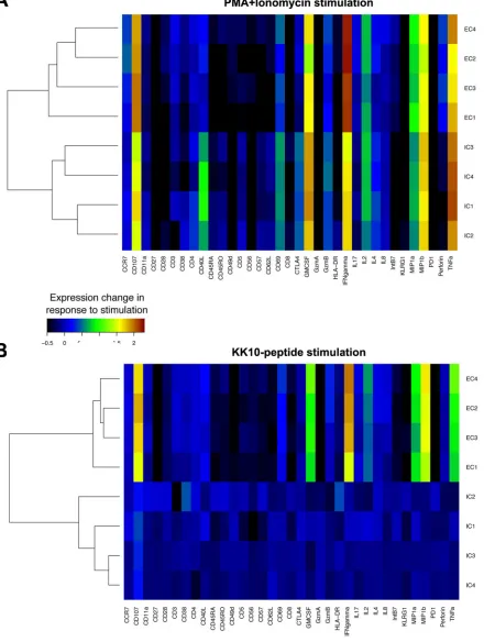
[image:3.585.41.371.84.194.2]Phenotypic profiles of KK10-specific effective and ineffective clonotypes.
We
next used CyTOF to assess potential associations between CD8
⫹T-cell clone function
and TCR-dependent and TCR-independent activation-induced expression of 20
pheno-typic and 13 functional markers (18). Data were analyzed in a manner similar to that
used for standard flow cytometry data by FlowJo software as shown in representative
scatter plots (Fig. 2). The expression of phenotypic and functional markers was first
analyzed by CyTOF after stimulation for 3 h with phorbol myristate acetate (PMA) and
ionomycin, a TCR-independent activation signal that directly triggers downstream
signal pathways. Effective CD8
⫹T-cell clones segregated together by cluster analysis,
as did ineffective CD8
⫹T-cell clones, although the differences between effective and
ineffective clones in the TCR-independent induction of expression of these markers
were modest (Fig. 3A). In contrast, following antigen-specific TCR stimulation for 3 h
with cognate KK10 peptide-loaded HLA-B
*
2705-expressing GXR cells, differences in
phenotypic and functional markers between effective and ineffective clonotypes were
readily apparent after subtracting the background from stimulations with GXR cells not
TABLE 1Clonotypes of HLA-B*2705-restricted KK10-specific CD8⫹T-cell clonesClone (original
designation)a TCRBVb CDR3 TCRBJc
% Specific lysisd
EC1 (S-C003) 25.1 CASSEADFEAF 1.1 74
EC2 (S-T001) 18 CASSPGQFSHEQY 2.7 77
EC3 (B3) 4.3 CASRPGLASNEQF 2.1 75
EC4 (B5) 6.5 CASRPGQGATEAF 1.1 78
IC1 (S-C007) 20.1 CSARDGGEQY 2.7 30
IC2 (B6) 20.1 CSARDRGTREVADNYGYT 1.2 22
IC3 (002) 5.6 CASGGGTVYEQY 2.7 26
IC4 (013) 2 CASSAGPGQYGNTIY 1.3 16
IC5 (S-T002) 7.9 CASSLDRLEQF 1.1 ND
aThe original designations are from reference 1. bTCRBV, TCR-chain variable region. cTCRBJ, TCR-chain joining (J) region.
dThe ability of KK10-specific effective CD8⫹T-cell clones (EC) and ineffective CD8⫹T-cell clones (IC) to kill GXR cells infected with NL4-3 wild-type virus was tested in the standard 4-h chromium release assay, and percent specific lysis was calculated as described in Materials and Methods. CD8⫹T-cell clones EC1, EC2, IC1, and IC5 were obtained from elite controller CTR203, CD8⫹T-cell clones EC3, EC4, and IC2 were obtained from elite controller FW56, and CD8⫹T-cell clones IC3 and IC4 were obtained from chronic progressor CR540 (1). The ineffective functional phenotype of IC5 (as S-T002) has previously been established and reported (1). ND, not done.
TCR Clonotype Modulation of CD8⫹T-cell Function Journal of Virology
March 2017 Volume 91 Issue 6 e02412-16 jvi.asm.org 3
on November 7, 2019 by guest
http://jvi.asm.org/
loaded with peptide. The effective and ineffective clonotypes did not differ significantly
in their expression of regulatory markers such as PD-1, CTLA-4, and KLRG-1, activation
markers such as CD38, CD57, CD69, and HLA-DR, and differentiation markers such as
CD45RA, CD45RO, CD27, CD28, CD62L, and CCR7. In contrast, we observed significantly
increased production of the functional cytokines gamma interferon (IFN-
␥
),
macro-phage inflammatory protein 1

(MIP-1

), MIP-1
␣
, tumor necrosis factor alpha (TNF-
␣
),
and granulocyte-macrophage colony-stimulating factor (GM-CSF) and the
degranula-tion marker CD107a in the effective CD8
⫹T-cell clones compared to the responses
FIG 2Representative CyTOF scatter plots. The effective CD8⫹T-cell clone EC1 and ineffective CD8⫹T-cell clone IC2 were incubated with either unloaded
HLA-B*2705-expressing GXR cells, KK10 peptide-loaded HLA-B*2705-expressing GXR cells or PMA and ionomycin (Iono) for 3 h. The cells were then stained with
the indicated metal-tagged antibody specific for IFN-␥and CD107 (upper panels), CD40L and GM-CSF (middle panels), and IL-2 and TNF-␣(lower panels) and
analyzed by mass cytometry. The data are presented as dot plots.
FIG 1Antiviral function of KK10-specific clonotypes. The ability of KK10-specific CD8⫹T-cell clones to
recognize NL4-3 WT and variant viruses was tested in the standard 4-h chromium release assay with virally infected (wild-type and variant strains as indicated) HLA-B*2705-encoding green fluorescent protein (GFP) reporter GXR cells at an effector/target cell ratio of 1:1. Viable infected (GFP-positive) GXR cells were sorted by a FACSAria cell-sorting instrument after infection for 5 days and used as target cells.
on November 7, 2019 by guest
http://jvi.asm.org/
[image:4.585.61.352.73.228.2] [image:4.585.43.543.420.698.2]FIG 3CyTOF characterization of functional and phenotypic markers. Stimulation-induced changes in 20 phenotypic and 13 functional markers
on clonotypic CD8⫹T cells were measured by CyTOF after incubation for 3 h with PMA and ionomycin (A) or KK10 peptide-loaded
HLA-B*2705-expressing GXR cells (B). Stimulation-induced changes in intensity of each marker were averaged, and background was subtracted. Similarity between clonotypes was compared by hierarchical cluster analysis. Similarities between the profiles of each marker tested were also clustered.
TCR Clonotype Modulation of CD8⫹T-cell Function Journal of Virology
March 2017 Volume 91 Issue 6 e02412-16 jvi.asm.org 5
on November 7, 2019 by guest
http://jvi.asm.org/
[image:5.585.48.488.83.667.2]seen in the ineffective CD8
⫹T-cell clones (
P
⬍
0.001) (Fig. 3B). These results indicate
that although effective and ineffective clonotypes segregate based on both
TCR-independent and TCR-dependent stimulation, the latter is associated with much
greater differences in a subset of phenotypic and functional markers associated
with antiviral functions.
TCR signal transduction of KK10-specific clonotypes.
We next assessed
immedi-ate effector function following cognimmedi-ate epitope recognition by each CD8
⫹T-cell clone
expressing a different TCR clonotype, using phosphoflow cytometry to assess the
kinetics and amplitude of the phosphorylation of proteins crucial for TCR signal
transduction, such as the p44/42 mitogen-activated protein kinase (MAPK) extracellular
signal-regulated kinase 1/2 (ERK1/2) (19). Ten minutes after stimulation with KK10
peptide, the phosphorylated protein forms of p44/42 MAPK in the effective CD8
⫹T-cell
clones were more rapidly mobilized than in the ineffective CD8
⫹T-cell clones (Fig. 4A
and B) (
P
⫽
0.028), whereas the effective and ineffective CD8
⫹T-cell clones displayed
a similar mobilization rate of phosphorylated MAPK following TCR-independent
stim-ulation with PMA and ionomycin (
P
⫽
0.28). These data indicated marked differences
in the kinetics of antigen-specific TCR-initiated signaling events between effective and
ineffective CD8
⫹T-cell clones.
Subsequent flow-cytometric analysis of dynamic intracellular cytokine profiles
dem-onstrated that a representative effective CD8
⫹T-cell clone, EC1, rapidly upregulated
antiviral cytokines and chemokines such as IFN-
␥
, MIP-1

, and TNF-
␣
, an upregulation
that was paralleled by declining levels of perforin expression (Fig. 4C), indicating the
rapid transition from efficient initial lytic to sustained nonlytic function. In contrast to
FIG 4TCR signal transduction and cytokine kinetics. (A) A representative flow cytometry histogram of phosphorylated protein forms of p44/42 MAPK examined by phosphoflow cytometry at the 10-min time point following stimulation with KK10 peptides or PMA and ionomycin of EC1
and IC1 CD8⫹T-cell clones. (B) Comparison of phosphorylation of p44/42 MAPK between effective and ineffective CD8⫹T-cell clones was made
with the Mann-Whitney test. (C and D) Functional cytokine and chemokine expression was examined by flow cytometry after incubation with
KK10 peptide-loaded HLA-B*2705-expressing GXR cells for 0.5 h, 1 h, 2 h, 3 h, and 5 h for effective CD8⫹T-cell clone EC1 (C) and ineffective CD8⫹
T-cell clone IC1 (D).
on November 7, 2019 by guest
http://jvi.asm.org/
[image:6.585.43.513.70.381.2]the effective CD8
⫹T-cell clone, only a fraction of the representative ineffective IC1
CD8
⫹T-cell clone cells displayed loading of lytic granules and upregulation of these
antiviral cytokines and chemokines, and this response was delayed (Fig. 4D). Early
perforin expression in the effective clonotypes declined and was followed by significant
expression of degranulation marker CD107a (Fig. 4C), consistent with previous findings
that effective clonotypes rapidly load and deliver perforin (1), mediating target cell lysis,
which is associated with the acquisition of cell surface CD107a (20).
Direct expression of KK10-specific TCRs from representative effective and
ineffective clonotypes using TCR expression lentiviral vectors.
To more precisely
determine the contribution of the TCR clonotype to the differences observed in CD8
⫹T-cell clones specific for the same viral peptide but segregated into the effective or
ineffective CD8
⫹T-cell clones, we directly evaluated the functional activity of TCR
clonotypes by cloning the full-length TCR
␣
and

chains from representative effective
(EC1) and ineffective (IC1 and IC5) CD8
⫹T-cell clones (Table 1). These CD8
⫹T-cell
clones were isolated from the same elite controller (CTR203) and were specific for KK10
and its L6M variant but not the R2T and R2T/L6M variants (1). The TCR
␣
and

chains
cloned from the EC1, IC1, and IC5 CD8
⫹T-cell clones were expressed in a lentiviral
vector (Fig. 5A) encoding a single transcript linked by a P2A self-cleaving peptide that
produces equimolar quantities of both TCR chains in transduced T cells and an internal
ribosome entry site (IRES)-driven green fluorescent protein (GFP) marker gene to enable
identification and quantification of transduced T cells, as described previously (21). A
FIG 5Phenotypic and functional analysis of the EC1, IC1, and IC5 TCRs expressed by the transduced Jurkat/MA cells. (A) The TCR␣andgenes
of effective (EC1) and ineffective (IC1 and IC5) CD8⫹T-cell clones were cloned into a lentiviral transfer vector driven by a spleen focus-forming
virus (SFFV) viral promoter. TCR␣andgenes were linked by a self-cleaving P2A peptide followed by an IRES-driven GFP marker gene to allow
visualization of the transduced cells by flow cytometry. (B) Efficiency of Jurkat/MA cell transduction by the GFP-expressing EC1-TCR, ICI-TCR, and IC5-TCR lentiviral vectors and their expression of a KK10-specific TCR were determined by flow-cytometric analysis 72 h after transduction by staining with HLA-B*2705-KK10 dextramer. (C) TCR responsiveness was measured by incubating mock-transduced and EC1-TCR-, IC1-TCR-, or
IC5-TCR-transduced Jurkat/MA cells with CTR0075 cells pulsed with the indicated peptide (10M) for 16 h and quantifying luciferase activity in
cellular lysates. (D) Functional activity of the EC1-TCR, IC1 TCR, and IC5-TCR expressed by the Jurkat/MA cells was determined by stimulating the
transduced cells with CTR0075 cells pulsed with the indicated concentration of KK10 peptide or a control influenza virus peptide (10M) for 16
h and measuring luciferase activity. The data shown in panels C and D are normalized relative luciferase unit (RLU) values from 3 individual experiments performed in duplicate. We normalized the data from 3 experiments to aid in accurate comparison between individual experiments
using the formulaXi,0 to 1⫽(Xi⫺Xmin)/(Xmax⫺Xmin), whereXirepresents each data point,Xminis the minimum among all data points in an
experiment,Xmaxis the maximum among all data points in an experiment, andXi,0 to 1represents the data pointinormalized between 0 and 1.
The data shown are the mean normalized RLU⫾standard errors of the means (SEM) from 3 experiments performed in duplicate. Statistical
significance was evaluated using the 2-way analysis of variance (ANOVA) test.*,P⬍0.05;****,P⬍0.0001.
TCR Clonotype Modulation of CD8⫹T-cell Function Journal of Virology
March 2017 Volume 91 Issue 6 e02412-16 jvi.asm.org 7
on November 7, 2019 by guest
http://jvi.asm.org/
[image:7.585.46.498.76.323.2]modified CD8
⫹Jurkat cell line, Jurkat/MA, that does not express endogenous surface
TCR, was transduced with the TCR-expressing lentivirus and evaluated for the
expres-sion and functional properties of the lentivirus-encoded TCR (22). Greater than 90% of
the Jurkat/MA cells were transduced with the individual TCR lentiviruses as indicated by
expression of GFP and of surface TCR, which specifically bound to the HLA-B
*
2705-KK10
dextramer (Fig. 5B). Based on the geometric mean fluorescent intensity (MFI) of
dextramer binding, EC1-TCR (MFI
⫽
40,616 relative fluorescence units [RFU]) and
IC1-TCR (MFI
⫽
30,072 RFU) were expressed at moderately higher densities than
IC5-TCR (MFI
⫽
13,759 RFU), which may be due to the differential stability of different
TCR
␣
and

proteins (23).
Functional activity of the expressed TCR from effective versus ineffective
clonotypes.
The stable integration of the TCR-induced NFAT luciferase reporter
con-struct in the Jurkat/MA cell line permitted this cell line to be used to quantify the
amplitude of the antigen-specific signal transduced by defined TCRs (21). Jurkat/MA
cells were transduced with the indicated lentivirus and incubated with CTR0075 cells,
allogeneic HLA-B
*
2705-expressing B-LCL cells, loaded with either the KK10 peptide, the
KK10-L6M variant peptide, or an HLA-B27 control influenza virus peptide (influenza A
virus nucleopeptide, SRYWAIRTR) for
⬃
16 h (24). The responsiveness of the
lentivirus-encoded TCR to its cognate pMHC complex was quantified by measuring the amplitude
of increased luciferase activity compared to that in mock-transduced cells. In contrast
to the minimal reactivity of the Jurkat/MA cells expressing EC1-TCR, IC1-TCR, or IC5-TCR
to CTR0075 cells pulsed with the control influenza virus peptide, CTR0075 cells pulsed
with the KK10 peptide or the KK10-L6M variant peptide markedly stimulated luciferase
activity in the Jurkat/Ma cells expressing EC1-TCR, IC1-TCR, or IC5-TCR (Fig. 5C). The
level of luciferase activity induced after EC1-TCR KK10-specific activation was not
significantly greater than after IC1-TCR activation (
P
⫽
0.30) but was significantly higher
than after the IC5-TCR activation (
P
⬍
0.05).
We further evaluated the functional activities of the expressed EC1-TCR, IC1-TCR,
and IC5-TCR by incubating Jurkat/MA cells transduced with the EC1-TCR, IC1-TCR, or
IC5-TCR lentivirus with CTR0075 cells loaded with no peptide, a control influenza virus
peptide (10
M), or KK10 peptide at concentrations that ranged from 0.001
M to 100
M. The Jurkat/MA cells transduced with EC1-TCR, IC1-TCR, or IC5-TCR displayed a
similar dose-response activation of the TCR-responsive luciferase reporter construct in
the Jurkat/MA cells, and there were no differences in magnitude between EC1-TCR and
IC1-TCR (Fig. 5D). However, at the higher peptide doses, EC1-TCR and IC1-TCR
express-ing Jurkat/MA cells displayed reproducibly higher maximal activation of the
TCR-responsive luciferase reporter construct than IC5-TCR expressing Jurkat/MA cells, albeit
the magnitude of differences was small. Overall, these data indicate a lack of significant
differences in TCR-mediated activation between an effective clonotype (EC1) and one
of the ineffective clonotypes (IC1), demonstrating that TCR alone does not account for the
functional differences observed in the parental clones IC1 and IC5. The difference observed
in the comparison of EC1 to IC5 is difficult to interpret, given that the clone with the lower
activation (IC5) also had the lowest TCR expression levels.
Primary human CD8
ⴙT cells transduced with lentivectors encoding the EC1,
IC1, or IC5 TCR clonotypes display equivalent anti-HIV-1 activity.
We have
previ-ously reported that primary naive CD8
⫹T cells can be reprogrammed into HIV-specific
CD8
⫹cells after transduction with lentivirus encoding the TCR
␣
and TCR

genes from
an HLA-A
*
02-restricted SL9-specific clone (21). We used this approach to evaluate the
potency of anti-HIV activity conferred by the TCR clonotypes derived from the
repre-sentative effective and ineffective HLA-B
*
27-restricted CD8
⫹T-cell clones by
transduc-ing primary CD8
⫹T cells isolated from two different HIV-1-naive donors with the EC1,
IC1, or IC5-TCR-encoding lentivirus.
After transduction of primary CD8
⫹T cells with an EC1 or IC1 lentivirus that utilized
an IRES to express the GFP marker gene, we observed consistently low levels of
transduction and binding of the HLA-B
*
2705-KK10 dextramer by GFP
⫹transduced cells
(Fig. 6). To increase transduction efficiency and TCR expression, we replaced the IRES
on November 7, 2019 by guest
http://jvi.asm.org/
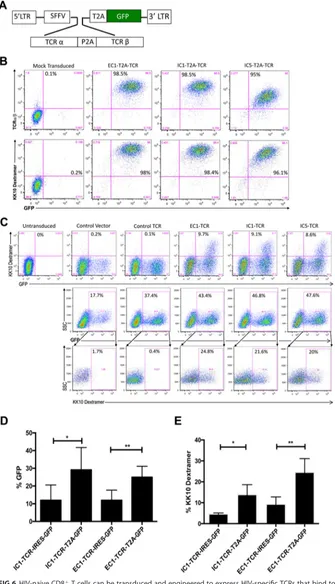
FIG 6HIV-naive CD8⫹T cells can be transduced and engineered to express HIV-specific TCRs that bind to
KK10-specific dextramer. (A) The TCR␣andgenes of effective (EC1) and ineffective (IC1 and IC5) CD8⫹
T-cell clones were cloned into a lentiviral transfer vector driven by a spleen focus-forming virus (SFFV) viral
promoter. TCR␣andgenes were linked by a self-cleaving P2A peptide followed by a T2A self-cleaving
peptide-linked GFP reporter cassette to allow visualization of the transduced cells by flow cytometry. (B) Jurkat/MA cells were transduced with lentiviral vectors expressing EC1-TCR, ICI-TCR, IC5-TCR, or a control glutamic acid decarboxylase peptide (residues 555 to 567)-specific TCR linked with a T2A sequence to a GFP reporter gene or a control lentiviral vector expressing only the GFP reporter gene. Expression of a KK10-specific TCR was determined by flow-cytometric analysis 48 h after transduction after staining with
(Continued on next page)
TCR Clonotype Modulation of CD8⫹T-cell Function Journal of Virology
March 2017 Volume 91 Issue 6 e02412-16 jvi.asm.org 9
on November 7, 2019 by guest
http://jvi.asm.org/
[image:9.585.43.378.73.657.2]sequence expressing the GFP marker gene with a 2A ribosomal “skip” peptide (T2A
[Thosea asigna virus 2A]) (Fig. 6A) reported to increase the expression of multiple
proteins by a single vector (25). Based on flow cytometric analysis, greater than 95% of
Jurkat/MA cells transduced with EC1-TCR-, IC1-TCR-, or the IC5-TCR-encoding
lentivi-ruses linked with a T2A peptide to the GFP marker gene expressed GFP and expressed
TCR that bound the HLA-B
*
2705-KK10 dextramer (Fig. 6B). Primary CD8
⫹T cells were
efficiently transduced with lentivirus encoding EC1, IC1, and EC5 TCR and expressed
TCR that bound the HLA-B
*
2705-KK10 dextramer (Fig. 6C). The transduction efficiency
and HLA-B
*
2705-KK10 dextramer binding in primary CD8
⫹T cells transduced with the
EC1-TCR- and IC1-TCR-encoding lentivirus, which used a T2A-linked GFP marker gene
(Fig. 6C), was significantly higher than with the EC1-TCR- and IC1-TCR-encoding
lenti-virus, which used an IRES (
P
⬍
0.05) (Fig. 6D and E). Multiple factors contribute to the
discrepancy in transduced CD8
⫹primary T cells between GFP
⫹reporter gene
expres-sion and the expresexpres-sion of sufficient surface TCR for MHC-KK10 dextramer staining,
including TCR expression cassette design, TCR
␣
and TCR

chain gene codon
optimi-zation and orientation, the more-rapid degradation of the TCR
␣
chain, the varying
stability of individual TCR
␣
and TCR

chains, and mispairing with the endogenous TCR
␣
and TCR

chains (23).
The anti-HIV-1 activity of the primary CD8
⫹T cells transduced with the EC1, IC1, and
IC5-TCR lentivectors was evaluated by using HIV-infected, HLA-B
*
2705-expressing GXR
cells infected with HIV-LucR, an infectious HIV-1 molecular clone expressing a
Renilla
luciferase reporter gene, as target cells as described previously (26). CD8
⫹T cells
transduced with EC1-TCR, IC1-TCR, and IC5-TCR lentivirus displayed equivalently potent
HIV-1-specific inhibitory activity (
⬃
80 to 90%) compared to mock-transduced CD8
⫹T
cells and CD8
⫹T cells transduced with a control lentivirus expressing the GFP gene
alone (Fig. 7A and B). To further evaluate the functional inhibitory capacity of EC1-TCR
and IC1-TCR, we used syngeneic HLA-B
*
2705 CD8
⫹T-cell-depleted peripheral blood
mononuclear cells (PBMC) infected with HIV-LucR as the target HIV-1-infected cells.
Primary CD8
⫹T cells transduced with lentivirus expressing the EC1-TCR and IC1-TCR
equivalently inhibited HIV-1 infection (
⬃
60%) in the primary syngeneic B
*
2705 CD8
⫹T-cell-depleted PBMC (Fig. 7C). These results indicate that some ineffective CD8
⫹T-cell
clones express TCR with the intrinsic capacity to mediate potent suppression of HIV-1
infection when expressed on primary CD8
⫹T cells.
DISCUSSION
Multiple studies have sought to define quantitative and qualitative differences in
CD8
⫹T-cell responses that may correlate with different outcomes in terms of disease
course and immune control of HIV-1 (2). Simple quantitative measures of HIV-specific
CD8
⫹T-cell populations have shown little correlation with viral control (27, 28),
suggesting that qualitative features of HIV-specific CD8
⫹T cells may correlate with
in
vivo
control. Qualitative factors potentially modulating CD8
⫹T-cell responses include,
among others, polyfunctionality (29), antigen sensitivity or functional avidity (30, 31),
FIG 6Legend (Continued)
HLA-B*2705-KK10 dextramer or anti-human TCR␣antibody. (C) Primary human CD8⫹T cells, isolated
from PBMC of HIV-naive donors, were activated and transduced with lentiviral vectors expressing EC1-TCR,
IC1-TCR, or IC5-TCR linked with a T2A sequence to a GFP reporter gene. After 8 days of culture, the CD8⫹
T cells were evaluated by flow cytometry for transduction by quantifying the fraction of GFP⫹cells. After
gating on the GFP⫹cells, their expression of the KK10-specific TCR was determined by measuring their
binding to HLA-B*2705-KK10 dextramer. Results shown are representative of 3 experiments with 2 different
donors. SSC, side scatter. (D) Primary human CD8⫹T cells were activated and transduced with the lentiviral
vectors expressing EC1-TCR or IC1-TCR linked to a GFP reporter gene by a T2A peptide or expressed by an
IRES-driven GFP reporter gene. After 4 days of culture, the CD8⫹T cells were evaluated by flow cytometry
for transduction by quantifying the fraction of GFP⫹cells. The values presented are the means⫾SEM of
the percentage of GFP-positive CD8⫹T cells from 9 different experiments using primary CD8⫹T cells from
3 different HIV-naive donors. (E) After gating, the GFP⫹cells were evaluated for their expression of the
KK10-specific TCR by measuring their binding to HLA-B*2705-KK10 dextramer. The values presented are the
means⫾SEM of the percentage of HLA-B*2705-KK10 dextramer binding-CD8⫹T cells from 3 different
experiments using primary CD8⫹T cells from 2 different HIV-naive donors.*,P⬍0.05;**,P⬍0.01.
on November 7, 2019 by guest
http://jvi.asm.org/
proliferative capacity (32), loading of lytic granules (33, 34), specific targeting of
conserved regions (35, 36), immunoregulatory mechanisms, including CD8
⫹T-cell
exhaustion (37–39), concurrent responses to multiple epitopes restricted by different
HLA alleles (40), and CD8
⫹T-cell-associated mutations that impair viral fitness (41, 42)
and immune escape (43). Some studies also suggest that properties of the TCR-pMHC
interaction may play a role in CD8
⫹T-cell functional activity (44, 45).
We previously reported that HLA-B
*
27-restricted and HLA-B
*
57-restricted CD8
⫹T-cell clones targeting the same epitope in elite controllers and progressors can be
clearly differentiated based on potency and cross-reactivity of TCR recognition of HIV-1
and viral variants, which is in turn related to specific TCR clonotypes that are selected
during natural HIV-1 infection (1). One factor that may contribute to the elite controller
phenotype is the “fortunate” selection and expansion of anti-HIV CD8
⫹T cells
express-ing broadly reactive TCRs durexpress-ing the evolution of the KK10-specific CD8
⫹T-cell
response that does not occur in progressors. Supporting this scenario was our isolation
of effective clonotypes such as EC2 and EC4, which were also characterized by broad
cross-reactivity against viral variants; these broadly reactive CD8
⫹T cells are well
FIG 7Antiviral capacity of TCR-engineered CD8⫹T cells. CD8⫹T cells were isolated from PBMC, activated, andmock transduced or transduced with lentivirus expressing EC1-TCR, IC1-TCR, or IC5-TCR and a GFP reporter linked by a T2A peptide or a control lentivirus expressing only the GFP reporter gene. (A) Five days after transduction, the
cells were added to GXR cells previously infected with HIV-LucR for 24 h at a CD8⫹T cell/GXR cell ratio of 1:1. After
3 days, the cells were harvested and the luciferase activities in the cellular lysates were determined and reported
as RLU⫾SEM. Results from a representative experiment performed in quadruplicate are shown. (B) The individual
means of 4 experiments performed as described for panel A with the data for the experiments normalized as
percent suppression of HIV-1 infection and the total means from the 4 independent experiments⫾SEM are shown.
(C) CD8⫹T cells mock transduced or transduced with lentiviral vectors expressing EC1-TCR or IC1-TCR were added
to syngeneic HLA-B27-expressing activated CD8⫹T cell-depleted PMBC 2 days after they were infected with
HIV-LucR. Six days later, the luciferase activity in the cellular lysates was determined and reported as RLU⫾SEM.
Statistical significance was evaluated using the ordinary one-way ANOVA test. ns,P⬎0.05;**,P⬍0.01;****,P⬍
0.0001.
TCR Clonotype Modulation of CD8⫹T-cell Function Journal of Virology
March 2017 Volume 91 Issue 6 e02412-16 jvi.asm.org 11
on November 7, 2019 by guest
http://jvi.asm.org/
[image:11.585.43.404.74.401.2]represented in controllers but rare or absent in progressors, whereas ineffective
clono-types such as IC1, IC2, IC3, IC4, and IC5 are present in both progressors and controllers.
However, other factors likely contribute to the effective CD8
⫹T-cell phenotype as
indicated by EC1, which reacted with only the KK10 and L6M variant, and EC3, which
was predominately reactive with the unmutated KK10 peptide (Fig. 1). We and others
have further demonstrated that the antiviral efficacy and cross-reactivity of the
clono-types were determined by different recognition and binding patterns among
TCR-pMHC interactions by crystallographic and computational studies (6, 8). All-atom
molecular dynamics simulations of TCR-KK10-MHC complexes revealed a structural
association with the clonotypic differences in CD8
⫹T-cell phenotypes and functions.
Although both effective and ineffective clonotypes bind to the N- and C-terminal
portions of the KK10-MHC through similar salt bridges, specific hydrophobic side chain
interactions with the TCR are the major force associated with the observed superior
antiviral efficacy of certain TCR clonotypes (6, 8).
The nature of the TCR-pMHC interactions directs the physical recruitment of
signal-ing pathways differentially inside the lymphocyte, which impacts on the kinetics of
signal propagation in various segments of the TCR signaling network that ultimately
influence the responsiveness of T cells (46–48), but the mechanistic basis for this
difference in signal transduction due to TCR variants is not established. We observed
that effective and ineffective clones were more greatly differentiated by KK10-specific
activation than by TCR-independent activation and that effective clones
phosphory-lated p44/42 MAPK more rapidly than ineffective clones when stimuphosphory-lated with their
cognate pMHC (Fig. 4A). This correlates with our previous findings that the effective
CD8
⫹T-cell clones are able to rapidly upregulate perforin and granzyme B, polarize
them at the immunologic synapse, and deliver them to the infected target cells after
incubation with HIV-1-infected HLA-B
*
2705-encoding GXR cells for 30 min (1). This also
explains why we did not observe much perforin and granzyme B in effective clonotypes
by CyTOF after culture with KK10 peptide-loaded HLA-B
*
2705-encoding GXR cells for 3
h; this time point likely followed the rapid delivery of perforin and granzyme B, which
is indicated by the rapid upregulated expression of CD107a that we observed on the
effective CD8
⫹T-cell clones following stimulation for 3 h (Fig. 4C). CD107a expression
is associated with loss of intracellular perforin, can be observed as early as 30 min
following stimulation of primary CD8
⫹T cells, and reaches maximum expression by 4
h after activation (20). During this phase, effective CD8
⫹T-cell clones upregulate
secretion of antiviral cytokines and chemokines, such as IFN-
␥
, MIP-1

, MIP-1
␣
, and
TNF-
␣
, and readily transit from efficient initial lytic to sustained nonlytic function,
whereas the ineffective CD8
⫹T-cell clones lack this ability.
In order to more precisely address the specific roles of TCR clonotypes in the
observed functional differences, we directly cloned and expressed the TCRs from one
effective CD8
⫹T-cell clone and two ineffective CD8
⫹T-cell clones from a single donor,
all with an identical specificity for WT KK10 and L6M KK10 but not R2T KK10. We
demonstrated using Jurkat/MA cells transduced with TCR-expressing lentiviral vectors
that the EC1 and IC1 TCR clonotypes displayed comparable TCR responsiveness to
HLA-B
*
2705-expressing target cells loaded with the KK10 peptide or the KK10 L6M
variant peptide, while the maximal response of the IC5 TCR clonotype was less than
that of EC1 and IC1 (Fig. 5C and D). Although this may reflect true TCR-specific
modulation of effector function between the effective and ineffective clonotypes, it
may also be due to the lower levels of expression of IC5 TCR than of EC1 and IC1 TCR
(Fig. 6B), which may be a consequence of differential intrinsic stability of different TCR
␣
and

chains (23). Our data do indicate that the phenotype of an ineffective
clonotype is not transferred by the TCR alone in all cases, since there were no functional
differences observed between cells transfected with the EC1 and IC1 TCRs, using either
TCR-transduced Jurkat/MA cells (Fig. 6) or primary CD8
⫹T cells (Fig. 7).
While we clearly show that a TCR cloned from an ineffective clone has equivalent
function to that of a TCR cloned from an effective clone, a limitation of this study is that
it evaluates only cloned TCRs from one effective and two ineffective clones from the
on November 7, 2019 by guest
http://jvi.asm.org/
same individual. Another limitation is that we studied the function of HIV-specific TCRs
expressed by lentiviral vectors in primary CD8
⫹T cells, which may not precisely
recapitulate the behavior of HIV-specific CD8
⫹T cells because of inherent restrictions
of lentivirus-encoded TCR expression, including vector design, orientation of the
ex-pression cassette, and the impact of mispairing with the endogenous TCR
␣
and

chains of the transduced CD8
⫹T cell (23). The impact of TCR chain mispairing likely
contributed to the disparity in TCR expression between Jurkat/MA cells, which do not
express endogenous TCR and among which almost all GFP
⫹cells bound to the
HLA-B
*
2705-KK10 dextramer (Fig. 6B), and the GFP
⫹primary CD8
⫹T cells, of which
only about 20 to 25% bound dextramer (Fig. 6C). Nevertheless, an adequate number of
primary CD8
⫹T cells expressed sufficient lentivirus-encoded TCR to enable us to
demonstrate that the EC1 TCR and IC1 TCR, and perhaps to a lesser extent the IC5 TCR,
conferred upon primary CD8
⫹T cells the capacity to potently inhibit HIV-1 infection.
Alternate TCR-independent mechanisms may modulate CD8
⫹T-cell function,
in-cluding the introduction of epigenetic changes into gene-regulatory elements of CD8
⫹T-cell differentiation and function that can be transmitted to daughter cells and
triggered by acute viral infection (49–53). For example, commitment of virus-specific
CD8
⫹T cells to an exhausted phenotype is reinforced by persistent demethylation of
the PD-1 locus as described for lymphocytic choriomeningitis virus (LCMV) and HIV
infections (54–56). Thus, the effectiveness of the antiviral response of CD8
⫹T cells
experiencing sustained high levels of TCR signaling during chronic virus infection may
be compromised by epigenetic modifications that permanently repress transcription,
which may be overcome by the lentivector-mediated expression of the ineffective
CD8
⫹T-cell clone TCR in CD8
⫹T cells from an HIV-1-naive donor that are not carrying
these epigenetic modifications. It is possible that the stimulation of epigenetic
modi-fications by chronic
in vivo
stimulation that confer an ineffective phenotype upon CD8
⫹T cells may preferentially occur in CD8
⫹T cells expressing some TCR clonotypes but not
others, enabling them to continue to function as effective CD8
⫹T cells.
The results reported here also support the capacity of lentiviral vectors to transform
naive CD8
⫹T cells into potent HIV-specific CD8
⫹T cells as a possible adjunctive
therapy to control and eliminate HIV-infected cells, potentially including reactivated
latently infected cells, thereby contributing to achieving a functional cure. For optimal
therapeutic efficacy, an approach needs to be developed to maximize expression of the
lentivirus-encoded TCR in the transduced primary CD8
⫹T cells. Treatment of
HIV-1-infected individuals with infusions of autologous CD8
⫹T cells transduced
ex vivo
with
lentivectors encoding broadly reactive HIV-1-specific TCRs isolated from a broadly
directed CD8
⫹T-cell clone such as EC2 and EC4 would be expected to delay the
emergence of immune escape variants and thereby to confer increased immune
control as observed in elite controllers. It is also possible that TCRs derived from these
effective CD8
⫹T-cell clones may display prolonged
in vivo
anti-HIV-1 activity due to
resistance to inhibitory and regulatory mechanisms, which would compromise their
function as reported for the association between increased functional avidity of
HIV-specific CD8
⫹T cells and T-cell exhaustion (57).
Taken together, these findings suggest that factors other than TCR structure likely
contribute to the ineffective phenotype of some CD8
⫹T-cell clones. Determining these
factors may enable the development of
in vivo
treatments to convert ineffective
HIV-specific CD8
⫹T cells into effective CD8
⫹T cells and to convert infected individuals
who are chronic progressors into elite controllers.
MATERIALS AND METHODS
Generation of CD8ⴙT-cell clones.PBMC were isolated from HIV-infected individuals and stained
with fluorophore-labeled HLA-B*2705 KK10 tetramer (ProImmune, Oxford, UK) and fluorophore-labeled
anti-CD8 and anti-CD3 antibodies. Tetramer-positive, CD8⫹T cells were sorted on a FACSAria cell-sorting
instrument (BD Biosciences, San Jose, CA) at 70 lb/in2, and single cells were placed into each well of
96-well plates, using irradiated allogeneic PBMC and CD3-specific monoclonal antibody (MAb) 12F6 to stimulate T-cell proliferation (58). Developing HLA-B*2705-restricted, KK10-specific clones were identified
by IFN-␥enzyme-linked immunospot (ELISPOT) assays after stimulation with optimal epitopes and by
tetramer staining. Cloned HLA-B*2705-restricted, KK10-specific CD8⫹T cells were maintained by
restimu-TCR Clonotype Modulation of CD8⫹T-cell Function Journal of Virology
March 2017 Volume 91 Issue 6 e02412-16 jvi.asm.org 13
on November 7, 2019 by guest
http://jvi.asm.org/
lation every 14 to 21 days with an anti-CD3 MAb and irradiated allogeneic PBMC in RPMI 1640 medium
with added heat-inactivated fetal bovine serum (10%, vol/vol), 100 U/ml penicillin, 10g/ml
strepto-mycin, 2 mM glutamine, and 10 mM HEPES buffer (R-10) supplemented with 50 units/ml recombinant interleukin-2 (IL-2) (R-10-50) (58). TCR clonotypes were determined by TCR sequencing, as described previously (59).
Cell lines and isolation of CD8ⴙT cells from human peripheral blood mononuclear cells.The
Jurkat/MA cells are molecularly engineered Jurkat T cells that do not express endogenous surface TCR and express CD8 and an NFAT-regulated luciferase reporter gene (22) and were cultured as described previously (24). CTR0075 is an Epstein-Barr virus (EBV)-transformed B cell line isolated from an HIV-infected patient with the HLA type of A*0201/0301, B*1501/2705, Cw*0102/0304. GXR is an HLA-B*2705-expressing CEM-derived cell line stably transduced with a plasmid encoding green fluorescent protein (GFP) driven by the long terminal repeat of HIV-1 and was constructed as described previously (60).
Purified CD8⫹T cells were isolated from peripheral blood mononuclear cells by immunomagnetic sorting
using CD8 microbeads (Miltenyi Biotec, San Diego, CA) according to the manufacturer’s instructions.
Chromium release assay.GXR cells were infected at the specified multiplicity of infection (MOI) with
HIV-1NL4-3wild-type or designated HIV-1NL4-3KK10-variant viruses generated by site-directed
mutagen-esis to introduce one or more mutations in the region of the gene encoding the Gag p24 KK10 peptide (KRWIILGLNK, Gag amino acids [aa] 263 to 272) (61). On day 5 after infection, viable GFP-expressing
HIV-1-infected cells were isolated on a FACSAria cell sorter and loaded with radiolabeled51chromium for
1 h at 37°C. CD8⫹T-cell clones were then mixed with the target GXR cells at the indicated effector-target
ratios, and cytotoxic activity of the CD8⫹ T-cell clones was determined using a standard 4-hour
chromium release assay as described previously (62). Percent specific lysis was calculated as [(mean
experimental cpm⫺mean spontaneous cpm)/(mean maximum cpm⫺mean spontaneous cpm)]⫻100,
where cpm is counts per minute. Spontaneous and maximum releases were determined by incubating the labeled target cells with medium alone or 2% Triton X-100, respectively.
Phosphoflow cytometry. KK10-specific CD8⫹ T-cell clones were stimulated with
HLA-B*2705-expressing GXR cells loaded with KK10 peptides (1M) for 10 min and fixed in cold Cytofix buffer (BD
Biosciences). Cells were permeabilized using Perm/Wash buffer III (BD Biosciences), stained with the appropriate phospho-specific antibodies (Cell Signaling Technology, Danvers, MA), washed thoroughly, and then analyzed on an LSRFortessa flow cytometer (BD Biosciences).
Flow cytometry.Cells were stained with fluorochrome-labeled antibodies to the indicated pheno-typic markers for 30 min at room temperature, washed, and fixed using the Cytofix/Cytoperm kit (BD Biosciences) according to the manufacturer’s instructions. Following fixation, the cells were washed twice with the Cytofix/Cytoperm wash buffer and stained with antibodies against intracellular proteins. Following staining, the cells were fixed by resuspension in phosphate-buffered saline (PBS) containing 2% paraformaldehyde. Cellular fluorescence was evaluated by an LSRFortessa cytometer (BD Biosci-ences), and the data were analyzed with the FlowJo software package (FlowJo, Ashland, OR).
Mass cytometry (CyTOF).Stimulation and staining of the KK10-specific CD8⫹T-cell clones and
analysis of data were performed as described previously (18). Briefly, CD8⫹T-cell clones were cultured
for 3 h in R-10 medium containing brefeldin (5g/ml), monensin (5g/ml), and anti-CD107a (2.5g/ml).
For KK10 peptide stimulation, CD8⫹T-cell clones were cultured with HLA-B*2705-expressing GXR cells
preloaded with KK10 peptide (1M) for 45 min at an effector cell/stimulator cell ratio of 1:1 at 37°C or
GXR cells not loaded with peptide as a negative control. At the end of the 3-h stimulation, cells were washed, resuspended in flow cytometry buffer (PBS– 0.05% sodium azide–2 mM EDTA–2% fetal calf serum), and stained for 30 min on ice with a prepared cocktail of metal-conjugated surface marker
antibodies. After surface staining, cells were washed, resuspended in DM-115 (20M; Fluidigm, San
Francisco, CA) for 30 min on ice, washed three times in flow cytometry buffer, and then resuspended in PBS containing 2% paraformaldehyde (Electron Microscopy Sciences, Hatfield, PA). After fixation at 4°C overnight, the cells were washed twice in intracellular staining permeabilization buffer (eBioscience, San Diego, CA), stained with a cocktail of antibodies to phenotypic and functional markers (Table 2) on ice for 45 min, washed twice in flow cytometry buffer, and labeled for 20 min at room temperature with 250 nM iridium interchelator (Fluidigm) suspended in PBS containing 2% paraformaldehyde. The cells were washed twice in flow cytometry buffer, twice in PBS, and twice in distilled water before dilution to the
appropriate concentration required to achieve an acquisition rate of⬍500 events/second on the CyTOF
instrument. Data were analyzed in a manner similar to that used for standard flow cytometry data by FlowJo software (FlowJo). Bioexponential transformed intensity values of each marker were averaged,
and similarity between KK10-specific CD8⫹T-cell clonotypes was analyzed by hierarchical cluster analysis
(using Matlab and Euclidean distance). After the average intensity values were plotted as a heat plot (maximal intensity in red, minimal intensity in blue), the differences in average between stimulated and unstimulated cells were normalized to the largest observed differences for each plot and represented as heat plots (large positive differences in red, small/negative differences in blue).
Cloning of TCR ␣and chains and construction of lentiviral vectors for clonotype TCR expression. The HLA-B*2705-restricted, KK10-specific TCR gene families and variable regions were sequenced from RNA isolated from a representative effective (EC1, previously reported as clone S-C003)
and two ineffective CD8⫹T-cell clones (IC1 and IC5, previously reported as clones S-C007 and S-T002)
cloned from KK10-specific CD8⫹T cells obtained from the same elite controller (CTR203) (1). The
ineffective functional phenotype of IC5 (as S-T002) has previously been established and reported (1). EC1
(TRAV8-4*03/TRBV-25*01), IC1 (TRAV8-4*03/TRBV20*01), and IC5 (TRAV8-4*03/TRBV7-9*03) TCR␣and
TCRsequences were cloned as a single transcript linked by a “self-cleaving” picornavirus 2A (P2A)
peptide, which permits equimolar translation of the TCR␣and TCRchains regulated by the spleen
on November 7, 2019 by guest
http://jvi.asm.org/
focus-forming virus (SFFV) promoter, followed by an IRES-driven or T2A peptide-linked GFP marker gene as previously described (21, 63, 64). As a control, we used a lentiviral vector expressing a human TCR specific for a peptide derived from the type 1 diabetes autoantigen glutamic acid decarboxylase (GAD555–567) linked to a GFP marker gene by a T2A peptide (65).
Third-generation vesicular stomatitis virus pseudotyped glycoprotein (VSV-g) lentiviral vectors ex-pressing the TCR in transduced cells were generated by calcium-mediated cotransfection of 293T cells with four plasmids: a TCR expression cassette, a construct expressing Rev, a packaging construct
expressing thegagandpolgenes, and a construct expressing a cytomegalovirus (CMV) promoter-driven
VSV-g envelope as described previously (66).
Transduction of Jurkat/MA cells and evaluation of TCR expression by flow cytometry.Jurkat/MA
cells were plated in 24-well plates (5⫻105/well) and the indicated lentivirus at an MOI of 10 in R-10. The
plate was spinoculated at 24,000 rpm for 30 min at 24°C and incubated at 37°C overnight. Fresh R-10 was added, and the transduced cells were cultured for an additional 48 h. TCR expression by transduced Jurkat/MA cells was evaluated by flow cytometric analysis on an LSRII flow cytometer (BD Biosciences) using Flowjo software after staining with phycoerythrin (PE)-labeled-HLA*B2705-KK10 dextramer
(Immu-dex, Copenhagen, Denmark) for 10 min at room temperature or PE-labeled anti-TCR␣ antibody
(Biolegend, San Diego, CA) for 30 min at 4°C and washing with PBS.
Evaluation of TCR function using Jurkat/MA cells expressing an NFAT-regulated luciferase reporter gene.The functional activity of the KK10-specific TCR clonotypes was evaluated by quantifi-cation of the activation of the NFAT-luciferase reporter gene in response to the KK10 (KRWIILGLNK) or KK10-L6M variant peptide (KRWIIMGLNK) (variation underlined) as described previously (21). Jurkat/MA
cells were plated (2.5⫻105cells/well) in a 48-well tissue culture plate or a 96-well plate (1⫻105
cells/well) in R-10 and mock transduced or transduced with EC1-TCR-, IC1-TCR-, or IC5 TCR-expressing lentivirus and cocultured with an equivalent number of CTR0075 HLA-B*2705 antigen-presenting cells
pulsed with 10M the indicated peptide (24). For the peptide titration, the transduced Jurkat/MA cells
were plated in a 96-well plate (1⫻105cells/well) in R-10 and mock transduced or transduced with
[image:15.585.42.374.84.462.2]either the EC1-TCR-, IC1-TCR-, or IC5-TCR-expressing lentivirus and cocultured with an equivalent
TABLE 2Specific antibodies used for CyTOF staining and analysis
Antibody Label atomic mass Ab clone; source
CD3 QDot (112, 114, etc.) S1.4; Invitrogen Qdot655
IL-8 139 E8N1; Biolegend
CD45RA 141 HI100; eBioscience
CD69 142 MCA1442; AbD Serotec
CD5 143 UCHT2; Biolegend
CD45RO 144 UCHL1; Biolegend
CD57 145 HCD57; Biolegend
CD8 146 HIT8a; Biolegend
GM-CSF 147 BVD2-21C11; Biolegend
CD11a 148 HI111; Biolegend
CD4 149 SK3; Biolegend
MIP-1 150 D21-1351; BD custom order
Granzyme B 151 2CF/F5; BD
TNF-␣ 152 MAb11; eBioscience
CD107a/b 153 H4A3, H4B4; BD
CD27 154 LG.7F9; eBioscience
PD1 155 EH12.2H7; Biolegend
CD13 156 WM15; Biolegend
CD19 157 HiB19; Biolegend
KLRG1 158 13F12F2; kind gift from Hanspeter Pircher
CD56 159 HiB19; Biolegend
CD28 160 CD28.2; BD
CD38 161 HIT2; eBioscience
IL-4 162 MP4-25D2
cCAS3 163 C92-605; BD
IL-17 164 BL168; Biolegend
CD40L 165 24-31; Biolegend
IL-2 166 MQ1-17h12; eBioscience
Integrin B7 167 FIB504; Biolegend
CCR7 168 150503; R&D
MIP-1␣ 176 11A3; BD custom order
IFN-␥ 170 4S.B4; eBioscience
HLA-DR 171 L243; BD
CD49d 172 9F10; Biolegend
CTLA-4 173 BN13; Biolegend
CD62L 174 DREG-56; BD
Perforin 175 B-D48; AbCam
Granzyme A 176 CB9; Biolegend
TCR Clonotype Modulation of CD8⫹T-cell Function Journal of Virology
March 2017 Volume 91 Issue 6 e02412-16 jvi.asm.org 15
on November 7, 2019 by guest
http://jvi.asm.org/
number of CTR0075 HLA-B*2705 antigen-presenting cells pulsed with 10-fold dilutions of the
indicated KK10 peptide (24). Cells from duplicate cultures were harvested⬃16 h later, and the
luciferase activity in the cellular lysates was determined using the firefly luciferase assay system (Promega, Madison, WI) and quantified using a Luminat Plus luminometer (Berthold Technologies, Oak Ridge, TN).
Evaluation of the capacity of the TCR clonotypes to inhibitin vitroHIV-1 infection.CD8⫹T cells
isolated by immunomagnetic sorting (Miltenyi Biotec) from PBMC from an HIV-naive donor were plated
in a 24-well plate (5⫻105cells/well) and activated with anti-CD28 antibody (1g/ml), anti-CD3 antibody
(100 ng/ml; Orthoclone OKT3), and IL-2 (100 units/ml). Two days later, the indicated lentivirus (MOI, 10)
and Polybrene (4g/ml) were added to each well, and the plate was spinoculated at 24,000 rpm for 60
min at 24°C and then cultured at 37°C overnight. R-10-100 was added the following day, and the cells
were cultured for an additional 3 days with a transduction efficiency ranging from⬃35% to 45% based
on GFP expression. KK10-specific TCR expression by the transduced CD8⫹T cells was determined by
sequential staining with PE-labeled HLAB*2705/KK10 dextramer for 10 min at room temperature and with Pacific Blue-labeled anti-CD8a antibody (BD Biosciences) for 20 min at 4°C followed by washing with
PBS and analysis by flow cytometry. In parallel, HLA-B*2705-expressing GXR cells (1⫻105cells/well) were
infected in 96-well plates with HIV-LucR (MOI,⬃0.5), an infectious HIV-1 molecular clone that expresses
the HIV-1JR-CSFEnv and aRenilla reniformisluciferase (LucR) reporter gene (67, 68). One day later, the
HIV-1-infected GXR cells and either mock-transduced or lentivirus-transduced CD8⫹T cells were added
at an effector-to-target ratio of 1:1 in quadruplicate and cultured for an additional 3 days. HIV-1 infection was quantified by harvesting the cells, lysing them, and measuring luciferase activity in the cellular lysate using the Promega Renilla Luciferase Assay system (26).
Alternatively, PBMC were obtained from an HIV-naive HLA-B*2705 donor, CD8⫹T cells were isolated
by immunomagnetic sorting and transduced with the indicated lentivirus as described above, and the
CD8⫹T cell-depleted fraction was activated with phytohemagglutinin (PHA; 4g/ml), cultured (1⫻105
cells/well) in R-10-100 in a 96-well plate, and infected with HIV-LucR (MOI,⬃0.5). Two days after infection,
the mock-transduced and lentivirus-transduced syngeneic CD8⫹T cells (1⫻105cells) were added to
triplicate wells containing the infected CD8⫹T-cell-depleted PBMC. Six days later, HIV-1 infection was
quantified by measuring luciferase activity in the cellular lysates.
Study approval.All the studies were performed under protocols approved by the Institutional Review Boards at the Albert Einstein College of Medicine and the Massachusetts General Hospital in compliance with the human experimentation guidelines of the U.S. Department of Health and Human Services.
ACKNOWLEDGMENTS
This work was supported by the National Institute of Drug Abuse at the National
Institutes of Health (DA033788, R01DA036171 to H.G.), the National Institute of Allergy
and Infectious Diseases at the National Institutes of Health (UM1AI26617 to H.G.,
T32-AI007501 to T.D.G.), the Charles Michael Chair in Autoimmune Diseases (to H.G.),
the National Institute of Allergy and Infectious Diseases at the National Institutes of
Health (AI030914 to B.D.W.), and the Howard Hughes Medical Institute (B.D.W.). Support
was also provided by the Einstein-Rockefeller-CUNY Center for AIDS Research
(P30-AI124414) and the Harvard University Center for AIDS Research (P30-AI060354), which
are supported by the following NIH Co-Funding and Participating Institutes and
Centers: NIAID, NCI, NICHD, NHBL, NIDA, NIMH, NIA, FIC, and OAR.
Christina Ochsenbauer (University of Alabama at Birmingham) kindly provided the
plasmid for the LucR reporter gene-expressing HIV-1 infectious molecular clone.
REFERENCES
1. Chen H, Ndhlovu ZM, Liu D, Porter LC, Fang JW, Darko S, Brockman MA, Miura T, Brumme ZL, Schneidewind A, Piechocka-Trocha A, Cesa KT, Sela J, Cung TD, Toth I, Pereyra F, Yu XG, Douek DC, Kaufmann DE, Allen TM, Walker BD. 2012. TCR clonotypes modulate the protective effect of HLA
class I molecules in HIV-1 infection. Nat Immunol 13:691–700.https://
doi.org/10.1038/ni.2342.
2. Feinberg MB, Ahmed R. 2012. Born this way? Understanding the immu-nological basis of effective HIV control. Nat Immunol 13:632– 634.
https://doi.org/10.1038/ni.2351.
3. Tubo NJ, Pagan AJ, Taylor JJ, Nelson RW, Linehan JL, Ertelt JM, Huseby
ES, Way SS, Jenkins MK. 2013. Single naive CD4⫹T cells from a diverse
repertoire produce different effector cell types during infection. Cell
153:785–796.https://doi.org/10.1016/j.cell.2013.04.007.
4. Iglesias MC, Almeida JR, Fastenackels S, van Bockel DJ, Hashimoto M, Venturi V, Gostick E, Urrutia A, Wooldridge L, Clement M, Gras S, Wil-mann PG, Autran B, Moris A, Rossjohn J, Davenport MP, Takiguchi M, Brander C, Douek DC, Kelleher AD, Price DA, Appay V. 2011. Escape from
highly effective public CD8⫹ T-cell clonotypes by HIV. Blood 118:
2138 –2149.https://doi.org/10.1182/blood-2011-01-328781.
5. Janbazian L, Price DA, Canderan G, Filali-Mouhim A, Asher TE, Ambrozak DR, Scheinberg P, Boulassel MR, Routy JP, Koup RA, Douek DC, Sekaly RP, Trautmann L. 2012. Clonotype and repertoire changes drive the func-tional improvement of HIV-specific CD8 T-cell populations under condi-tions of limited antigenic stimulation. J Immunol 188:1156 –1167.
https://doi.org/10.4049/jimmunol.1102610.
6. Ladell K, Hashimoto M, Iglesias MC, Wilmann PG, McLaren JE, Gras S, Chikata T, Kuse N, Fastenackels S, Gostick E, Bridgeman JS, Venturi V, Arkoub ZA, Agut H, van Bockel DJ, Almeida JR, Douek DC, Meyer L, Venet A, Takiguchi M, Rossjohn J, Price DA, Appay V. 2013. A molecular basis
for the control of preimmune escape variants by HIV-specific CD8(⫹) T
cells. Immunity 38:425– 436. https://doi.org/10.1016/j.immuni.2012
.11.021.
7. Mendoza D, Royce C, Ruff LE, Ambrozak DR, Quigley MF, Dang T, Venturi V, Price DA, Douek DC, Migueles SA, Connors M. 2012. HLA B*5701-positive
on November 7, 2019 by guest
http://jvi.asm.org/
long-term nonprogressors/elite controllers are not distinguished from
pro-gressors by the clonal composition of HIV-specific CD8⫹T cells. J Virol
86:4014 – 4018.https://doi.org/10.1128/JVI.06982-11.
8. Xia Z, Chen H, Kang SG, Huynh T, Fang JW, Lamothe PA, Walker BD, Zhou R. 2014. The complex and specific pMHC interactions with diverse HIV-1 TCR clonotypes reveal a structural basis for alterations in CTL function.
Sci Rep 4:4087.https://doi.org/10.1038/srep04087.
9. Stinchcombe JC, Griffiths GM. 2003. The role of the secretory
immuno-logical synapse in killing by CD8⫹ CTL. Semin Immunol 15:301–305.
https://doi.org/10.1016/j.smim.2003.09.003.
10. Pipkin ME, Lieberman J. 2007. Delivering the kiss of death: progress on understanding how perforin works. Curr Opin Immunol 19:301–308.
https://doi.org/10.1016/j.coi.2007.04.011.
11. Prakken B, Wauben M, Genini D, Samodal R, Barnett J, Mendivil A, Leoni L, Albani S. 2000. Artificial antigen-presenting cells as a tool to exploit
the immune ‘synapse’. Nat Med 6:1406 –1410.https://doi.org/10.1038/
82231.
12. Lin J, Weiss A. 2001. T cell receptor signalling. J Cell Sci 114:243–244. 13. van Leeuwen JE, Samelson LE. 1999. T cell antigen-receptor signal
transduction. Curr Opin Immunol 11:242–248.https://doi.org/10.1016/
S0952-7915(99)80040-5.
14. Myung PS, Boerthe NJ, Koretzky GA. 2000. Adapter proteins in
lympho-cyte antigen-receptor signaling. Curr Opin Immunol 12:256 –266.https://
doi.org/10.1016/S0952-7915(00)00085-6.
15. Cannons JL, Schwartzberg PL. 2004. Fine-tuning lymphocyte regulation: what’s new with tyrosine kinases and phosphatases? Curr Opin Immunol
16:296 –303.https://doi.org/10.1016/j.coi.2004.03.011.
16. Marie-Cardine A, Schraven B. 1999. Coupling the TCR to downstream signalling pathways: the role of cytoplasmic and transmembrane
adap-tor proteins. Cell Signal 11:705–712. https://doi.org/10.1016/S0898
-6568(99)00047-9.
17. Gett AV, Sallusto F, Lanzavecchia A, Geginat J. 2003. T cell fitness
determined by signal strength. Nat Immunol 4:355–360.https://doi.org/
10.1038/ni908.
18. Newell EW, Sigal N, Bendall SC, Nolan GP, Davis MM. 2012. Cytometry by time-of-flight shows combinatorial cytokine expression and
virus-specific cell niches within a continuum of CD8⫹ T cell phenotypes.
Immunity 36:142–152.https://doi.org/10.1016/j.immuni.2012.01.002.
19. Stefanova I, Hemmer B, Vergelli M, Martin R, Biddison WE, Germain RN. 2003. TCR ligand discrimination is enforced by competing ERK positive and SHP-1 negative feedback pathways. Nat Immunol 4:248 –254.
https://doi.org/10.1038/ni895.
20. Betts MR, Brenchley JM, Price DA, De Rosa SC, Douek DC, Roederer M, Koup RA. 2003. Sensitive and viable identification of antigen-specific
CD8⫹T cells by a flow cytometric assay for degranulation. J Immunol
Methods 281:65–78.https://doi.org/10.1016/S0022-1759(03)00265-5.
21. Joseph A, Zheng JH, Follenzi A, DiLorenzo T, Sango K, Hyman J, Chen K, Piechocka-Trocha A, Brander C, Hooijberg E, Vignali DA, Walker BD, Goldstein H. 2008. Lentiviral vectors encoding human immunodefi-ciency virus type I (HIV-1)-specific T-cell receptor genes efficiently con-vert peripheral blood CD8 T lymphocytes into cytotoxic T lymphocytes with potent in vitro and in vivo HIV-1-specific inhibitory activity. J Virol
82:3078 –3089.https://doi.org/10.1128/JVI.01812-07.
22. Calogero A, Hospers GA, Kruse KM, Schrier PI, Mulder NH, Hooijberg E, de Leij LF. 2000. Retargeting of a T cell line by anti MAGE-3/HLA-A2 alpha beta TCR gene transfer. Anticancer Res 20:1793–1799.
23. Banu N, Chia A, Ho ZZ, Garcia AT, Paravasivam K, Grotenbreg GM, Bertoletti A, Gehring AJ. 2014. Building and optimizing a virus-specific T cell receptor library for targeted immunotherapy in viral infections. Sci
Rep 4:4166.https://doi.org/10.1038/srep04166.
24. Scholten KBJ, Schreurs MWJ, Ruizendaal JJ, Kueter EWM, Kramer D, Veenbergen S, Meijer C, Hooijberg E. 2005. Preservation and redirection of HPV16E7-specific T cell receptors for immunotherapy of cervical
cancer. Clin Immunol 114:119 –129.https://doi.org/10.1016/j.clim.2004
.11.005.
25. de Felipe P, Luke GA, Hughes LE, Gani D, Halpin C, Ryan MD. 2006. E unum pluribus: multiple proteins from a self-processing polyprotei