Characterization of a Novel Conformational GII.4 Norovirus Epitope:
Implications for Norovirus-Host Interactions
Noelia Carmona-Vicente,aSusana Vila-Vicent,aDavid Allen,b,cRoberto Gozalbo-Rovira,aMiren Iturriza-Gómara,c,dJavier Buesa,a,e Jesús Rodríguez-Díaza,e
Department of Microbiology, School of Medicine, University of Valencia, Valencia, Spaina
; Virus Reference Department, Public Health England, London, United Kingdomb ; NIHR Health Protection Research Unit in Gastrointestinal Infections, University of Liverpool, Liverpool, United Kingdomc
; Institute of Infection and Global Health, University of Liverpool, Liverpool, United Kingdomd
; Institute for Clinical Research of the Hospital Clínico Universitario (INCLIVA), Valencia, Spaine
ABSTRACT
Human noroviruses (NoVs) are the main etiological agents of acute gastroenteritis worldwide. While NoVs are highly diverse
(more than 30 genotypes have been detected in humans), during the last 40 years most outbreaks and epidemics have been
caused by GII.4 genotype strains, raising questions about their persistence in the population. Among other potential
explana-tions, immune evasion is considered to be a main driver of their success. In order to study antibody recognition and evasion in
detail, we analyzed a conformational epitope recognized by a monoclonal antibody (3C3G3) by phage display, site-directed
mu-tagenesis, and surface plasmon resonance. Our results show that the predicted epitope is composed of 11 amino acids within the
P domain: P245, E247, I389, Q390, R397, R435, G443, Y444, P445, N446, and D448. Only two of them, R397 and D448, differ
from the homologous variant (GII.4 Den-Haag_2006b) and from a previous variant (GII.4 VA387_1996) that is not recognized
by the antibody. A double mutant derived from the VA387_1996 variant containing both changes, Q396R and N447D, is
recog-nized by the 3C3G3 monoclonal antibody, confirming the participation of the two sites in the epitope recogrecog-nized by the
anti-body. Furthermore, a single change, Q396R, is able to modify the histo-blood group antigen (HBGA) recognition pattern. These
results provide evidence that the epitope recognized by the 3C3G3 antibody is involved in the virus-host interactions, both at the
immunological and at the receptor levels.
IMPORTANCE
Human noroviruses are the main cause of viral diarrhea worldwide in people of all ages. Noroviruses can infect individuals who
had been previously exposed to the same or different norovirus genotypes. Norovirus genotype GII.4 has been reported to be
most prevalent during the last 40 years. In the present study, we describe a novel viral epitope identified by a monoclonal
anti-body and located within the highly diverse P domain of the capsid protein. The evolution of this epitope along with sequential
GII.4 variants has allowed noroviruses to evade previously elicited antibodies, thus explaining how the GII.4 genotype can
per-sist over long periods, reinfecting the population. Our results also show that the epitope participates in the recognition of host
receptors that have evolved over time, as well.
N
oroviruses (NoVs) are the predominant etiological agents of
acute gastroenteritis worldwide, causing both outbreaks and
sporadic cases (
1–3
). In many countries, NoVs have become the
main cause of infantile gastroenteritis since the introduction of
rotavirus vaccines (
4–7
), and they have also been recognized
glob-ally as the main cause of associated foodborne diseases (
8
,
9
).
NoVs belong to the family
Caliciviridae
, and currently, they are
classified into 6 genogroups (GI to GVI) (
10
) subdivided into
more than 30 genotypes based on the capsid protein sequence
diversity. Nevertheless, most human NoV infections are caused by
genogroups GI and GII. Furthermore, in the last 2 decades,
geno-type GII.4 has been the causative agent of
⬎
95% of NoV
gastro-enteritis outbreaks, with globally distributed epidemic viral
vari-ants emerging every 2 to 3 years (
11
,
12
).
NoVs are small nonenveloped viruses with a nonsegmented
single-stranded positive-sense RNA genome, which encodes the
viral structural and nonstructural proteins in three open reading
frames (ORFs). ORF1 encodes the six nonstructural proteins,
in-cluding the viral protease and the RNA-dependent RNA
polymer-ase (RdRp), while ORF3 encodes a small basic protein, VP2, which
interacts with VP1 and stabilizes the virion (
13
). ORF2 encodes
the major structural protein VP1, which is further organized into
the N-terminal (N), shell (S), and protruding (P) domains. The P
domain can be further divided into two subdomains, P1 and P2
(
14
). The P1 subdomain forms the anchoring portion of the P
dimer, connecting it to the S domain and promoting the
sta-bility of the viral particle, while the P2 subdomain is exposed
on the surface of the capsid protein and is the most variable
region of the virus (
11
). Both the main epitopes for
immu-norecognition and the histo-blood group antigen (HBGA)
binding domains reside within this P2 subdomain. The
emer-Received24 May 2016Accepted8 June 2016
Accepted manuscript posted online15 June 2016
CitationCarmona-Vicente N, Vila-Vicent S, Allen D, Gozalbo-Rovira R, Iturriza-Gómara M, Buesa J, Rodríguez-Díaz J. 2016. Characterization of a novel conformational GII.4 norovirus epitope: implications for norovirus-host interactions. J Virol 90:7703–7714.doi:10.1128/JVI.01023-16.
Editor:S. López, Instituto de Biotecnologia/UNAM
Address correspondence to Jesús Rodríguez-Díaz, [email protected], or Javier Buesa, [email protected].
N.C.-V. and S.V.-V. contributed equally to this work.
Copyright © 2016, American Society for Microbiology. All Rights Reserved.
on November 7, 2019 by guest
http://jvi.asm.org/
gence and accumulation of mutations within the P2
sub-domain are the main drivers of evolution for GII.4 strains and
result in new epidemic strains with altered antigenicity and
HBGA binding properties (
15–18
).
The GII.4 genotypes associated with the majority of NoV
pan-demics have been GII.4 US1995_1996, Farmington_Hills_2002,
GII.4 Hunter_2004, GII.4 Den Haag_2006b, GII.4 New
Or-leans_2009, and, most recently, GII.4 Sydney_2012. Of these six
pandemic strains, it has been postulated that the first four are the
result of the mutational evolution of the P domain capsid, whereas
the two most recent variants display additional recombination
events between ORF1 and ORF2 (
12
,
19
).
Despite recent advances in norovirus culture
in vitro
(
20
), the
historical lack of an
in vivo
model (that mimics the disease) and of
a reproducible
in vitro
replication system have hampered the
study of NoVs, including a definitive explanation of the
evolution-ary success of GII.4 strains. Despite these challenges, several
alter-natives and surrogate systems have been successfully applied to
the study of the immunogenicity and receptor binding
proper-ties of NoV strains and their variants. Virus-like particle
(VLPs) expressed in mammalian or insect cells (
21
) and P
par-ticles expressed in
Escherichia coli
(
22
) show structural properties
similar to those of the native virus and maintain the antigenic
properties and HBGA binding ability, and their use has led to the
identification of several epitopes and HBGA binding domains (
15
,
23–26
).
In order to further characterize the impact of NoV GII.4
evo-lution on immune evasion, we analyzed the functionality of the
epitope recognized by a monoclonal antibody (MAb) (3C3G3)
directed against a NoV GII.4 strain, using phage display and
site-directed mutagenesis. The epitope recognized is composed of 11
amino acids, two of them, R397 and D448, implicated in the
fold-ing of the epitope and in the recognition patterns for different
HBGAs.
MATERIALS AND METHODS
Expression and purification of NoV VLPs.VLPs of NoV strains GI.1 Norwalk, GII.3, GII.4_1999 (v0), GII.4_2004 (v2), and GII.4 Den Haag_2006b were expressed in insect cells after infection with recombi-nant baculoviruses, as previously described (15).
Expression and purification of recombinant NoV P particles and P domains.P particles from NoV GI.1 strain Norwalk, strain GII.9 VA207, and GII.4 variants VA387_1996, Den Haag_2006b, and Sydney_2012, as well as five mutants of the VA387_1996 variant (M1 to M5 [see below]), were produced and purified inE. coliBL21 as previously described (27). The GII.9 VA207 synthetic gene was purchased as a synthetic gene (GeneArt; Invitrogen). The Den Haag_2006b P particle was subcloned from a previous VP1 construction available in our laboratory (28) using the primers P524 and P590 described previously (22), and the GII.4 Sydney_2012 variant was cloned from a clinical sample using P-Sydney
forward (5=GCACGGATCCTCAAGAACTAAACCATTCTCTG3=) and
reverse (5=GCATGCGGCCGCTTAGCAAAAGCAATCGCCACGGCAA
TCGCATACTGCACGTCTACGCCCCGTTCC3=) primers. The P
do-main of the GII.4 strain Apeldoorn_2007 was also produced and purified as previously described (28). This construction is referred to as a P domain and not a P particle because it lacks the cysteine-rich peptide that stabilizes the formation of P particles.
After the affinity chromatography step, 10 mM EDTA was added to the resulting P particles to chelate the coeluted nickel, and the mixture was loaded into a preparative HiPrep 16/60 Sephacryl S-300 HR size exclusion chromatography column (GE Healthcare Life Sciences, Uppsala, Sweden) equilibrated with phosphate-buffered saline (PBS). The fractions
corre-sponding to the P particles (molecular masses between 750 and 1,100 kDa) were pulled and stored at⫺20°C in PBS containing 10% glycerol.
Antibodies utilized in the present study.A newly developed mono-clonal antibody (3C3G3) was obtained by immunizing a 6-week-old fe-male BALB/c mouse with NoV VLPs from GII.4 Den Haag_2006b via intraperitoneal injection with Freund’s adjuvant. Three days after the final boost injection, the mouse spleen lymphocytes were fused with Sp2/0-Ag14 myeloma cells, and hybridomas were screened by enzyme-linked immunosorbent assay (ELISA) and subcloned by limiting dilution. One of the growing hybridomas produced an anti-GII.4_2006b VLP MAb (3C3G3), which was purified using HiTrap protein A Sepharose columns (GE Healthcare). Two previously obtained and characterized monoclonal antibodies were also used in the present study: the anti-v0.8 MAb raised against the preepidemic GII.4 v0_2000 variant and the anti-v2.5 MAb raised against the GII.4 v2_2004 variant (15). Two polyclonal antisera were also utilized in the present study. They were obtained by immunizing rabbits, following standard methods, either with GII4 VA387_1996 P par-ticles (P-PAb) or with a mixture of VLPs GII.4 v0_2000, GII.4 v2_2004, and GII.3 (HPA-PAb).
Characterization of antibodies by ELISA.The 96-well microtiter plates (Corning, NY) were coated with the different VLP variants, P par-ticles, or P domain (see above) at 1g/ml in carbonate/bicarbonate buffer (pH 9.2) and incubated overnight at 4°C. The plates were blocked by incubation for 1 h at 37°C in PBS-0.05% Tween (PBST) with 3% bovine serum albumin (BSA). The primary antibodies (anti-v0.8 MAb, anti-v2.5 MAb, 3C3G3 MAb, P-PAb, and HPA-PAb) were added to the coated plates at 2-fold dilutions, starting at 1/1,000 up to 1/128,000 dilution in PBST containing 1% BSA. Binding was detected with either horse-radish peroxidase (HRP)-conjugated anti-mouse IgG at 1/4,000 or anti-rabbit IgG at the same dilution (Santa Cruz Biotechnology), as appropriate. The reactions were developed by the addition of OPD (o-phenylenediamine dihydrochloride; Sigma) and stopped after 10 min of incubation with 3 M H2SO4. Absorbance was measured at 492
nm in a Multiskan Spectrum microplate reader (Thermo Fisher Scien-tific, Vantaa, Finland). Assays were performed in triplicate, and nega-tive and blank controls were included. The mean value of neganega-tive controls (without primary antibody) plus 3 standard deviations (SD) was used as the cutoff value.
Saliva binding blocking assay.Microtiter plates (Corning, NY) were coated with saliva from one secretor-positive donor diluted 1/500 in car-bonate/bicarbonate buffer (pH 9.2) and incubated at 37°C for 1 h and at 4°C overnight. The plates were washed three times with PBST and blocked with PBS containing 3% BSA for 1 h at 37°C. The GII.4 2006b P particles (1g/ml) were incubated for 1 h with serial dilutions of the 3C3G3 MAb in PBST at 37°C. The mixture was added to the coated plate and incubated for 1 h at 37°C. After three washes, the P-PAb was added at a dilution of 1/1,000. Finally, an HRP-conjugated anti-rabbit antibody at 1/4,000 (Santa Cruz Biotechnology) was added. The OPD substrate was used to develop the reactions, which were stopped after 20 min by adding 3 M H2SO4. The absorbance was measured at 492 nm. Assays were performed
in triplicate, and the blocking of the binding was expressed as percentages of signal in relation to the negative blocking control optical density at 492 nm (OD492) (P particles without any blocking agent).
3C3G3 epitope characterization by phage display.Ph.D.-C7C phage display peptide libraries (1.5⫻1013PFU/ml) and the host bacterial strain
E. coliER2738 were purchased from New England BioLabs (Beverly, MA, USA).
Panning was carried out in 96-well microtiter plates by direct target coating, mainly referenced from the Ph.D.-C7C library kit manual. Briefly, a 96-well plate was coated with purified 3C3G3 MAb (100g/ml) with 150l of carbonate buffer (0.1 M NaHCO3, pH 8.6) and incubated
overnight at 4°C under gentle agitation. Nonspecific binding was blocked by incubation with 300l of blocking buffer (0.1 M NaHCO3, pH 8.6,
containing BSA at 5 mg/ml) for 1 h at 4°C. For the panning-elution pro-cedure, approximately 2⫻1011PFU/ml phages diluted with 0.1% TBST
on November 7, 2019 by guest
http://jvi.asm.org/
were incubated with the 3C3G3-coated plate for 45 min at room temper-ature with shaking. Unbound phages were removed by washing with 0.01% TBST. The 3C3G3 MAb-bound phages were eluted with 100l of glycine (0.2 M glycine-HCl, pH 2.2) containing 1 mg/ml of BSA and then neutralized with 15l of 1 M Tris-HCl, pH 9.1. The eluted phages were amplified and purified to be used for subsequent rounds of selection and to infectE. coliER2738 bacteria for amplification and titration. In the second and third rounds of panning, the stringency of washing was aug-mented by increasing the number of washes and the amount of Tween 20 in consecutive rounds.
After three rounds of panning-elution selection, individual positive clones were picked up from LB/IPTG/X-Gal
(5-bromo-4-chloro-3-indo-lyl--D-galactopyranoside) plates, amplified, and submitted for DNA se-quencing. The primer used for sequencing was 96gIII (5=-CCCTCATAG TTAGCGTAACG-3=), provided in the kit. The sequences encoding 7 amino acids were identified and used to predict the epitope.
Epitope modeling.Conformational modeling of the epitope was car-ried out using the Pepitope server (29). The crystal structure of the NoV strain GII.4 VA387_1996 was used as the model to localize the epitope recognized by MAb 3C3G3 (Protein Data Bank [PDB] ID,2OBR). The Modeller (http://salilab.org/modeller/) program (version 9.15) was used for homology and comparative modeling of three-dimensional protein structures (30). We used the GII.4 VA387_1996 strain structure as the template (PDB ID 2OBR) and provided an adequate alignment with the
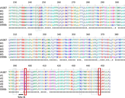
[image:3.585.78.508.63.400.2]FIG 1Alignment of the deduced amino acid residues from GII.4 VA387_1996, the M1 to M5 mutants, and the Den Haag_2006b variant. The 11 residues forming the predicted 3C3G3 epitope are boxed (R397 and D448 in red).
TABLE 1Primers used for site-directed mutagenesis
Primer Sequence Mutant(s)
MUT_1_FW 5=-CAGGATGGTAATAACCACAGGAATGAACCCCAGCAATG-3= M1 and M4
MUT_1_RV 5=-CATTGCTGGGGTTCATTCCTGTGGTTATTACCATCCTG-3= M1 and M4
MUT_2_FW 5=-CGGGTATCCCAACATGGACCTGGATTGCCTACTC-3= M2, M4, and M5
MUT_2_RV 5=-GAGTAGGCAATCCAGGTCCATGTTGGGATACCCG-3= M2, M4, and M5
MUT_3_1_FW 5=-CGTCATCCAGGATGGTAGCACCCACCAAAATGAACCCC-3= M3 and M5
MUT_3_1_RV 5=-GGGGTTCATTTTGGTGGGTGCTACCATCCTGGATGACG-3= M3 and M5
MUT_3_2_FW 5=-CCAGGATGGTAGCACAACCCACCAAAATGAACC-3= M3 and M5
MUT_3_2_RV 5=-GGTCCTACCATCGTGTTGGGTGGTTTTACTTGG-3= M3 and M5
MUT_1_3_FW 5=-CGTCATCCAGGATGGTAGCACAACCCACAGGAATGAACCCCAGCAATG-3= M5
MUT_1_3_RV 5=-CATTGCTGGGGTTCATTCCTGTGGGTTGTGCTACCATCCTGGATGACG-3= M5
on November 7, 2019 by guest
http://jvi.asm.org/
sequences of mutants M1 to M5 (see below). The Modeler software built the required number of models of the target and estimated the quality parameters of the models. The model with the best dope score was chosen for each mutant. The structures were visualized with the PyMOL program (PyMOL Molecular Graphics System, version 0.99; Schrodinger, LLC).
Site-directed mutagenesis.To confirm the amino acid residues form-ing part of the 3C3G3 epitope, the GII.4 VA387_1996 strain (not recog-nized by the antibody) was used as a scaffold to incorporate the amino acids present in the predicted epitope. Five mutants were constructed by introducing the following mutations into the GII.4 VA387_1996 se-quence: M1 (Q396R), M2 (N447D), M3 (NN-393 and 394 STT), M4 (M1 plus M2), and M5 (M1 plus M2 plus M3) (Fig. 1). The GeneArt site-directed mutagenesis system (Thermo Fisher) was applied following the manufacturer’s instructions to incorporate the mutations. The primers utilized to create the mutants are listed inTable 1. M1 and M2 were produced in one step, but M3 was constructed in two steps, first using the MUT_3_1_FW and MUT_3_1_RV primers and then the MUT_3_2_FW and MUT_3_2_RV primers in a second mutagenesis reaction. The M4 construction (a double mutant incorporating M1 and M2) was made using the M1 construction as the template and incorporating the M2 mutation afterward. To create the M5 mutant, the M3 construction was used as the template, using primers MUT_1_3_FW and MUT_1_3_RV to incorporate M1 without changing the M3 mutation that was already pres-ent, followed by the addition of the M2 mutation.
Transformants were analyzed by PCR and sequencing. Positive trans-formants were transferred to theE. coliBL21 strain GroES/EL, and mutant P particles were produced and purified as previously described (27).
SPR.The abilities of P particles (GII.4 VA387_1996, M1, M2, M3, M4, M5, and Den Haag_2006b) to bind to three different MAbs (3C3G3, anti-V0.8, and anti-V2.5) was tested by surface plasmon resonance (SPR) using a Biacore T100 instrument (Biacore; GE Healthcare). An His anti-body (Clontech) was immobilized on the surfaces of CM5 chips (GE Healthcare) and used as a capture antibody for P particles. Immobiliza-tion was achieved using an amine coupling kit (GE Healthcare). Briefly, 5,000 anti-His MAb resonance units (RU) were immobilized in channel 2, leaving channel 1 as a reference. Each P particle was captured by flowing a solution of 100g/ml for 100 s at a flow rate of 5l/min in HBS-EP⫹ buffer (GE Healthcare) to reach a capture level of⬃100 RU. Several dilu-tions of each MAb (100 nM, 50 nM, 25 nM, 12.5 nM, and 6.125 nM) were injected, starting with the most dilute and finishing with the most con-centrated, on the CM5 chip at a flow rate of 30l/min in HBS-EP⫹buffer (GE Healthcare) at 25°C in single-cycle kinetics experiments. The binding time was 60 s, with a final dissociation time of 600 s. Each interaction was tested in three independent experiments. Binding and kinetics evaluations were performed with the Biacore evaluation software.
P particle binding to neoglycoconjugates.The binding of P particles to five different neoglycoproteins (Table 2) with the oligosaccharide structures of antigen H (blood group O), blood group A, blood group B, Lewis X (Lex), and sialyl Lewis X (SiLex) was assayed by ELISA. All the
neoglycoproteins were obtained from Isosep AB (Tullinge, Sweden). The oligosaccharides were linked to human serum albumin (HSA) through an acetylphenylenediamine (APD) or aminophenylethyl (APE) spacer, with 10 to 30 oligosaccharides per protein molecule.
[image:4.585.40.288.78.145.2]Microtiter plates (Corning, NY) were coated with the different neogly-coconjugates at 1g/ml in carbonate/bicarbonate buffer (pH 9.2) and incubated at 37°C for 1 h and at 4°C overnight. The plates were washed three times with PBST and blocked with PBS containing 3% BSA for 1 h at 37°C. The P particles were added at 1g/ml in PBST, and the plates were incubated for 90 min at 37°C. After three washes, P-PAb was added to the plates at a dilution of 1/1,000. Binding was detected with HRP-conjugated anti-rabbit at 1/4,000 (Santa Cruz Biotechnology). The reactions were developed by the addition of OPD and stopped after a 20-min incubation with 3 M H2SO4. Absorbance was measured at 492 nm. Assays were per-formed in triplicate, and negative (nonfunctionalized HSA at 1g/ml)
TABLE 2Neoglycoconjugates used in the present study
Designation Oligosaccharide structure
H-type 1-HSA Fuc␣2Gal3GlcNAc3Gal4Glcc-APD
SiLex-HSA Neu5Ac␣3Gal4(Fuc␣3)GlcNAc3Gal4Glcc-APD
Lex-HSA Gal4(Fuc␣3)GlcNAc3Gal4(Fuc␣3)GlcNAc-APD
A-tri-HSA GalNAc␣3(Fuc␣2)Gal-O-APE B-tri-HSA Gal␣3(Fuc␣2)Gal-O-APE
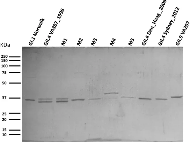
FIG 2SDS-12% PAGE gel stained with Coomassie blue showing the 10 different P particles used in the present study. The molecular masses are indicated with bars on the left.
on November 7, 2019 by guest
http://jvi.asm.org/
[image:4.585.135.449.467.701.2]and blank controls were included. The binding was expressed as percent-ages of signal in relation to the higher OD492.
Ethics statement.In this study, BALB/c mice were employed to obtain monoclonal antibodies. The Animal Welfare and Ethics Committee of the University of Valencia approved all the protocols conducted here, accord-ing to applicable national and international guidelines. J.B. possesses ac-creditation by the Conselleria de Agricultura, Generalitat Valenciana, to design and perform experiments with laboratory animals.
RESULTS
Production and purification of norovirus P particles.
With the
aim of studying the immunogenicities and the binding abilities of
different NoV strains to several HBGAs, a total of 10 P particles
were produced, one corresponding to the GI.1 Norwalk genotype,
four corresponding to different GII NoVs (strains GII.9 VA207,
GII4 VA387_1996, GII.4 Den Haag_2006b, and GII4 Sydney_
2012), and the remainder obtained by site-directed mutagenesis of
the GII.4 VA387_1996 P particle (M1 to M5). All the proteins
showed a molecular mass close to the expected mass (36 kDa) but
with small differences. The GII4 VA387_1996 variant showed a
typical double band for the strain (
22
,
27
). This duplet was also
present in the M1 (Q396R) mutant. The M2 mutant (N447D)
migrated as a single band, as did all the other constructions, except
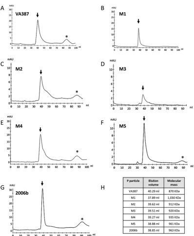
FIG 3(A to G) Chromatograms showing the size exclusion chromatography of GII.4 VA387_1996, the M1 to M5 mutants, and Den Haag_2006b P particles. The arrows indicate the elution peaks corresponding to P particles with an expected molecular mass of 840 kDa. The asterisks mark the elution peaks of nonparticulate proteins. (H) Elution volumes and calculated molecular masses of P particle preparations.
on November 7, 2019 by guest
http://jvi.asm.org/
[image:5.585.93.491.59.543.2]the M4 double mutant, which displayed lower mobility in the gel
(
Fig. 2
). The P particles were further purified by size exclusion
chromatography. The results showed that the majority (more
than 80%) of the proteins produced formed particles, as can be
observed in
Fig. 3
. The elution volume was slightly different for
every preparation, ranging from 37.89 ml (M1) to 40.29 ml
(VA387), which corresponds to 1,030 kDa to 870 kDa (
Fig. 3
).
Altogether, we were able to produce and purify 10 different P
particles, which were used in ELISA, SPR, and binding assays.
Characterization of MAbs by ELISA.
In order to elucidate the
reactivity patterns of each of the MAbs against different NoV
genogroups, genotypes, and variants, the initial characterization
of the 3C3G3, anti-v0.8, and anti-v2.5 MAbs was performed by
ELISA using a set of NoV antigens (
Table 3
and
Fig. 4
). As a
control, the HPA-PAb was utilized. The 3C3G3 antibody
rec-ognized only its homologous VLP (GII.4 Den Haag_2006b)
and the most closely related GII.4 Apeldoorn_2007 P domain.
Surprisingly, it did not recognize the GII.4 v2_2004 VLPs and
GII.4 VA387_1996 P particle, two different variants within the
same genotype. This result is similar to those obtained with the
anti-v0.8 and anti-v2.5 MAbs. The anti-v0.8 MAb efficiently
recognized its homologous GII.4 v0_1999 VLP and the GII.4
VA387_1996 P particle, while the anti-v2.5 MAb recognized only
its homologous GII.4 v2_2004 VLP and the closer GII.4 2006b
antigen, but not the GII.4 Apeldoorn_2007 P domain. The HPA
polyclonal antibody was able to recognize all the tested antigens
except the GI.1 P particles (
Table 3
). These results confirm that the
fast evolution of NoV GII.4 variants seems to be driven by the
antibody pressure in the host, favoring the emergence of antibody
escape variants.
[image:6.585.41.544.77.250.2]The 3C3G3 MAb blocks the binding of P particles to saliva.
The main aim of the present study was to characterize the epitope
of a MAb directed to the viral capsid protein to provide an
expla-nation of how antibody evasion occurs in NoV GII.4 variants. It
TABLE 3Antibody reactivities against different norovirus antigens by ELISA and affinity constants (KD) estimated by surface plasmon resonance
Norovirus antigen
Antibody reactivitya(K D[nM])
HPA-PAb P-PAb 3C3G3 MAb Anti-v0.8 MAb Anti-v2.5 MAb
VLP GI.1 Norwalk ⫹ ND ⫺ ⫺ ⫺
VLP GII.4-1999 ⫹ ⫹ ⫺ ⫹ ⫺
VLP GII.4-2004 ⫹ ⫹ ⫺ ⫺ ⫹
VLP GII.3 ⫹ ⫹ ⫺ ⫺ ⫺
P-GI.1 Norwalk ⫺ ND ⫺ ⫺ ⫺
P-GII.4-1996 ⫹ ⫹ ⫺ ⫹(47⫾35) ⫺
P-GII.4-1996 M1 ⫹ ⫹ ⫺ ⫹(72⫾49) ⫺
P-GII.4-1996 M2 ⫹ ⫹ ⫺ ⫹(7⫾1) ⫺
P-GII.4-1996 M3 ⫹ ⫹ ⫺ ⫹(12⫾3) ⫺
P-GII.4-1996 M4 ⫹ ⫹ ⫹(153⫾74) ⫹(7⫾1) ⫺
P-GII.4-1996 M5 ⫹ ⫹ ⫹(25⫾12) ⫹(12⫾3) ⫺
P-GII.4-2006b ⫹ ⫹ ⫹(2.1⫾1) ⫺ ⫹(11⫾3)
P-GII.4-2012 ⫹ ⫹ ⫺ ⫺ ⫺
P domain GII.4-2007 ⫹ ⫹ ⫹ ⫺ ⫺
a⫹, positive recognition;⫺, negative recognition; ND, not determined.
FIG 4Properties of binding of 4 antibodies, anti-v0.8, anti-v2.5, 3C3G3, and HPA PAb, to a panel of norovirus antigens (VLPs, P particles, and P domain) analyzed by ELISA. For clarity, only the 1/1,000 dilutions are represented. The error bars indicate the standard deviations of three replicates. The horizontal line indicates the cutoff value of the ELISA.
on November 7, 2019 by guest
http://jvi.asm.org/
[image:6.585.140.450.492.689.2]was important to know if the 3C3G3 MAb was able to block the
binding of NoV to its receptors. For this reason, a saliva binding
blocking assay was performed, and the results confirmed that the
3C3G3 MAb is able to block the binding of GII.4 Den Haag_2006b
P particles to secretor-positive saliva in a dose-dependent manner
(
Fig. 5
).
Characterization of the 3C3G3 MAb epitope by phage
dis-play and site-directed mutagenesis.
With the aim of
characteriz-ing the epitope recognized by the 3C3G3 MAb, the phage display
technique was applied, followed by site-directed mutagenesis.
Af-ter three rounds of panning, a consensus sequence of 11 amino
acids was obtained. The predicted epitope was formed by the
fol-lowing amino acids: P245, E247, I389, Q390, R397, R435, G443,
Y444, P445, N446, and D448 of the GII.4 Den Haag_2006 variant
(
Fig. 1
). Three of these amino acids are within the P2 subdomain
(I389, Q390, and R397), and the other eight are within the P1
subdomain (
Fig. 6
).
To confirm the epitope, site-directed mutagenesis was
per-formed, using the P particle of the GII.4 VA387_1996 variant as a
scaffold. This variant was not recognized by the 3C3G3 MAb, and
only 2 residues were different between the two proteins in the
predicted epitope, R397 and D448 (
Fig. 1
). Five mutants were
produced as described in Materials and Methods (M1 [Q396R],
M2 [N447D], M3 [NN-393, 394 STT], M4 [M1 plus M2], and M5
[M1 plus M2 plus M3]), and the reactivity of the 3C3G3 MAb
against the mutants was tested by ELISA. In addition to the
mu-tants, two new P particles were added to the experiment: the GII.4
Den Haag_2006 variant P particles as the positive control and the
GII.4 Sydney_2012 variant P particles to evaluate the performance
of the antibody toward the newer GII.4 epidemic variant. The
ELISA results, summarized in
Table 3
and
Fig. 7A
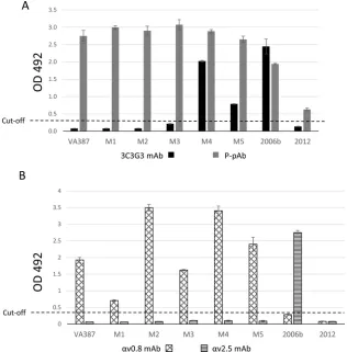
, found that
none of the single mutants, M1, M2, or M3, was recognized by the
antibody. Interestingly, both the double mutant, M4, and the
multiple mutant, M5, were recognized by the antibody,
confirm-ing that at least R397 and D447 form part of the epitope
recog-nized by the 3C3G3 antibody. These results show that the
anti-genic site B (STT 393,394,395) does not seem to be involved in the
formation of the 3C3G3 epitope, since M3 was not recognized by
the 3C3G3 MAb. It is also interesting that the later epidemic
vari-ant, GII.4 Sydney_2012, is not recognized by the MAb even
though it shares the 11 residues of the predicted epitope (
Table 4
).
This indicates that other residues not identified in this study might
be implicated in the formation of the 3C3G3 epitope.
To determine if this newly described epitope affects the
bind-ing of other GII.4-directed MAbs, the reactivity of the anti-v0.8
and anti-v2.5 MAbs against the different P particles was also
as-sayed by ELISA. The results show that the anti-v2.5 epitope might
be different from the 3C3G3 epitope, since none of the mutants
were recognized by the MAb (
Fig. 7B
). Interestingly, this antibody
was also independent of the previously described antigenic site B
present here in the M3 and M5 constructions (
15
). In contrast, the
anti-v0.8 MAb reacted against each of the mutants, albeit at
different levels. This may indicate that the selected residues exert
an influence on the folding of the epitope recognized by the
anti-body, since both mutagenized residues are conserved in the
VA387_1996 and V0_1999 variants (
Table 4
). According to our
results, this MAb showed a dependence on the antigenic site B, as
previously described (
15
).
Characterization of the 3C3G3 MAb epitope by surface
plas-mon resonance.
To quantify the strength of the interaction
be-tween the tested MAbs and the different P particles recognized by
them, an SPR approach was applied, and affinity constants (
K
D)
were obtained in at least three independent experiments. Of all the
interaction pairs, the highest affinity (lowest
K
Dvalue) was
ob-tained in the 3C3G3-GII4 Den Haag_2006b interaction pair (
Ta-ble 3
and
Fig. 8
). Although M4 gave higher signal in ELISA (
Fig.
7A
), the SPR experiments revealed that the M4 mutant had a
K
D(153 nM) 6 times higher than that of the M5 mutant (25 nM)
(
Table 3
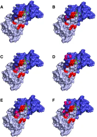
). After modeling the structures of the different mutants,
using the P domain structure of the VA387_1996 as a scaffold
(PDB ID, 2OBR), we observed that the R397 and D448 residues
probably present different conformations in M4 and M5 (
Fig. 3C
and
D
). In the M5 model, the D448 anionic carboxylate (RCOO
⫺)
and R397 cationic ammonium (RNH3
⫹) were closer. This
in-creased the possibility of saline bridge formation, which allowed
3C3G3 epitope stabilization.
R397 is involved in HBGA recognition.
The binding of the
VA387_1996, Den Haag_2006b, and Sydney_2012 variants, as
well as of the five mutants (M1 to M5), to five different HBGAs
(SiLe
x, Le
x, blood group O [H antigen], blood group A, and blood
group B) was analyzed in order to study if the residues involved in
their immunogenicity also had an impact on their receptor
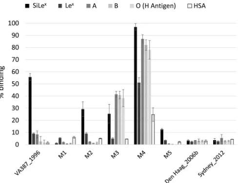
bind-ing. Our results showed that there was a change in the recognition
pattern between the VA387_1996 and the Den Haag_2006b and
Sydney_2012 variants. While the VA387_1996 strain possessed
great ability to bind to the SiLe
xantigen, the Den Haag_2006b and
Sydney_2012 variants did not recognize any of the assayed
anti-gens (
Fig. 9
). Interestingly, M1, incorporating the single mutation
Q396R, lost its ability to bind to the tested HBAs, showing the
same recognition pattern as the 2006b and 2012 variants. The M2
mutant maintained binding to SiLe
xsimilar to that of the
wild-type VA387_1996. M3 and M4 increased their ability to recognize
blood group A, B, and O antigens. Moreover, M4 recognized the
FIG 5Blockade of GII.4 Den Haag_2006b P particle binding to secretor-positive saliva by the 3C3G3 MAb. The error bars indicate the standard devi-ations of three replicates. The numbers on thexaxis indicate the concentra-tions of the 3C3G3 MAb.
on November 7, 2019 by guest
http://jvi.asm.org/
[image:7.585.41.285.65.279.2]nonfunctionalized HSA. Finally, the M5 mutant demonstrated a
residual binding of 10% to SiLe
x.
DISCUSSION
Due to the relevance of antibody escape variants in NoV GII.4
persistence during the last 40 years, we decided to resolve the
epitope recognized by the 3C3G3 MAb, which is a binding
block-ing antibody. The results of the present study shed light on how
NoV escapes antibody neutralization. The antibody was not able
to recognize any of the tested antigens by Western blotting,
indi-cating that the target epitope should be conformational. For this
reason, a phage display approach was chosen. Of the 11 residues
suspected to form part of the epitope (see Results), two, R397 and
D448, were confirmed to be part of the epitope by site-directed
mutagenesis of VA387_1996 P particles. When different P
parti-cles were produced, differences in electrophoretic mobility were
observed. These differences in electrophoretic mobility have been
previously reported for the VA387 variant; they were associated
with P particle formation (P dimers versus P particles) (
22
) and
might reveal structural changes. In the present study, all the
de-signed P particles were found to be successfully constructed, with
estimated molecular masses ranging from 870 to 1,030 kDa.
Besides the R397 and D448 residues, another difference
be-tween VA387_1996 and Den Haag_2006b sequences was a change
FIG 6Structural models showing the surfaces of the different mutants produced in the present study. The P1 subdomain is shown in gray and the P2 subdomain in blue. Blood group trisaccharide type A was located according to PDB ID 2OBS and can be seen in a stick representation. (A) Structure of the VA387_1996 wild type with the residues forming the 3C3G3 epitope in red. (B, C, and D) Single mutants M1, M2, and M3, respectively. (E) Model of the M4 double mutant. (F) Model of the M5 multiple mutant. All the mutations are shown in pink. Only mutant M5 has the D448 anionic carboxylate (RCOO⫺) and R397 cationic ammonium (RNH3⫹) in a close conformation that increases the possibility of saline bridge formation, which allows 3C3G3 epitope stabilization.
on November 7, 2019 by guest
http://jvi.asm.org/
[image:8.585.137.453.68.513.2]involving amino acids 393 to 395 (STT in the Den Haag_2006b
variant and N-N in the VA387_1996 variant). This change was
incorporated in the M3 and M5 mutants, because they were in the
near vicinity of R397. They had been previously reported to form
an important epitope in GII.4 NoVs (
15
). Nevertheless, these
three residues did not rescue the binding ability of the 3C3G3
antibody (M3 was not recognized by 3C3G3), but they helped
stabilize the 3C3G3 epitope, since 3C3G3 had 6 times more
affin-ity for M5 than for M4. After the molecular modeling of the 5
mutants (
Fig. 6
), we could predict the formation of a saline bridge
between R397 and D448 only in M5 and not in M4. That could
explain the higher stability of the 3C3G3 epitope in M5, while STT
residues 393 to 395 did not seem to be part of the 3C3G3 epitope
themselves.
As shown in
Table 4
, it seems that residues R397 and D448 have
been important in the successive GII.4 variants. In order to
eluci-date if they are part of epitopes recognized by other MAbs raised
against different GII.4 variants, we also characterized two
previ-ously obtained MAbs, anti-v0.8 and anti-v2.5, that were tested
against the same antigens and mutants as the 3C3G3 MAb. We
confirmed by ELISA and SPR that the anti-v0.8 MAb was
depen-dent on antigenic site B (393 to 395, -NN to STT), as previously
described (
15
). The epitope recognized by anti-v0.8 was also
af-fected in the M1 and M2 mutants, indicating that amino acids 397
and 448 might also be implicated in the epitope conformation.
Furthermore, the results with all three MAbs showed that there
was cross-reactivity only between the closer variants and that none
of the more distant variants shared any reactivity (
Table 3
). The
anti-v2.5 MAb recognized only the closer P particle
correspond-ing to the GII.4 Den Haag_2006b variant. All the mutants were
made using the VA387_1996 variant as a scaffold, which is not
recognized by the antibody. Only if R397 and D448 were present
in its epitope would recognition by the antibody be recovered. The
only conclusion that we can come to is that these residues might
not be part of the anti-v2.5 MAb epitope.
These results have important implications from an
evolution-ary point of view, since they demonstrate that the MAbs against
the older variants do not recognize the newer ones. This explains
why the same genotype can produce successive pandemics.
More-over, the HPA-PAb is able to recognize the GI.1 VLPs but not
the GI.1 P particles, supporting the idea that the cross-reactive
epitopes between the two NoV genogroups are present only in
the shell domain of VP1, which is not present in the P particles
(
31
,
32
).
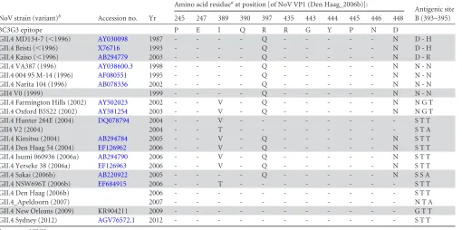
When the 3C3G3 epitopes in several NoVs GII.4 variants were
compared (
Table 4
), the 2 residues identified in the present study,
R397 and D448, were always conserved. All the variants prior to
2004 possessed the combination Q397-N448, and all the variants
that emerged since 2006 contain the duplet R397-D448. There was
a transition period between 2004 and 2006 in which both duplets
FIG 7Binding of different antibodies to wild-type and mutant P particles analyzed by ELISA. (A) Recognition by the P-PAb and 3C3G3 antibodies. (B) Recognition by anti-v0.8 and anti-v2.5 MAbs. Only the 1/1,000 dilution is shown for clarity. The error bars indicate the standard deviations of three replicates. The horizontal line indicates the cutoff value of the ELISA.
on November 7, 2019 by guest
http://jvi.asm.org/
[image:9.585.134.450.67.388.2]were present, but always in the same combination. In the
VA387_1996 structure and in the structural models in
Fig. 6
, it was
observed that these two amino acids were located very close to
each other, and in M5, they seemed to interact through a saline
bridge. This physical interaction might explain why these two
res-idues were in fixed combinations throughout the different
vari-ants. We could hypothesize that the residues might additionally be
involved in the stabilization of the P particle itself, not just in the
3C3G3 epitope. Indeed, we observed changes in the
electropho-retic mobility of the different P particles when these residues were
mutagenized within VA387_1996, M1, and M4, with P particles
being the most heterogeneous migrating particles. Nevertheless,
the Den Haag_2006b and Sydney_2012 variants, which possess
the R397-D448 pair, along with the 393 to 395 STT version of
antigenic site B (which favors the formation of the R397-D448
saline bridge), were more homogeneous than the 1996 variant.
It is known that GII.4 noroviruses have strain-specific HBGA
recognition patterns (
17
,
18
,
33
). To evaluate whether the 3C3G3
epitope was involved in the interaction with receptors, we
ana-lyzed the binding of the different GII.4 P particles to several
HBGAs. Our results indicated that the change in R397 seemed to
be the key in the different recognition patterns seen in the newer
GII.4 variants compared to those of the older ones. The
conclu-sion reached was that this change might have had an effect on the
folding of the fucose-binding pocket, which is not in contact with
this residue (
Fig. 6
).
[image:10.585.42.552.80.334.2]Interestingly, the M4 mutant that includes both substitutions
(Q396R and N447D) has increased binding ability, including high
TABLE 43C3G3 epitope sequences in different variants of the GII.4 norovirus
NoV strain (variant)b Accession no. Yr
Amino acid residueaat position [of NoV VP1 (Den Haag_2006b)]:
Antigenic site B (393–395)
245 247 389 390 397 435 443 444 445 446 448
3C3G3 epitope P E I Q R R G Y P N D
GII.4 MD134-7 (⬍1996) AY030098 1987 - - - - Q - - - N D - H
GII.4 Bristi (⬍1996) X76716 1993 - - - - Q - - - N D - H
GII.4 Kaiso (⬍1996) AB294779 2003 - - - - Q - - - N D - R
GII.4 VA387 (1996) AY038600.3 1998 - - - - Q - - - N N - N
GII.4 004 95 M-14 (1996) AF080551 1995 - - - - Q - - - N N - N
GII.4 Narita 104 (1996) AB078336 2002 - - - - Q - - - N N - N
GII4 V0 (1999) 1999 - - - - Q - - - N N - N
GII.4 Farmington Hills (2002) AY502023 2002 - - V - Q - - - N N G T
GII.4 Oxford B5S22 (2002) AY581254 2003 - - V - Q - - - N N G T
GII.4 Hunter 284E (2004) DQ078794 2004 - - V - - - S T T
GII4 V2 (2004) 2004 - - T - - - S T A
GII.4 Kimitsu (2004) AB294784 2005 - - V - Q - - - N S T T
GII.4 Den Haag 54 (2004) EF126962 2006 - - V - Q - - - N S T T
GII.4 Isumi 060936 (2006a) AB294790 2006 - - V - Q - - - N S T T
GII.4 Yerseke 38 (2006a) EF126963 2006 - - V - Q - - - N S T T
GII.4 Sakai (2006b) AB220922 2005 - - - - Q - - - N S S A
GII.4 NSW696T (2006b) EF684915 2006 - - T - - - S T T
GII.4 Den Haag (2006b) 2006 - - - S T T
GII.4_Apeldoorn (2007) 2007 - - - N T A
GII.4 New Orleans (2009) KR904211 2009 - - - G T T
GII.4 Sydney (2012) AGV76572.1 2012 - - - S T T
a-, same as 3C3G3.
b
Variants⬍1996, 1999, 2004, and 2009 are shaded.
FIG 8Representation of the different affinity constants (KD) in molar (M) obtained by surface plasmon resonance experiments. The error bars indicate the standard deviations of three replicates. The arrow indicates the best interaction pair.
on November 7, 2019 by guest
http://jvi.asm.org/
[image:10.585.136.449.557.701.2]binding to the HSA (negative control). In
Table 4
, it can be seen
that this amino acid combination (R396 and D447) is always
ac-companied by the antigenic site B triplet and not the duplet that is
present in M4. The combination of the antigenic site B duplet with
R396 and D447 has not been found in wild-type viruses so far,
which may reflect a negative selection process, probably due to its
impaired binding, as is seen with M4 mutant.
This was the first time that phage display has been applied to
study GII.4 NoV epitopes. In previous studies, evolutionary and
structural approaches were used to identify putative epitopes,
in-cluding sites A (amino acids 296 to 298) and B (amino acids 393 to
395) (
16
). These epitopes were confirmed using chimeras between
different NoV GII4 variants and monoclonal antibodies (
15
). A
similar approach showed that site A should be formed by amino
acids 294, 368, and 372, in addition to 296 to 298 (
17
). It has also
been shown that epitope B has an influence on the changes in the
HBGA binding abilities of different GII4 variants (
17
,
18
). Using
the phage display technique, we have been able to identify 2
resi-dues (397 and 448) that were not as exposed as the previously
described epitopes, making it difficult to predict their relevance
after structural analysis. We have shown that these residues play
an important role in antibody recognition and HBGA interactions
and that they have evolved from ancestral to modern variants.
Altogether, we were able to study the epitope recognized by the
3C3G3 antibody and have shown that this epitope was implicated
in virus-host interactions. On one hand, the two amino acids R397
and D448 seemed to be involved in evasion of the host antibody
response, showing how small changes in the amino acid sequence
could render huge benefits to the virus in terms of antibody
eva-sion. On the other hand, we have demonstrated that a single
change in position 396 of the 1996 variant (397 in the 2006b
vari-ant) could be enough to modulate the binding of noroviruses to
HBGAs.
ACKNOWLEDGMENTS
The views expressed are those of the authors and not necessarily those of the NHS, the NIHR, the Department of Health, or Public Health England.
J.R.-D., J.B., S.V.-V., and N.C.-V. conceived and designed the study. N.C.-V., S.V.-V., R.G.-R., J.R.-D., J.B., D.A., and M.I.-G. designed the experiments. N.C.-V., S.V.-V., R.G.-R., and J.R.-D. performed the exper-iments. All authors contributed to data analysis and manuscript prepara-tion.
FUNDING INFORMATION
This work, including the efforts of Jesús Rodríguez-Díaz, was funded by Ministerio de Economía y Competitividad (MINECO) (RYC-2013-12442 and AGL2014-52996-C2-2-R). This work, including the efforts of Javier Buesa, was funded by Ministerio de Economía y Competitividad (MI-NECO) (SAF2012-38368). This work, including the efforts of Noelia Car-mona-Vicente, was funded by Universitat de València (V Segles fellow-ship).
REFERENCES
1.Tam CC, Rodrigues LC, Viviani L, Dodds JP, Evans MR, Hunter PR, Gray JJ, Letley LH, Rait G, Tompkins DS, O’Brien SJ.2012. Longitu-dinal study of infectious intestinal disease in the UK (IID2 study): inci-dence in the community and presenting to general practice. Gut61:69 –77. http://dx.doi.org/10.1136/gut.2011.238386.
2.Gastanaduy PA, Hall AJ, Curns AT, Parashar UD, Lopman BA.2013. Burden of norovirus gastroenteritis in the ambulatory setting—United States, 2001–2009. J Infect Dis207:1058 –1065.http://dx.doi.org/10.1093 /infdis/jis942.
3.Patel MM, Widdowson MA, Glass RI, Akazawa K, Vinje J, Parashar UD.2008. Systematic literature review of role of noroviruses in sporadic gastroenteritis. Emerg Infect Dis14:1224 –1231.http://dx.doi.org/10.3201 /eid1408.071114.
4.Bucardo F, Lindgren PE, Svensson L, Nordgren J.2011. Low prevalence of rotavirus and high prevalence of norovirus in hospital and community wastewater after introduction of rotavirus vaccine in Nicaragua. PLoS One6:e25962.http://dx.doi.org/10.1371/journal.pone.0025962. 5.Williams DJ, Edwards KM, Payne DC, Manning J, Parashar UD,
Lopman BA.2012. Decline in gastroenteritis-related triage calls after ro-tavirus vaccine licensure. Pediatrics130:e872– e878.http://dx.doi.org/10 .1542/peds.2012-0330.
6.Koo HL, Neill FH, Estes MK, Munoz FM, Cameron A, DuPont HL, Atmar RL.2013. Noroviruses: the most common pediatric viral enteric pathogen at a large university hospital after introduction of rotavirus vac-cination. J Pediatric Infect Dis Soc2:57– 60.http://dx.doi.org/10.1093 /jpids/pis070.
7.Payne DC, Vinje J, Szilagyi PG, Edwards KM, Staat MA, Weinberg GA, Hall CB, Chappell J, Bernstein DI, Curns AT, Wikswo M, Shirley SH, Hall AJ, Lopman B, Parashar UD.2013. Norovirus and medically at-tended gastroenteritis in U.S. children. N Engl J Med368:1121–1130.http: //dx.doi.org/10.1056/NEJMsa1206589.
8.Buesa J, Rodriguez Diaz J.2006. Molecular virology of enteric viruses (with emphasis on caliciviruses), p 43–100.InGoyal S (ed), Viruses in foods. Springer, New York, NY.
9.Kirk MD, Pires SM, Black RE, Caipo M, Crump JA, Devleesschauwer B, Dopfer D, Fazil A, Fischer-Walker CL, Hald T, Hall AJ, Keddy KH, Lake RJ, Lanata CF, Torgerson PR, Havelaar AH, Angulo FJ. 2015. World Health Organization estimates of the global and regional disease burden of 22 foodborne bacterial, protozoal, and viral diseases, 2010: a data synthesis. PLoS Med12:e1001921.http://dx.doi.org/10.1371/journal .pmed.1001921.
10. Vinje J.2015. Advances in laboratory methods for detection and typing of norovirus. J Clin Microbiol53:373–381.http://dx.doi.org/10.1128/JCM .01535-14.
11. Hoa Tran TN, Trainor E, Nakagomi T, Cunliffe NA, Nakagomi O.
2013. Molecular epidemiology of noroviruses associated with acute spo-radic gastroenteritis in children: global distribution of genogroups, geno-types and GII.4 variants. J Clin Virol56:185–193.http://dx.doi.org/10 .1016/j.jcv.2012.11.011.
12. White PA.2014. Evolution of norovirus. Clin Microbiol Infect20:741– 745.http://dx.doi.org/10.1111/1469-0691.12746.
13. Vongpunsawad S, Venkataram Prasad BV, Estes MK.2013. Norwalk virus minor capsid protein VP2 associates within the VP1 shell domain. J Virol87:4818 – 4825.http://dx.doi.org/10.1128/JVI.03508-12.
14. Prasad BV, Hardy ME, Dokland T, Bella J, Rossmann MG, Estes MK. FIG 9Percentages of binding of seven different P particles (VA387_1996, M1
to M5, and Den Haag_2006b) to five different HBGAs (SiLexantigen, Lex
antigen, and blood group A, B, and O H antigens) determined by ELISA. Each P particle binding was normalized with the higher OD492value. The error bars
indicate the standard deviations. HSA was used as the control.
on November 7, 2019 by guest
http://jvi.asm.org/
[image:11.585.43.287.65.253.2]1999. X-ray crystallographic structure of the Norwalk virus capsid. Science
286:287–290.http://dx.doi.org/10.1126/science.286.5438.287.
15. Allen DJ, Noad R, Samuel D, Gray JJ, Roy P, Iturriza-Gomara M.2009. Characterisation of a GII-4 norovirus variant-specific surface-exposed site involved in antibody binding. Virol J6:150.http://dx.doi.org/10.1186 /1743-422X-6-150.
16. Allen DJ, Gray JJ, Gallimore CI, Xerry J, Iturriza-Gomara M.2008. Analysis of amino acid variation in the P2 domain of the GII-4 norovirus VP1 protein reveals putative variant-specific epitopes. PLoS One3:e1485. http://dx.doi.org/10.1371/journal.pone.0001485.
17. Debbink K, Donaldson EF, Lindesmith LC, Baric RS. 2012. Genetic mapping of a highly variable norovirus GII.4 blockade epitope: potential role in escape from human herd immunity. J Virol86:1214 –1226.http: //dx.doi.org/10.1128/JVI.06189-11.
18. Shanker S, Choi JM, Sankaran B, Atmar RL, Estes MK, Prasad BV.
2011. Structural analysis of histo-blood group antigen binding specificity in a norovirus GII.4 epidemic variant: implications for epochal evolution. J Virol85:8635– 8645.http://dx.doi.org/10.1128/JVI.00848-11. 19. Eden JS, Tanaka MM, Boni MF, Rawlinson WD, White PA. 2013.
Recombination within the pandemic norovirus GII.4 lineage. J Virol87:
6270 – 6282.http://dx.doi.org/10.1128/JVI.03464-12.
20. Jones MK, Watanabe M, Zhu S, Graves CL, Keyes LR, Grau KR, Gonzalez-Hernandez MB, Iovine NM, Wobus CE, Vinje J, Tibbetts SA, Wallet SM, Karst SM.2014. Enteric bacteria promote human and mouse norovirus infection of B cells. Science346:755–759.http://dx.doi.org/10 .1126/science.1257147.
21. Jiang X, Wang M, Graham DY, Estes MK. 1992. Expression, self-assembly, and antigenicity of the Norwalk virus capsid protein. J Virol
66:6527– 6532.
22. Tan M, Jiang X.2005. The P domain of norovirus capsid protein forms a subviral particle that binds to histo-blood group antigen receptors. J Virol
79:14017–14030.http://dx.doi.org/10.1128/JVI.79.22.14017-14030.2005. 23. Siebenga JJ, Vennema H, Renckens B, de Bruin E, van der Veer B, Siezen RJ, Koopmans M.2007. Epochal evolution of GGII.4 norovirus capsid proteins from 1995 to 2006. J Virol81:9932–9941.http://dx.doi.org /10.1128/JVI.00674-07.
24. Tan M, Huang P, Meller J, Zhong W, Farkas T, Jiang X.2003. Muta-tions within the P2 domain of norovirus capsid affect binding to human
histo-blood group antigens: evidence for a binding pocket. J Virol77:
12562–12571.http://dx.doi.org/10.1128/JVI.77.23.12562-12571.2003. 25. Lindesmith LC, Donaldson EF, Baric RS.2011. Norovirus GII.4 strain
antigenic variation. J Virol 85:231–242. http://dx.doi.org/10.1128/JVI .01364-10.
26. Lindesmith LC, Donaldson EF, Lobue AD, Cannon JL, Zheng DP, Vinje J, Baric RS.2008. Mechanisms of GII.4 norovirus persistence in human populations. PLoS Med5:e31. http://dx.doi.org/10.1371/journal.pmed .0050031.
27. Rubio-del-Campo A, Coll-Marques JM, Yebra MJ, Buesa J, Perez-Martinez G, Monedero V, Rodriguez-Diaz J.2014. Noroviral P-particles as an in vitro model to assess the interactions of noroviruses with probiotics. PLoS One
9:e89586.http://dx.doi.org/10.1371/journal.pone.0089586.
28. Carmona-Vicente N, Fernandez-Jimenez M, Ribes JM, Tellez-Castillo CJ, Khodayar-Pardo P, Rodriguez-Diaz J, Buesa J. 2015. Norovirus infections and seroprevalence of genotype GII.4-specific antibodies in a Spanish population. J Med Virol87:675– 682.http://dx.doi.org/10.1002 /jmv.24112.
29. Mayrose I, Penn O, Erez E, Rubinstein ND, Shlomi T, Freund NT, Bublil EM, Ruppin E, Sharan R, Gershoni JM, Martz E, Pupko T.2007. Pepitope: epitope mapping from affinity-selected peptides. Bioinformat-ics23:3244 –3246.http://dx.doi.org/10.1093/bioinformatics/btm493. 30. Sali A, Blundell TL.1993. Comparative protein modelling by satisfaction
of spatial restraints. J Mol Biol234:779 – 815.http://dx.doi.org/10.1006 /jmbi.1993.1626.
31. Parra GI, Azure J, Fischer R, Bok K, Sandoval-Jaime C, Sosnovtsev SV, Sander P, Green KY.2013. Identification of a broadly cross-reactive epitope in the inner shell of the norovirus capsid. PLoS One8:e67592. http://dx.doi.org/10.1371/journal.pone.0067592.
32. Crawford SE, Ajami N, Parker TD, Kitamoto N, Natori K, Takeda N, Tanaka T, Kou B, Atmar RL, Estes MK.2015. Mapping broadly reactive norovirus genogroup I and II monoclonal antibodies. Clin Vaccine Im-munol22:168 –177.http://dx.doi.org/10.1128/CVI.00520-14.
33. Tan M, Xia M, Cao S, Huang P, Farkas T, Meller J, Hegde RS, Li X, Rao Z, Jiang X.2008. Elucidation of strain-specific interaction of a GII-4 norovirus with HBGA receptors by site-directed mutagenesis study. Vi-rology379:324 –334.http://dx.doi.org/10.1016/j.virol.2008.06.041.