This is a repository copy of
Uncertainty driven pooling network for microvessel
segmentation in routine histology images
.
White Rose Research Online URL for this paper:
http://eprints.whiterose.ac.uk/150424/
Version: Accepted Version
Proceedings Paper:
Fraz, M.M., Shaban, M., Graham, S. et al. (2 more authors) (2018) Uncertainty driven
pooling network for microvessel segmentation in routine histology images. In: Stoyanov,
D., Taylor, Z., Ciompi, F., Xu, Y., Martel, A., Maier-Hein, L., Rajpoot, N., van der Laak, J.,
Veta, M., McKenna, S., Snead, D., Trucco, E., Garvin, M.K., Chen, X.J. and Bogunovic, H.,
(eds.) Computational Pathology and Ophthalmic Medical Image Analysis. COMPAY 2018 :
International Workshop on Computational Pathology, 16 Sep - 20 Oct 2018, Granada,
Spain. Lecture Notes in Computer Science (11039). Springer Nature , pp. 156-164. ISBN
9783030009489
https://doi.org/10.1007/978-3-030-00949-6_19
This is a post-peer-review, pre-copyedit version of an article published in COMPAY 2018
Proceedings. The final authenticated version is available online at:
http://dx.doi.org/10.1007/978-3-030-00949-6_19
[email protected] https://eprints.whiterose.ac.uk/ Reuse
Items deposited in White Rose Research Online are protected by copyright, with all rights reserved unless indicated otherwise. They may be downloaded and/or printed for private study, or other acts as permitted by national copyright laws. The publisher or other rights holders may allow further reproduction and re-use of the full text version. This is indicated by the licence information on the White Rose Research Online record for the item.
Takedown
If you consider content in White Rose Research Online to be in breach of UK law, please notify us by
Microvessel Segmentation in Routine Histology
Images
M.M.Fraz1,2,4
M.Shaban1
S.Graham1
S.A.Khurram5
N.M.Rajpoot1,2,3
1
Dept. of Computer Science, University of Warwick, Coventry, United Kingdom
2
The Alan Turing Institute, London, United Kingdom
3
University Hospitals Coventry and Warwickshire, NHS Trust, United Kingdom
4
National University of Sciences and Technology, Islamabad, Pakistan
5
The University of Sheffield, United Kingdom
Abstract. Lymphovascular invasion (LVI) and tumor angiogenesis are
correlated with metastasis, cancer recurrence and poor patient survival. In most of the cases, the LVI quantification and angiogenic analysis is based on microvessel segmentation and density estimation in immuno-histochemically (IHC) stained tissues. However, in routine H&E stained images, the microvessels display a high level of heterogeneity in terms of size, shape, morphology and texture which makes microvessel segmenta-tion a non-trivial task. Manual delineasegmenta-tion of microvessels for biomarker analysis is labor-intensive, time consuming, irreproducible and can suf-fer from subjectivity among pathologists. Moreover, it is often beneficial to account for the uncertainty of a prediction when making a diagnosis. To address these challenges, we proposed a framework for microvessel segmentation in H&E stained histology images. The framework extends DeepLabV3+ by using an improved dice coefficient based custom loss function and also incorporating an uncertainty prediction mechanism. The proposed method uses an aligned Xception model, followed by atrous spatial pyramid pooling for feature extraction at multiple scales. This ar-chitecture counters the challenge of segmenting blood vessels of varying morphological appearance. To incorporate uncertainty, random transfor-mations are introduced at test time for a superior segmentation result and simultaneous uncertainty map generation, highlighting ambiguous regions. The method is evaluated using 1167 images of size 512x512 pix-els, extracted from 13 WSIs of oral squamous cell carcinoma (OSCC) tissue at 20x magnification. The proposed net-work achieves state-of-the-art performance compared to current semantic segmentation deep neural networks (FCN-8, U-Net, SegNet and DeepLabV3+).
Keywords: Microvessel detection· Tumor angiogenesis ·
Lymphovas-cular invasion ·Separable convolution· Pyramid pooling based neural network·Uncertainty quantification
1
Introduction
2 Fraz et al.
Fig. 1. Representative images of microvessels in H&E stained histology images of
OSCC tissue, illustrating their shape variability. The microvessel boundary annotation is shown in red color. (a-c) Microvessel with varying red cells density, (c) Microvessels at different sizes, (d) Keratinization, which appears similar to microvessel
vessels by intravasation allowing them to circulate through the intravascular stream. This lamphovascular invasion (LVI) can lead to the proliferation of tu-mor cells at another site in the body. This phenomena is tu-more commonly referred to as metastasis. The lymphatic or vascular invasion by the primary tumor is considered as a sign of aggressive disease and is usually accompanied by metas-tases to the regional lymph nodes and to distant sites. The formation of new blood vessels is important for growth, survival and metastatic spread of tumor cells [2]. Tumor neoangiogenesis leads to formation of new blood vessels in the tumor tissues, with an initial purpose to facilitate the transport of nutrients and oxygen to help the tumor cells survive [2]. In diagnostic clinical pathology, most of the currently existing tissue datasets and pathways recommend com-menting on the presence or absence of LVI however the degree of angiogenesis is not routinely examined or reported. In the existing research literature, there is strong evidence that microvessel density in tumor tissue is directly correlated with an increased risk of cancer spread, an increased incidence of disease recur-rence and poor patient survival [10]. Recent studies have re-ported LVI detection and quantification as an important risk factor in disease progression particularly in breast, cervical and lung cancers [10]. However, most of the results in these studies are based on manual localization of microvessels in the subjectively de-fined regions of tissue whole slide image (WSI). The subjective identification of LVI and angiogenic regions in the tissues is irreproducible, time consuming and often requires clinical knowledge. In routine pathological practice, accurate seg-mentation of microvessels can assist pathologists in identification of LVI which would otherwise be very time-consuming. Furthermore, objective quantification of LVI and tumor angiogenesis from multi giga pixel histopathology images will provide extremely valuable big data aiding prediction of tumor behavior and prognosis.
compara-tively expensive than H&E staining. Due to this cost, it is not commonly used in routine clinical practice. Likewise, the IHC-stained histology images are rarely available in public datasets, which is a major hindrance in the development of automated methods to investigate the role of microvessels in tumor prognosis and therapy response.
Deep learning has recently been successfully used for the automated analysis of histopathology images. Specifically, deep learning based architectures have been proposed for the detection, segmentation and classification of histopatho-logical structures in WSI images including deep learning based architectures are proposed for detection and classification of nuclei [14], mitoses [12], lymphocytes [13], tumor and stromal regions [15] and glandular structure segmentation [7]. A few studies have explored automatic quantification of tumor angiogenic hotspots by the detection of microvessels in IHC stained histology images only [8]. Most recently, a fully convolutional neural network (FCN) based method for microves-sel detection in H&E stained images is presented [16]. However, the methods for microvessel segmentation particularly in H&E stained images are limited.
In this paper, we present a framework for precise segmentation of microvessels in H&E stained histology images at multiple scales and resolutions by using an uncertainty aware spatial pyramid pooling deep neural network architecture. The Deeplabv3+ [4] architecture is extended by using an improved dice coefficient minimization based custom loss function and by accounting for the uncertainty of a prediction. The proposed network aims to solve the key challenges posed by automated microvessel segmentation. The method uses a modified Aligned Xception model [5] followed by an atrous spatial pyramid pooling (ASPP) unit [3] for feature extraction at multiple scales. This overcomes a major challenge of segmenting vessels of various sizes. Moreover, despite achieving state-of-the art performance in semantic segmentation, the deep networks typically do not inherently model the segmentation uncertainty. For this purpose, we apply ran-dom transformations to the images during test time, as a method to generate the approximate predictive distribution. Taking the average of these predictions of transformed images yields a superior segmentation and enables us to observe ambiguous areas, where the network is uncertain in a decision. The method-ology is evaluated on 1167 images of size 512x512 pixels, extracted from 13 WSIs of oral squamous cell carcinoma (OSCC) tissue at 20x magnification, and demonstrated promising results. Moreover, the proposed network achieves state-of-the-art performance compared to current semantic segmentation deep neural networks (FCN-8 [9], U-Net [11], SegNet [1] and DeepLabv3+ [4]).
2
Proposed Method
4 Fraz et al.
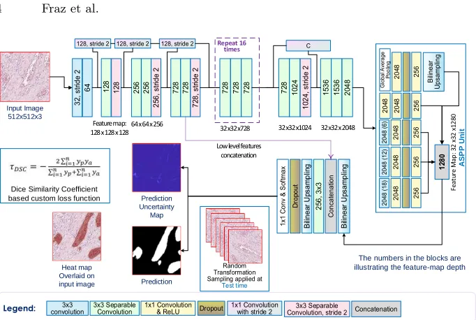
Feature map: 128 x 128 x 128
64 x 64 x 256 32 x32 x728 32 x32 x1024 32 x32 x 2048
ASP P Uni t F e a tu re M a p : 3 2 x 3 2 x 1 2 8 0 Repeat 16 times Prediction Heat map Overlaid on input image Prediction Uncertainty Map Legend:
Dice Similarity Coefficient based custom loss function
Low level features concatenation
The numbers in the blocks are illustrating the feature-map depth Input Image
[image:5.612.138.479.91.320.2]512x512x3
Fig. 2. The Proposed Framework: The legend represents the color coding of various
types of convolutional layers. The values inside the blocks represents the depth of cor-responding feature map. In ASPP Unit, the value in brackets denotes the dilation rates of 6, 12 and 18. To obtain the prediction uncertainty map, Random Transformation Sampling at test time is applied. The network uses custom loss function based on minimization of ve of Dice Similarity Coefficient.
(AlexNet, VGG, Google Net, ResNet e.t.c.) used for image classification has in-herited limitations to model geometric transformations due to fixed geometric structures in their building modules. Moreover, these networks use a hierarchical combination of maxpooling and convolutions to increase the receptive field size. This results in loss of image information which may be very significant for precise object segmentation. In order to deal with these issues, the feature extraction process should be invariant to geometric transformations and retain the low level image information. The Xception model [5] has demonstrated promising perfor-mance in image classification task on ImageNet in terms of speed and accuracy. Xception has been modified to incorporate geometric transformations modeling capability for feature extraction. Further to this, Chen et al. [4] proposed the
replacement of all max pooling operations with depthwise separable convolu-tions with striding. This allows the application of atrous separable convoluconvolu-tions for multiscale feature extraction at arbitrary resolution. Atrous convolution is an extension of the standard convolution operation, which provide us with the ability to explicitly control the resolution of features computed by deep convolu-tional neural networks and adjust filters receptive field for capturing multiscale information. The use of separable convolutions reduces the number of convolu-tional parameters, hence increasing the computaconvolu-tional efficiency. Subsequntly, this is more suitable for processing of multi giga pixel WSIs.
deforma-tion invariance. Incorporating multiscale and geometric transformadeforma-tion invariant features allow us to perform accurate microvessel segmentation. Atrous spatial pyramid pooling [3] with varying dilation rates (6, 12 and 18) is applied at the end of the encoder, to aggregate the multilevel features. This pooling module allows our proposed network to segment microvessels of varying shape and sizes. Global average pooling has been used to incorporate the global level context. Moreover, a 1x1 convolution is performed before each operation, followed by a dropout layer and another 1x1 convolution for dimensionality reduction. The features from each dilation operation is concatenated to give a powerful representation of high level image contextual information. The low level image information for precise delineation of microvessel boundaries is taken from the shallow layers of the deep network and concatenated with the feature map obtained after bilinear upsampling by a factor of 4. The feature map size is illustrated at each block level. The output is upsampled twice by a factor of 4 to obtain the final output after applying the softmax layer. We have used the loss function τDSC based
on minimizing the negative of dice similarity coefficient (DSC) for training the network. The custom loss function is explained in Eq. 1
τDSC=−
2Pni=1ypya
Pn i=1yp+
Pn i=1ya
(1) where,yprepresents the value of softmax predicted segmentationmap∈[0, . . . ,1],
andya is the ground at each pixeli.
Traditional deep learning models are capable of learning discriminative fea-tures and have the ability to accurately map the high dimensional input data to expected output. However, the models do not quantify that how certain the model is its prediction. A Bayesian approach in machine learning can model the uncertainty, but current deep learning models do not represent the prediction uncertainty. Recently, a number of methods for uncertainty quantification by estimating the posterior distribution have been proposed [6]. We estimate the model uncertainty by applying random transformations [7] to the test input im-ages. With this we are able to capture the noise inherent in the input data and visualize the regions where the segmentation network is uncertain in its predic-tion. To obtain the predictive distribution, we apply a random transformation
δ(x) to a set of m images, whereδperforms median blur, Gaussian blur, rotation, flipping or Gaussian noise. Taking the mean of this ample gives the refined pre-diction and the variance within the sample gives the uncertainty in prepre-diction. The prediction and the uncertainty can be defined as;
µ=−1
m
m
X
i
f(δi(x);W) ; σ=−
1
m
m
X
i
f(δi(x);W−µ)
2
(2)
where,µis the prediction of microvessel segmentation,σis the prediction uncer-tainty andm in the number of applied transformations.δi denotes the random
transformation applied to the input imagex. Taking the average of the prediction of transformed images give better segmentation.
3
Experiments and Results
3.1 Materials6 Fraz et al.
Table 1.Quantitative performance measures of miscrovessel segmentation, compared
with FCN-8[9], U-Net[11], SegNet[1] and DeepLabv3+ [4].
Jaccard Index DSC Accuracy Sensitivity Specificity Precision Recall FCN-8 0.8562 0.9225 0.9612 0.9086 0.9791 0.9368 0.9086 U-Net 0.8561 0.9225 0.9616 0.9010 0.9818 0.9442 0.9017 SegNet 0.8431 0.9148 0.9569 0.9100 0.9729 0.9524 0.8854 DeepLabv3+ 0.8741 0.9329 0.9667 0.9085 0.9834 0.9540 0.9089 Proposed 0.8851 0.9390 0.9694 0.9261 0.9862 0.9558 0.9225
for microvessels is validated by two independent pathologists. The dataset is split into training, validation sets such that 686 training and 226 validation images are obtained from 10 WSIs. The test set is comprises of 255 images taken from remaining 3 WSIs. The training and validation images are augmented with random rotation, elastic distortion, random flip, median blur and Gaussian blurring.
3.2 Experimental Settings
The framework is implemented in Keras 2.2 with TensorFlow backend and trained on workstation equipped with Nvidia GeForce GTX 1080 Ti for 175 epochs (35000 iterations). We have used Adam optimizer, the learning rate was initialized at 10-4, the input image to the network is 512x512x3 and the batch size is 2. As explained in section 2, we have used a custom loss function based on minimizing the dice score.
3.3 Evaluation
The model is quantitative evaluated using Jaccard index, Dice Similarity Coef-ficient (DSC), Accuracy, Sensitivity, Specificity, Precision and Recall. Further-more, several state-of-the-art segmentation methods including FCN-8 [9], U-Net [11], SegNet [1] and DeepLabv3+ [4] are implemented for comparative analysis, which is presented in Table 1.
4
Discussion and Conclusion
Fig. 3. Visual illustration of microvessel segmentation results shown as miscrovessel prediction heatmap obtained by FCN-8[9], U-Net[11], SegNet[1], DeepLabv3+[4] and the proposed approach, overlaid on the original images. The miscrovessel boundary is marked on the original images in the 1st column.
proposed framework successfully localizes and segments the microvessels of dif-ferent shapes and sizes with mercurial density of red cells. The segmentation of microvessels in the histology images is the first step in automated quantification of LVI and estimation of tumor angiogenesis. In routine pathological practice, the microvessels can be identified using IHC stained histology images with as-sociated time and cost implications. We have present a method for the precise segmentation of microvessels in H&E stained histology images. The proposed method uses a modified aligned Xception model, atrous spatial pyramid pooling and a customized dice coefficient minimization based loss function to segment microvessels of various shapes and size. Random transformations at test time are used to incorporate the predictive uncertainty. Taking the average of these predictions gives a superior segmentation. The visual results and quantitative performance measures illustrate that the proposed method is able to precisely segment the microvessels in challenging cases.
References
1. Badrinarayanan, V., Kendall, A., Cipolla, R.: Segnet: A deep convolu-tional encoder-decoder architecture for image segmentation. arXiv preprint arXiv:1511.00561 (2015)
8 Fraz et al.
3. Chen, L.C., Papandreou, G., Kokkinos, I., Murphy, K., Yuille, A.L.: Deeplab: Se-mantic image segmentation with deep convolutional nets, atrous convolution, and fully connected crfs. IEEE transactions on pattern analysis and machine intelli-gence40(4), 834–848 (2018)
4. Chen, L.C., Zhu, Y., Papandreou, G., Schroff, F., Adam, H.: Encoder-decoder with atrous separable convolution for semantic image segmentation. arXiv preprint arXiv:1802.02611 (2018)
5. Chollet, F.: Xception: Deep learning with depthwise separable convolutions. arXiv preprint pp. 1610–02357 (2017)
6. Gal, Y., Ghahramani, Z.: Dropout as a bayesian approximation: Representing model uncertainty in deep learning. In: international conference on machine learn-ing. pp. 1050–1059 (2016)
7. Graham, S., Chen, H., Dou, Q., Heng, P.A., Rajpoot, N.: Mild-net: Minimal in-formation loss dilated network for gland instance segmentation in colon histology images. arXiv preprint arXiv:1806.01963 (2018)
8. Kather, J.N., Marx, A., Reyes-Aldasoro, C.C., Schad, L.R., Z¨ollner, F.G., Weis, C.A.: Continuous representation of tumor microvessel density and detection of angiogenic hotspots in histological whole-slide images. Oncotarget 6(22), 19163 (2015)
9. Long, J., Shelhamer, E., Darrell, T.: Fully convolutional networks for semantic segmentation. In: Proceedings of the IEEE conference on computer vision and pattern recognition. pp. 3431–3440 (2015)
10. Noma, D., Inamura, K., Matsuura, Y., Hirata, Y., Nakajima, T., Yamazaki, H., Hirai, Y., Ichinose, J., Nakao, M., Ninomiya, H., et al.: Prognostic effect of lym-phovascular invasion on tnm staging in stage i non–small-cell lung cancer. Clinical lung cancer19(1), e109–e122 (2018)
11. Ronneberger, O., Fischer, P., Brox, T.: U-net: Convolutional networks for biomedi-cal image segmentation. In: International Conference on Medibiomedi-cal image computing and computer-assisted intervention. pp. 234–241. Springer (2015)
12. Saha, M., Chakraborty, C., Racoceanu, D.: Efficient deep learning model for mitosis detection using breast histopathology images. Computerized Medical Imaging and Graphics64, 29–40 (2018)
13. Saltz, J., Gupta, R., Hou, L., Kurc, T., Singh, P., Nguyen, V., Samaras, D., Shroyer, K.R., Zhao, T., Batiste, R., et al.: Spatial organization and molecular correlation of tumor-infiltrating lymphocytes using deep learning on pathology images. Cell reports23(1), 181 (2018)
14. Sirinukunwattana, K., Raza, S.E.A., Tsang, Y.W., Snead, D.R., Cree, I.A., Ra-jpoot, N.M.: Locality sensitive deep learning for detection and classification of nuclei in routine colon cancer histology images. IEEE transactions on medical imaging35(5), 1196–1206 (2016)
15. Xu, J., Luo, X., Wang, G., Gilmore, H., Madabhushi, A.: A deep convolutional neural network for segmenting and classifying epithelial and stromal regions in histopathological images. Neurocomputing191, 214–223 (2016)

![Table 1. Quantitative performance measures of miscrovessel segmentation, comparedwith FCN-8[9], U-Net[11], SegNet[1] and DeepLabv3+ [4].](https://thumb-us.123doks.com/thumbv2/123dok_us/1965800.157434/7.612.131.485.145.216/quantitative-performance-measures-miscrovessel-segmentation-comparedwith-segnet-deeplabv.webp)

