Copyright © 1999, American Society for Microbiology. All Rights Reserved.
Human Immunodeficiency Virus Type 1 (HIV-1) Vpr Functions
as an Immediate-Early Protein during HIV-1 Infection
MOHAMMED HRIMECH, XIAO-JIAN YAO, FRANC¸OIS BACHAND, NICOLE ROUGEAU,ANDE´RIC A. COHEN*
Laboratoire de Re´trovirologie Humaine, De´partement de Microbiologie et Immunologie, Faculte´ de Me´decine, Universite´ de Montre´al, Montre´al, Que´bec, Canada H3C 3J7
Received 30 October 1998/Accepted 29 January 1999
Human immunodeficiency virus type 1 (HIV-1) Vpr is a virion-associated protein which facilitates HIV-1 infection of nondividing cells by contributing to the nuclear transport of the preintegration complex (PIC). Vpr was also shown to induce a cell cycle G2arrest in infected proliferating cells that optimizes HIV-1 long terminal
repeat (LTR)-directed gene expression and viral production. However, it is unclear whether this activity is mediated primarily early by virion-associated Vpr or alternatively late during infection when Vpr is de novo expressed. We report here that in the absence of de novo expression, virion-associated Vpr induces a transient G2arrest that can subsequently lead to cell killing by apoptosis. Interestingly, the induction of both cell cycle
G2arrest and apoptosis by virion-associated Vpr requires viral entry but not viral replication, since reverse
transcriptase and protease inhibitor treatments do not prevent these Vpr effects. These results raise the possibility that in vivo both infectious and noninfectious viruses contribute to the dysfunction and killing of CD41cells. In addition, our results reveal that virion-associated Vpr stimulates viral replication in prolifer-ating cells after establishing a cell cycle G2arrest by increasing LTR-directed gene expression. Importantly,
this Vpr-mediated LTR activation appears to be a requirement for subsequent optimal Tat transactivation. Taken together, these results strongly suggest that in addition to participating in the HIV PIC nuclear transport in nondividing cells, virion-associated Vpr activates HIV-1 LTR-directed gene expression by manip-ulating the host cell cycle. From this, we conclude that Vpr functions as an immediate-early protein during HIV-1 infection.
Human immunodeficiency virus type 1 (HIV-1), the etiolog-ical agent of AIDS, infects and ultimately incapacitates critetiolog-ical cellular components of the immune system. Part of the expla-nation for the complex HIV-host interaction lies in the com-plex genetic organization of the viral genome. In addition to gag, pol, and env structural gene products, HIV encodes six regulatory or accessory proteins that are not found in the other classes of retroviruses. Some of these products (Tat and Rev) are essential for HIV replication, while others (Vif, Vpr, Vpu, and Nef) appear to optimally modulate the infection and rep-lication processes (7, 9). Vpr is a 14-kDa, 96-amino-acid pro-tein that is highly conserved among HIV-1, HIV-2, and simian immunodeficiency virus. The importance of Vpr during HIV infection and pathogenesis has been suggested by a number of in vitro and in vivo studies (reviewed in references 7 and 9). Vpr was shown to be packaged in significant quantities into viral particles (4, 47). These observations suggested that Vpr may play a role in early events during infection. Indeed, recent studies have demonstrated that Vpr participates in the active nuclear translocation of the HIV-1 preintegration complex (PIC) in nondividing cells by interacting with the nuclear trans-port pathway (12, 17, 29, 34, 41). This function of Vpr appears essential for HIV infection of macrophages under conditions that closely mimic the in vivo situation (39). A recently discov-ered second function of Vpr is to promote cell differentiation and growth arrest at the G2/M phase of the cell cycle (16, 21,
25, 35, 36). This Vpr-mediated cell cycle arrest activity is con-served among divergent HIV and simian immunodeficiency viruses (32). Recently, several studies have provided evidence that Vpr up-regulates HIV replication during infection of di-viding T cells and primary macrophages as a result of its cell cycle-modulating activity (14, 39, 44). Thus, it is conceivable that Vpr may contribute to HIV persistence in the host by optimizing viral production during the short life span of in-fected cells in vivo (14).
Recent studies indicate that the turnover of both HIV and infected CD41cells is extremely rapid in HIV-infected
indi-viduals (18, 31, 42). However, whether active HIV-1 replica-tion predominantly kills infected cells or induces the death of uninfected cells (bystander cell killing) remains controversial. While it has been shown that HIV replication directly kills CD41T cells, other studies have reported that a marked
de-crease of the CD41 cell population occurs even if the
fre-quency of HIV-infected cells detected in vivo is low (2, 11, 13). Although the mechanisms involved in HIV-mediated cytopath-icity are not fully understood, many studies have indicated that apoptosis is involved in direct and indirect HIV-mediated CD41cell depletion in vivo (2, 11, 15, 19, 20). Interestingly,
Vpr was shown to differentially regulate the occurrence of apoptosis. During active HIV replication, Vpr expression in-duces apoptosis of infected cells (38, 44). On the other hand, under certain conditions such as low-level expression, Vpr was shown to act as a negative regulator of T-cell-induced apopto-sis (1, 6).
In this study, we examined the effect of virion-associated Vpr during the early stages of HIV infection in dividing T cells by using an efficient vesicular stomatitis virus G glycoprotein (VSV-G)-pseudotyped HIV infection system (3, 44). Our re-* Corresponding author. Mailing address: Laboratoire de
Re´trovi-rologie Humaine, De´partement de Microbiologie et Immunologie, Faculte´ de Me´decine, Universite´ de Montre´al, Montre´al, Que´bec, Canada H3C 3J7. Phone: (514) 343-5967. Fax: (514) 343-5995. E-mail: eric.cohen@umontreal.ca.
4101
on November 9, 2019 by guest
http://jvi.asm.org/
sults indicate that virion-associated Vpr induces cell cycle ar-rest of infected cells in G2 and stimulates HIV expression.
However, these effects are transient without de novo Vpr ex-pression. In addition, we provide evidence indicating that after viral entry, virion-associated Vpr can induce cell killing by apoptosis even in the presence of anti-HIV agents.
MATERIALS AND METHODS
Cell lines, antisera, and chemicals.The Jurkat T-lymphoid cell line and the human embryonic kidney 293T cell line were maintained in RPMI 1640 medium or Dulbecco modified Eagle medium containing 10% fetal calf serum. The rabbit polyclonal anti-Vpr serum and the monoclonal anti-HIV p24 antibody were described previously (24, 46). Propidium iodide, actinomycin D, Polybrene, and AZT (39-azido-39-deoxythymidine) were purchased from Sigma Chemical Inc. (Mississauga, Ontario, Canada). The annexin V-fluorescein isothiocyanate (FITC) kit (no. 1828681) was purchased from Boehringer Mannheim Inc. (Laval, Quebec, Canada).
HIV molecular clones and expressors.The HIV proviral constructs HxBRUR1 or HxBRUR2and the envelope-defective HIV-1 proviral plasmids used in this study, including HxBRUR1/Env2, HxBRUR2/Env2, and HxBRURR80A/Env2, were described previously (39, 45). The Vpr expressors SVCMV-VPR, SVCMVRR80A, and the negative control plasmid SVCMVR2were constructed by PCR amplification of the HxBRU Vpr sequence as described elsewhere (45). The VSV-G expressor SVCMV–VSV-G was also described previously (44). The chloramphenicol acetyltransferase (CAT) expressor pCEP4IIICAT was con-structed by inserting a XhoI-BamHI fragment containing the HIV long terminal repeat (LTR) and the CAT gene into an episomal plasmid pCEP4 polylinker (Invitrogen, Carlsbad, Calif.). This XhoI-BamHI fragment was derived from an HIV- LTR-driven CAT expressor plasmid (IIICAT) (37).
Production of pseudotyped viruses and infection.VSV-G-pseudotyped HIV-1 virus stocks were generated by cotransfection of 293T cells (53106) with 12.5mg
of envelope-defective HIV-1 proviral DNA and 25mg of VSV-G expression plasmid SVCMV–VSV-G by using the calcium phosphate coprecipitation method. VSV-G-pseudotyped Vpr2HIV containing trans-incorporated Vpr pro-tein were generated from 293T cells cotransfected with HxBRUR2/Env2(12.5
mg), SVCMV-VPR (18mg), and SVCMV–VSV-G (25mg) expressors. Vpr2 HIV containing trans-incorporated Vpr protein were prepared as described above, except that HxBRUR2was used instead of HxBRUR2/Env2. The Vpr1 and Vpr2HIV stocks were produced by transfection of 293T cells with HxBRUR1 or HxBRUR2proviral plasmids. At 72 h posttransfection (p.i.), cell supernatants were collected, clarified, and ultracentrifuged at 45,000 rpm in a Beckman 60Ti rotor for 1 h to pellet pseudotyped or HIV virions. Each viral stock was resus-pended in RPMI 1640 medium and filtered through a 0.45-mm-pore-size filter (Costar, Cambridge, Mass.). Virus stocks were subjected to titer determination by the multinuclear activation of galactosidase indicator (MAGI) assay (22).
To infect Jurkat cells, 0.53106cells were incubated with either different
VSV-G pseudotyped viruses or wild-type HIV at multiplicities of infection (MOI) of 10 for 8 h in the presence of 10mg of Polybrene per ml. Infected cells were then washed and cultured at a density of 0.53106cells/ml. To monitor viral
production, infected-cell supernatants were collected at different time intervals. Virus levels in the supernatants were determined by the HIV reverse transcrip-tase (RT) assay, as described previously (44).
Immunoblot analysis.To examine whether viruses incorporate Vpr at com-parable levels, similar amounts of each virus preparation (with the same RT activity) were lysed in Laemmli buffer and viral proteins were separated on sodium dodecyl sulfate–12.5% polyacrylamide gels. After electrophoresis, the proteins were transferred to nitrocellulose filters (pore size, 0.45-mm; Schleicher & Schuell) by electroblotting. The blots were incubated first with monoclonal antibody against HIV p24 or the rabbit polyclonal anti-Vpr antibodies and then with a horseradish peroxidase-linked donkey anti-mouse or anti-rabbit antibody. After several washes, the blots were developed by using the 3,39 -diaminobenzi-dine detection system as recommended by the manufacturer (Sigma Chemical Inc.).
Cell cycle profile, cell growth, and apoptosis analyses.Cells were harvested at several time points p.i. and tested for DNA content by flow cytometry analysis (44). Briefly, the cells were washed and resuspended in 80% ethanol on ice. Following additional washes, the cells were treated with 180 U of RNase A per ml in 1 ml of phosphate-buffered saline at 37°C for 30 min and subsequently stained with 30mg of propidium iodide (PI) per ml. The cellular DNA content was then analyzed with a FACScan apparatus and Consort 30 software. In parallel, the viable cell number was determined every 12 h postinfection by trypan blue exclusion assay to monitor cell growth.
The occurrence of apoptotic cells was detected by the annexin V-FITC assay performed as recommended by the manufacturer (Boehringer Mannheim Inc.). Briefly, 0.253106infected cells were washed once with phosphate-buffered
saline and then resuspended in annexin V binding buffer (2.5mg of annexin V-FITC per ml, 10 mM HEPES–NaOH [pH7.4], 150 mM NaCl, 5 mM KCl, 1 mM MgCl2, 1.8 mM CaCl2, 1mg of PI per ml). After 10 to 15 min of incubation,
stained cells were washed twice with binding buffer, resuspended in binding buffer containing 1% paraformaldehyde, and analyzed with a FACScan apparatus.
Effect of Vpr on LTR-directed CAT expression.Jurkat T cells were transfected with the pCEP4IIICAT plasmid by the standard electroporation method. After 48 h, hygromycin (500mg/ml) was added to the culture for positive selection. After 10 days, hygromycin-resistant cells were harvested and used for infection as described above. The CAT assay was performed as described previously (5).
RESULTS
A transient cell cycle G2arrest is mediated by
virion-asso-ciated Vpr during the early stage of infection.To investigate the functional role of virion-associated Vpr in HIV infection of dividing T cells, we used a previously described VSV-G-pseudotyped HIV-1 one-cycle infection system (3, 44). This system allows a highly efficient infection of Jurkat T cells, achieving simultaneous infection of over 95% of cells in the culture (3, 44). The VSV-G-pseudotyped Vpr1or Vpr2HIV
particles were produced from 293T cells cotransfected with HIV proviral plasmid HxBRUR1/Env2expressing Vpr in cis
or HxBRUR2/Env2 and a VSV-G expressor, SVCMV–
VSV-G. In parallel, Vpr2HIV containing trans-incorporated
Vpr protein were generated by cotransfection of 293T cells with the Vpr2HIV proviral plasmid HxBRUR2/Env2, a
wild-type Vpr expressor, SVCMV-VPR, and SVCMV–VSV-G. Since the resulting pseudotyped viruses do not encode a func-tional Vpr gene (the ATG initiation codon is mutated), infec-tion with these viruses delivers trans-incorporated Vpr into cells without subsequent de novo Vpr expression. Therefore, these viruses were designated Vpr1-trans (Vpr1-T) viruses.
To determine the levels of virion-associated Vpr in Vpr1
and Vpr1-T viruses, the same amount of viral particles, as
determined by virion-associated RT activity, was analyzed by Western blotting with an anti-HIV p24 monoclonal antibody and an anti-Vpr polyclonal antibody, as described in Materials and Methods. The results in Fig. 1A show that similar amounts of HIV p24gag were detected in the different virus samples
(lanes 2 to 4). Moreover, abundant amounts of Vpr were detected in Vpr1-T and Vpr1viral particles (Fig. 1B, lanes 2
and 4) but not in Vpr2viruses (lane 3). Densitometric analysis
of p24gagand Vpr bands revealed that the relative amount of
Vpr incorporated in Vpr1and Vpr1-T viral particles was
sim-ilar (data not shown).
To investigate whether virion-associated Vpr could mediate a cell cycle G2arrest, Jurkat T cells were infected with Vpr1,
Vpr2, or Vpr1-T viruses at a multiplicity of infection (MOI) of
10. At different time points p.i., the cell cycle profile of infected cells was evaluated by PI staining of cellular DNA and flow cytometric analysis. The results reveal that at 20 h p.i., the cell cycle profile of cells infected with Vpr2virus was
indistinguish-able from that of mock-infected cells (G2-M/G1ratio of 0.29
for Vpr2and 0.28 for mock-infected cells) (Fig. 2A). In
con-trast, Jurkat T cells infected with either Vpr1 or Vpr1-T
pseudotyped viruses displayed a drastic redistribution of their cell cycle profile (G2-M/G1ratio of 0.8 for Vpr1and 0.85 for
Vpr1-T) (Fig. 2A). Interestingly, at 40 h p.i., the G
2-M/G1
ratio in Vpr1HIV-infected cells had increased to 1.4 whereas
the G2-M/G1 ratio in Vpr1-T HIV-infected cells had
de-creased to 0.4, a value comparable to that obtained in Vpr2
HIV-infected cells or mock-infected cells (Fig. 2A). These results suggest that at early times following infection (20 h p.i.), virion-associated Vpr derived from both Vpr1 and Vpr1-T
pseudotyped viruses induced a G2 arrest. However, unlike in
the Vpr1viral infection, the G
2arrest mediated by the
virion-incorporated Vpr (Vpr1-T) was transient since, at 40 h, the G
2
arrest phenotype diminished substantially. This effect is prob-ably due to the relative stability of the Vpr protein, with a reported half-life of more than 20 h (28), and the lack of de novo expression of Vpr in these Vpr1-T HIV-infected cells.
on November 9, 2019 by guest
http://jvi.asm.org/
To further evaluate the cell cycle-interfering effect of Vpr, we also monitored the number of viable cells at different time points after infection by a trypan blue exclusion assay. As shown in Fig. 2B, the number of mock-infected and Vpr2
HIV-infected cells increased throughout the time course of the experiment. In contrast, a cell growth arrest was observed in both Vpr1and Vpr1-T HIV-infected cells during the first 24 h
p.i. After 24 h p.i., the Vpr1HIV infection resulted not only in
cell growth arrest but also in cell killing, since the number of viable cells continue to decrease with time. However, the Vpr1-T HIV-infected cells started to grow at a rate similar to
that of the Vpr2 HIV-infected cells. Taken together, these
results indicate that the growth arrest mediated by virion-associated Vpr is transient and lasts for only approximately 24 h. However, sustained de novo expression of Vpr in HIV-infected cells is required to maintain the G2arrest state and
ultimately leads to cell killing.
Virion-associated Vpr-mediated cell cycle G2arrest requires
viral entry but is not dependent on viral replication.To inves-tigate whether Vpr must be translocated in cells to mediate this
G2 arrest, we infected Jurkat T cells with Vpr1/VSV-G2or
Vpr1-T/VSV-G2viral particles that do not contain VSV-G or
HIV envelope glycoproteins. In contrast to infection with the VSV-G pseudotyped Vpr1viruses, which induced a G
2arrest,
incubation of Jurkat T cells with Vpr1/VSV-G2or Vpr1-T/
VSV-G2viral particles did not reveal any cell cycle G
2arrest
phenotype (Table 1). Hence, cell cycle arrest requires both Vpr and viral entry into cell. These results also rule out the possi-bility that the observed cell cycle arrest resulted from contam-inating soluble Vpr proteins present in our virion preparations. We also tested whether viral replication is required for viri-on-associated Vpr-mediated cell cycle arrest. Jurkat T cells pretreated with 5 mM AZT (for 2 h) were infected with pseudotyped Vpr1, Vpr1-T, or Vpr2viruses and cultured in
the presence of AZT. The 5mM AZT treatment was shown to efficiently inhibit viral production in this system, since viral RT activity was not detected in the supernatants of infected cells (data not shown). Analysis of the cell cycle profile revealed that even in the presence of AZT, Vpr1 and Vpr1-T viral
infection still induced a G2arrest at 20 h p.i. and that the extent
of cell cycle arrest was comparable to that in the absence of AZT (Table 1). Similar results were obtained with viruses produced from transfected cells treated with a protease inhib-itor, Palinavir (data not shown). However, at 40 h p.i., the G2
arrest phenotype induced in both Vpr1 and Vpr1-T
virus-infected cells was not maintained in the presence of AZT. To rule out the possibility that the cell cycle arrest phenotype occurs only with VSV-G pseudotyped HIV, we infected Jurkat T cells with wild-type, Vpr1-T/Env1, Vpr1/Env1, and Vpr2/
Env1HIV in the presence and absence of AZT. The cell cycle
measurements were performed at 20 h p.i. before the occur-rence of HIV-1 envelope glycoprotein-mediated syncytium for-mation. The results revealed that the infection mediated by Vpr1-T/Env1and Vpr1/Env1viruses also efficiently induced
cell cycle G2arrest at 20 h p.i. even in the presence of AZT
(Table 1). From these results, we conclude that virion-associ-ated Vpr can induce a G2arrest as long as viral entry is not
prevented. Inhibition of subsequent steps of the infection cycle does not inhibit the G2 cell cycle arrest mediated by
virion-associated Vpr.
[image:3.612.74.268.70.414.2]Virion-associated Vpr induces apoptosis in infected Jurkat T cells in the presence of AZT. We next examined whether
FIG. 1. Detection of HIV-1 Vpr and p24gagproteins in Vpr2, Vpr1, and Vpr1-T VSV-G-pseudotyped HIV-1. Each viral stock was produced from 293T cells cotransfected with the corresponding HIV provirus, SVCMV–VSV-G and SVCMV-VPR1expressors, as described in Materials and Methods. Similar amounts of viral particles, as determined by virion-associated RT activity, were lysed in Laemmli buffer. Viral proteins were then run onto a sodium dodecyl sulfate-polyacrylamide gel and transferred to nitrocellulose. The presence of HIV p24gagand Vpr in the viral particles was detected by immunoblotting with a monoclonal anti-HIV p24 antibody (A) or with a rabbit anti-Vpr serum (B).
TABLE 1. Effect of AZT treatment on virion-associated Vpr-mediated cell cycle arrest at G2a
Pseudotyped virus G2-M/G1ratio at:
20 h 40 h
Mock 0.18 0.16
Vpr1/VSV-G2 0.2 NDc
Vpr1-T/VSV-G2 0.23 ND
Vpr2/VSV-G1 0.22 0.20
Vpr1/VSV-G1 1.01 1.25
Vpr1-T/VSV-G1 0.90 0.32
Vpr2/VSV-G11AZTb 0.21 0.34
Vpr1/VSV-G11AZT 0.99 0.4
Vpr1-T/VSV-G11AZT 0.92 0.38
HxBRUR2/Env11AZT 0.23 ND
HxBRUR1/Env11AZT 0.9 ND
HxBRUR1-T/Env11AZT 0.87 ND
aThe cell-associated DNA content of infected Jurkat cells was analyzed by PI DNA staining as described in Materials and Methods. The G2arrest ability of
each pseudotyped virus was determined by calculating the G2-M/G1ratio. b1AZT, AZT (5mM) was added to Jurkat cell cultures 2 h prior to infection and was maintained during the course of infection.
cND, not determined.
on November 9, 2019 by guest
http://jvi.asm.org/
[image:3.612.313.553.91.237.2]virion-associated Vpr induces apoptosis of infected cells. After 24 and 48 h p.i., double staining of Jurkat T cells infected with VSV-G-pseudotyped Vpr1, Vpr2, or Vpr1-T viruses was
per-formed with annexin V-FITC and propidium iodide, as de-scribed in Materials and Methods. At 24 h p.i., no obvious apoptotic cells were detected in any infected cultures com-pared with the mock-infected culture (data not shown). At 48 h p.i., a significant number of apoptotic cells were detected in Jurkat T cells infected with VSV-G-pseudotyped Vpr1 and
Vpr1-T viruses. Mock-infected as well as Vpr2HIV-infected
cultures exhibited detectable apoptosis in a small proportion of cells (less than 5%). Interestingly, in contrast to the Vpr1HIV
infection, Vpr1-T HIV infection resulted in a significant but
smaller proportion of cells becoming apoptotic (Fig. 3) (15%, compared to 27% in Vpr1viral infection). These results
[image:4.612.55.551.87.644.2]sug-gest that virion-associated Vpr indeed induces apoptosis in a small percentage of infected Jurkat T cells late during infection (48 h p.i.). The lower percentage of apoptotic cells in Vpr1-T
FIG. 2. Virion-associated Vpr induces a transient cell cycle arrest at G2.
Jurkat T cells were infected with VSV-G-pseudotyped Vpr2Vpr1or Vpr1-T HIV. At 20 and 40 h p.i., the cell-associated DNA content of mock-infected and virus-infected Jurkat T cells was analyzed by PI staining as described in Materials and Methods. (A) Histograms of flow cytometric analysis of DNA content in mock-infected cells as well as in all pseudotyped virus-infected cells. The ability of each virus to induce G2arrest was determined by calculating the G2-M/G1
ratio. (B) In parallel, at different times, p.i., the number of viable cells was counted by the trypan blue exclusion assay. The values represent means of duplicated samples. Similar results were obtained in three independent experi-ments.
on November 9, 2019 by guest
http://jvi.asm.org/
virus-infected Jurkat cells than of Vpr1 virus-infected cells
probably reflects the lack of Vpr de novo expression. To test whether AZT treatment could block this virion-associated Vpr-mediated apoptosis, Jurkat T cells were pre-treated with 5 mM AZT (for 2 h) and infected with pseudotyped Vpr1, Vpr1-T, or Vpr2viruses and cultured in
the presence of AZT. After 48 h p.i., double-staining analysis with annexin V-FITC and propidium iodide showed that in the presence of AZT, approximately 14% of apoptotic cells were detected in both Vpr1 and Vpr1-T HIV infected cultures
while only 4 to 5% of apoptotic cells were found in mock-infected and Vpr2HIV-infected culture (Table 2). These
re-sults indicate that AZT does not prevent apoptosis mediated by virion-associated Vpr after virus entry. However, it appears that a threshold level of Vpr is required to induce apoptosis, since the expression of de novo Vpr appears to increase the number of cells becoming apoptotic.
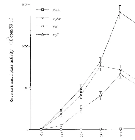
Virion-associated Vpr elevates viral production early in HIV infection.Recent studies have reported that the presence of Vpr stimulates viral production by a mechanism which is func-FIG. 3. Virion-associated Vpr induces apoptosis of Jurkat T cells during single-cycle infection. Jurkat T cells were infected with VSV-G pseudotyped Vpr2, Vpr1, or Vpr1-T HIV. At 48 h p.i., mock-infected and virus-infected cells were costained with annexin V and PI and analyzed by flow cytometry. The axes represent the cell-associated fluorescence intensity of annexin V (x) and PI (y). The percentage of cells in each quadrant is indicated above each graph. Similar results were obtained in two independent experiments.
on November 9, 2019 by guest
http://jvi.asm.org/
[image:5.612.106.496.63.556.2]tionally related to Vpr cell cycle G2 arrest activity (14, 44).
Since virion-associated Vpr induces a transient G2arrest, we
tested whether virion-associated Vpr stimulated viral produc-tion. Jurkat T cells were infected with different VSV-G-pseudotyped Vpr1, Vpr1-T, or Vpr2 viruses. At different
times p.i., supernatants from each infected culture were col-lected and viral production was evaluated by measuring virion-associated RT activity. As shown in Fig. 4, at early times p.i. (within 24 h), Jurkat T cells infected with Vpr1pseudotyped
viruses produced approximately twice as much virus as did Jurkat T cells infected with Vpr2pseudotyped virus.
Interest-ingly, similar levels of virus were produced in Vpr1-T
HIV-infected cells to those in Vpr1HIV-infected cultures (Fig. 4).
After 24 h p.i., the RT activity in Vpr1HIV-infected culture
continued to increase and reached a plateau at 30 to 48 h p.i. whereas the RT activity from the Vpr1-T HIV-infected culture
declined to a level similar to that detected in the Vpr2
HIV-infected culture. This data clearly indicate that after viral entry, virion-associated Vpr has a stimulating effect on viral produc-tion. However, in the absence of de novo expression of Vpr, this early activation of viral production cannot be sustained.
Stimulation of LTR-driven CAT gene expression.To inves-tigate whether the Vpr stimulation of viral production resulted from protein transactivation activity, we tested the effect of virion-associated Vpr on the expression of a CAT reporter gene driven by the HIV-1 LTR. Briefly, Jurkat T cells were transfected with an HIV-1 LTR-CAT expressor (pCEP4IIICAT), which contains a hygromycin resistance gene. After 10 days of hygromycin selection, cells were infected with VSV-G pseudotyped Vpr1-T, Vpr1, or Vpr2virus. At 12, 15, and 18 h
p.i., infected cells were lysed and the CAT activity in these cell lysates was measured. The results in Fig. 5A show that, at 12 h p.i., indicator cells infected with Vpr1and Vpr1-T virus
ex-hibited a five fold increase in CAT activity, as compared to Vpr2virus infected cells. The CAT activity continued to
in-crease in Vpr1and Vpr1-T virus-infected cells by twofold at
15 h p.i. and by fourfold at 18 h p.i. This continued increase in CAT activity most probably results from initiation of Tat ex-pression, since it was not observed upon treatment with AZT (data not shown). Interestingly, CAT activity increased only by about twofold between 12 and 18 h p.i. in Vpr2virus-infected
cells. At 18 h p.i., CAT activity was ninefold higher in Vpr1
and Vpr1-T virus-infected cells than in Vpr2 virus-infected
cells. We next tested a Vpr C-terminal mutant, R80A, which was previously shown to be incapable of inducing a cell cycle G2arrest. As shown in Fig. 5B, pseudotyped virus containing
the R80A mutant (VprR80A-T) could not stimulate
LTR-di-rected CAT activity upon infection of the indicator cell line. To confirm that virion-associated Vpr itself was responsible for the early stimulation of HIV LTR-directed gene
expres-sion, we performed similar experiments in the presence or absence of AZT (5mM). Figure 5C and D reveals that in the presence of AZT, LTR-directed CAT activity was detected in Vpr1-T and Vpr1virus-infected cells (lanes 3 and 4) but not in
cells infected with Vpr2virus (lane 2). At 12 h p.i. the levels of
CAT activity detected in Vpr1-T and Vpr1virus-infected cells
in the presence or absence of AZT were similar (Fig. 5C, compare lanes 3 and 4 with lanes 7 and 8), confirming that at early time points p.i., virion-associated Vpr itself stimulated LTR-directed CAT expression. At 30 h p.i. and in the absence of AZT, an increase in CAT activity was detected in Vpr2
virus-infected cells (Fig. 5D, lane 6), presumably through the activating effect of de novo-expressed Tat. Interestingly, a more pronounced stimulation of CAT activity was detected in Vpr1-T and Vpr1virus-infected cells (Fig. 5D, lanes 7 and 8),
suggesting that virion-associated Vpr may exert a positive ef-fect on Tat transactivation. Overall, these results indicate that virion-associated Vpr can stimulate LTR-directed gene expres-sion. This early stimulation of gene expression, which appears to potentiate Tat transactivation, correlates with the ability of the protein to mediate cell cycle arrest in G2.
DISCUSSION
Like structural gag and env gene products, the HIV acces-sory protein Vpr is expressed late in the HIV replication cycle and is efficiently packaged into progeny viral particles. The virion-associated Vpr actively participates in the nuclear trans-port of the HIV PIC in nondividing cells. In addition, the expression of Vpr, even in the absence of any other viral proteins, induces a cell cycle arrest in G2and increases gene
[image:6.612.317.544.71.306.2]expression from a variety of viral promoters (4, 21, 36). One central question is why a protein like Vpr, that stimulates LTR-directed gene expression by manipulating the host cell cycle, is expressed in the late stages of viral replication and FIG. 4. Virion-associated Vpr stimulates HIV production during the early stage of infection. At each time interval after Jurkat T cells were infected with VSV-G-pseudotyped Vpr2, Vpr1or Vpr1-T HIV (as indicated), supernatant from each infected culture was collected and the amount of virion-associated RT activity was determined. Values represent means of duplicate samples. Similar results were obtained in three independent experiments.
TABLE 2. Effect of AZT on Vpr-induced apoptosis
Pseudotyped virus % of apoptotic cellsa
Mock... 4
Vpr2/VSV-G1... 5
Vpr1-T/VSV-G1... 14
Vpr1-T/VSV-G11AZTb... 15
Vpr1/VSV-G1... 25
Vpr1/VSV-G11AZT ... 13 aThe percentage of apoptotic cells in each infected culture was determined by costaining of infected cells with annexin V- FITC and PI at 48 h p.i. and analysis by flow cytometry.
b1AZT, AZT (5mM) was added to Jurkat cell cultures 2 h prior to infection and maintained during the course of infection.
on November 9, 2019 by guest
http://jvi.asm.org/
[image:6.612.57.294.80.156.2]packaged in substantial amounts in progeny viral particles. Is this function of the protein required only late in infection, or, alternatively, is it possible that, in addition to participating in the nuclear translocation of the HIV PIC, virion-associated Vpr has a positive effect on viral gene expression early in infection? Until now, the molecular mechanism(s) regulating the basal transcriptional activity of integrated HIV LTR is still not fully resolved. Whether a viral factor(s) contributes to the immediate-early activation of the LTR before de novo expres-sion is initiated remains to be determined.
Our results clearly demonstrate that virion-associated Vpr induces a G2 arrest in host cells following viral entry. This
effect was observed at a MOI as high as 10 (as described in the present study) but also at a MOI as low as 0.01 (data not shown). Indeed, titer determination studies of VSV-G-pseudotyped HIV particles indicate that (i) the G2-M/G1ratio
of cells infected at a MOI of 1 is comparable to that of cells infected at a MOI of 10, suggesting that once a threshold level of Vpr is translocated into a target cell, an efficient cell cycle G2arrest is initiated (data not shown); (ii) infection of cells at
MOIs ranging from 0.5 to 0.01 still induced a detectable cell cycle arrest in G2, although not as efficiently as infection at
MOIs ranging from 1 to 10 (data not shown). These results support data recently reported by Poon et al. (33) showing that at a MOI of 0.15, the level of arrest induced by virion-associ-ated Vpr alone is lower than that observed with wild-type virus capable of de novo Vpr production, probably because higher levels of Vpr are expressed in cells de novo.
Moreover, our results indicate that the establishment of a cell cycle arrest at G2stimulates the activity of the HIV LTR
since virion-associated Vpr mutant R80A, which has lost its cell cycle-modulating activity, was unable to activate CAT ex-pression from the LTR. This early effect of Vpr on LTR-directed gene expression was shown to lead to an increase in viral production. Interestingly, in our experimental system, the positive effect of Vpr on the HIV-1 LTR appears to potentiate Tat transactivation. It is likely that Vpr-mediated transactiva-tion, by increasing LTR-directed Tat expression, stimulated Tat transactivation. Alternatively, but without excluding the former possibility, optimal Tat transactivation may require a minimum level of LTR basal transcriptional activity. Such ac-tivation of basal transcription from the integrated LTR may be provided early in the infection by virion-associated Vpr via its cell cycle arrest at G2. Indeed, a recent study indicates that Tat
FIG. 5. Virion-associated Vpr stimulates HIV LTR-directed CAT gene expression. Jurkat T cells transfected with pCEP4III LTR-CAT were selected with hygromycin (500mg/ml). After 10 days of selection, hygromycin-resistant Jurkat T cells were infected with VSV-G-pseudotyped Vpr2, Vpr1, or Vpr1-T HIV (as indicated). (A) At 12, 15, and 18 h p.i., CAT activity was determined. The transactivation level in each VSV-G pseudotyped HIV-infected cell sample is expressed as fold increase in CAT activity. The CAT activity value obtained in Vpr2-infected Jurkat T cells at 12 h p.i. was arbitrarily set at 1 (A). (B) CAT activity detected in pCEP4III LTR-CAT-transfected Jurkat cells following infection with VSV-G-pseudotyped Vpr2, Vpr1, Vpr1-T, and VprR80A-T viruses. (C and D)
Hygromycin-resistant Jurkat cells were infected with VSV-G-pseudotyped Vpr2, Vpr1, or Vpr1-T HIV in the presence (lanes 1 to 4) or absence (lanes 5 to 8) or 5mM AZT. At 12 h p.i. (C) and 30 h p.i. (D), CAT activity in each VSV-G-pseudotyped, HIV-infected cell sample was measured.
on November 9, 2019 by guest
http://jvi.asm.org/
functions after the formation of a specific transcription initia-tion complex and that Tat transactivainitia-tion is accompanied by a remodeling of chromatin structure of integrated LTR (8). In-terestingly, we have recently reported that Vpr, via its cell cycle arrest activity at G2, cooperates with p300/CBP, a
transcrip-tional coactivator that regulates the activity of NF-kB as well as of a variety of transcription factors presumably through its ability to regulate chromatin structure by histone acetylation (10). Overall, our results support the notion that virion-asso-ciated Vpr, in addition to being involved in the nuclear trans-location of the PIC, actively participates in the immediate-early activation of the HIV LTR by a molecular mechanism that is not fully understood. This newly identified activity of Vpr is reminiscent of that of VP16, a herpes simplex virus immediate-early gene product, even though the molecular mechanisms of their respective transactivation activity are likely to be distinct. VP16 is a transcription factor found in herpes simplex virus particles that selectively transactivates a class of viral promoters that control the expression of early gene products (40).
Apoptosis is one of the main cell-killing mechanisms in-volved in HIV-mediated direct and/or indirect CD41cell
de-pletion in vivo (2, 11, 15, 19, 20). Several gene products, such as Tat, Nef, gp120, and Vpr, induce apoptosis of HIV-infected cells in different systems (16, 23, 38, 43, 44). Our results clearly indicate that virion-associated Vpr by itself can induce apopto-sis of HIV infected cells. However, this Vpr-induced apoptoapopto-sis occurs in a relatively small number of infected cells (15% at 48 h p.i.), suggesting that only a subpopulation of infected Jurkat cells is susceptible to Vpr-induced apoptosis. Interest-ingly, Ayyavoo et al. reported that the Vpr-modulated T-cell receptor triggered apoptosis in a manner similar to that of glucocorticoids (1). In the absence of T-cell receptor-mediated activation, Vpr induced apoptosis, whereas in the presence of such stimuli, Vpr interrupted the expected induction of apo-ptosis. By analogy to the latter results, it is possible that a subpopulation of cells in the Jurkat cell line are in a state that makes them more susceptible to Vpr-induced apoptosis. Our data also show that Vpr de novo expression increases the frequency of apoptotic cells in the culture (27% at 48 h), suggesting that a sustained threshold level of Vpr may be required for efficient induction of apoptosis.
The results obtained with Vpr1-T virus or Vpr1 virus in
presence of AZT indicate that the effect of virion-associated Vpr on the cell cycle and on viral production is transient and lasts up to 24 h. This probably reflects the relative stability of the Vpr protein, which has been reported to have a half-life of more than 20 h (28). It is clear from our results that the maintenance of a G2cell cycle arrest and the resulting positive
effect on viral replication requires Vpr de novo expression. However, as discussed above, once a threshold level of Vpr is reached, cell killing by apoptosis may occur late during infec-tion, at least in our in vitro system. Vpr is expressed late in the infection cycle and is packaged into viral particles presumably via an interaction with the p6 domain of the Gag precursor polyprotein (Pr55gag) (27, 30). It is tempting to speculate that
the binding of Vpr to Pr55gagand the protein targeting to the
site of viral assembly may regulate the amount of free Vpr in the cell and thereby control the delicate balance between the optimization of viral gene expression and production and the induction of cytopathic effects.
Previous studies have indicated that in plasma, the ratio of noninfectious to infectious virus particles is approximately 100,000:1 (26). However, the relevance of the presence of such a large number of noninfectious virus particles in vivo is not clear. During the preparation of this paper, Poon et al.
dem-onstrated that HIV-1 Vpr packaged in virions, including those rendered defective for infection by RT or protease inhibitors, were capable of inducing cell cycle arrest (33). Interestingly, our results further indicate that virion-associated Vpr not only mediated cell cycle arrest at G2but also could induce apoptosis
in some cells. In agreement with the observation by Poon et al., our results indicate that virion-associated Vpr still induced a transient G2arrest as well as apoptosis during HIV infection of
Jurkat T cells in the presence of AZT as well as in Jurkat cells exposed to noninfectious virus particles produced from trans-fected cells treated with a protease inhibitor, Palinavir (Fig. 4 and Table 1; data not shown for Palinavir). Thus, these results raise the possibility that Vpr incorporated in infectious as well as noninfectious or defective virus particles contributes to the host immune system suppression in vivo by disturbing the cell cycle progression and/or by inducing apoptosis of HIV target cells. This could also at least partly account for the “bystander cell-killing” effect reported during HIV infection (11, 13).
Overall, in this study, we provided biological evidence that in addition to its PIC nuclear targeting activity in nondividing cells, virion-associated Vpr plays an important role in the im-mediate-early transcription of the HIV-1 genome during HIV infection. Moreover, the induction of cell cycle arrest at G2and
apoptosis by virion-associated Vpr, independent of viral repli-cation, strongly suggest a role of Vpr in HIV-mediated CD41
-T-cell depletion and immune system dysfunction. Thus, ther-apeutic approaches directed against virion-associated Vpr function may strongly attenuate HIV infection and replication and reduce HIV-mediated CD41-T-cell dysfunction.
ACKNOWLEDGMENTS
We thank Serge Senechal for the flow cytometric analysis. We also thank Daniel Lamarre and BioMega-Boehringer Ingelheim for the generous gift of the HIV protease inhibitor Palinavir.
E.A.C. is a recipient of a Medical Research Council of Canada (MRC) scientist award. This work was supported by grants from MRC and from the Fonds pour la Formation de Chercheurs et l’Aide a` la Recherche (FCAR).
REFERENCES
1. Ayyavoo, V., A. Mahboubi, S. Mahalingam, R. Ramalingam, S. Kudchodkar, W. V. William, D. R. Green, and D. B. Weiner.1997. HIV-1 Vpr suppresses immune activation and apoptosis through regulation of nuclear factor kappa B. Nat. Med. 3:1117–1123.
2. Baltimore, D., R. T. Gandhi, B. K. Chen, S. E. Strauss, J. K. Dale, and J. Lenardo.1998. HIV-1 directly kills CD41T cells by a Fas-independent mechanism. J. Exp. Med. 187:1113–1122.
3. Bartz, S. R., M. E. Rogel, and M. Emerman. 1996. Human immunodeficiency virus type 1 cell cycle control: Vpr is cytostatic and mediates G2
accumula-tion by a mechanism which differs from DNA damage checkpoint control. J. Virol. 70:2324–2331.
4. Cohen, E. A., G. Dehni, J. G. Sodroski, and W. A. Haseltine. 1990. Human immunodeficiency virus vpr product is a virion-associated regulatory protein. J. Virol. 64:3097–3099.
5. Cohen, E. A., E. F. Terwilliger, Y. Jalinoos, J. Proulx, J. G. Sodroski, and W. A. Haseltine.1990. Identification of HIV-1 Vpr product and function. J. Acquired Immune Defic. Syndr. 1:11–18.
6. Conti, L., G. Rainaldi, P. Matarrese, B. Varano, R. Rivabene, S. Columba, A. Sato, F. Belardelli, W. Malorni, and S. Gessani.1998. The HIV-1 Vpr protein acts as a negative regulator of apoptosis in human lymphoblastoid T cell line: possible implications for pathogenesis of AIDS. J. Exp. Med. 187:403–413.
7. Cullen, B. R. 1998. HIV-1 auxiliary proteins: making connections in dying cell. Cell 93:685–692.
8. El Kharroubi, A., G. Piras, R. Zensen, and M. A. Martin. 1998. Transcrip-tional activation of the integrated chromatin-associated human immunode-ficiency virus type 1 promoter. Mol. Cell. Biol. 18:2535–2544.
9. Emerman, M., and M. H. Malim. 1998. HIV-1 regulatory/accessory genes: keys to unraveling viral and host cell biology. Science 280:1880–1883. 10. Felzien, L. K., C. Woffendin, M. O. Hottiger, R. A. Subbramanian, E. A.
Cohen, and G. J. Nabel.1998. HIV transcriptional activation by the acces-sory protein, Vpr, is mediated by the p300 co-activator. Proc. Natl. Acad. Sci. USA 95:5281–5286.
on November 9, 2019 by guest
http://jvi.asm.org/
11. Finkel, T. H., G. Tudor-Williams, N. K. Banda, M. F. Cotton, T. Curiel, C. Monks, T. W. Baba, R. M. Ruprecht, and A. Kupfer.1995. Apoptosis occurs predominantly in bystander cells and not in productively infected cells of HIV and SIV-infected lymph nodes. Nat. Med. 1:129–134.
12. Fouchier, R. A., B. E. Meyer, J. H. Simon, U. Fisher, A. V. Albright, F. Gonzalez-Scarano, and M. H. Malim.1998. Interaction of the human im-munodeficiency virus type 1 Vpr protein with the nuclear pore complex. J. Virol. 72:6004–6013.
13. Glynn, J. M., D. L. McElligott, and D. E. Mosier. 1996. Apoptosis induced by HIV infection in H9 T cells is blocked by ICE-family protease inhibition but not by a Fas (CD95) antagonist. J. Immunol. 157:2754–2758. 14. Goh, W. C., M. E. Rogel, C. M. Kinsey, S. F. Michael, P. N. Fultz, M. A.
Nowak, B. H. Hahn, and M. Emerman.1998. HIV-1 Vpr increases viral expression by manipulation of the cell cycle: a mechanism for selection of Vpr in vivo. Nat. Med. 4:65–71.
15. Gougeon, M. L., A. G. Laurent-Crawford, A. G. Hovanessian, and L. Mon-tagnier.1993. Direct and indirect mechanisms mediating apoptosis during HIV infection: contribution to in vivo CD4 T cell depletion. Semin. Immu-nol. 5:187–194.
16. He, J., S. Choe, R. Walker, P. Di Marzio, D. O. Morgan, and N. R. Landau. 1995. Human immunodeficiency virus type 1 viral protein R (Vpr) arrests cells in the G2phase of the cell cycle by inhibiting p34cdc2activity. J. Virol. 69:6705–6711.
17. Heinzinger, N. K., M. I. Bukrinsky, S. A. Haggerty, A. M. Ragland, V. Kewalramani, M. A. Lee, H. E. Gendelman, L. Ratner, M. Stevenson, and M. Emerman.1994. The Vpr protein of human immunodeficiency virus type 1 influences nuclear localization of viral nucleic acids in nondividing host cells. Proc. Natl. Acad. Sci. USA 91:7311–7315.
18. Ho, D. H., A. U. Neumann, A. P. Perelson, W. Chen, J. M. Leonard, and M. Markowitz.1995. Rapid turnover of plasma virions and CD4 lymphocytes in HIV-1 infection. Nat. Med. 373:123–126.
19. Hovanessian, A. G., A. G. Laurent-Crawford, B. Krust, S. Muller, Y. Riviere, M. A. Rey-Cuille, J. M. Bechet, and L. Montagnier.1991. The cytopathic effect of HIV is associated with apoptosis. Virology 185:829–839. 20. Hovanessian, A. G., E. Jacotot, C. Callebaut, J. Blanco, Y. Riviere, and B.
Krust.1995. HIV envelope glycoprotein-induced cell killing by apoptosis is enhanced with increased expression of CD26 in CD41T cells. Virology 223:318–330.
21. Jowett, J. B., V. Planelles, B. Poon, N. P. Shah, M. L. Chen, and I. S. Chen. 1995. The human immunodeficiency virus type 1 vpr gene arrests infected T cells in the G21M phase of the cell cycle. J. Virol. 69:6304–6313.
22. Kimpton, J., and M. Emerman. 1992. Detection of replication-competent and pseudotyped human immunodeficiency virus with a sensitive cell line on the basis of activation of an integratedb-galactosidase gene. J. Virol. 66: 2232–2239.
23. Kolesnitchenko, V., L. M. Wahl, H. Tian, I. Sunila, Y. Tani, D. P. Hartmann, J. Cossman, M. Raffeld, J. Orenstein, and L. E. Samelson.1995. Human immunodeficiency virus 1 envelope-initiated G2 phase programmed cell
death. Proc. Natl. Acad. Sci. USA 92:11889–11893.
24. Lavalle´e, C. X-J. Yao, A. Ladha, H. Go¨ttlinger, W. A. Haseltine, and E. A. Cohen.1994. Requirement of the Pr55gagprecursor for incorporation of the Vpr product into human immunodeficiency type 1 viral particles. J. Virol. 68:1926–1934.
25. Levy, J. A. 1993. Pathogenesis of human immunodeficiency virus infection. Microbiol. Rev. 57:183–289.
26. Levy, J. A. 1998. HIV and the pathogenesis of AIDS. ASM Press, Washing-ton, D.C.
27. Lu, Y. L., P. Spearman, and L. Ratner. 1993. Human immunodeficiency virus type 1 viral protein R localization in infected cells and virions. J. Virol. 67:6542–6550.
28. Mahalingam, S., S. A. Khan, M. A. Jabbar, C. E. Monken, R. G. Collman, and A. Srinivasan.1995. Identification of residues in the N-terminal acidic domain of HIV-1 Vpr essential for virion incorporation. Virology 207:297– 302.
29. Nie, Z., D. Bergeron, R. Subbramanian, X.-J. Yao, F. Checroune, N.
Rougeau, and E. A. Cohen.1998. The putative alpha helix 2 of human immunodeficiency virus type 1 Vpr contains a determinant which is respon-sible for the nuclear translocation of the proviral DNA in growth-arrested cells. J. Virol. 72:4104–4115.
30. Paxton, W., R. I. Connor, and N. R. Landau. 1993. Incorporation of Vpr into human immunodeficiency virus type 1 virions: requirement for the p6 region of Gag and mutational analysis. J. Virol. 67:7229–7237.
31. Perelson, A. S., A. U. Neuman, M. Markowitz, J. M. Leonard, and D. D. Ho. 1996. HIV-1 dynamics in vivo: virion clearance rate, infected cell life-span, and viral generation time. Science 271:1582–1586.
32. Planelles, V., J. B. Jowett, Q. X. Li, Y. Xie, B. Hahn, and I. S. Chen. 1996. Vpr-induced cell cycle arrest is conserved among primate lentiviruses. J. Vi-rol. 70:2516–2524.
33. Poon, B., K. G. Ferbas, S. A. Stewart, and I. S. Y. Chen. 1998. Cell cycle arrest by Vpr in HIV-1 virions and insensitivity to antiviral agents. Science 281:266–268.
34. Popov, S., M. Rexach, G. Zybarth, N. Reiling, M.-A. Lee, L. Ratner, C. M. Lane, M. S. Moore, G. Blobel, and M. Bukrinsky.1998. Viral protein R regulates nuclear import of HIV-1 pre-integration complex. EMBO J. 17: 909–917.
35. Re, F., D. Braaten, E. K. Franke, and J. Luban. 1995. Human immunode-ficiency virus type 1 Vpr arrests the cell cycle in G2by inhibiting the
activa-tion of p34cdc2-cyclin B. J. Virol. 69:6859–6864.
36. Rogel, M. E., L. I. Wu, and M. Emerman. 1995. The human immunodefi-ciency virus type 1 Vpr gene prevents cell proliferation during chronic in-fection. J. Virol. 69:882–888.
37. Sodroski, J., C. Rosen, and F. Wong-Staal. 1985. Trans-acting transcriptional regulation of human T-cells leukemia virus type III long terminal repeat. Science 227:171–173.
38. Stewart, S. A., B. Poon, J. B. M. Jowett, and I. S. Chen. 1997. Human immunodeficiency virus type 1 Vpr induces apoptosis following cell cycle arrest. J. Virol. 71:5579–5592.
39. Subbramanian, R. A., A. Kessous-Elbaz, R. Lodge, J. Forget, X-J. Yao, D. Bergeron, and E. A. Cohen.1998. Human immunodeficiency virus type 1 Vpr is a positive regulator of viral transcription and infectivity in primary human macrophages. J. Exp. Med. 187:1103–1111.
40. Triezenberg, S. J., K. L. Lamarco, and S. L. McKnight. 1998. Evidence of DNA:protein interactions that mediate HSV-1 immediate early gene activa-tion by VP16. Genes Dev. 2:730–742.
41. Vodicka, M. A., D. M. Koepp, P. S. Silver, and M. Emerman. 1998. HIV-1 Vpr interacts with the nuclear transport pathway to promote macrophage infection. Genes Dev. 12:175–185.
42. Wei, X., S. K. Ghosh, M. E. Taylor, V. A. Johnson, E. A. Emini, P. Deutsch, J. D. Lifson, S. Bonhoeffer, M. A. Nowak, B. H. Hahn, M. S. Saag, and G. M. Shaw.1995. Viral dynamics in human immunodeficiency virus type 1 infec-tion. Nat. Med. 373:117–122.
43. Westendorp, M. O., R. Frank, C. Ochsenbauer, K. Stricker, J. Dhein, H. Walczak, K. M. Debatin, and P. H. Krammer.1995. Sensitization of T cells to CD95-mediated apoptosis by HIV-1 Tat and gp 120. Nat. Med. 375:497– 500.
44. Yao, X.-J., A. J. Mouland, R. A. Subbramanian, J. Forget, N. Rougeau, D. Bergeron, and E. A. Cohen.1998. Vpr stimulates viral expression and in-duces cell killing in human immunodeficiency virus type 1 infected dividing Jurkat T cells. J. Virol. 72:4686–4693.
45. Yao, X.-J., R. A. Subbramanian, N. Rougeau, F. Boisvert, D. Bergeron, and E. A. Cohen.1995. Mutagenic analysis of human immunodeficiency virus type 1 Vpr: role of a predicted N-terminal alpha-helical structure in Vpr nuclear localization and virion incorporation. J. Virol. 69:7032–7044. 46. Yao, X.-J., S. Garzon, F. Boisvert, W. A. Haseltine, and E. A. Cohen. 1993.
The effect of Vpu-induced syncytia formation. J. Acquired Immune Defic. Syndr. 6:135–141.
47. Yu, X. F., M. Matsuda, M. Essex, and T. H. Lee. 1990. Open reading frame vpr of simian immunodeficiency virus encodes a virion-associated protein. J. Virol. 64:5688–5693.