0022-538X/97/$04.00
1
0
Copyright
q
1997, American Society for Microbiology
High-Efficiency Transduction of Human Lymphoid Progenitor Cells
and Expression in Differentiated T Cells
DONG SUNG AN,
1,2YOSHIO KOYANAGI,
2JIA-QI ZHAO,
1RAMESH AKKINA,
3GREG BRISTOL,
1NAOKI YAMAMOTO,
2JEROME A. ZACK,
1ANDIRVIN S. Y. CHEN
1*
Departments of Microbiology & Immunology and Medicine, UCLA School of Medicine, Los Angeles, California 90095-1678
1;
Department of Microbiology, Tokyo Medical & Dental University, Bunkyo-ku, Tokyo 113, Japan
2; and
Department of Pathology, Colorado State University, Fort Collins, Colorado 80523
3Received 16 August 1996/Accepted 1 November 1996
Gene therapy strategies for humans have been limited by low transduction efficiencies and poor expression
of retroviral vectors in differentiated progeny cells carrying the transduced vector. Here we describe a strategy
utilizing a cell surface reporter gene, murine
thy-1.2
, selectable by fluorescence-activated cell sorting (FACS),
to achieve higher gene marking efficiencies. Human CD34-positive cells were transduced by a murine retroviral
vector bearing the
thy-1.2
marker and pseudotyped with vesicular stomatitis virus G protein, followed by FACS
to enrich for CD34-positive cells that express Thy-1.2 on the cell surface. Gene marking and expression after
differentiation into thymocytes were assessed in a SCID-hu Thy/Liv mouse model for human lymphoid
progenitor cell gene therapy. We found that virtually all of the differentiated T-cell progeny were marked with
vector sequences. It is of particular importance that reconstitution with the selected cells resulted in expression
of Thy-1.2 in up to 71% of donor-derived thymocytes. It is of note that the donor-derived thymocytes that did
not express Thy-1.2 still harbored vector
thy-1.2
sequences, suggesting repression of transgene expression in
some cells during progenitor cell differentiation into thymocytes. These studies provide a proof of concept for
efficient expression of transgenes through T-lymphoid differentiation and a potential basis for utilizing similar
strategies in human gene therapy clinical trials.
One of the key elements of effective stem or progenitor cell
gene therapy is development of vectors for efficient
transduc-tion and expression of transduced genes in the differentiated
progeny of progenitor cells (10, 13, 27, 35). Testing of vector
efficiency is complicated by relatively poor transduction
effi-ciencies of CD34
1progenitor cells and relatively poor
expres-sion in the mature cells derived from the transduced
progen-itor cells (4, 5, 7, 12, 16, 17). We have utilized the severe
combined immunodeficient (SCID)-hu Thy/Liv mouse model
(23), a SCID mouse implanted with human fetal thymus and
liver tissues, to develop an effective system for rapid
experi-mental testing of gene therapy vectors and approaches in a
setting in which hematopoietic progenitor cells can be
trans-duced and used to reconstitute a human hematolymphoid
or-gan (Thy/Liv). We have previously shown that an amphotropic
murine leukemia virus (MuLV)-based retrovirus vector, LNL6
(25), can transduce human CD34
1cells and that the
geneti-cally marked cells can reconstitute T-cell development in the
SCID-hu Thy/Liv mouse (1).
In this study, we utilized this model system to demonstrate a
proof of principle for a novel vector-reporter gene system. We
demonstrated that immunoselection for transduced human
progenitor cells, using a cell surface reporter gene, results in
efficient reconstitution of the T-cell compartment of the
thy-mus, where a significant proportion of the cells express the
vector.
MATERIALS AND METHODS
Construction of retroviral vectors.The MuLV-based vectors SRaLluc and SRaLthy were derived from LXSN (25). The simian virus 40 (SV40) promoter and the neomycin resistance gene were removed from LXSN and replaced with the firefly luciferase or the murine thy-1.2 cDNA (11, 14, 32). The SRasequence
was inserted before the 59Moloney murine sarcoma virus (MSV) long terminal repeat (LTR) for vector amplification in COS cells (26, 34). LlucSN was con-structed from LXSN and IISVluc. LXSN was digested with BamHI, filled in with Klenow fragments, and then excised by XhoI. The resulting 7-kb fragment was ligated to a 1.7-kb fragment of IISVluc, which contains the luciferase gene, digested with MluI, filled in with a DNA polymerase Klenow fragment, and digested with XhoI. SRaLlucSN was constructed from LlucSN and pcDL-SRa -296 (34). LlucSN was digested with SacII and AccI and filled in with T4 DNA polymerase. The resulting 5.3-kb fragment was ligated to a 3.2-kb fragment of pcDL-SRa-296, which contains the SRa sequence digested with XhoI and BamHI, and filled in with T4 DNA polymerase. Transcription of the 59MSV LTR is in the same orientation as the promoter in SRasequences. SRaLluc was constructed from SRaLlucSN. SRaLlucSN was digested with BamHI and RsrII and filled in with T4 DNA polymerase, and the resulting 7.5-kb fragment was religated to remove the SV40 promoter and the neomycin resistance gene from the vector. SRaLthy was constructed from SRaLlucSN and LthySN (32). SRaLlucSN was digested with RsrII, filled with T4 DNA polymerase, and excised with EcoRI. The resulting 5.8-kb fragment was ligated to a 0.5-kb fragment of LthySN, which contains the murine thy-1.2 cDNA, digested with BamHI, filled in with T4 polymerase, and digested with EcoRI.
Retroviral vector production.Vesicular stomatitis virus G protein (VSV-G)-pseudotyped retroviral vectors were recovered following electroporation of three plasmids: retroviral vectors SRaLluc or SRaLthy, SVC2env2MLV (19), and pHCMV-G (6). Twenty micrograms of each plasmid was added to 107
COS cells in 500ml of RPMI with 20% fetal calf serum. Electroporation was achieved by a single pulse from a Bio-Rad Gene Pulser apparatus equipped with a capaci-tance extender unit (960mF, 230 V). Transfected COS cells were cultured in 10 ml of Dulbecco’s modified Eagle medium with 10% calf serum, 100 U of peni-cillin per ml; and 100mg of streptomycin per ml, and the medium was changed on day 1 postelectroporation. Supernatants (240 ml) were collected on days 2 and 3 postelectroporation, filtered with a 0.45-mm-pore-size filter, treated with RNase-free DNase (2mg/ml; Worthington) for 30 min at room temperature (20 to 258C) in the presence of 0.01 M MgCl2, and subjected to ultracentrifugation
in a Beckman L3-50 centrifuge with an SW28 rotor at 50,0003g (25,000 rpm) and 48C for 90 min. The pellet was resuspended in 200ml of 0.1% Hanks’ balanced salt solution for 24 h at 48C. Concentrated supernatants were stored at 2708C until use.
Infection and immunoselection of gene-transduced CD34-positive cells by fluorescence-activated cell sorting (FACS). Human CD34-positive cells were purified from a male fetal liver as previously described (1). Cells were cultured in Iscove’s modified Dulbecco’s medium with 100 ng each of interleukin 3 (IL-3), IL-6, and stem cell factor per ml, 20% fetal calf serum, 100 U of penicillin per ml, and 100mg of streptomycin per ml.
CD34-positive cells (53105) were infected with SRaLluc
VSV-G-pseudo-* Corresponding author. Phone: (310) 825-4793. Fax: (310)
794-7682.
1397
on November 9, 2019 by guest
http://jvi.asm.org/
typed virus by incubation with concentrated virus in the presence of Polybrene (8 mg/ml) at 378C for 2 to 3 h on days 1, 2, and 3 after CD34-positive cell purifi-cation from fetal liver. SRaLluc-infected CD34-positive cells (23105) were
injected into each female Thy/Liv implant of irradiated animals on day 4 after CD34-positive cell purification from fetal liver.
CD34-positive cells (23106) were infected with SRaLthy
VSV-G-pseudo-typed virus by incubating concentrated virus in the presence of Polybrene (8 mg/ml) at 378C for 2 to 3 h on days 1 and 2 after CD34-positive cell purification from fetal liver. On day 3 after CD34-positive cell purification from fetal liver, thy-1.2-transduced CD34-positive cells were stained with monoclonal antibodies (MAbs) to murine Thy-1.2 (Caltag) and human CD34 (Becton-Dickinson) con-jugated with fluorescein isothiocyanate (FITC) or phycoerythrin (PE), respec-tively. CD34 and Thy-1.2 double-positive cells were sorted on a FACStarplus
(Becton-Dickinson). Female Thy/Liv implants of irradiated animals (400 rads) were directly injected with 23105
Thy-1.2 and CD34 double-positive cells on day 4 after CD34-positive cell purification from fetal liver.
Luciferase assay.To analyze luciferase expression, 106cells were washed twice
with phosphate-buffered saline and lysed with 100ml of 13luciferase lysis buffer (Promega Corp.). Ten microliters of each lysate was subjected to the luciferase assay (Promega Corp.) with a Monolight 2010 luminometer (Analytical Lumi-nescence Laboratory, San Diego, Calif.).
Quantitative PCR assay.Each PCR amplification was performed as previously described (1, 3, 15, 18, 21, 29, 30, 38). In brief, to detect luc or thy-1.2 sequences, one of the oligonucleotide primers for each pair used was end labeled with32P,
and 25 ng was included in the reaction mixture (usually 53106to 13107cpm).
The second oligonucleotide primer was not labeled, and 50 ng was incorporated into each reaction mixture. Each reaction mixture contained a 0.25 mM concen-tration of each of the four deoxynucleoside triphosphates, 50 mM NaCl, 25 mM Tris-HCl (pH 8.0), 5 mM MgCl2, 100mg of bovine serum albumin per ml, and
1.25 U of Taq DNA polymerase (Promega). The reaction mixture was overlaid with 25ml of mineral oil and then subjected to 25 cycles of denaturation for 1 min at 948C and polymerization for 2 min at 658C. The reaction was performed on a Perkin-Elmer thermocycler. Amplified products resulting from the PCR were analyzed by electrophoresis on 6% nondenaturing polyacrylamide gels and visu-alized by direct autoradiography of the dried gels. Quantitative analysis of the amplified products was performed with a radioanalytic imager (Ambis, San Diego, Calif.). The nucleotide sequences of the oligonucleotide primers used for luc DNA detection were derived from the nucleotide sequence of the luc cDNA (9) and are as follows: Luc1, 59-CTCTAGAGGATGGAACCGCTG-39; Luc2, 59-CAGCCCATATCGTTTCATAGC-39. The nucleotide sequences of the oli-gonucleotide primers used for murine thy-1.2 cDNA detection were derived from the nucleotide sequence of the murine thy-1.2 sequence (12) and are as follows: thy2, 59-CTAGCCAACTTCACCACCAAGGA-39; thy3, 59-CTTATGCCGCCA CACTTGACCAG-39. A pair of oligonucleotide primers complementary to the first exon of the humanb-globin gene (28) was used in each reaction mixture in PCR analyses to normalize the total amount of human cellular DNA present. A pair of oligonucleotide primers complementary to the sequence of the human Y chromosome (38) was used in each reaction mixture in PCR analyses to deter-mine the amount of donor cell-derived cellular DNA present. During PCR amplification, either labeledb-globin- or Y chromosome-specific oligonucleo-tides were incorporated into the reaction at 53106
to 13107
cpm per reaction. Quantitation of luc or murine thy-1.2 DNA during PCR amplifications was performed by analyzing a standard curve of dilution of linearized SRaLluc or SRaLthy plasmid DNA digested with HindIII, which does not cleave vector sequences. These DNAs were diluted in 0.1mg of human peripheral blood mononuclear cell DNA per ml. The copy number of luc or murine thy-1.2 included in the standard curve ranged from 54 to 14,000 or 200 to 48,000 copies, respectively. Standard curves for humanb-globin and Y chromosome DNAs were obtained by amplification of 0.001 to 0.1mg of human cellular DNA (100 to 10,000 cell equivalents) from human male CD34-positive cells.
Flow cytometric analysis.Thymocytes from Thy/Liv implants were stained with MAbs to human CD3, CD4, CD5, CD8, CD45 (Becton-Dickinson), or CD1 (Coulter) or murine Thy-1.2 (Caltag) directly conjugated with FITC, PE, or peridinin chlorophyll protein. Samples were run on a FACScan flow cytometer, and data were analyzed with the Cell Quest program (Becton-Dickinson). Ten thousand events were acquired for Fig. 2A, a and b; Fig. 3A, a and b; and Fig. 3B, a to f.
Thymocyte culture for in vitro activation studies.Thymocytes were cultured at a concentration of 106
/ml in flat-bottom culture plates. Culture plates were coated with goat anti-mouse immunoglobulin G (IgG) (GAM; Tago, Burlin-game, Calif.). GAM (10mg/ml) in phosphate-buffered saline (PBS; pH 7.4) was added to wells and incubated for 2 h at 378C. Plates were then washed three times with PBS. Anti-human CD3 MAbs (T3; Coulter) were added at 4mg/ml in PBS, and the mixture was incubated at 378C for 1 h. The immobilized GAM provides a solid phase for binding of anti-human CD3 MAbs, as previously described (20, 36). After washing, thymocytes obtained from Thy/Liv implants of SCID-hu mice were cultured in the presence of IL-2 (1mg/ml), an anti-human CD28 MAb (1mg/ml), and an immobilized anti-human CD3 MAb in RPMI–20% fetal calf serum supplemented with 100 U of penicillin per ml and 100mg of streptomycin per ml. Control thymocytes were cultured in parallel in the absence of IL-2, anti-CD28 MAbs, and immobilized anti-CD3 MAbs. After 2 days of stimulation in vitro, thymocytes were analyzed by flow cytometry for murine
Thy-1.2 and human CD25 cell surface markers as already described. Dead cells were excluded by seven-amino-acid–actinomycin D staining as previously de-scribed (33). [3H]thymidine incorporation was measured by pulse-labeling cells
for 6 h on day 2 as previously described (38).
RESULTS
VSV-G-pseudotyped MuLV-based vectors bearing reporter
genes.
To facilitate the testing of different vector strategies, we
utilized a transient transfection system to provide helper virus
functions for the vector. We introduced the reporter gene,
either firefly luc or murine thy-1.2, into MuLV-based vectors to
allow sensitive detection of expression in cells (Fig. 1).
Quan-titative PCR was utilized to assess infection and transduction
efficiencies (1, 3, 15, 18, 21, 29, 30, 38). Each of these vectors
was pseudotyped with VSV-G and MuLV gag and pol,
pro-vided in trans through cotransfection. The VSV-G
pseudo-types are highly efficient for transduction of a variety of human
cell types, including transformed T-cell lines, peripheral blood
lymphocytes, and CD34-positive hematopoietic cells derived
from fetal liver. Since VSV-G pseudotypes can be
concen-trated by ultracentrifugation without loss of infectivity (2, 6,
37), we could achieve approximately 10- to 100-fold higher
titers in human cells than with the same vectors pseudotyped
with the amphotropic MuLV envelope, as measured by
lucif-erase activity on human HeLa, Rd, MT-2, SupT1, and primary
CD34
1cells. Furthermore, as previously reported,
VSV-G-pseudotyped virions could be concentrated and stored as
fro-zen stocks, facilitating standardization between different
exper-iments (6, 37).
Transduction of CD34-positive cells and reconstitution of
irradiated Thy/Liv implants of SCID-hu mice.
In initial
stud-ies, we tested the ability of CD34-positive cells transduced with
murine retroviral vectors bearing a luc reporter gene and
pseudotyped with VSV-G to reconstitute T-cell compartments
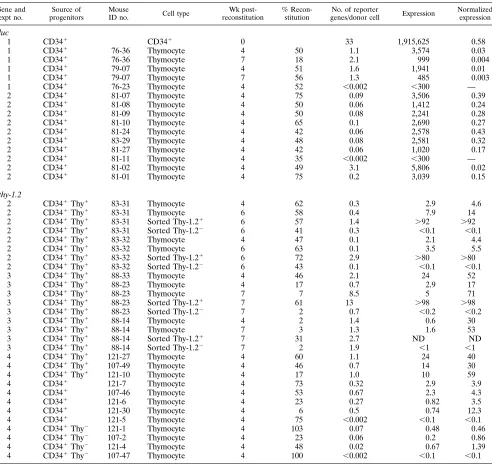
in SCID-hu mice (Table 1). Transduced CD34-positive cells
from male donors were injected into female-derived Thy/Liv
implants of irradiated SCID-hu mice. Reconstitution
efficien-cies varied from 18 to 75%, as assessed by PCR for Y
chro-mosome sequences 4 to 7 weeks postreconstitution. All of the
FIG. 1. Maps of luciferase and Thy-1.2 vectors. MuLV-based vectors SRaLluc and SRaLthy were developed from LXSN (25). The SV40 promoter and the neomycin resistance gene were removed from LXSN and replaced with firefly luc or murine thy-1.2 cDNA (11, 14, 32). The SRasequence was inserted before the 59 MSV LTR for vector amplification in COS cells (25, 34). SVC2env2MLV is an MuLV gag and pol expression construct kindly provided by Dan R. Littman (19), and pHCMV-G is a VSV-G expression construct kindly provided by Jane C. Burns (6, 37). SV40 ori is an SV40 origin sequence.C2 indicates deletion of the retroviral packaging signal, env2means deletion of the envelope gene, and CMV is cytomegalovirus.
on November 9, 2019 by guest
http://jvi.asm.org/
TABLE 1. Luciferase and Thy-1.2 gene transduction and expression efficiency in CD34
1cells and thymocytes in
SCID-hu mouse Thy/Liv implants
aGene and expt no.
Source of progenitors
Mouse
ID no. Cell type
Wk post-reconstitution
% Recon-stitution
No. of reporter
genes/donor cell Expression
Normalized expression
luc
1
CD34
1CD34
10
33
1,915,625
0.58
1
CD34
176-36
Thymocyte
4
50
1.1
3,574
0.03
1
CD34
176-36
Thymocyte
7
18
2.1
999
0.004
1
CD34
179-07
Thymocyte
4
51
1.6
1,941
0.01
1
CD34
179-07
Thymocyte
7
56
1.3
485
0.003
1
CD34
176-23
Thymocyte
4
52
,
0.002
,
300
—
2
CD34
181-07
Thymocyte
4
75
0.09
3,506
0.39
2
CD34
181-08
Thymocyte
4
50
0.06
1,412
0.24
2
CD34
181-09
Thymocyte
4
50
0.08
2,241
0.28
2
CD34
181-10
Thymocyte
4
65
0.1
2,690
0.27
2
CD34
181-24
Thymocyte
4
42
0.06
2,578
0.43
2
CD34
183-29
Thymocyte
4
48
0.08
2,581
0.32
2
CD34
181-27
Thymocyte
4
42
0.06
1,020
0.17
2
CD34
181-11
Thymocyte
4
35
,
0.002
,
300
—
2
CD34
181-02
Thymocyte
4
49
3.1
5,806
0.02
2
CD34
181-01
Thymocyte
4
75
0.2
3,039
0.15
thy-1.2
2
CD34
1Thy
183-31
Thymocyte
4
62
0.3
2.9
4.6
2
CD34
1Thy
183-31
Thymocyte
6
58
0.4
7.9
14
2
CD34
1Thy
183-31
Sorted Thy-1.2
16
57
1.4
.
92
.
92
2
CD34
1Thy
183-31
Sorted Thy-1.2
26
41
0.3
,
0.1
,
0.1
2
CD34
1Thy
183-32
Thymocyte
4
47
0.1
2.1
4.4
2
CD34
1Thy
183-32
Thymocyte
6
63
0.1
3.5
5.5
2
CD34
1Thy
183-32
Sorted Thy-1.2
16
72
2.9
.
80
.
80
2
CD34
1Thy
183-32
Sorted Thy-1.2
26
43
0.1
,
0.1
,
0.1
3
CD34
1Thy
188-33
Thymocyte
4
46
2.1
24
52
3
CD34
1Thy
188-23
Thymocyte
4
17
0.7
2.9
17
3
CD34
1Thy
188-23
Thymocyte
7
7
8.5
5
71
3
CD34
1Thy
188-23
Sorted Thy-1.2
17
61
13
.
98
.
98
3
CD34
1Thy
188-23
Sorted Thy-1.2
27
2
0.7
,
0.2
,
0.2
3
CD34
1Thy
188-14
Thymocyte
4
2
1.4
0.6
30
3
CD34
1Thy
188-14
Thymocyte
7
3
1.3
1.6
53
3
CD34
1Thy
188-14
Sorted Thy-1.2
17
31
2.7
ND
ND
3
CD34
1Thy
188-14
Sorted Thy-1.2
27
2
1.9
,
1
,
1
4
CD34
1Thy
1121-27
Thymocyte
4
60
1.1
24
40
4
CD34
1Thy
1107-49
Thymocyte
4
46
0.7
14
30
4
CD34
1Thy
1121-10
Thymocyte
4
17
1.0
10
59
4
CD34
1121-7
Thymocyte
4
73
0.32
2.9
3.9
4
CD34
1107-46
Thymocyte
4
53
0.67
2.3
4.3
4
CD34
1121-6
Thymocyte
4
23
0.27
0.82
3.5
4
CD34
1121-30
Thymocyte
4
6
0.5
0.74
12.3
4
CD34
1121-5
Thymocyte
4
75
,
0.002
,
0.1
,
0.1
4
CD34
1Thy
2121-1
Thymocyte
4
103
0.07
0.48
0.46
4
CD34
1Thy
2107-2
Thymocyte
4
23
0.06
0.2
0.86
4
CD34
1Thy
2121-4
Thymocyte
4
48
0.02
0.67
1.39
4
CD34
1Thy
2107-47
Thymocyte
4
100
,
0.002
,
0.1
,
0.1
a
Experiments 1 to 4 were performed at different times with different CD34-positive cells from male fetal livers (gestation ages, 16 to 24 weeks). The source of progenitor cells is the cell type injected into irradiated Thy/Liv implants of SCID-hu mice. For a given experiment, the retrovirus vector stocks were the same. Cell type is the type of cells analyzed. CD341is CD34-positive cells infected with SRaLluc. Thymocytes are cells biopsied from reconstituted Thy/Liv implants of SCID-hu mice. Sorted Thy-1.21and sorted Thy-1.22mean murine Thy-1.2-positive and -negative thymocytes sorted by the FACStarplus
(Becton-Dickinson), respectively. The mouse identification (ID) number preceding the hyphen refers to the Thy/Liv implant before reconstitution. Cells were analyzed at 0, 4, 6, and 7 weeks postreconstitution (0 refers to the time prior to CD34-positive cell injection into irradiated Thy/Liv implants). Percent reconstitution was calculated by dividing the values determined by Y chromosome PCR by the values determined byb-globin PCR. The number of reporter genes per donor cell was calculated by dividing the values determined by luc or thy-1.2 PCR by the values determined by Y chromosome PCR. For luc gene experiments, expression is reported in relative light units per 105
CD34-positive cells or total thymocytes, and for thy-1.2 gene experiments, expression is reported as percent Thy-1.2 expression in total thymocytes from Thy/Liv implants. Normalized expression (relative light units per unit of luc DNA for luc gene experiments and percent Thy-1.2 in donor thymocytes for thy-1.2 gene experiments) was calculated by the following formulas: RLU/U of luc DNA5(RLU/105
total cells)/no. of luc DNA copies in 105
total cells and % Thy-1.2 in donor thymocytes5(% Thy-1.2 in total thymocytes/% reconstitution)3102
). ND, not done. —, no significant level of luciferase activity. Most PCR experiments were done twice. The coefficient of variation for the values of PCR quantification was generally less than 30% (18). The percentages of Thy-1.2 and CD34 double-positive cells of each SRaLthy-infected CD34-positive cell were 10% in experiment 2, 53% in experiment 3, and 24% in experiment 4. The levels of sorting purity of Thy-1.2 and CD34 double-positive cells were 75% in experiment 2, 97% in experiment 3, and 98% in experiment 4. Sorting purity was determined by flow cytometric analysis of sorted cells. The lower levels of Thy-1.2 expression and the lower sorting purity in experiment 2 may have resulted in the lower levels of thy-1.2 reporter gene per donor cell following reconstitution in thymocytes.
on November 9, 2019 by guest
http://jvi.asm.org/
on November 9, 2019 by guest
http://jvi.asm.org/
reconstituted animals showed typical CD4-positive,
CD8-pos-itive, and CD4-CD8 double-positive thymocyte profiles (data
not shown). We achieved multiplicities of infection in
CD34-positive cells of greater than 1 (Table 1), most likely as a result
of using highly concentrated VSV-G-pseudotyped viruses. In
the thymocytes derived from the transduced CD34-positive
cells, we found that the transgene level was approximately one
per cell, consistent with the majority of thymocytes harboring
the vector. Detectable luc expression was present in
thymo-cytes of 11 of 13 reconstituted Thy/Liv implants of SCID-hu
mice, ranging from 485 to 5,806 relative light units (RLU)/10
5total thymocytes (Table 1). The calculated levels of luc
expres-sion (RLU per luc DNA copy were much lower than those of
either the input CD34-positive cells (Table 1) or those of in
vitro infections (typically, 1 to 10 RLU per luc DNA copy in
human transformed cell lines; data not shown). These low
levels of transgene expression are consistent with the low levels
of neo
rexpression that we previously reported (1) and with the
general experience of others engaged in stem cell gene therapy
experimentation (4, 5, 7, 12).
Immunoselection of CD34-positive cells tranduced with a
thy-1.2
-bearing vector and reconstitution of irradiated Thy/Liv
implants of SCID-hu mice.
We undertook a different strategy
that enabled selection by FACS to enrich transduced and
transgene-expressing cell populations. The luc gene was
re-placed with the murine thy-1.2 gene, which we previously
showed to be effective for monitoring of gene expression in
individual cells by flow cytometry (32). One additional step was
introduced into the reconstitution protocol whereby, following
transduction of CD34-positive cells, the cells were purified by
FACS on the basis of Thy-1.2 positivity and CD34 surface
expression (Fig. 2A). In this manner, Thy/Liv implants of
SCID-hu mice should be reconstituted with CD34-positive
cells that are highly enriched for the transgene. At 4 to 6 weeks
after introduction of CD34-positive cells into Thy/Liv implants,
the levels of vector DNA ranged from 0.1 to 8.5 copies of
thy-1.2 per donor-derived thymocyte (Table 1 and Fig. 2B). We
confirmed Thy-1.2 expression in human cells by demonstrating
a distinct population of cells positive for both Thy-1.2 and
CD45, a marker for human lymphocytes (Fig. 2A). When
nor-malized to the percentage of donor cells, calculated by PCR for
human Y chromosome versus
b
-globin sequences, the
propor-tion of cells expressing Thy-1.2 ranged from 4.4 to 71% (Table
1). In other experiments, we compared reconstitution and
ex-pression of an unfractionated CD34-positive cell population
with the same cells sorted into positive and
Thy-1.2-negative subpopulations. As expected, we detected little or no
thy-1.2 expression in thymocytes derived from reconstitution of
CD34-positive, Thy-1.2-negative cells, low levels (
,
3%) in
thy-mocytes derived from unfractionated CD34-positive cells, and
much greater levels (
,
25%) in thymocytes derived from
CD34-positive, Thy-1.2-positive cells (Table 1, experiment 4).
The Thy-1.2-positive, CD34-positive cells selected by FACS
should have been highly enriched for Thy-1.2-containing cells,
yet there appeared to be a proportion of donor-derived
thy-mocytes in all Thy/Liv implants which harbored thy-1.2 DNA
but did not express the transgene at a level detectable by flow
cytometry. We confirmed this by sorting the thymocytes into
Thy-1.2-positive and Thy-1.2-negative subpopulations and
de-termining the levels of vector DNA in the two populations.
Our results show that vector DNA was maintained in both
Thy-1.2-positive and Thy-1.2-negative donor cell populations,
although the Thy-1.2-positive population generally harbored
higher levels, and apparently multiple copies, of thy-1.2 DNA
per cell than the Thy-1.2-negative population (Table 1 and Fig.
2B).
Three-color flow cytometric analysis determined that when
thymocytes were first gated on Thy-1.2-positive cells and
sub-sequently analyzed for expression of CD4 and CD8, the
distri-bution of CD4- and CD8-positive cells was similar to that of
the ungated population, indicating that Thy-1.2 is expressed at
similar proportions on the different CD4- and CD8-positive
cell subpopulations Fig. 3A, compare c with d; also see Fig.
3B). In two Thy/Liv implants, we also analyzed the
CD1-pos-itive immature and CD3-posCD1-pos-itive and CD5-posCD1-pos-itive mature
thymocyte subpopulations (Fig. 3B). In general, Thy-1.2 was
expressed equally in all subpopulations, indicating vector
ex-pression throughout thymopoiesis. In one Thy/Liv implant,
Thy-1.2 was preferentially expressed in the CD1-expressing
immature thymocyte subpopulation (Fig. 3B), but no
differ-ence was observed in the second Thy/Liv implant (data not
shown).
Activation of thymocytes does not induce increased Thy-1.2
expression.
Since a proportion of the cells harboring thy-1.2
DNA were negative for Thy-1.2 expression, we tested whether
we could induce Thy-1.2 expression following activation of the
cells. Thymocytes from 2 Thy/Liv implants harboring luciferase
vectors and 14 Thy/Liv implants harboring thy-1.2 vectors were
stimulated in vitro with IL-2 or anti-CD3 or anti-CD28 MAbs
for 2 or 3 days. The rate of thymidine incorporation of the
stimulated cells increased approximately 10-fold over that of
nonstimulated cells, and CD25 expression increased from
un-detectable levels to 6 to 18% of the total thymocyte population.
Thy-1.2 expression remained unchanged in the stimulated
thy-mocytes (Table 2). Therefore, at least by this means of
stimu-FIG. 2. (A) (a and b) Selection by FACS of cells positive for human CD34 and murine Thy-1.2. (a) SRaLthy-infected, CD34-positive cells were stained for murine Thy-1.2 (Thy1.2) and human CD34 (CD34) markers, and Thy-1.2 and CD34 double-positive cells were selected by FACS. The representative data shown are from experiment 3 of Table 1, in which 53% of the cells were Thy-1.2 and CD34 double positive, relative to the gates set by using the isotype control. The R2gate used for
sorting was set tightly and represented 39% of Thy-1.2 and CD34 double-positive cells. Sorting purity was 97%. (b) Isotype control. The same cells as those used for panel a were also stained with MAbs for mouse IgG2b (IgG2b) and mouse IgG1 (IgG1), conjugated with FITC or PE, respectively, to quantify nonspecific staining. (c
and d) Flow cytometric analysis of murine Thy-1.2 and human CD45 of thymocytes from reconstituted SCID-hu Thy/Liv implants. The representative data shown are from experiment 3 of Table 1. Biopsies were obtained at 4 and 7 weeks postinjection of CD34-positive, Thy-1.2-positive cells (shown in panel a) into irradiated SCID-hu Thy/Liv implants. Thymocytes were stained for murine Thy-1.2 (Thy 1.2) and human CD45 (CD45). (c) Thymocytes taken from the reconstituted Thy/Liv implant of mouse 88-33 (see Table 1) at 4 weeks postinjection. Twenty-four percent of CD45-positive cells expressed Thy-1.2. (d) Thymocytes taken from the reconstituted Thy/Liv implant of mouse 88-23 at 7 weeks postinjection. Five percent of CD45-positive cells expressed Thy-1.2. (B) Representative murine thy-1.2 (thy1.2) and human Y chromosome (Y-Chromosome) quantitative PCRs. The data shown are from experiments 3 (mice 88-33 and 88-23) and 2 (mouse 83-31) of Table 1. DNA from thymocytes obtained by biopsy at 6 weeks (6W) and 7 weeks (7W) postreconstitution was analyzed for thy-1.2 gene transduction by PCR (left panels). Thymocytes were stained and sorted based on the Thy-1.2 surface marker and analyzed for thy-1.2 gene transduction in the Thy-1.2-positive (Thy1) and -negative (Thy2) thymocytes and unsorted (unsort) populations (middle panels). The sorting purities of Thy-1.2-positive and -negative thymocytes were 80 to 98% and 99%, respectively, as determined by flow cytometric analysis of the sorted cells (Table 1). Quantitative thy-1.2 DNA and male cell DNA standards (Male Cell Equivalents) were assayed in parallel (right panels). Mouse cell DNA (mouse DNA) and human female cell DNA (female DNA) were analyzed in parallel as negative controls. The thy-1.2-specific signal was compared with that of amplified Y chromosome sequences to determine the number of thy-1.2 gene copies per donor cell (Reporter gene per donor cell; see Table 1). In other PCR analyses (data not shown), the Y chromosome-specific signal was compared with that of amplified humanb-globin sequences to determine the number of donor cells per total thymocyte population (%Reconstitution; see Table 1). FL, fluorescence.
on November 9, 2019 by guest
http://jvi.asm.org/
on November 9, 2019 by guest
http://jvi.asm.org/
lation, it appears that activation of the thymocytes is
insuffi-cient to induce greater levels of Thy-1.2 expression.
DISCUSSION
In human progenitor cell transductions with amphotropic
retrovirus vectors reported to date, transduction efficiencies in
CD34-positive cells have been estimated to be 10 to 40%, and
progeny cells harbor transduced genes at 0.01 to 5% (4, 5, 7,
12, 16, 17). We previously showed, by using the SCID-hu Thy/
Liv mouse model (23), that an amphotropic retrovirus vector
commonly utilized for gene transfer purposes, LNL6 (25), gave
results consistent with these low efficiencies (1). Here we
dem-onstrate that the majority of thymocytes derived from
recon-stitution with CD34-positive cells previously transduced with
VSV-G-pseudotyped retroviral vectors can harbor vector DNA
sequences. This is presumably because of the greater titers
achievable through concentration of VSV-G-pseudotyped
vi-ruses, although we cannot exclude potential differences in cell
tropism between VSV-G and the MuLV amphotropic
enve-lope. Thus, one approach to the introduction of a transgene
efficiently into at least the T-cell compartment is through use
of high-titer VSV-G pseudotypes.
We also demonstrate that a complementary approach,
per-haps more suitable in situations in which high titers are not
achievable or not desired, would be to utilize vectors bearing a
reporter gene encoding an immunoselectable cell surface
marker (22, 31). In this manner, transduced cells can be
phys-ically separated from nontransduced cells by virtue of
expres-sion of a cell surface marker. Use of this approach may be most
effective when utilizing amphotropic and/or gibbon ape
leuke-mia virus pseudotypes (24, 25), where the efficiencies of
CD34-positive cell infection are relatively low. Although the
ap-proach described here could have application for human gene
therapy, a number of potential obstacles need to be addressed.
For future clinical applications, it would be advantageous to
develop immunoselection methods that do not rely upon
FACS, such that larger numbers and more rapid purifications
of transduced progenitor cells would be possible. In addition, a
cell surface reporter gene may be immunogenic and lead to
rejection of reconstituted cells expressing the transgene.
Clin-ical application of such an approach would likely require use of
small cell surface proteins that consist of nonimmunogenic
epitopes or, alternatively, vectors that do not express the
se-lectable transgene after reconstitution.
A similar approach was described in a murine gene therapy
system (31). Another recent report described the use of a
vector bearing murine CD2 and transduction of CD34-positive
cord blood cells injected directly into a human fetal thymus
fragment in SCID mice (SCID-hu thymus) (9). The approach
differs from ours in that our SCID-hu Thy/Liv mice contain a
relatively long-lived, self-renewing organ that is irradiated
prior to reconstitution. Thus, our system may be more closely
representative of a human stem cell gene therapy protocol in
which reconstitution of an established site of hematopoiesis is
undertaken after irradiation to deplete endogenous stem cells.
Furthermore, our experiments involved selection by FACS of
vector-expressing progenitor cells, which was not performed in
the previous study.
Either one or a combination of these two approaches should
allow reconstitution with 100% transduced CD34-positive
cells, thus ensuring that all of the differentiated progeny would
harbor the vector. In some situations, it may be desirable that
only a proportion of the cells be transduced. Use of thy-1.2 or
a comparable reporter gene would allow accurate control of
the proportion of transduced cells relative to nontransduced
cells. Perfection of either of these two approaches would solve
one of the major technical hurdles in human stem cell gene
therapy.
The other major hurdle in gene therapy applications is the
maintenance of efficient expression in differentiated cells
de-rived from progenitor cells (8, 13; reviewed in references 10,
27, and 35). Consistent with the experience of other in vivo
gene therapy applications, our results indicate that the
MuLV-based vector expresses genes poorly in the majority of
thymo-cytes following differentiation, in contrast to the same vectors
in cultured T cells. It is of note that selection by FACS of the
transduced CD34-positive population not only ensured
effi-cient transduction but also resulted in a significant proportion
of cells expressing Thy-1.2. Nevertheless, a proportion of
do-nor-derived thymocytes harboring the transgene are unable to
express thy-1.2 at a level detectable by flow cytometry,
appar-ently independent of activation state or the stage of
thymopoi-esis. Use of this model to understand the factors responsible
for the higher levels of gene expression in some cells will have
important implications for the effective use of gene transfer as
a therapeutic modality. The SCID-hu mouse model, in
combi-TABLE 2. Activation of thymocytes with anti-CD3 and anti-CD28
mAbs and IL-2 does not induce increased Thy-1.2 expression
aMouse ID no.
Stimulated thymocytes Unstimulated thymocytes
% Thy-1.2
expres-sion
% CD25
expres-sion
[3
H]thymi-dine incor-poration
(cpm)
% Thy-1.2
expres-sion
% CD25
expres-sion
[3
H]thymi-dine incor-poration
(cpm)
121-27 20.6 18.3 3,258 24.6 1.1 304
107-49 13.0 17.7 1,622 13.4 0.7 170
121-10 10.9 11.8 1,115 10.1 0.38 128
107-46 3.9 16.2 2,234 5.6 1.0 222
121-30 1.2 6.4 636 0.8 0.08 173
121-1 0.7 9.2 4,962 0.6 0.9 776
aThymocytes (106) obtained from reconstituted Thy/Liv implants were
cul-tured for 2 days in the presence of IL-2, anti-human CD28 MAbs, and immo-bilized anti-human CD3 MAbs. Control thymocytes were cultured in parallel in the absence of growth-stimulating agents. After 2 days of stimulation in vitro, thymocytes were analyzed by flow cytometry for murine Thy-1.2 and human CD25 cell surface markers. Dead cells were excluded by 7-amino-acid–actino-mycin D staining. [3H]thymidine incorporation was measured by pulse-labeling
of cells for 6 h on day 2. This experiment was repeated twice in independent mouse experiments, and results of a representative experiment are shown. Mouse identification ID numbers correspond to those in Table 1.
FIG. 3. Flow cytometric analysis of thy-1.2-transduced thymocytes from reconstituted Thy/Liv implants of SCID-hu mice. (A) The representative data shown are from experiment 3 of Table 1. The reconstituted Thy/Liv implant of mouse 88-33 was biopsied at 4 weeks postinjection of Thy-1.2 and CD34 double-positive cells. Thymocytes were analyzed by three-color flow cytometry for murine Thy-1.2 (Thy1.2) and human CD4 (CD4) and CD8 (CD8) markers. (a) Data for CD4 and Thy-1.2. Twenty-four percent of CD4-positive cells expressed Thy-1.2. The Thy-1.2-positive population was gated by the region shown as R2 for analysis as shown in panel c. (b) Data for CD8 and Thy-1.2. Twenty-five percent of CD8-positive cells expressed Thy-1.2. (c) R2-gated Thy-1.2-positive population analyzed for CD4 and CD8. Two thousand events were analyzed for direct comparison with panel d. (d) CD4 and CD8 staining of total thymocytes. Two thousand events were analyzed for direct comparison with panel c. (B) The representative data shown are from experiment 3 of Table 1. The reconstituted Thy/Liv implant of mouse 88-23 was biopsied at 7 weeks postinjection of Thy-1.2 and CD34 double-positive cells. Thymocytes from this implant preferentially expressed Thy-1.2 in the subpopulation of cells with higher CD1 levels. FL, fluorescence.
on November 9, 2019 by guest
http://jvi.asm.org/
[image:7.612.317.557.532.646.2]nation with the reporter genes we described here, will provide
a rapid means to assess different vector strategies.
ACKNOWLEDGMENTS
We thank Q.-X. Li, G. Feuer, E. Withers-Ward, J. Jowett, L.
Pedroza-Martins, N. Shah, and C. Uittenbogaart for critical review of
the manuscript; D. Bockstoce and N. Neago for cell sorting and flow
cytometry analysis; Y. Nagata for technical assistance; W. Aft for
preparation of the manuscript; and V. Planelles and N. Shaw for
providing LlucSN, A. D. Miller for LXSN, N. Arai for pCDLSR
a
296,
J. C. Burns for pHCMV-G, and D. R. Littman for SV
C
2env
2MLV.
This work was supported by the University of California
Universi-tywide AIDS Research Program, the UCLA Center for AIDS
Re-search (NIH grant AI28697), NIH grants AI36555 and AI36554, the
McCarthy Family Foundation, and the Japanese Human Science
Foundation. I.S.Y.C. was a Scholar of the Leukemia Society of
Amer-ica. J.A.Z. is an Elizabeth Glaser Scientist supported by the Pediatric
AIDS Foundation.
REFERENCES
1. Akkina, R. K., J. D. Rosenblatt, A. G. Campbell, I. S. Y. Chen, and J. A.
Zack.1994. Modeling human stem cell gene therapy in the SCID-hu mouse. Blood 84:1393–1398.
2. Akkina, R. K., R. M. Walton, M. L. Chen, Q.-X. Li, V. Planelles, and I. S. Y.
Chen.1996. High-efficiency gene transfer into CD341cells with a human immunodeficiency virus type 1-based retroviral vector pseudotyped with vesicular stomatitis virus envelope glycoprotein G. J. Virol. 70:2581–2585. 3. Arrigo, S. J., S. Weitsman, J. A. Zack, and I. S. Y. Chen. 1990.
Character-ization and expression of novel singly spliced RNA species of human immu-nodeficiency virus type 1. J. Virol. 64:4585–4588.
4. Bordignon, C., L. D. Notarangelo, N. Nobili, G. Ferrari, G. Casorati, P.
Panina, E. Mazzolari, D. Maggioni, C. Rossi, P. Servida, A. G. Ugazio, and F. Mavilio.1995. Gene therapy in peripheral blood lymphocytes and bone marrow for ADA2immunodeficient patients. Science 270:470–475. 5. Brenner, M. K., D. R. Rill, M. S. Holladay, H. E. Heslop, R. C. Moen, M.
Buschle, R. A. Krance, V. M. Santana, W. F. Anderson, and J. N. Ihle.1993. Gene marking to determine whether autologous marrow infusion restores long-term haemopoiesis in cancer patients. Lancet 342:1134–1137. 6. Burns, J. C., T. Friedmann, W. Driever, M. Burrascano, and J.-K. Yee. 1993.
Vesicular stomatitis virus G glycoprotein pseudotyped retroviral vectors: concentration to very high titer and efficient gene transfer into mammalian and nonmammalian cells. Proc. Natl. Acad. Sci. USA 90:8033–8037. 7. Cassel, A., M. Cottler-Fox, S. Doren, and C. E. Dunbar. 1993.
Retroviral-mediated gene transfer into CD34-enriched human peripheral blood stem cells. Exp. Hematol. 21:585–591.
8. Challita, P.-M., D. Skelton, A. El-Khoueiry, X.-J. Yu, K. Weinberg, and D. B.
Kohn.1995. Multiple modifications in cis elements of the long terminal repeat of retroviral vectors lead to increased expression and decreased DNA methylation in embryonic carcinoma cells. J. Virol. 69:748–755.
9. Champseix, C., V. Marechal, I. Khazaal, O. Schwartz, S. Fournier, N.
Schle-gel, G. Dranoff, O. Danos, P. Blot, E. Vilmer, J.-M. Heard, B. Peault, and P. Lehn.1996. A cell surface marker gene transferred with a retroviral vector into CD341cord blood cells is expressed by their T-cell progeny in the SCID-hu thymus. Blood 88:107–113.
10. Crystal, R. G. 1995. Transfer of genes to humans: early lessons and obstacles to success. Science 270:404–410.
11. de Wet, J. R., K. V. Wood, M. DeLuca, D. R. Helinski, and S. Subramani. 1987. Firefly luciferase gene: structure and expression in mammalian cells. Mol. Cell. Biol. 7:725–737.
12. Dunbar, C. E., M. Cottler-Fox, J. A. O’Shaughnessy, S. Doren, C. Carter, R.
Berenson, S. Brown, R. C. Moen, J. Greenblatt, F. M. Stewart, S. F. Leitman, W. H. Wilson, K. Cowan, N. S. Young, and A. W. Nienhuis.1995. Retrovirally marked CD34-enriched peripheral blood and bone marrow cells contribute to long-term engraftment after autologous transplantation. Blood 85:3048– 3057.
13. Friedmann, T. 1996. Human gene therapy—an immature genie, but certainly out of the bottle. Nat. Med. 2:144–147.
14. Giguere, V., K.-I. Isabe, and F. Grosveld. 1985. Structure of the murine Thy-1 gene. EMBO J. 4:2017–2024.
15. Go, A. S., J. A. Zack, S. J. Arrigo, and I. S. Y. Chen. 1990. Quantitative polymerase chain reaction, p. 2–33. In A. Aldovini and B. D. Walker (ed.),
Techniques in HIV research. Stockton Press, New York, N.Y.
16. Hughes, P. F. D., J. D. Thacker, D. Hogge, H. J. Sutherland, T. E. Thomas,
P. M. Lansdorp, C. J. Eaves, and R. K. Humphries.1992. Retroviral gene transfer to primitive normal and leukemic hematopoietic cells using clinically applicable procedures. J. Clin. Invest. 89:1817–1824.
17. Kohn, D. B., K. I. Weinberg, J. A. Nolta, L. N. Heiss, C. Lenarsky, G. M.
Crooks, M. E. Hanley, G. Annett, J. S. Brooks, A. El-Khoureiy, K. Lawrence, S. Wells, R. C. Moen, J. Bastian, D. E. Williams-Herman, M. Elder, D. Wara, T. Bowen, M. S. Hershfield, C. A. Mullen, R. M. Blaese, and R. Parkman.1995. Engraftment of gene-modified umbilical cord blood cells in neonates with adenosine deaminase deficiency. Nat. Med. 1:1017–1023. 18. Krogstad, P., and J. A. Zack. 1995. Detection of viral DNA by the
polymer-ase chain reaction (PCR), p. 143. In J. Karn (ed.), HIV: a practical approach, vol. 1. Oxford University Press, Oxford, England.
19. Landau, N. R., and D. R. Littman. 1992. Packaging system for rapid pro-duction of murine leukemia virus vectors with variable tropism. J. Virol.
66:5110–5113.
20. Lorre, K., A. Kasran, F. Van Vaeck, M. de Boer, and J. L. Ceuppens. 1994. Interleukin-1 and B7/CD28 interaction regulate interleukin-6 production by human cells. Clin. Immunol. Immunopathol. 70:81–90.
21. Masuda, T., V. Planelles, P. Krogstad, and I. S. Y. Chen. 1995. Genetic analysis of HIV-1 integrase and the U3 att site: unusual phenotype of mu-tants in the zinc finger-like domain. J. Virol. 69:6687–6696.
22. Mavilio, F., G. Ferrari, S. Rossini, N. Nobili, C. Bonini, G. Casorati, C.
Traversari, and C. Bordignon.1994. Peripheral blood lymphocytes as target cells of retroviral vector-mediated gene transfer. Blood 83:1988–1997. 23. McCune, J. M., R. Namikawa, H. Kaneshima, L. D. Shultz, M. Lieberman,
and I. L. Weissman.1988. The SCID-hu mouse: murine model for the analysis of human hematolymphoid differentiation and function. Science
241:1632–1639.
24. Miller, A. D., J. V. Garcia, N. von Suhr, C. M. Lynch, C. Wilson, and M. V.
Eiden.1991. Construction and properties of retrovirus packaging cells based on gibbon ape leukemia virus. J. Virol. 65:2220–2224.
25. Miller, A. D., and G. J. Rosman. 1989. Improved retroviral vectors for gene transfer. BioTechniques 7:980–987.
26. Muller, A. J., J. C. Young, A.-M. Pendergast, M. Pondel, N. R. Landau, D. R.
Littman, and O. N. Witte.1991. BCR first exon sequences specifically acti-vate the BCR/ABL tyrosine kinase oncogene of Philadelphia chromosome-positive human leukemias. Mol. Cell. Biol. 11:1785–1792.
27. Mulligan, R. C. 1993. The basic science of gene therapy. Science 260:926– 932.
28. Nakahori, Y., K. Hamano, M. Iwaya, and Y. Nakagome. 1991. Sex identifi-cation of polymerase chain reaction using X-Y homologous primer. Am. J. Med. Genet. 39:472–473.
29. O’Brien, W. A., A. Namazi, H. Kalhor, S.-H. Mao, J. A. Zack, and I. S. Y.
Chen.1994. Kinetics of human immunodeficiency virus type 1 reverse tran-scription in blood mononuclear phagocytes are slowed by limitations of nucleotide precursors. J. Virol. 68:1258–1263.
30. Pang, S., Y. Koyanagi, S. Miles, C. Wiley, H. Vinters, and I. S. Y. Chen. 1990. The structure of HIV DNA in brain and blood of AIDS patients. Nature
343:85–90.
31. Pawliuk, R., R. Kay, P. Lansdorp, and R. K. Humphries. 1994. Selection of retrovirally transduced hematopoietic cells using CD24 as a marker of gene transfer. Blood 84:2868–2877.
32. Planelles, V., A. Haislip, E. S. Withers-Ward, S. A. Stewart, Y. Xie, N. P.
Shah, and I. S. Y. Chen.1995. A new reporter system for detection of viral infection. Gene Ther. 2:369–376.
33. Schmid, I., C. H. Uittenbogaart, W. J. Krall, J. Braun, and J. V. Giorgi. 1992. Dead cell discrimination with 7-amino-actinomycin D in combination with dual color immunofluorescence in single laser flow cytometry. Cytometry
13:204–208.
34. Takebe, Y., M. Seiki, J.-I. Fujisawa, P. Hoy, K. Yokota, K.-I. Arai, M.
Yoshida, and N. Arai.1988. SRapromoter: an efficient and versatile mam-malian cDNA expression system composed of the simian virus 40 early promoter and the R-U5 segment of human T-cell leukemia virus type 1 long terminal repeat. Mol. Cell. Biol. 8:466–472.
35. Verma, I. 1994. Gene therapy: hopes, hypes and hurdles. Mol. Med. 1:2–3. 36. Verwilghen, J., M. L. Baroja, F. Van Vaeck, J. Van Damme, and J. L.
Ceuppens.1991. Differences in the stimulating capacity of immobilized anti-CD3 monoclonal antibodies: variable dependence on interleukin-1 as a helper signal for T-cell activation. Immunology 72:269–276.
37. Yee, J.-K., A. Miyanohara, P. Laforte, K. Bouic, J. C. Burns, and T.
Fried-man.1994. A general method for the generation of high-titer, pantropic retroviral vectors: highly efficient infection of primary hepatocytes. Proc. Natl. Acad. Sci. USA 91:9564–9568.
38. Zack, J. A., S. J. Arrigo, S. R. Weitsman, A. S. Go, A. Haislip, and I. S. Y.
Chen.1990. HIV-1 entry into quiescent primary lymphocytes: molecular analysis reveals a labile, latent viral structure. Cell 61:213–222.