JOURNAL OFVIROLOGY, 0022-538X/97/$04.0010
Mar. 1997, p. 2500–2504 Vol. 71, No. 3
Copyrightq1997, American Society for Microbiology
Structure-Based Rationale for the Rescue of Systemic
Movement of Brome Mosaic Virus by Spontaneous
Second-Site Mutations in the Coat Protein Gene
STANISLAW FLASINSKI,1ALEKSANDRA DZIANOTT,2JEFFREY A. SPEIR,3JOHN E. JOHNSON,3
ANDJOZEF J. BUJARSKI2*
Plant Biology Division, S. R. Noble Foundation, Inc., Ardmore, Oklahoma 734021; Plant Molecular Biology Center
and the Department of Biological Sciences, Northern Illinois University, De Kalb, Illinois 601152; and Department of
Molecular Biology, Scripps Research Institute, La Jolla, California 920373
Received 8 April 1996/Accepted 19 November 1996
We describe spontaneous second-site reversions within the coat protein open reading frame that rescue the systemic-spread phenotype and increase virion stability of a mutant of brome mosaic virus. Based on the crystal structure of the related cowpea chlorotic mottle virus, we show that the modified residues are spatially clustered to affect the formation of hexamers and pentamers and therefore virion stability.
Translocation of plant viruses involves both cell-to-cell and long-distance movement (9, 13, 18). The involvement of coat protein (CP) in cell-to-cell transport differs among different virus groups. For instance, tobamoviruses and bromoviruses can move from cell to cell without the CP gene (2, 10, 16) while cucumber mosaic virus, tobacco etch virus, and potato virus X require CP for cell-to-cell movement (6–8, 14, 15, 27, 28). The requirement for CP in long-distance movement through sieve elements of the phloem tissue has been shown for a variety of plant viruses (2, 8, 16–18, 21, 23).
There are two models of CP involvement in virus movement. One mechanism assumes that the CP is needed solely because systemic (long-distance) spread but not necessarily cell-to-cell spread requires intact virions (18, 21, 24). The second model suggests that CP functions as a helper during the spread of viral RNA, not necessarily through the formation of stable virions but rather through interactions with other viral and/or host components (18, 21, 24).
Bromoviruses are a group of tripartite, single-stranded, pos-itive-sense plant RNA viruses whose RNA 1 and RNA 2 seg-ments encode, respectively, replicase proteins 1a and 2a (1). RNA 3 encodes the 3a movement protein and the CP. The CP is translated from the subgenomic RNA 4. Both CP and move-ment protein are required for long-distance movemove-ment of brome mosaic virus (BMV) (2, 23). The flexibility of the BMV RNA genome due to RNA recombination has been studied extensively (reviewed in reference 7). Earlier experiments on BMV and on cowpea chlorotic mottle virus (CCMV) revealed that systemic infection requires compatibility between CP and the plant host (2, 11, 12, 22, 23). We have identified at least two regions on the BMV CP that are involved in virus systemic spread in a green variety ofChenopodium hybridum: the 7-ami-no-acid N terminus and a central core area (16). The former was necessary for leaf-to-leaf spread in C. hybridum but its absence did not affect virion stability. In contrast, mutation within the core region (designated SP1 [Fig. 1]) inhibited long-distance movement and destabilized BMV virions (16). This suggests that CP is necessary for long-distance movement,
through affecting either the RNA packaging or interactions with important viral and/or host factors.
To further study the CP functions in BMV and, in particular, to test the linkage between virion stability and systemic spread, in this work we analyzed spontaneous BMV pseudorevertants that were isolated from plants infected with the SP1 mutant. The second-site mutations present in pseudorevertants re-stored both the stability of virions and systemic movement inC. hybridum. We discuss our results in relation to the CP confor-mation based on crystal structure of CCMV (5).
In our experiments we used a variety ofC. hybridumwhich has green stems and petioles. This green variety supports both local lesion formation and systemic spread of BMV (16). In contrast, the variety with purple stems and petioles supports only large local lesions with BMV (5). Barley (Hordeum vulgare
cv. Morex) andC. hybridum(green variety) plants were main-tained under greenhouse conditions (16). Plasmids pB1TP3, pB2TP5, and pB3TP7 (a generous gift from Paul Ahlquist, University of Wisconsin, Madison), which contained cDNA copies of M1 strain BMV RNA 1, 2, and 3, respectively (19), were used in this work. The mutant SP1 (Fig. 1) was generated by site-directed mutagenesis of pB3TP7 as described before (16). In vitro transcription, plant inoculation with transcribed RNAs, and dot blot analysis of virion RNA accumulation were done as described in reference 16.
The location of modified residues in the three-dimensional structure of BMV was inferred from the 3.2-Å X-ray structure of CCMV and the alignment of coat protein sequences of CCMV and BMV (25, 26). The model of BMV CP conforma-tion was constructed with the program “0” (20) by “mutating” residues in the CCMV three-dimensional model to the aligned residues from BMV. The sequence identity between CCMV and BMV CPs is 70%, implying that the aligned residues are in comparable locations within the three-dimensional structure.
To recover a high number of pseudorevertants, the original SP1 infection was passaged seven times through barley and then seven times throughC. hybridum. While the former pro-vided lower selection pressure for amplification of the emerg-ing variants, the latter selected for the rescued systemic-spread phenotype. The experiment has been done twice, as two series of passages were done in parallel to analyze the profiles of SP1 pseudorevertants. The formation of systemic symptoms was observed on newly developing leaves. After the first passage * Corresponding author. Mailing address: Plant Molecular Biology
Center, Northern Illinois University, Montgomery Hall, De Kalb, IL 60115. Phone: (815) 753-0601. Fax: (815) 753-7855. E-mail: t80jjb1 @wpo.cso.niu.edu.
2500
on November 9, 2019 by guest
http://jvi.asm.org/
(SP1/1), the systemic symptoms were observed in 1 and 2 of 10
C. hybridumplants in the first and the second passage series, respectively (Fig. 1). This suggested that the SP1 mutation undergoes some type of reversion. Subsequent passages grad-ually increased the intensity of systemic symptoms inC. hybri-dumand in barley. After the third passage (SP1/3) cycle all 10
C. hybridum plants showed systemic, though delayed, symp-toms in both passage series. After seven passages (SP1/7) no difference between the SP1-derived and wild-type (wt) virus-derived phenotypes in both series (Fig. 1B) was observed.
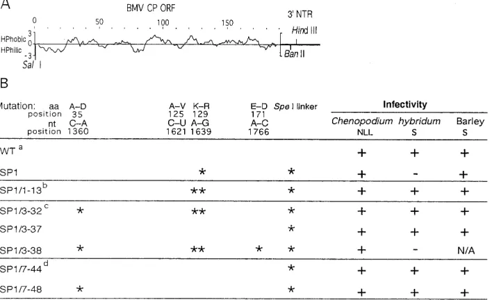
Viral RNA was extracted from systemically infectedC. hy-bridumleaves after the first, third, and seventh passages, and the CP gene was amplified as cDNA by reverse transcription-PCR, cloned, and sequenced. Two oligonucleotide primers were used: primer 1, 59CAGTGAATTCTGGTCTCTTTTAG AG39, complementary to the last 15 nucleotides (nt) of all BMV RNAs (the primer-derived 59overhang is shown in ital-ics), and primer 2, 59CTAACAAGCTCGGTCCATTC39, rep-resenting nt 1015 to 1035 of BMV RNA 3. The cDNA products were digested withEcoRI and cloned into pUC19 vector be-tween SmaI and EcoRI restriction sites. The nucleotide se-quences of the entire CP open reading frames were deter-mined in 10 clones resulting from each amplification reaction. This demonstrated that the SP1 mutant gave rise to second-site reversions within the CP gene. Specifically, in one passage series, in 50% of clones sequenced the first passage (SP1/1) generated a double mutant, SP1/1-13, which had the original SP1 mutation and Ala-125 changed to Val due to a C (1621)-to-U transition (Fig. 1). To test the properties of SP1/1-13 during infection, the PCR-generated subclones were used to obtain full-length cDNA clones (by replacing the correspond-ing fragment in plasmid pB3TP7) from which pseudorevertant RNA 3s could be synthesized. To eliminate the possibility of contamination with wt BMV, all constructs had a 39 marker mutation (SpeI linker) ligated at theBanII position (Fig. 1A), nt 1835 to 1840, after removal of the AGCC 39overhang with T4 DNA polymerase. SP1/1-13 spread systemically inC.
hybri-dum(Fig. 1B), but the spread was delayed by nearly 1 week compared to that of wt BMV.
The third (SP1/3) passage (Fig. 1) generated a triple CP mutant, SP1-3/32, which had the C-to-U transition at position 1621 and a C-to-A transversion at position 1360, the latter causing the Ala-35-to-Asp transition. Also, a quadruple mu-tant, SP1/3-38, was isolated which had the same mutations as SP1/3-32 plus an upstream A (1766)-to-C transversion, which caused Glu-171 to be replaced by Asp. A wt revertant (SP1/3-37) was also identified in this virus progeny. Except the qua-druple mutant SP1/3-38, all variants were found in similar ratios in the second passage series.
After the seventh passage (SP1/7), one passage series pro-duced the wt revertant SP1/7-44 as well as a single-nucleotide mutant (SP1/7-48) with A-to-C transversion that replaced Ala-35 with Asp. This substitution was also present in triple mutant SP1/3-32, isolated after the third passage, which may indicate that SP1/7-48 originated from SP1/3-32. The second passage series generated only wt revertants similar to SP1/7-44. In general, the similarity of pseudorevertant profiles in both passage series strengthens the importance of all these sites, most likely reflecting the existence of preferences in the strat-egies for the reversion of CP in order to rescue virus systemic movement and more stable virions.
Dot blot hybridization assays were used to analyze the ac-cumulation of revertant viral RNA in systemic tissue during passages (Fig. 2A and B). Previously, we reported a 50- to 100-fold decrease in viral RNA accumulation of the SP1 mu-tant in barley plants and no systemic spread inC. hybridumin contrast to the wt virus (16). Since the first passage did not always rescue the systemic symptoms inC. hybridum, the ac-cumulation of SP1 progeny was analyzed only after the third and seventh passages (Fig. 2A and B). These analyses revealed that the rescue of systemic movement in C. hybridum was accompanied by wt levels of BMV RNAs in barley and in C. hybridumhosts.
[image:2.612.131.485.75.291.2]The phenotypes of the identified pseudorevertants were
FIG. 1. Properties of BMV CP mutants studied in this work. (A) Standard hydropathy plot of BMV CP encoded by the open reading frame (ORF). Hydrophilic (HPhilic) and hydrophobic (HPhobic) areas are represented by the curves below and above the central line, respectively. SP1 and its pseudorevertant derivatives have a marker mutation (SpeI linker) at the 39nontranslated region (39NTR) ligated at theBanII site. (B) Properties of the BMV CP mutant and pseudorevertants in vivo. Locations of nucleotide and amino acid changes alongside the CP sequence in SP1 and in pseudorevertants (above) are marked by asterisks. NLL, necrotic local lesion; S, systemic infection; N/A, not analyzed;1, virus detected;2, no virus detected; aa, amino acid; wt, primary inoculation.
on November 9, 2019 by guest
http://jvi.asm.org/
tested separately by using the biologically active reconstructed full-length cDNA clones. The corresponding transcribed RNA 3s were inoculated together with wt RNA 1 and 2 on barley and onC. hybridumplants, and virus accumulation was monitored by dot blot hybridization (Fig. 2C and D). SP1/3-32 accumu-lated to the wt level in systemic barley leaves (Fig. 2C) and spread systemically, though at lower levels, inC. hybridum(Fig. 2D). SP1/3-32 was unstable, as it converted to the wt virus after two additional passages. In contrast, SP1/3-38 exhibited low infectivity similar to that of SP1 and did not spread systemically inC. hybridum(Fig. 1B). To confirm that there were no more mutations present in SP1/3-38, its Asp-172 was mutated back to the wt Glu. This recreated a triple mutant, with a phenotype similar to that described for SP1/3-32 (data not shown). Like-wise, SP1/7-48 accumulated at high levels in barley (Fig. 2C) and to lower levels in systemically infectedC. hybridumleaves (Fig. 2D). SP1/7-48 was stable after two additional passages. The true revertants, SP1/3-37 and SP1/7-44, behaved identi-cally to wt BMV (data not shown). This result demonstrated that the 39SpeI linker insertion had no or little effect on BMV infection in barley and inC. hybridum.
The assay for BMV infectivity involves measuring the RNA content away from the site of inoculation, and thus it reflects the combination of virus replication and virus spread. To dis-tinguish whether the reduced levels of BMV RNA observed in SP1 infections are due to reduced RNA replication or reduced virus spread, we studied viral RNA replication in barley pro-toplasts (data not shown). This demonstrated that the SP1 mutant accumulated to levels similar to those of the pseudor-evertants SP1/3-32 and SP1/7-48 and the wt virus. It further means that the SP1 mutation in the CP molecule did not participate in a detectable way in BMV RNA replication, and thus virus movement is a factor which is more likely to be responsible for the observed effects.
To determine the stability of virions, crude viral prepara-tions were extracted from infected tissue and incubated with-out and with RNase A, followed by electrophoresis of virion
RNAs in 1% agarose gel, as described before (16). These treatments revealed an increased degradation of virion RNA with both the RNase A and endogenous RNAses for the SP1 preparations, compared to the wt BMV (Fig. 3). Pseudorever-tants SP1/3-32 and SP1/7-48 displayed restored protection against degradation. The increased RNA degradation in SP1 virions could be due to the virus falling apart and releasing the RNA and/or due to the virion swelling and thus making the RNA more accessible to the RNases.
Examination of the BMV CP spatial folding, which has been generated on the basis of the CCMV X-ray structure (25, 26), provides a structural rationale for the observed phenotype of SP1. Lys-129 of BMV aligns with Lys-131 of CCMV (Fig. 4A) and is involved in the interactions within pentamers and at hexamers (26). The proper ratio between pentamers and hex-amers is crucial for T53 (180-subunit) particles. If hexamers become too stable (or pentamers become too unstable), the formation of the hexagonal sheets is expected. Alternatively, if pentamers become too stable, T51 (60-subunit) particles can be formed. In both cases the likelihood of assembly of T53 particles would be reduced. Lys-131 (Lys-129 in BMV) is one of the major determinants of the stability of hexamers versus pentamers in CCMV (Fig. 4A). Replacing Lys-129 with Arg in SP1 would force a significant reorganization of the hydrogen bonding pattern in the hexamers and pentamers which, in turn, would result in less stable particles.
Based on the above considerations we explain the observed effects with pseudorevertants as follows. The phenotype of the SP1/1-13 revertant is delayed movement. The residues changed from the wt sequence are Lys-129-Arg and Ala-125-Val (Fig. 4A). Ala-125 in BMV corresponds to Ala-127 in CCMV, which is buried at subunit interfaces between hexameric and penta-meric units (Fig. 4B). By homology modeling we predict that this may alter the stability between hexamers and pentamers to more closely resemble stability in the wt. In contrast, SP1/3-32 and SP1/7-48 mutants display wt levels of long-distance move-ment and virion stability. Changes in SP1/3-32 are the same as those in SP1/1-13 with an additional change of Ala-35-Asp that corresponds to Ala-37 in CCMV. Ala-37 is located within the
[image:3.612.353.518.68.214.2]FIG. 2. Accumulation of SP1-derived progeny (A and B) and reconstructed (cloned) SP1 pseudorevertants (C and D) in barley and in C. hybridum, as determined by dot blot hybridization. The first leaf of barley or the third and the fourth leaves ofC. hybridumwere inoculated with progeny of SP1 (SP1/3 or SP1/7) or with transcribed BMV RNAs (SP1/3-32 and SP1/7-48), as described before (16). Total RNA was extracted 12 days postinoculation from a leaf (0.5 g of tissue) above the inoculated barley leaf or from the fourth leaf above the inoculatedC. hybridumleaf. The extract was diluted 1, 10, or 100 times (as indicated on the left), immobilized on a nylon (Hybond N1) membrane, and hybridized to a32P-labeled riboprobe complementary to the last 200 nt of the 39 end of BMV RNA.
FIG. 3. Effect of RNase A treatment on the protection of viral RNAs in the BMV virions against degradation. Virus preparations were extracted from in-fected barley leaves with a sodium acetate buffer, precipitated with 4% polyeth-ylene glycol and 0.25 M NaCl, and resuspended in sodium phosphate buffer, pH 7.4 (16). Subsequently, the preparations were incubated for 24 h at room tem-perature alone (lanes marked “24h”) or with 10 ng of RNase A per ml (lanes marked “RNase A”). A control treatment included the incubation of virions in the same buffer for 24 h at 48C (lanes marked “0”). After the incubation, virion RNA was extracted and the RNA content was analyzed by electrophoresis in 1% agarose gels. The nomenclature of BMV mutants (shown above each lane) is described in the text.
2502 NOTES J. VIROL.
on November 9, 2019 by guest
http://jvi.asm.org/
b hexamer, and we predict that this mutation increases the stability of hexamers and pentamers and the proper ratio be-tween them. Yet another mutant, SP1/3-38, has lost the long-distance movement phenotype, and it has the changes of
SP1/3-32 plus Glu-171-Asp. Glu-171 (Glu-174 in CCMV) is important in CP dimer formation (25, 26). The Glu-171-Asp mutation probably destabilizes particle formation because hex-amers and penthex-amers may not be joined.
FIG. 4. Location of BMV pseudorevertants on the structure of the CCMV CP. (A) Single subunit of CCMV viewed from the side. The particle exterior is at the top, the inside is at the bottom, and the residues on the left (27 to 40) form thebhexamer that stabilizes the sixfold interactions of subunits. The residues displayed as space-filling models (and numbered) are those that are discussed in the text. Note that residues 37, 127, and 131 are clustered and probably alter the relative stability of hexamers and pentamers. The mechanism of action of the 174 mutation is more likely to effect dimer formation. (B) Hexamer of subunits viewed directly down a sixfold particle axis. Shading designates C subunits (light) or B subunits (dark) (25). The original single-site mutation (Lys-131-Arg) is seen coming off thebF-bG loop and circles the six shortbstrands of thebhexamer with Asp-132. The reversion mutations (Ala-127-Val, Ala-35-Asp, and Glu-171-Asp) are also shown as space-filling residues further within the subunit fold. The view is from the outside the particle looking in.
on November 9, 2019 by guest
http://jvi.asm.org/
In this communication we demonstrate that systemic spread of a BMV CP mutant can be rescued by second-site amino acid substitutions. Both RNase A treatments and computer mod-eling suggest that these pseudoreversions do correlate with restored virion assembly and/or with greater stability of virions. Currently available data do not distinguish between the two possibilities. One experiment to determine if the additional amino acid changes in pseudorevertants are compensating for the Lys-129-Arg effect on the SP1 virion assembly (3, 4) would be to analyze the CP aggregates in vitro: anomalously high levels of hexameric aggregates, monomers, or T51 particles in the SP1 mutant and increased levels of T53 particles in the pseudorevertants would confirm our theory.
In general, whether analyzed at the assembly or the stability level, the observed correlations imply that BMV utilizes virions as the long-distance movement form inC. hybridumand thus further support the first model of CP involvement in virus movement (16, 23). However, we cannot also exclude the pos-sibility that changes in such properties of CP as interactions with other viral and/or with host components contribute to the observed effects.
S.F., A.D., and J.J.B. were supported by grants from the National Institute for Allergy and Infectious Diseases (3RO1 AI26769) and the National Science Foundation (MCB-9630794) and by the Plant Mo-lecular Biology Center at Northern Illinois University. J.A.S. and J.E.J. were supported by a grant from NIH (GM54076).
REFERENCES
1.Ahlquist, P., R. Allison, W. De Jong, M. Janda, P. Kroner, R. Pacha, and P. Traynor.1991. Bromovirus RNA replication and host specificity. NATO ASI Ser.212:11–21.
2.Allison, R., C. Thompson, and P. Ahlquist.1990. Regeneration of a func-tional RNA virus genome by recombination between deletion mutants and requirement for cowpea chlorotic mottle virus 3a and coat genes for systemic infection. Proc. Natl. Acad. Sci. USA87:1820–1824.
3.Bancroft, J. B., G. E. Brocker, and G. W. Wagner.1969. Structures derived from cowpea chlorotic mottle and brome mosaic virus proteins. Virology 38:324–335.
4.Bancroft, J. B.1970. The self-assembly of spherical plant viruses. Adv. Virus Res.16:99–134.
5.Bancroft, J. B.1972. A virus made from parts of the genomes of brome mosaic and cowpea chlorotic mottle viruses. J. Gen. Virol.14:223–228. 6.Boccard, F., and D. C. Baulcombe.1993. Mutational analysis of cis-acting
sequences and gene function in RNA3 of cucumber mosaic virus. Virology 193:563–578.
7.Bujarski, J. J., P. D. Nagy, and S. Flasinski.1994. Molecular studies of genetic RNA-RNA recombination in brome mosaic virus. Adv. Virus Res. 43:275–302.
8.Chapman, S., G. Hills, J. Watts, and D. Baulcombe.1992. Mutational anal-ysis of the coat protein gene of potato virus X: effects on virion morphology
and viral pathogenicity. Virology191:223–230.
9.Citovsky, V., and P. Zambryski.1991. How do plant virus nucleic acids move through intercellular connections? Bioassays8:373–379.
10. Dawson, W. O., P. Bubrick, and G. L. Grantham.1988. Modifications of tobacco mosaic virus coat protein gene affecting replication, movement and symptomatology. Phytopathology78:783–789.
11. De Jong, W., and P. Ahlquist.1991. Bromovirus host specificity and systemic infection. Semin. Virol.2:97–105.
12. De Jong, W., and P. Ahlquist.1995. Host-specific alterations in viral RNA accumulation and infection spread in a brome mosaic virus isolate with an expanded host range. J. Virol.69:1485–1492.
13. Deom, C. M., M. Lapidot, and R. N. Beachy.1992. Plant virus movement proteins. Cell69:221–224.
14. Dolja, V. V., R. Haldeman-Cahill, A. E. Montgomery, K. A. Vandenbosch, and J. C. Carrington.1995. Capsid protein determinants involved in cell-to-cell and long distance movement of tobacco etch potyvirus. Virology206: 1007–1016.
15. Dolja, V. V., R. Haldeman, N. L. Robertson, W. G. Dougherty, and J. Car-rington.1994. Distinct functions of capsid protein in assembly and move-ment of tobacco etch potyvirus in plants. EMBO J.13:1482–1491. 16. Flasinski, S., A. Dzianott, S. Pratt, and J. J. Bujarski.1995. Mutational
analysis of the coat protein gene of brome mosaic virus: effects on replication and movement in barley and inChenopodium hybridum. Mol. Plant-Microbe Interact.8:23–31.
17. Hilf, M. E., and W. O. Dawson.1993. The tobamovirus capsid protein functions as a host-specific determinant of long-distance movement. Virol-ogy193:106–114.
18. Hull, R.1991. The movement of viruses within plants. Semin. Virol.2:89–95. 19. Janda, M., R. French, and P. Ahlquist.1987. High efficiency T7 polymerase synthesis of infectious RNA from cloned brome mosaic virus cDNA and effects of 59extensions on transcript infectivity. Virology158:259–262. 20. Jones, T. A., J.-Y. Zhou, S. W. Cowan, and M. Kjeldaard.1991. Improved
methods for building protein models in electron density maps and the loca-tion of errors in these models. Acta Crystallogr.A47:110–119.
21. Maule, A. J.1991. Virus movement in infected plants. Crit. Rev. Plant Sci. 9:457–473.
22. Mise, K., R. F. Allison, M. Janda, and P. Ahlquist.1993. Bromovirus move-ment protein genes play a crucial role in host specificity. J. Virol.67:2815– 2825.
23. Sacher, R., and P. Ahlquist.1989. Effects of deletions in the N-terminal basic arm of brome mosaic virus coat protein on RNA packaging and systemic infection. J. Virol.63:4545–4552.
24. Seron, K., and A. L. Haenni.1996. Vascular movement of plant viruses. Mol. Plant-Microbe Interact.9:435–442.
25. Speir, J. A., S. Munshi, G. Wang, T. S. Baker, and J. E. Johnson.1995. Structures of the native and swollen forms of cowpea chlorotic mottle virus determined by X-ray crystallography and cryo-electron microscopy. Struc-ture3:63–78.
26. Speir, J. A.1994. The 3.2A resolution structure of the polymorphic cowpea chlorotic mottle virus ribonucleoprotein particle. Ph.D. thesis. Purdue Uni-versity, West Lafayette, Ind.
27. Suzuki, M., S. Kuwata, J. Kataoka, C. Masuta, N. Nitta, and T. Takanami. 1991. Functional analysis of deletion mutants of cucumber mosaic virus RNA 3 using anin vitrotranscription system. Virology183:106–113.
28. Taliansky, M. E., and F. Garcia-Arenal.1995. Role of cucumovirus capsid protein in long-distance movement within the infected plant. J. Virol.69: 916–922.
2504 NOTES J. VIROL.