DISSERTATION ON
Evaluation of Clinical and Functional Outcome Of
Closed Reduction/Open Reduction and Internal
Fixation with Intra Medullary Interlocking Nailing
and ‘Poller’ Blocking Screws in Tibial
Metaphyseal Fractures
Submitted to
THE TAMILNADU
DR. M.G.R. MEDICAL UNIVERSITY CHENNAI, TAMILNADU
As fulfillment of the regulations for the award of the degree
M.S. (ORTHOPAEDIC SURGERY) BRANCH II
MADURAI MEDICAL COLLEGE MADURAI
ACKNOWLEDGEMENT
I wish to express my sincere thanks to our Dean DR.N.MOHAN
M.S., F.I.C.S,, Madurai Medical College, Madurai, for having allowed me to
conduct this study.
It is my proud privilege to express my sincere thanks to my
beloved and kind hearted Chief PROF.DR.P.V.PUGALENTHI.,M.S
Ortho.,D.Ortho.,HOD & Professor of Orthopaedics & Traumatology
Madurai Medical College and Govt Rajaji Hospital, for his total support in all
my endeavours. I wish to express my sincere gratitude and heartfelt thanks to
our PROF&HOD for his guidance and support during the conduct of the
study.
I wish to express my sincere gratitude and heartfelt thanks to
PROF.DR.A.Rajamani M.S.Ortho.,D.Ortho., Professor of Orthopaedics Spine, for his support and encouragement. I wish to express my sincere
gratitude and heartfelt thanks to PROF.DR.T.C.Chandra Prakasam
M.S.Ortho.,D.Ortho., Professor of Hand surgery, for his support and
encouragement. I wish to express my sincere gratitude and heartfelt thanks to
PROF.DR.Shanmuganathan M.S.Ortho., D.Ortho., Professor of Orthopaedics & Traumatology for his support and guidance.
PROF.DR.L.D.Thulasiram M.S.Ortho.,D.Ortho., for his support and
encouragement
I wish to express my sincere gratitude and heartfelt thanks to
PROF.DR.R.Sivakumar M.SOrtho.,D.Ortho., for his support and
encouragement. I wish to express my sincere gratitude and heartfelt thanks to
my Co-Guide Dr.T.C.Premkumar M.SOrtho.,D.Ortho., for his
guidance,timely advice,support and encouragement.
I am deeply indepted to Dr.Ravichandran M.S.Ortho.,
Dr.Ramanathan, M.S.Ortho., Dr. M.N.Karthi M.SOrtho., Dr.K.P.Saravanakumar M.SOrtho., Dr.P.V.Thirumalaimurugan M.S Ortho.,D.N.B Ortho., Dr.T.C.Premkumar. M.S.Ortho., D.Ortho., Dr.Pathiarasakmar M.S.Ortho., Dr.Maheshwaran. M.S.Ortho., D.Ortho., Dr,Saravanamuthu, M.S.Ortho., Dr.V.A.Prabhu M.S.Ortho Dr.Gnanaprakasm M.S.ortho., D.Ortho for their immense help, continuous
motivation, expert guidance, timely advice during the course of my study and
for the preparation of this dissertation.
Last but not the least I sincerely thank all the patients involved in this
CERTIFICATE
Certified that the dissertation on “Evaluation of Clinical and
Functional outcome Of Open Reduction/Closed Reduction with Intra Medullary Interlocking Nailing and ‘Poller’ Blocking Screws in Tibial Metaphyseal Fractures” is a bonafide work done by
Dr.K.SHANMUGANATHAN, Postgraduate, in the Department of
Orthopaedic Surgery and Traumatology, Madurai Medical college. &Govt
Rajaji Hospital, Madurai, under my guidance and supervision in fulfilment
of the regulations of The Tamilnadu Dr. M. G. R. Medical University
for the award of M.S. Degree Branch II (Orthopaedic Surgery) during
the academic period of May 2010– April 2013
PROF.DR.P.V.PUGALENTHI
M.S.Ortho.,D.Ortho.,
PROF&HOD., Dept.ofOrthopaedics,
Traumatology&Rehablitation, Madurai Medical College& Govt.Rajaji Hospital,
Madurai.
DECLARTATION
I, declare that this dissertation “Evaluation of Clinical and
Functional Outcome of Open Reduction/Closed Reduction with Intra
Medullary Interlocking Nailing and ‘Poller’ Blocking Screws in Tibial
Metaphyseal Fractures” has been conducted by me at the Department of
Orthopaedic Surgery&Traumatology, Madurai Medical College & Govt
Rajaji Hospital, Madurai, under the guidance and supervision of my
respected Chief PROF.DR. P.V.PUGALENTHI M.S.Ortho,D.Ortho,
Madurai Medical College& Govt. Rajaji Hospital, Madurai.
It is submitted as part of fulfilment of the award of the
degree in M. S. Orthopaedic surgery for the April-2013 examination to be
held under The Tamilnadu Dr. M. G. R Medical University, Chennai. This
has not been submitted previously by me for the award of any degree or
diploma from any other university.
Dr.K.SHANMUGANATHAN
M.S.ORTHO P.G.
TITLE PAGE
Sl.No CONTENTS PAGE NO.
1. INTRODUCTION 1
2 AIM OF THE STUDY 4
3 TIBIAL METAPHYSEAL FRACTURES 5
4 LITERATURE REVIEW 29
5 MAIN OUTCOME MEASUREMENTS 51
6 PATIENTS AND MATERIALS 52
7 OPERATIVE PROTOCOL 54
8 POST OPERATIVE PROTOCOL 61
9 RESULTS-ANALYSIS 64
10 DISCUSSION 84
11 CONCLUSION 89
BIBILOGRAPHY
CASE ILLUSTRATIONS PROFORMA
CONSENT IN TAMIL MASTER CHART
ETHICAL CLEARANCE
ANTI PLAGIARISM CERTIFICATE
1. INTRODUCTION
Due to the increasing number of road traffic accidents, long bone fractures are more common nowadays. Fracture of tibia is one of the most
commonly occurring fractures due its superficial location. Proper treatment of
these fractures is paramount importance. Among the treatment of tibial
fractures, the treatment of metaphyseal fractures of tibia remains challenging,
because of the sagittal and coronal malalignment which is mainly due to the
mismatch between medullary canal diameter between the two fragments of the
metaphyseal tibial fractures and anatomy of the metaphyseal region.
Establishment of length and prevention of the coronal, sagittal, rotational
malalignment is the at most importance during fixation. The treatment options
for the metaphyseal tibial fractures are conservative management, open
reduction with plate osteosynthesis, external fixators and recently the
intramedullary interlocking nailing. Each treatment has its own advantages and
disadvantages. The conservative management has the high level complications.
These are the delayed union, malunion, rarely non union. Due to the knee and
ankle joints are having predominantly single plane functional movements the
effects of the malunion and delayed union are high with secondary
osteoarthritis of the joints. Then open reduction with plate osteosynthesis has
perfect anatomic reduction in this type treatment. The external fixation method
having the complications of the post traumatic complications like high
incidence of post traumatic joint stiffness. So nowadays most authors consider
the intramedullary interlocking nailing is the most effective treatment of
choice.Futher it is important to do advice early mobilisation of knee and ankle
to achieve normal range of movements in the joints. It is possible only in
intramedullary nailing only
Interlocking nailing of tibial fractures are most desirable because these are
load sharing devices in compare to load bearing plate osteosynthesis implants.
Biological fixation of the fracture is possible in the method of nailing without
opening the fracture site, further it is possible to spare the extra osseous blood
supply and we can avoid extensive soft tissue dissection. Nailing of
metaphyseal fractures with short proximal or distal fragment is associated with
an increase in malalignment particularly in coronal plane, so there is always a
chance for mal union, rarely non union and need for secondary procedures to
achieve union. The cause has been attributed to both displacing muscular
forces and medullary canal anatomical factors. As there is always a mismatch
between the diameters of the nail and the medullary canal, with no nail-cortex
contact, the nail may translate coronally or sagittaly particularly in proximal
tibial fractures. Due to this there is a increased stress on the locking screws to
maintain fracture alignment after surgery, which further leads to periprosthetic
improve nailing procedures for the metaphyseal fractures these are, “poller”
blocking screws, temporary unicortical plating, different nail designs with
different proximal bends in case of proximal third fractures and fibular plating
in case of distal third fractures.
Poller screws are named after a traffic guiding device used in
European countries to guide the traffic on the roads. They are used for the
following three purposes:
a) Achievement of the fracture alignment, by using screw as a reduction
tool.
b) Improvement of the stability of the bone – implant Construct by
reducing the medullary canal width.
2. AIM OF THE STUDY
3. TIBIAL
METAPHYSEAL FRACTURES
Anatomy of Tibia-Metaphysis
Shaft of tibia is triangular in cross section. It widens at both the
proximal and distal metaphyseal region to articulate with the femur in the
proximal part and with the talus and fibula in the distal part thereby support
body’s weight at the knee and ankle joint .Proximal and distal end of tibia is
shaped like a rectangular box with a bony protuberance. Theses are forming the
tibial condyles proximally and medial malleoli distally.
Anterior aspect of Proximal Metaphysis
with attachments
Tibial tuberosity
Medial condyle Gerdy’s tubercle
Lateral condyle
Patellar Tendon Lateral Collateral ligament
Illio Tibial Tract
Capsule Semi
Membranosus
Medial Collateral Ligament Gracilis
Sartorius,
Posterior aspect of Proximal Metaphysis
with soft tissue attachments
Head of Fibula
Soleal Line Groove for
Semimembranosus Groove for
Popliteus
Semimembranosus
Popliteus
Anterior aspect of Distal Metaphsis
With soft tissue attachments
Lateral Malleolus Medial Malleolus
Physeal Line Capsular
Posterior aspect of Distal Metaphysis
Soft tissue attachments
Medial Malleolus
Groove for Tibialis Posterior
Groove for peroneus tendons
Lateral Malleolus
Physeal line
Capsular attachment Physeal
Vascular Anatomy
Proximal metaphysis is supplied by the metaphyseal vessels which are branches of the genicular artery anatomoses around the knee. Distal tibia is
supplied by vascular anostomoses formed by the arteries arising from the both t
anterior tibial and posterior tibia arteries which are located in periosteal region
forming an anastomoses. There is a water shed area at the junction of the middle
and distal third of the tibia. So whenever excessive soft dissection is being done
in the distal tibia delayed union or the nonunion may occur.
Epidemiology of metaphyseal fractures
Incidence metaphyseal tibial fracture is currently in increasing trend.
Tibial fractures are most common fractures in compare to all other long bones.
The annual incidence of the tibial fractures is 26 fracture per 100000 population
Proximal metaphyseal fracture contributes for the 7% of the tibial fractures
There is increasing number of open fractures as the one third of the bone is
superficial. Distal tibia fractures are extraarticular fractures of the metaphysis.
In high violence injuries it may lead to intra articular tibial plafond or pilon
fractures. One study reported incidence rates of metaphyseal ankle fractures that
varied considerably by age and gender 1. Incidence ranged from a low of 3 per
10,000 per year among 30 to 34-year-old women to a high of 28 per 10,000 per
Mechanism of injury
Most commonly due to the road traffic accidents, direct trauma, gun shot
injuries, fall from the height, sports injuries, Injuries always are to be assessed
regarding the violence of the injry.The prognosis depends upon the soft tissue
injury status. It is sufficient to say here that the more violent the trauma, the
more likely the presence of major soft tissue damage.
Clinical evaluation
Diagnosis of the fracture is usually obvious. Evaluation regarding the compartment syndrome particularly in case of the proximal metaphyseal
fractures has the paramount importance. The management options mainly
depend on the soft tissue injury. The classical clinical symptoms of the
compartment syndrome are pain out proportion, tense pulseless and paralysed
Hg of the diastolic pressure is the indication for the emergency fasciotomy.
Always distal neurological status is to be evaluated .Common peroneal nerve
injury is most common in case fracture neck of fibula when it is associated
with proximal tibial metaphyseal fractures. Then the fracture pattern and
displacements is to be assessed. Due to the gastronemius acting posteriorly,
tibialis anterior acting anterolateraly and patellar tendon acting anteriorly, there
is always a tendency for valgus and antecurvatum malalignment of the proximal
fragment
.
Tibial fractures most commonly associated with the ligament injuriesin the knee. So, clinical evaluation regarding the ligament is always important.
Stability of the knee/ankle joint always is to be examined in the proximal/distal
metaphyseal fractures respectively.
Radiological evaluation
Anteroposterior, lateral and the full length tibia views should be obtained
to assess the bone as well as soft tissue status.Oblique radiographic views at 45
degree sometimes are required to detect a non displaced spiral fracture.
Radiographs of the contra lateral tibia sometimes are necessary to evaluate the
length in fractures with severe comminution or bone loss. Metapyhseal fractures
always associated with extension of the fracture into the joint, So always the X
ray views should include the knee and the ankle joints. CT is scan some time
needed to assess the intra articular extension of the farcture.MRI scan also
some time is needed to rule out the ligament injuries particularly in proximal
Classifications
There are TWO type of classifications: I. AO/OTA Classification
II. Taylor and Martin SUD classification
In the AO/OTA classification, the metaphyseal regions are designed as follows:
Proximal metaphysis - 41
Distal metaphysis - 43.
AO/OTA Classification of proximal tibial metaphysis 41.
Type A - Extraarticular,
Type B - Partialy articular
Type C - Completely articular.
A – Extra articular fracture
A1 – Extra articular, avulsion
A1.1 Of the fibular head
A1 .2 Of the tibial tuberosity
A1. 3 Of the cruciate insertion
A2 - Extra articular fracture, Metaphyseal simple
A2 .1. Oblique in frontal plane
A2 .2. Oblique in sagittal plane
A 2 .3 .Transverse
A3 - Extra articular fracture, Metaphyseal multifragmentary
A3.1. Intact wedge
A3 .2. Fragmented wedge
A3.3. Complex
B – Partial articular fracture
B1 – Partial articular fracture, Pure split
B1.1 Of the lateral surface
B1 .2 Of the medial surface
B1.3 Oblique, involving the tibial spines and one of the
surfaces
B2 - Partial articular fracture, Pure depression
B2.1 Lateral total
B2.2 Lateral limited
B2. .3 Medial
B3 - Partial articular fracture, Split depression
B3 .1 Lateral
C – Complete articular fracture
C1 – Complete articular fracture, articular simple, metaphyseal
simple
C1.1 Slight displacement
C1.2 One condyle displaced
C1.3 Both condyles displaced
C2 - Complete articular, articular simple, metaphyseal
multifragmentary
C2.1 Intact wedge
C2.2 Fragmented wedge
C2.3 Complex
C3 – Complete articular fracture, multifragmentary
C3.1 Lateral
C3.2 Medial
C3.3 Lateral and medial
2.AO/OTA Classification of Distal tibial metaphysis. 43.
A – Extra articular fracture
A1 – Extra articular, Metaphyseal simple
A1.1 Spiral
A1.2 Oblique
A2 - Extra articular fracture, Metaphyseal wedge
A2.1 Posterolateral impaction
A2.2 Anteromedial wedge
A2.3 Extending into the diaphysis
A3 - Extra articular fracture, Metaphyseal complex
A3.1 Three intermediate fragments
A3.2 > 3 intermediate fragments
A3.3 Extending into the diaphysis
B – Partial articular fracture
B1 – Partial articular fracture, Pure split
B1.1 Frontal
B1.2 Sagittal
B1.3 Metaphyseal multifragmentary
B2 - Partial articular fracture, split depression
B2.1 Frontal
B2.2 Sagittal
B2.3 Of the central fragment
B3 - Partial articular fracture, multifragmentary depression
B3.1 Frontal
B3.2 Sagittal
C – Complete articular fracture
C1 – Complete articular fracture, articular simple, metaphyseal
simple
C .1 Without depression
C1.2 With depression
C1.3 Extending into diaphysis
C2 - Complete articular, articular simple, metaphyseal
multifragmentary
C2.1 With asymmetric impaction
C2.2 Without asymmetric impaction
C2.3 Extending into diaphysis
C3 – Complete articular fracture, multifragmentary
C3 .1 Epiphyseal
C3 .2 Epiphyseo - metaphysis
C3 .3 Epiphyseo - metaphyseo – diaphyseal
II. Taylor and Martin SUD classification of metaphyseal fractures
Taylor and Martin proposed a classification of metaphyseal fractures
(SUD) in which the main fracture is characterised as stable (S), unstable (U) or
with diaphyseal extension (D). These are further divided into three subtypes 4.
S-Stable
S.0 - extra articular
S.1 - <2mm displacement
S.2 - >2mm displacement
U-Unstable
U.0 - extra articular
U.1 - <2mm displacement
U.2 - >2mm displacement.
D-Diaphyseal Extension
D.0 - extra articular
D.1 - <2mm displacement
D.2 - >2mm displacement
According to Taylor and Martin classification with progression from
type’ S’ to type’ D’, treatment shifts toward external fixator and away from
open reduction. Conversely with progression from subgroup ‘0’ to subgroup’ 2’
open reduction is indicated.
Treatment options
The GOALS of the treatment:
- To obtain a healed, well-aligned fracture.
The optimal treatment method should assist in meeting these goals, while
minimizing the complications, especially infection. The presence of hinge joints
at the knee and the ankle allows no adjustment for rotatory deformity after
fracture, and special care is necessary during reduction to correct such
deformities
Treatment and prognosis depends on
1. . . . The state of the soft tissues:
The risk of complications and the progress to fracture healing are
directly related to the amount and type of soft tissue damage. Closed fractures
are best described using Tscherne’s method. For open fractures, Gustilo’s
grading is more useful. The incidence of severe soft tissue injury ranges from 1
percent in Gustilo-anderson type I to 30 percent for type IIIC.
2. The severity of the bone injury:
High-energy fractures are more damaging and take longer time to heal
than low-energy fractures. This is regardless of whether the fracture is open or
closed. Low energy injuries are typically closed or Gustilo I or II and spiral in
which fracture union takes comparatively less time. High energy fractures are
usually caused by direct trauma and tend to be open (Gustilo III A-C),
transverse or comminuted with bone loos in which delayed union and non union
3. Stability of the fracture:
It is considered when the weight bearing can be allowed. More stable
fractures with less displacement can be managed conservatively. In case of
internal fixation, stable fracture can be easily fixed which in turn avoid the
unnecessary soft t tissue dissection. Severely comminuted fractures are the least
stable of all, and the most likely to need mechanical fixation, soft tissue
dissection.
4. Degree of contamination:
In open fractures this is an important additional issue. The limb should be
carefully examined for signs of soft-tissue damage, bruising, severe swelling,
crushing or tenting of the skin, and open wounds gas gangrene.
Treatment options
1. Conservative method
2. External fixator.
3. Open reduction with plate osteostnthesis
4. Open reduction/Closed reduction internal fixation with intramedullary
interlocking nailing.
Conservative method
Indications:
1. Deblitated patients
Demerits of conservative management:
1. High incidence of post traumatic stiffness
2. Mal union/Delayed union/Non union
3. Secondary Osteoarthritis of knee /Ankle joints
Sarmiento et al in 1989 concluded that bracing was contraindicated in
fractures with excessive initial shortening or ones showing increasing angular
deformity while in cast. Most series of closed treatment have reported 25 to
40% incidence of ankle and subtalar joint stiffness after prolonged casting and
immobilisation.
External fixation
Indications
:
• Compound fractures with severe contamination • Fractures with acute compartment syndrome
• Severely comminuted / unstable fractures with articular
extension with severe soft tissue damage.
• Fractures with very short proximal/distal fragments
• Polytrauma patients with haemodynamic instability
Methods:
• Hybrid fixator
• Knee spanning or ankle spanning external fixators
Hybrid external fixator:
As the metaphyseal fractures having the short proximal and distal
fragments in proximal and distal metaphyseal fractures respectively, it is very
difficult to put two pins in the longitudinal axis so the pins are inserted in the
tranverse axis and a special ¾ or ½ ring is used to stablise the short fragment.
Then the diaphyseal fragment is stabilised with longitudinal pins with AO rods.
Then the two fragments interfixed with two more AO rods.
Demerits of external fixators:
• They are not a definitive treatment option.
• Exchange nailing or plating is to be done within 2-3 weeks
otherwise pin tract infection will occur.
• Knee/ankle spanning leads to post traumatic joint stiffness in long
standing cases
Rationale for open reduction in metaphyseal fractures
manipulation, an extremely large periarticular gap is formed. If treated non operatively, the distal/proximal fragment may tend to displace into that gap in the post reduction period, necessitating multiple reductions. Also, since the compression fractures has been disimpacted, and since cancellous bone heals poorly under such conditions, fracture healing may be delayed. This, in turn, will require prolonged immobilization of the limb, with resultant of poor post traumatic joint function. Sarmiento et al. and Waddell and Reardon reported ankle stiffness in 20% to 30% of patients who had closed treatment. Digby,
Holloway, and Webb reported that 27% of patients treated conservatively were
unable to run, even several years after the fracture had healed, because of ankle
and subtalar stiffness. Angular deformity of more than 5 degrees occurs in 10%
to 55% of fractures treated with a cast or brace, and shortening of at least 12 to
14 mm occurs in 5% to 27% of patient. Sarmiento's series of carefully selected
fractures had the best results, whereas other series with more unstable fractures
reported poorer results.
Methods of surgical management
1. Open reduction, internal fixation with plate osteosynthesis.
2. Closed reduction/open reduction with IMIL nailing.
1.Open reduction with plate osteosynthesis
Indications
:
Advantages:
1. Anatomic reduction with stable fixation
2. Feasiblity of bone grafting
3. Fixed angle fixation,bicolumnar fixation
4. Maintenance the length& alignment.
High incidence of soft tissue complications in the range of 10 to 15% are
reported in many series. But the recent advances in plating like indirect
reduction and percutaneous plating (LISS- Less Invasive stabilisation System)
is indicated in tibial metaphyseal fractures with periarticular metaphyseal
comminution. The use of standard incisions according to Collinge and Sanders
also improve the wound healing.
Locked compression plate:
The locked compression plate with MIPO(Minimally Invasive plate
ostesynthesis) technique eliminates the soft tissue problems associated with
Complications and disadvantages of plate osteosynthesis:
1. Soft tissue complications in conventional open reduction and plating
2. Development of superficial wound problems increases the risk deep
infection six fold
3. Malalignment is more frequent in percutaneous plating than with
other methods of fixation.
4. Fracture alignment could not be aided by the locked plate ( no lag
effect through the plate). It has to be restored before applying the
plate12.
5. Locked compression plating needs careful preoperative planning,
if applied without following the principles of plating and the
Order of putting the screws, failures are not uncommon17.
6. Late pain may occur over the distal tibial and fibular plate or screws1.
4. LITERATURE REVIEW
Intramedullary nailing
History:
The evolution of intramedullary nailing had started 500 years ago. In
the good old days Aztecs used wooden intramedullary nails19. In 1916 Hey
Groves introduced a solid nail for tibia19.In, 1930 German Orthopaedician
Gerhard Kuntscher invented the metalic intramedllary nail used for the femoral
fractures. Kuntscher called it as elastic nail as it expands after the insertion. He
did his first nailing in 1939 for subtrochantric fracture of femur. In 1950, he
invented the technique of the medullary canal reaming and closed nailing. Now,
this became as standard practice. In1950 Lottes one of the pioneers in tibial
nailing developed a rigid nail fort tibia20.In 1951 Herzog made a modification in
the Kuntscher nail, by adding a proximal bend to simplify the nail
insertion.Modney designed the first interlocking nail4.Kuntscher also designed
an interlocking nail ( The Detensor nail, 1968) which was then modified by
Klemm Schellumm initially and then by Kempf and Grosse later in 19724.In
1986 Bone & Johnson were the first to report interlocking nail in USA. They
used Grosse Kempf interlocking tibial nail in 28 fractures of tibial
shaft20.Charnley in his text “closed treatment of common fractures” stated that
the eventual solution to the tibial shaft fracture would be a non reamed
Of late, Comparative studies between conservative management and
intramedullary nailing show superior results with intramedullary interlocking
nailing than the conservative methods17. Bone et al.18, in their prospective
randomized series, stated only a 2% incidence of nonunion and malunion in
nailing group when comparing with the 10% nonunion and 26% malunion
rates in their conservatively managed group.
Principle of Intramedullary nailing:
As the nail extends from the one end of the bone to other end in the medullary canal, it act as an internal splint. It allows the axial forces to be
transmitted to the opposed ends of the fragments. It prevents the angulation,
translation,and to some extent rotatory movement. In this, the contact is
occurring between the bone and the nail in the three points. The entry point,
narrowest portion of the medullary canal(Isthmus) and at the cancellous
epiphyseal bone at the opposite end are the three points.(Three point fixation).
Indications:
Conventionally interlocking nail is the gold standard treatment of the fractures in a zone of 5cm below the knee and 5cm above the ankle in fractures
of tibia. As the fracture line extend into the metaphyseal zones of the tibia, the
stability provided by any nail decreases precipitously5. Recently the indication
of nailing is extended to the metaphyseal region also. Locked intramedullary
nailing currently is considered the treatment of choice for most type I, type II,
Advantages:
In intramedullary nailing the fracture haematoma and periosteal blood
supply is not disturbed which leads to early fracture healing. Eventhough the
entosteal blood supply is disturbed in the reamed nailing,it improve the
periosteal blood supply. These are the load sharing devices in compare to plate
which are load bearing devices. So there is no chance for periprosthetic
osteopenia and periprothetic fractures
Procedure of intramedullary nailing tibia:
• Patients should be supine position.
1. On traction table with knee in 900 flexion or
2. On radiolucent table with knee fully flexed or
3. On a padded knee support with the knee flexed as for as possible.
• Various approaches for the entry point of the nail are
1. Medial para patellar.
2. Patellar tendon splitting.
3. Lateral para patellar.
Ptaellar tendon splitting approach:
Incision started from the inferior margin of the patella to anterosuperior
aspect of tibia over the patellar tendon. After splitting the patellar tendon,
This should be at the level of fibular head and 1.5cm from the joint line.
Initially the bone awl should be pointed posteriorly, and then directed in line
with the medullary canal or crest of the tibia. The medullary canal is reamed in
incremental fashion, till 1.5 cm more than the measured nail size. Then the trial
reduction is done. With maintaining the reduction the proposed sized nail is
inserted and fixed with interlocking.
Reaming-Pathophysiology: Local changes :
Reaming of the medullary canal is causing damage to the endosteal
blood supply by the direct damage or by the intravasation of the marrow
elements in to the vasculature. Various animal studies states that e it is
reversible within 8-12 weeks. This disturbed blood supply may cause delay in
the fracture healing.But the exraosseous blood supply increases and the vessels
traverse into the endosteal region to revascularise the damaged endosteal
vasculature.During the early weeks after trauma reaming may cause increasing
risk of infection,especially in open tibial fractures. Because of infection rates as
high as 21%.Due to the reaming the local temperature may be increased as high
as 44.6 degree C. This leads to the cell enzymes to be damaged. The threshold
value for the thermal osteonecrosis is 47 degree C for one minute which far high
General changes:
These include pulmonary embolisation, temperature related changes of the coagulation system and humeral, neural and inflammatory reactions. The
development of post traumatic pulmonary failure following early femoral
nailing in the multiply injured patients is associated with the reaming
procedure.Wenda et al measuring intramedullary pressure intra operatively
while doing the reaming, founded values between 420 - 1510 mm Hg with
reaming procedures, as compared with 40-70 mm Hg in cases where solid nails
were used without reaming.
Unreamed nailing advantages:
• Smaller diameter implants –Easy to insert
• Less heat induced cell death,osteonecrosis
• Less disturbances of the endosteal blood supply.
• There is also considerably less bone necrosis, which appears to be
one of the risk factors for the development of post operative
infection
The effect of diameter on endosteal vasculature and mechanical parameters
studied in dog models by Hupel TM et al., following segmental osteotomy of
the tibia. They concluded that a loose fitting nail did not affect cortical
perfusion as much as tight fitting nail and it allowed more complete cortical
Reamed nailing advantgages:
• Tight fit nail can be inserted which giving the more stable nail-bone
construct(Hupel et al)
• Stable nail bone construct gives stable fixation.
• It increase the vascularity, perfusion of the periosteum and the
surrounding muscles
Reamed versus undreamed nailing
Reaming increase the diameter of the medulary canal,thereby increasing
the contact between the bone and the nail which inturn the stability of the nail-
bone construct. Keating et al. reported a randomized, prospective study
comparing reamed with unreamed locked nailing of open tibial fractures.
Forty-seven nails were inserted after reaming, and 41 were inserted without reaming.
The average time to union was 30 weeks for reamed nailing and 29 weeks for
unreamed nailing, and there was no difference in functional outcome between
the groups. Infection developed in two patients (4.3%) with reamed nailing and
in one patient (2.4%) with unreamed nailing. 9% percent of fractures treated
with reamed nailing did not unite compared with 12% of fractures treated with
unreamed nailing. Nail failure occurred in two (4.3%) reamed nailings and one
(2.4%) unreamed nailing, whereas screw breakage occurred in 9% of reamed
nailings and 29% of unreamed nailings. Overall, there was no statistically
significant difference in the results of treatment of open tibial fractures with
screw failure in the unreamed nailings. Problems with delayed union and
hardware failure with the smaller implants used in unreamed nailing have led to
emergence of the of reamed nailing in tibial fractures..
Blachut et al. also reported a randomized, prospective study comparing
73 fractures treated with reamed nailing with 64 fractures treated with unreamed
nailing. There were no significant differences in infection (0% for reamed, 1.6%
for unreamed), nail failure (1.4% reamed, 0% unreamed), malunion (4.1%
reamed, 3.2% unreamed), or fracture union (95% reamed, 89% unreamed).
Finkemeier et al., in a prospective, randomized study of 94 unstable tibial shaft
fractures, found that for open fractures there were no significant differences
between reamed and unreamed nailing in time to union or number of additional
procedures required to obtain union. A higher percentage of closed fractures
were healed at 4 months, however, after reamed nailing than after unreamed
nailing. But there was no difference at 6 and 12 months. More secondary
procedures were required to obtain union in fractures treated with unreamed
nailing. Bhandari et al stated that in their meta analysis the reamed nailing
potentially the 2/3 of the non union cases depending on the fracture
characteristics and suggested that there is increased risk of implant failure in
non reamed nailing when comparing to the unreamed nailing. These authors
concluded that reamed nailing of closed tibial shaft fractures led to earlier
determining fracture outcome than the choice of treatment. They recommended
reamed nailing for most closed unstable tibial fractures
Application Intramedullary nailing in fractures of tibia:
Conventionally the intramedullary interlocking nailing is the treatment
of choice in tibial fractures. More recent economic analysis of the management
strategies of the closed and compound grade I fractures by Buses et al
suggested that reamed intramedullary nailing is the treatment of choice.
Economically this method has less financial burden to the society while the
conservative methods causing higher financial burden with long duration of
the treatment and less functional achievement.
Intramedullary interlocking for diaphyseal fractures:
The intramedullary interlocking nailing is gold standard method of the
management, in diaphyseal fractures. Intramedullary nailing preserves the
soft-tissue sleeve around the fracture site and allows early fracture healing and early
mobilisation of the adjacent joints. The ability to lock the nails proximally and
distally provides control of length, alignment, and rotation in unstable fractures
and permits stabilization of fractures,
Intramedullary nailing in tibial metaphyseal fractures:
Intramedullary nailing is now widely accepted as a satisfactory treatment of choice for tibial metaphyseal fractures. But there are concerns about the use
the malalignment of the fragment and maintenance of the reduction till union
are the concerns here. Various supplementary procedures were used by different
authors to effectively manage the metaphyseal fractures of tibia with
intramedullary nailing.
Various methods to prevent the malalignment of fragments: • ‘Poller’ screws.
• Unicortical plating.
• Lateral entry of nail.
• Proximal bend(Herzog’bend).
• Fibular plating.
Proximal tibial metaphyseal fracture:
Due to the anterior pull of patellar tendon,posterior pull of gastronemius
tendon and the discrepancy in the size of the nail and medullary canal diameter
the short proximal fragment will go for malalignment. Antecurvatum and
valgus deformities are the most common deformities. Other deformities are
Valgus and antecurvatum deformity not corrected by the nail fixation which
could be corrected by the “poller” screws.
Biomechanism of deformities:
Valgus deformity results from the mismatchbetween the axis of the nail
insertion and the anatomic axis of the distal segment that contains the isthmus
of the medullary canal. This mismatch is primarilycaused by use of a starting
point that is located too far medially and to some extend by the shape of the
proximal part of the tibia. The antero-posterior width of the tibia is much
narrower on the medial half thanin the lateral half, so the medial cortex of the
tibiaforces the nail laterally. During the procedure of tibial nailing, if a medial
parapatellar incision is made and the patellar tendon is retracted laterally to
expose the entry site in addition to the shape of the metaphysis, the valgus
deformity occur. Furthermore in proximal metaphysis, the fracture beginsin the
lateral aspect of the proximal part of the tibia andextends medially and distally.
the nail aligned properly. Once the nail engages the distal segment, valgus
angulation occurs because of the mismatch between theso-called nail entrance
angle and shape of the proximal tibial canal.The origin of the musculature of
the anteriorcompartment also acts as a tether on the lateral tibial surface, which
may contribute to valgus angulations if any gapping of the fracture occurs
during the nailing procedure.
Antecurvatum deformity is due to four factors. These are the shape of
thenail, an eccentric starting point and entrance angle of the nail, insertion of
the nail with the knee flexed position of the knee and the deforming muscle
forces. The shape of the nail, specifically the location of the proximal bend,
contributes to anterior angulation and posterior translationaldeformities. When
the fracture is proximal to the bend in thenail, it can displace up to 1 cm, with
the distal fragment typically translating posteriorly. When the starting point is
placed eccentrically at the edge of the articular surface this point may be
anterior to the axis of the medullary canal in the sagittal plane. This eccentric
entry of the nail also causes the antecurvatum deformity. Further the pull of
patellar tendon anteriorly and the pull of the gastronemius posteriorly
contributes the deformity. The other less common deformities are varus and
What literature says?
In 1996,Tornetta et al advocated semi extended position to prevent the
anterior translation and antecurvatum.In 1997 Buchler et al and Tembcke et al
suggested lateral entry point to prevent the valgus deformity. In 2003
Laflamme et al proposed more oblique screws to maintain the alignment33.In
2006 Sean E Nork suggested temporary unicortical plating to achieve
alignment1. Laflamme et al and Sean E Nork explained the wedging effect,
when the proximal bend is distal to the fracture site which leads to
malalignment of the fragments. Hence they used nails with more proximal
bend33, 1. In a biomechanical study, Henley et al. found that medial to lateral
screws in one plane can allow the nail to slide on the screws, to centralise the
nail in the medullary cavity. They also found that a nail with a proximal bend
that is at or below the fracture site can cause anterior translation of the proximal
fragment when the nail wedges against the cortex. Locking the nail proximally
with the knee flexion causes extension of the proximal fragment owing to the
pull of the patellar tendon. Lang et al. examined 32 proximal-third tibial
fractures treated with locked nailing and reported angulation of more than 5
degrees in 84%, displacement at the fracture site of more than 1 cm in 59%, and
loss of fixation in 25%, most often associated with a single proximal locking
screw. Refinements in technique, including more precise placement of the entry
greatly reduced the frequency of this complication. Oh CW, Kim SJ16 et al in
the journal of Korean fracture society stated that the malalignment in proximal
tibial fracture can be prevented by the use of ‘poller’blocking screws. Peter
Schandelmaier, MD et al in 1997 with their OTA postars stated that poller
screws are useful in preventing the malalignment in proximal metaphyseal
tibial fractures.
Ricci et al. reported 12 proximal third tibial fractures treated with locked
intramedullary nailing and ‘poller’ blocking screws. In this study they reported
that malunion occurred in only one patient and concluded that the poller screw
technique is most effective,simple method. In 1998,Andrew H Schmidt, MD,
Christopher G Et al17 stated that ‘poller screws’ are the simplest technique to
prevent malalignment in proximal tibial metaphyael fractures.In 1999,
C.Krettek,C.Stephan et al stated that ‘poller’ are useful in correcting the
malalinment and to maintain the alignment till fracture union18 in proximal and
Intramedullary interlocking nailing in distal metaphyseal fractures:
Due to various soft tissue deforming forces the short distal fragment will
go for valgus or varus malalignment.
.
Valgus deformity varus deformity
What literature says?
Tarr et al. and Puno et al. demonstrated that distal tibial malalignment
causes more morbidity than the more proximal malalignment. In 1995
Robinson et al used percutaneous large reduction forceps to achieve the
alignment and maintain the same throughout the nailing procedure.He also
resected the distal few millimetres of the standard AO nail They used the distal
locking bolts as lag screw through the fracture site41.
In 1997 Thompson KA et al and Weber TG et al showed excellent results
when supplemented with fibular plating40.In 2006 Kenneth A Egol et al
advocated fibular plating and temporary unicortical plating34. Intramedullry
nailing of more distal fractures is possible, but the ability to maintain a
extends distally. In his study of 63 patients, all but five had satisfactory clinical
outcomes. In a biomechanical study, Gorczyca et al. determined that the fixation
strength achieved in fractures 4 cm from the tibiotalar joint with a shortened
nail (1 cm removed) was comparable to that of standard intramedullary nailing
of fractures. .In 2003 James Kellam stated that fibular plating or ‘poller screw’
were effective as supplementary techniques in intramedullary nailing of distal
tibial metaphyseal fractures35. In 1999,C.Krettek,C.Stephan et al stated that
‘poller’ screw method is useful, simple, less invasive than any other methods
in correcting the malalinment and to maintain the alignment till fracture union18
in proximal and distal metaphysea
l
fractures.In 2004 , Hans wernar stedfieild etal described the methods of using the ‘poller screws’ in distal tibial metaphyseal
fractures1. In 2007 L Dodd et al stated that the ‘poller screws’ are the most
useful and method in preventing the malalignment in open reduction and
fixation of the distal tibial malunion, when comparing with other methods
.
Poller screw
“
Poller” screws derived their name from a traffic guiding device used in European cities as it guide and direct the nail in the centre of the medullarycavity. Originally they are 4.5 mm sized cortical screws. They can be used in
anteroposterior direction in case of the coronal plane deformities or in
The 4.5 cortical screws designated as ‘Poller screws’
Purposes Used for:
1. Achievment of the fracture alignment, by using screw as a
reduction tool.
2. Improvement of the stability of the bone – implant construct by
reducing the medullary canal diameter.
3. Maintainence of the fracture alignment till union with poller
blocking screw preventing the displacement.
Biomechanism of function of poller blocking screw:
In proximal mataphyseal tibial fractures,the fracture is commonly
oriented from distal anterior to proximal posterior. Nails used in proximal
fractures are not forced anteriorly as occurred in case of midshaft fractures.
Further disparity in the medullary canal diameter between the fragments and the
anatomy of metaphysis leads nail to go eccentrically causing malalignment of
medullary canal diameter and act as an substitute for the laterl cortex guiding
the nail. Thus it guide the nail when the nail is displaced laterally due to the
shape of the medullary cavity and more medial entry of the nail. In the varus
deformity with the screw applied on the concave side of the deformity,it act as
substitute for the medial cortex thereby centralise the nail in the medullary
cavity.
In antecurvatum deformity, mediolaterally placed poller blocking
screw essentially functionsas a substitute for anterior cortex, keeping the nail
centrally in the medullary canal as it is maintained in a midshaft fracture.Thus,
the blocking screw placed in the anterior half ofthe proximal part of the tibia in
the sagittal plane blocks the nail from passing anteriorly and abolishing the
extension and translational forces in antecurvatum deformitry.The reverse is
applied for recurvatum deformity.
Similarly, in distal metaphyseal fractures as the nail passing into the
short distal fragment the disparity in the medullary canal diameter between the
short distal fragment and the long proximal fragment leads nail to go
eccentricaly causing the malalignment. An anteroposterior poller screwplaced
laterally or medialy decrease the medullary canal diameter and act as substitute
forthe lateral/medial cortex, keep the nail at midline, and prevent valgus/varus
The
rule of thumb for poller screw insertion“At concave side of the deformity in the short fragment”
Demonstration of function of poller screw:
In the above model, Malalignment exists due to
1. Disparity in medullary canal diameter and Off center of the entry point of
the nail
• The deformity is corrected with poller screw on the concave side of the
deformity in the short fragment.
• Demonstration of cases Proximal metaphysis
In recurvatum deformity:
The recurvatum deformity is corrected by the “poller” screw on the
mediolateral plane posterior to the nail
In valgus deformity:
Distal metaphyseal fractures In valgus deformity :
The anteroposterior “poller” screw is put on the lateral side of the nail on
the concave side of the deformity.One additional screw also inserted on the
medial side of the deformity.
In varus deformity:
The anteroposterior “poller” screw is put on the concave side of the
What literature says?
In 1994, Krettek et al is the first to describe the clinical applications of
blocking screws. He termed it as “poller” screws. It is functioning by
prevention of coronal and sagittal plane deformities of proximal and distal
metaphyseal fractures of tibia during intra medullary nailing. The same
technique can be used for femur and humerus fractures22,23. Ricci et al.
reported the results of the use of poller blocking screws for twelve
consecutive fractures of the proximal metaphysis of the tibia. All patients
had <5° of malalignment except for one who had a 6° valgus deformity,
Tornetta et al. reported on seventy-three proximal fractures for which they
had used an algorithm for the application of the poller screws in
metaphyseal fractures. In 2010 C.petrou,Baikousis stated that the distal
tibial fractures within 5 cm of ankle treated with intramedullary inter locking
nailing and ‘poller’ blocking screw showed the result of acceptable
radiographic alignment, which was defined as <5° of angulation in any
plane, in 135 patients (950.The poller screw maintained the fracture
reduction till union. Further they described that no non-unions or failures of
the implant with the ‘poller’ screw.
5. MAIN OUTCOME MEASUREMENTS
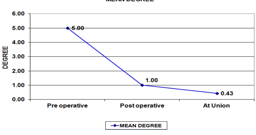
Alignment and reduction of the fracture pre operatively, post
operatively and at healing was the main outcome measured with an emphasis on
6. PATIENTS AND MATERIALS
This is a prospective study of 20 cases of tibial metaphyseal
fractures treated with open reduction/closed reduction with statically locked
intra medullary nailing an ‘ poller screws’ between August 2010 and September
2012 at Govt Rajaji Hospital, Madurai Medical College.
Patients
Inclusion criteria:
• Adult patients more than 20 years of age.
• Either proximal or distal metaphyseal fractures.
• Unstable, comminuted metaphyseal fractures of tibia with varying
soft tissue injuries.
• Segmental fractures with involvement of metaphysis.
Exclusion criteria:
• Adolescent patients <20 yrs of age.
• Very minimally (or) undisplaced fractures.
• Tibial metaphyseal fractures involving the articular surface.
• Associated with previous anatomic deformities.
• Fractures with wound at the nail entry site.
Materials
1. Intramedullary inter locking nail (In varying sizes 9,10,11):
2.“Poller” blocking screw:
Cortical screws in size of 4.5 mm,designated as “poller” screw
7. OPERATIVE PROTOCOL
Pre operative planning
• X ray of the injured leg in AP & Lateral views were taken.
• The fracture tendency for valgus or varus and antecurvatum or
recurvatum deformity was noted.
• The angle of malalignment was measured.
• Fracture was classified according to AO and Taylar& martin SUD system
• Fracture location from the proximal or distal articular surface was
measured.
• The length of fracture was also measured.
• The diameters of medullary canal at isthmus and at the level of fracture
were measured.
• Appropriate length of the nail was measured in one of the following
ways
1. From the contralateral normal limb, from the tibial tuberosity
to the medial malleolus.
2.Inra operative clinical method
Operative Technique
Methods:-
• All the cases were taken up for surgery under spinal anaesthesia.
• In three cases (4 weeks, 5 weeks,5weeks old ) tourniquet was used in
which open reduction and internal fixation was done with bone grafting
Technique:
Under spinal anaesthesia the patients were put in fracture table in
supine position on a padded knee support with the knee flexed to< 30 degrees.
For distal metaphyseal fractures the flexion maintained at 90 degrees.
Through the patellar tendon splitting approach incision started from the
inferior margin of the patella to antero superior aspect of tibia. The patellar
tendon splitting was done in line of the skin incision. Extra articular surface of
the tibia exposed. Entry point was made with bone awl at a point proximally
and laterally in case of proximal metaphyseal fractures. For distal metaphyseal
fractures it was done as usually. This was made at a point of 1.5cm from the
articular joint line. The bone awl initially pointed posteriorly, then directed in
line with the medullary canal or crest of the tibia. Then the trial reduction was
done under the C-Arm guidance. The tendency of the fragment towards for
which deformity is noted.The poller screw is inserted under the C-Arm guidance
metaphyseal fractures the reaming was done close to the anterior cortex.
Reaming was done till 1mm higher than the proposed nail size. Then the
proposed nail inserted and fixed with the static locking mode. Additional poller
screws was considered according to the correction deformities.
Positions of poller screws
Proximal metaphyseal fractures:Deformity Site of poller screw in short fragment
Antecurvatum Anterior to the nail
Recurvatum Posterior to the nail
Valgus Lateral to the nail, at concave side.
varus Medial to the nail, concave side.
Intraoperative demonstration of cases: Valgus deformity:
The valgus deformity is still not corrected
valgus deformity is corrected after inserting poller screw at concave side of the deformity lateral to the nail
Distal Metaphyseal fractures
Deformity Site of poller screw in short fragment
Valgus Deformity Lateral to the nail
Varus Deformity Medial to the nail
Poller screw is inserted on the concave side of the deformity
According the deformities presented in the fractures, the poller screws
were inserted in the all the patients in line with the above criteria. Additional
poller screws were inserted according the correction of the deformities. In all
the cases reamed nailing method was used. The medullary canal was reamed in
incremental manner till 1mm higher than the measured nail size. All the nails
were locked in static mode. The fracture alignment checked under the C-Arm.
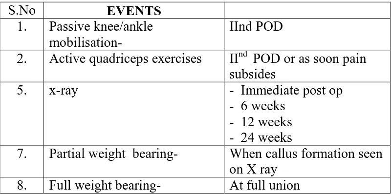
8. POST OPERATIVE PROTOCOL
Table of the time related events
S.No EVENTS
1. Passive knee/ankle
mobilisation-
IInd POD
2. Active quadriceps exercises IInd POD or as soon pain
subsides 5. x-ray
- Immediate post op - 6 weeks
- 12 weeks - 24 weeks
7. Partial weight bearing- When callus formation seen
on X ray
8. Full weight bearing- At full union
EOT : wound Examination On Table.
POD : Post Operative Day.
Follow-up
All the fractures were followed up till union of fracture with clinical and
radiological examination at 6 weeks 12 weeks and 24 weeks. The maximum
follow up was 28 weeks for the one case which complicated with delayed union.
On follow up axial alignment was assessed and functional analysis was
quantified using Karlstorm-Olerud score.Varus and antecurvatum were
expressed as positive values and valgus and recurvatum were expressed as
[image:67.595.98.498.191.390.2]Karlstrom-Olerud Score
1.
Residual angulation: ( 0 to 3 points)• 0° -- 0 point
• 1 to 3° -- 1 points
• 4 to 5° -- 2 points
• >5° -- 3 points
2.
Fracture healing: (0 to 3 points)• Union < 12 weeks -- 0 point
• Delayed union >12 weeks -- 1 point
• Delayed union requiring -- 2 points
secondary procedures
• Non union > 6 months -- 3 points
3. Cast support: (0 to 1 point )
• No cast support -- 0 point
• Cast support -- 1 point
Outcome:
• 0 & 1 Points - Excellent
• 2 & 3 Points - Good
• 4 Points - Satisfactory
• 5 Points - Fair
Patients were evaluated clinically and radiographicaly with the
anteroposterior and lateral x rays according to the above karlstorm-Olerud
scoring system.
In this study we have followed the definition of fracture union as
follows “when patient was able to bear full weight on the injured limb without
pain & support and when radiographs showed bridging callus in at least 3
cortices”.
9. RESULT - ANALYSIS
Method Used
In our study, the test used for data analysis is ANOVA test. In this method, repeated measures designs allow their own subject to act as control
This improves the precision of the experiment by reducing the size of the error
variance on many of the F-tests24. In our study there was no control group. So
the ANNOVA test is chosen for our study.
Multiple measurements are made on the same individual at different
point of times24. In line with this, the variables in our study were the angle at the
fracture site measured within the same subjects at different point of times.
Because few extreme values of variables of normal distribution should not
mislead the interpretation of analysis24, .95 % upper and lower confidence limits
were preferred over range to express the variables.Karlstorm-Olerud score
which was used to asses the functional outcome is an independent measurement,
not influenced by other co-morbid conditions and associated injuries25.
Data analysis
Age Distribution:
Table-1
Age No. of cases Percentage
<30 2 10%
31-40 2 10%
41-50 8 40%
51-60 4 20%
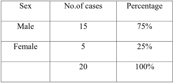
Sex Distribution:
Table-2
Sex No.of cases Percentage
Male 15 75%
Female 5 25%
20 100%
There were fifteen male and five female patients with the average age of
49.5 years,with 95% lower confidence limit(LCL) of 47.5years,and 95% upper
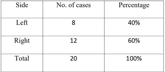
Side of the Injury:
Table-3
Side No. of cases Percentage
Left 8 40%
Right 12 60%
Total 20 100%
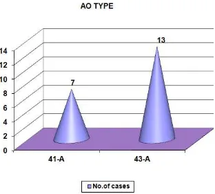
[image:73.595.133.463.190.335.2]Fracture Pattern-AO type:
Table-4
AO Type No.of cases Percentage
41-A 7 35%
43-A 13 65%
Total 20 100%
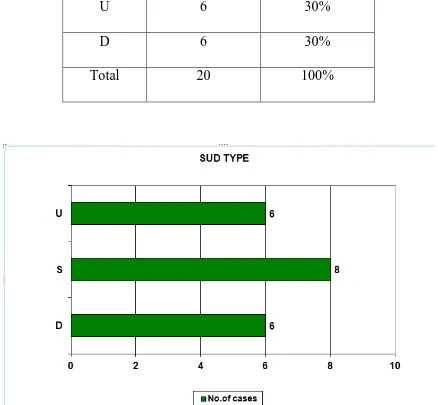
Fracture Pattern-Tayler and Martin SUD type:
Table-5
SUD Type No.of cases Percentage
S 8 40%
U 6 30%
D 6 30%
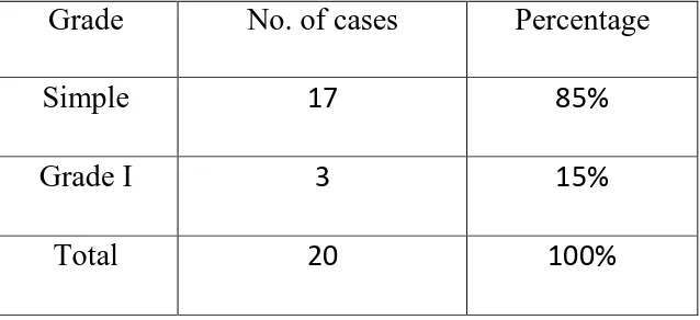
Gustilo-Anderson Pattern of fractures:
Table-6
Grade No. of cases Percentage
Simple 17 85%
Grade I 3 15%
Total 20 100%
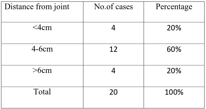
Distance from the Joint at which fracture occurred:
Table-7
Distance from joint No.of cases Percentage
<4cm 4 20%
4-6cm 12 60%
>6cm 4 20%
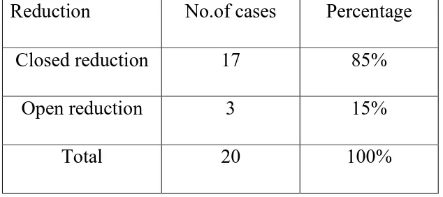
Open reduction/Closed reduction: <