STUDY ON PULMONARY TUBERCULOSIS IN THE
ADULT POPULATION - SOCIAL, CLINICAL,
RADIOLOGICAL PRESNTATION OF
SMEAR-POSITIVE TUBERCULOSIS
Dissertation submitted for
MD Degree (Branch I) General Medicine
March 2007
CERTIFICATE
This is to certify that this dissertation titled “STUDY ON PULMONARY TUBERCULOSIS IN THE ADULT POPULATION - SOCIAL, CLINICAL, RADIOLOGICAL PRESENTATION OF SMEAR-POSITIVE TUBERCULOSIS submitted by Dr .V.N.ALAGA VENKATESAN, (MD) to the faculty of General Medicine, The Tamilnadu Dr. M.G.R. Medical University, Chennai in partial fulfillment of the requirement for the award of MD degree Branch I (General Medicine) is a bonafide research work carried out by him under our direct supervision and guidance.
Dr. MOSES.K.DANIEL, M.D, Dr. NALINI GANESH, M.D, Professor of Medicine, Head of Department and Professor, Madurai Medical College, Department of Medicine,
Madurai. Madurai.
Place: Madurai Date :
DECLARATION
I, Dr. V.N.ALAGA VENKATESAN, solemnly declare that the dissertation titled “A STUDY ON PULMONARY TUBERCULOSIS IN THE ADULT POPULATION-SOCIAL, CLINICAL, RADIOLOGICAL PRESENTATION OF SMEAR-POSITIVE TUBERCULOSIS” has been prepared by me.
This is submitted to the Tamil Nadu Dr. M.G.R. Medical University, Chennai, in partial fulfillment of the regulations for the award of MD Degree Branch I (General Medicine).
Place : Madurai
ACKNOWLEDGEMENTS
At the foremost, I wish to express my sincere, heartfelt gratitude to my esteemed teacher and guide our unit chief Dr. Moses.K.Daniel.M.D, Professor Department of the Department of Medicine, Govt. Rajaji Hospital, Madurai for his continuous and understanding guidance throughout my postgraduate course and the period of this work.
It is my privilege and honour to extend my gratitude to the Dean Dr. Siva kumar M.S, Govt. Rajaji Hospital, Madurai, for permitting me to carry out this study and all the help rendered in the completion of this study.
I express my thanks to our Prof. Dr.Nalini Ganesh M.D, PROFESSOR AND Head of department, Medicine for valuable guidance and support in this study.
My heartful thanks to Assistant Professors Dr. S.Somasundaram, Dr. David Pradeep Kumar, Dr.K. Senthil, Dr .Rama krishnan &Dr.Vivekanandan, for their constant encouragement ,critical advice and timely suggestions in preparing this work.
ABBREVIATIONS AND ACRONYMS
ATT : Anti Tuberculous Therapy BMI : Body Mass Index
COPD : Chronic Obstructive Pulmonary Diseases DOTS : Direct Observed Therapy Short-term HIV : Human Immunodeficiency Virus PPD : Purified Protein Derivative
PAO2 : Alveolar oxygen concentration
RFLP : Restrictive Fragment Length Polymorphism
TU : Tuberculin Units
CONTENTS
Page No.
1. TITLE PAGE i
2. CERTIFICATE ii
3. DECLARATION iii
4. ACKNOWLEDGEMENT iv
5. ABBREVIATIONS AND ACRONYMS v
6. INTRODUCTION 1
7. AIM AND OBJECTIVES 6
8. REVIEW OF LITERATURE 7
9. MATERIALS AND METHODS 29
10. RESULTS 33
11. DISCUSSION 46
12. CONCLUSION 58
13. SUMMARY 60
14. BIBLIOGRAPHY
APPENDIX I - APPROVAL FROM ETHICAL COMMITTEE
62 vi
APPENDIX II – PRO FORMA viii
Introduction
Mycobacterium tuberculosis is as old as mankind, ubiquitous and is believed one amongst the oldest bacteria in earth. TB is still a problem in the world. It is a major health problem through out the world.
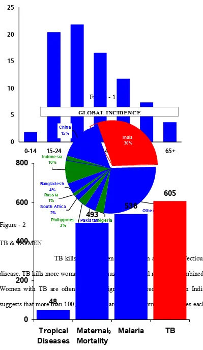
TB is a leading killer of adults among the infectious diseases. One third of TB in the world population is from India. India has more cases of TB than any other country in the world and twice as many cases as China which has next highest cases. Studies show an incidence rate of more than 200 per lakh among the highest in the world. About 8 million new cases of TB in a year with two million deaths reported.
The age above >15 to 44 is 57.3% (Male-363,876,219 & Female-340,181,764).In India about 0.6 million people die of TB every year. Most cases between 15-44 age group. India accounts for one-fourth of the global burden. (Grazybowsky)Ind.J.Tub.199542-201)
1.prevalance rate of TB infections in India about 30% 2.Annual incidence of rate of infection of average is 1%
Tuberculosis in Adult
MacGregor Rr study (Am J Med 58:221) shows Tuberculosis affects greater numbers of adult people. According to Park, about 65% of people within the age of 55 in India are affected with tuberculosis. TB is a leading killer of adult. TB kills more adults than any other diseases. Because it affects adults, tuberculosis causes enormous social and economic disruption. TB burden is hidden by stigma and poor diagnostic quality. It was viewed as a dreadful disease. Although the cause of the illness was already understood, the cure remains elusive not to arrive for at least a decade. Tuberculosis, although predominant among the poor and underfed and rampant among those living in unsanitary surroundings, did not discriminate.
Tuberculosis attack the young and middle age, and took its toll among healthy. Their immune system succeeded in fighting the germs to a draw. But the invader remains, content to live quietly in his host, temporarily encapsulated by antibodies but awaiting the opportunistic moment for return. . The majority of pulmonary tuberculosis, in older people is due to inactivated diseases. In one study by Liaw et al (1995) about 26.2% of elderly tuberculosis patients suffer previous tuberculosis disease.
Figure - 1
Figure - 2 TB & WOMEN
TB kills more women in India than any other infectious disease. TB kills more woman than all cause of maternal mortality combined. Women with TB are often severely stigmatized. A recent study in India suggests that more than 100,000 women are rejected from their families each
SMEAR POSTIVE – TB IN RELATION WITH AGE
0 5 10 15 20 25
year an account of TB.
Figure - 3 This present report deals with socioclinical dimension of adult tuberculosis.
.
TB AND POVERTY
TB is more common in poor and malnourished people, but spreads without regard for socio-economic status. TB treatment is effective
independent of nutritional or economic status. Adherence to
treatment is irregular, regardless of age, sex, religion, education, or severity of disease — therefore, directly observed treatment is
standard of care for all TB patients. Access to treatment is more difficult for the poor. Community-level treatment ensures cure of infectious patients and reduce spread of disease
Other names for tuberculosis disease
• TB (short for tuberculosis and also for Tubercle Bacillus)
• Consumption (TB seemed to consume people from within with its symptoms of bloody cough, fever, pallor, and long relentless wasting)
• Wasting disease
• White plague (TB sufferers appear markedly pale)
• Phthisis (Greek for consumption) and phthisis pulmonalis
• King's evil (so called because it was believed that a king's touch would heal scrofula)
• Pott's disease of the spine
• Miliary TB (x-ray lesions look like millet seeds)
• Tabes mesenterica (TB of the abdomen)
• Lupus vulgaris (the common wolf - TB of the skin)
• Prosector's wart, also a kind of TB of the skin, transmitted by contact
with contaminated cadavers, to anatomists, surgeons, butchers, etc.
• Koch's Disease named after Robert Koch who discovered the
tuberculosis bacilli.
DRUG RESISTANCE IN TUBERCULOSIS
a. Primary resistance It occurs when a patient develops tuberculosis after being infected by another patient who has resistant micro organisms
b. Initial resitance is seen in patients who deny history of previous treatment but might have taken anti tubercular drugs knowingly or unknowingly .It includes primary resistance and unknown amount of undisclosed acquired resistance
c. Acquired resistance is the one where the bacilli develop resitance to one or more antitubercular drugs during inadequate therapy
d. Multi drug resistance Mycobacterium tuberculosisresistant to isoniazid and Rifampicin with or without resistance to other drugs
Aim and Objectives
1) To study the socio-demographic pattern of adult tuberculosis. 2) To analyze their clinical presentation.
REVIEW OF LITERATURE
Global Epidemiology of Tuberculosis
In 1993, the World Health Organization
declared tuberculosis as a global emergency because
of the scale of the epidemic and the urgent need to
improve global tuberculosis control.
According to Tuberculosis case notification and rates by WHO in 2002, three regions dominates the world wise distribution & notification i.e., South East region 36%, African region 24% & Western Pacific region 20%. (Maher & Raviglione 2005).
reversal of the previous trend include increased poverty among marginalized group in inner city areas, immigration from areas with high tuberculosis prevalence, the impact of HIV and the failure to maintain the necessary public health infrastructure under the mistaken belief, tuberculosis was the disease of the past.
TABLE 1 TUBERCULOSIS CASE NOTIFICATION AND RATES BY WHO REGION IN 2005 .
WHO region No. of cases notified Proportion of global total (%)
South East Asia 1,487,985 36
Africa 992,054 24
Western Pacific 806,112 20
Americas 233,648 9
Eastern Mediterranean 188,458 6
European 373,497 5
SOURCE: CLINICS IN CHEST MEDICINE 2005
In 2002, there was an estimated 8.8 million new cases of tuberculosis world wide, with an incidence rate of 141 per 100,000 population.
Epidemiology of Tuberculosis in India
Tuberculosis continues to be a major public health problem in India. Since the national reporting system is defective, the only reliable source of information on the magnitude of the tuberculosis problem has been the population sample surveys.
Tuberculosis Institute, Bangalore undertook three longitudinal surveys at Delhi, Bangalore and Chengelpet. India ranks first in the estimated number of Tuberculosis cases i.e, 1,049,549 thousand cases. (Maher & Raviglione 2005).
The overall prevalence of infection (as judged by the standard tuberculin test) was about 30%, in males 35% and in females 25%. The prevalence was 4 cases per 1,000 population. The incidence of new cases was about 1.5 per 1000 population. According to experts it is safe to estimate that at least 50% of the population above the age of 20 years is infected and will remain at risk of disease throughout their lifetime (WHO 2002).
Gender difference in tuberculosis
Little has been written about gender differences in tuberculosis. In general, the notification rate is higher in men than women. Gender differences vary in different parts of the world. In their study, Chan-Yeung et al (2002) demonstrated that the tuberculosis rate was higher in men than in women in all age groups and the sex difference increases with age. In those aged 15-44, men are affected times more than women.
bias. Sutherland et al (1979) studied the risk of tuberculosis infection in the Netherlands from 1967 to 1979 and found that there was no differences in the annual rate of infection between boys and girls aged 6 – 12 years. However for those between 12 – 18 years, an excess in the annual rate of infection was found among males.
Duration of illness
There is no delay in diagnosing TB in adult patients who attended to the hospitals with symptoms. They present with productive cough, non productive cough more than 15 days duration, anorexia, weight loss, fever(low grade), night sweats & fatigue. In elderly people diagnosis is difficult because they present more commonly with non-specific complaints. Therefore the tuberculosis may be suspected initially and there maybe considerable early diagnosis in adult patients before they may present with the advanced stage of the disease. Children and adult are more likely to experience reactivation of disease and often triad of symptoms fever, weight loss and night sweats.
47.3% in patients less than 60 years of age against 38.6% in patients in elderly.
Pathogenesis & immunological aspects
The principal route of entry in tuberculosis is the lung. Inhaled tubercle bacilli are engulfed by alveolar macrophages and transported to regional lymph nodes. Infected macrophages and circulating monocytes secrete proteolytic enzymes, generating an exudative lesion and granuloma formation with activation of T cells. This leads to the onset of cell mediated immunity. The characteristic Ghon complex ultimately develops, tubercle bacilli ultimately restrain within caseous necrosis with eventual healing. A study by Zhmikrobiol et al (2002) reported that there is a decreased activity of natural killer cells in tuberculosis patients. The greatest decrease in the activity of natural killer cells was observed in patients with chronic fibro cavernous form of tuberculosis.
Interestingly, while these concomitant illnesses only produce a minor cellular deficiency, it is enough to make patients more susceptible to TB disease. Thus altered immune function might predispose the patients to potential reinfection with TB.
It is due to these defects in cellular immunity, patients who present with a chronic wasting or respiratory illness may not present in the classic form.
Clinical spectrum of Tuberculosis
Few adult patients may not present with the classic features of tuberculosis. Tuberculosis in the population may present clinically with cough, chronic fatigue, and anorexic or unexplained low grade fever.
According to Chan et al (1995), cough was the most common symptom in adult tuberculosis patients and old age patients. However non specific symptoms occur in 12% of the adult tuberculosis patients. Cough with expectoration is the commonest symptom in adult where as systemic symptoms dominates the picture in old age tuberculosis Chan et al(2002) &, that ‘breathlessness’ was the most common symptom in old age tuberculosis patients in Arora et al (1989) study.
age group as compared to the adult. Katz et al (1987) found no significant difference in the presentation with symptoms of fever and weight loss among the adult and elderly. They observed that the patients significantly present with hemoptysis, have cavitatory lesions on radiographs. Old age TB patients were more likely to present with dyspnoea as their main symptom common in people of Asian, African & Caribbean origin. Fever if present is usually low grade and is easily missed unless rectal temperature is measured in the late afternoon or evening (Brochlehurst et al 1992).
Smoking and Tuberculosis
“Some patients commit suicide by drowning but
many by smoking
”.
According to Arora et al (1989) about 68% of tuberculosis patients are smokers & chronic bronchitis occurs in about 64% of tuberculosis patients. This results in altered clinical presentation. The majority did not seek early medical relief as they attribute their complaints to smoking.
elderly tuberculosis patients present late and they wrongly attribute these symptoms to chronic obstructive pulmonary disease.
In patients of both sexes over the age of 30 suffering from pulmonary tuberculosis, it has been shown by Lowe et al (1956) that there is a highly significant deficiency of non smokers and light smokers compared with controls of the same as suffering from other diseases. Kahn et al (1966) shown that mortality from pulmonary tuberculosis among doctors has been shown to increase significantly with the number of cigarette smoked and the similar effect has been also shown in investigations.
Co morbid Illness
age patients suffer from concomitant diseases like chronic bronchitis, emphysema, diabetes, etc and their presence would complicate both the diagnosis and treatment of TB.
Diabetes & tuberculosis
In most studies on this subject, persons with
diabetes mellitus have two fold to four fold higher
incidence of active tuberculosis than non-diabetic
patients (Morse et al (1964). The predisposition of
diabetes to infection that are normally controlled by
cell mediated immunity may result from one or
more defects of pulmonary host defense, including
conditions that interfere with normal clearance
mechanisms or that impair pulmonary function. The
greatest difficulty in studying diabetes mellitus as an
independent risk factor for the development of
tuberculosis is the presence of potential confounding
medical conditions (eg. malnutrition, chronic renal
disease) and personal behaviors, such as smoking &
alcohol that may further weaken host defenses.
Koziel et al (1998))
Henry & Stableforth (1983) described this
finding from a group of diabetic TB patients living
in the United Kingdom as diabetic tuberculosis
patients presents with a higher incidence of
cavitatory disease and sputum positive states than
did a control group of Non Asian diabetic TB
patients. This was presumably due to higher
exposure risk of the Asians which appeared to be
associated with their living in settings that replicate
and concentrate the bacilli, the social behavior and
TB & HIV
HIV
infected persons are at great risk of TB. Without
HIV the life time risk of developing TB in TB
infected people is about 10%, compared with at least
50% in HIV infected TB infected people. The HIV
pandemic could rapidly increase the incidence of
TB. Diagnosis of TB in HIV infected person is more
difficult due to more non TB- Respiratory diseases,
more smear negative TB, extra pulmonary TB&
X-Rays are even less specific .Even among HIV
infected patients, TB can be cured in more than
90% of surviving HIV infected TB. Over all TB
patients, because of death from non-TB causes.
Miliary Tuberculosis
Miliary TB is common in immunocompramised
adults. Miliary refers to diffuse tiny nodule; similar
in size of millet seeds which are seen in x-rays. Skin
test is often negative. This presentation occurs in 1
in 100 cases.(Pathy 1993). It is one of the common
causes of pyrexia of unknown origin in the TB
patients. Usually sputum will be negative for
mycobacterium. According to Robert et al (1974),
the classical acute or sub acute high intermittent
fever and early onset of complicating meningitis or
serositis is often present in adult and absent in the
old. It presents as more chronic form of slowly
or low grade fever without any localizing symptoms
or signs. Examination may reveal only a
hepatosplenomegaly. Abnormal chest x-ray is quite
compatible with miliary tuberculosis (Kulpati).
Hematological abnormalities in tuberculosis.
Investigations in Tuberculosis
Tuberculin Intradermal Test
The tuberculin was discovered by Von Pirquet in 1907. A positive reaction to the test is generally accepted as evidence of present or past infection by M. tuberculosis. The tuberculin test is the only means of estimating the prevalence of infection in a population.
Tuberculin – Now purified protein derivative is used. It contains 50,000 tuberculin units per milligram. One TU is equal to 0.01 ml of O.T. or 0.0002 mg PPD (Youman et al 1980).
Dosage
a) First strength or 1 TU
b) Intermediate strength or 5 TU c) The second strength or 250 TU.
For routine testing, the vaccinating team in India use 1TU. (Comstoch et al 980.)
Mantoux test
In Tuberculin reaction induration is measured.
> 10 mm Positive < 6 mm Negative 6 – 9 mm Doubtful
(Source: Park’s Preventive & social medicine, 18th edn.)
A positive reaction indicates that the person is infected with M.tuberculosis, it does not prove that the person is suffering from the disease. (The Tuberculosis Association of India, New Delhi, 1981).
An excellent but inadequately noticed paper by Zybowski & Allen in 1964 documents the phenomenon of reversal of tuberculin reaction to negative over time while the role of reversal varies with age. It was shown to continue for the life span at a rate of about 5% per year.
According to Yokayama et al (2003) antibody to tuberculin skin test with purified protein derivative was evident in 7% of patient under 60 and 14% of those over 60 years of age.
antigen (i.e. candida, mumps or tetanus toxoid) diluted to a 1.5 concentration with phenol buffered diluent to assess the presence of anergy. A positive booster effect and therefore a positive tuberculin skin test reaction is a skin test of 10 mm or more and an increase of 6 mm or more over the first skin test reaction. The booster effect occurs in a person previously infected with M.tuberculosis but who has a false negative skin test, repeat test elicits a truly positive test. It is important to distinguish the booster phenomenon from a true tuberculin conversion. Conversion occurs in persons previously uninfected with M.tuberculosis and who had a true negative tuberculin test but who becomes infected within 2 years as demonstrated by a repeat skin test that is positive during this period. 15 mm or more is considered positive for a conversion activity in persons 35 years and older and in persons with no risk factors for tuberculosis (Hazzaro et al).
Chest X-ray
The chest x-ray is mandatory in the diagnosis of Tuberculosis. The radiological features of tuberculosis are often similar in younger patients and old age patients, but there is a greater prevalence of mid and lower zone shadowing in old age (Umeki et al 1989). The criteria used for interpretation is described in Milka-Cabanne et al 12 .
proportion of patients with lower lung fields lesions progressively increases with age where as the frequency of cavitations steadily decreases with age.
Upper lobe findings are common in adult tuberculosis. Juvekar et al(1998 IN J Tub 45,95) study observed that only 10% of urban and 21% rural patients were subjected to sputum examination. Majority of patients 78% urban, 56% rural reported have undergone x-ray before treating TB. Jagota (1995 IND J Tub 45 5) study showed even after National tuberculosis programme over diagnosis of TB patients which shows only 20% after sputum examination. Blind relay on x-rays need to be discouraged but if sputum facility is not available it is helpful. Aging leads to increased alveolar ventilation and reduced perfusion resulting in increased mismatch and increased PAO2. These changes should affect the lower lobes more than
the upper lobes because the latter have a higher ratio and higher PAO2.
Therefore age induced changes should favor multiplication of Mycobacterium tuberculosis in lower lung zones. Further more frequency of upper lung field lesion (with or without lower lung field lesion) was similar at all ages, suggesting that aging does not alter condition in upper lobes.
According to Yamaguchi et al (2001) the frequency of cavitations was higher in the middle aged group (87.4%) than old age patients (56.67%)
Sputum examination
Sputum examination for M.tuberculosis using smear
and culture is indicated for all patients who have
pulmonary symptoms and radiographic changes
compatible with tuberculosis. Sputum examination
is carried out by Ziehl Neelson & Kinyoun methods
which utilize the carbolfuschin method. Otherwise
auromine rhodamine dyes are used with uses
flurochrome methods.
For suspected tuberculosis, it is recommended that 3 fresh consecutive sputum specimen obtained in the morning be used for routine mycobacteriological study (American thoracic society, Diagnostic standards of classification of TB, 2000).
aspiration. Smear test for M.tuberculosis may require a minimum of 104-5
AFB per milliliter to be seen by light microscopy (Rajagopalan et al (2001)). Arora et al (2003) compared sputum profile of tuberculosis between younger and geriatric patients. He demonstrated that the ratio smear positive to smear negative patients was almost similar in the two age groups. Further the treatment outcomes of new smear positive adult TB patients in comparison to old age patients showed better sputum conversion. This same view was endorsed by a study in Hong Kong where the bacteriologically confirmed pulmonary tuberculosis cases increases with age from 59.8% in those < 20 years to 76.2% in those > 80 years. As against this Gaur et al (2004) showed that the percentage of old age tuberculosis patients found to be bacteriologically positive by direct smear was 59.6% as compared to 65.8% among the comparison group (35 – 50 years).
Culture
Immunoassay for mycobacterial antigen, serological tests for tuberculosis and detection of mycobacterial components or products by high performance liquid chromatography, gas chromatography and mass spectrometry are also important.
Henn et al (2001) evaluated the sensitivity and specificity of anti Lipoarabinomannan IgG antibodies for diagnosis of tuberculosis in cases of pulmonary, extra pulmonary or associated forms. She concluded that the test is easy and ideal for use in diagnosis of active tuberculosis in specific cases in conjugation with the other methods and permit to evaluate the humoral immunity. Rapid radiometric detection makes out mycobacterial growth (detection of 14 CO2 released) in 10 – 12 days. A further advantage is that the
DNA probe identification and antibiotic susceptibility tests can also be performed rapidly (Rajagopalan et al).
Other tests
Molecular techniques extend knowledge about TB.
Genetic finger printing using restriction fragment
length polymorphism patterns to identify isolates,
have proved that HIV seropositive individuals and
re-infected by TB. Molecular epidemiology again using
RFLP pattern has helped identifying transmission
network that the traditional epidemiology failed to
identify. Early detection of drug resistance by rapid
identification of genetic mutation and by
chemiluminescent methods so called as “Fire fly”.
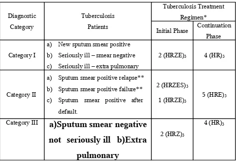
[image:33.612.85.559.386.711.2]Management of Adult Tuberculosis.
TABLE 2 Treatment categories in DOTS chemotherapy in India.
Diagnostic Category Tuberculosis Patients Tuberculosis Treatment Regimen*
Initial Phase Continuation Phase
Category I
a) New sputum smear positive b) Seriously ill – smear negative c) Seriously ill – extra pulmonary
2 (HRZE)3 4 (HR)3
Category II
a) Sputum smear positive relapse** b) Sputum smear positive failure** c) Sputum smear positive after
default.
2 (HRZES)3
1 (HRZE)3
5 (HRE)3
Category III
a)Sputum smear negative
not seriously ill b)Extra
pulmonary
2 (HRZ)3
non seriously ill
* The number before the letter refers to the number of months of treatment. The subscript after the letter refers to the number of doses per week.
** In rare and exceptional cases, patients who are sputum smear negative or who have extra pulmonary disease can have relapse or failure.
This diagnosis in all such cases should be made by a medical officer and should be supported by culture and histological evidence of current, active tuberculosis. In these cases the patient should be categorized as others and given category II treatment. (Source: Park’s Preventive and social medicine 18th edition).
Drug Adverse Reactions
1. INH GIT irritation, peripheral neuropathy*, blood dyscarsias, hyperglycemia, giddiness, mild
drowsiness & liver damage**
2.Rifampicin Hepatotoxicity,
3. Streptomycin Vestibular damage, nephrotoxicity. 4. Pyrazinamide Hepatotoxicity, Hyper uricemia 5. Ethambutol Retro bulbar neuritis.
(Source: Park’s Preventive & social medicine, 18th edition)
* Peripheral neuropathy can be prevented by pyridoxine 10 – 20 mg daily.
** A transient rise in serum enzymes to three times the normal may occur during INH therapy.
Therapeutic difficulties in adult patients
additional hepatotoxic effects. As renal function, hearing acuity, vestibular function declines with aging, ototoxicity and nephrotoxicity due to streptomycin is less in adult patients when compared with old age patients. Since some visual impairment is common in the elderly, a careful examination that includes visual acuity and color discrimination should be performed before initiating ethambutol therapy. Drug interactions must also be considered. Patients may be taking other drugs for other illness which may interact with antituberculosis drugs.
Materials and Methods
Study Design : Cross Sectional Study
Setting : Govt. Rajaji Hospital, Madurai Period of study : Aril 2005 to March 2006
Ethical clearance : Ethical committee approved the methodology of the study
and copy enclosed in annexure I
Consent : Consent was obtained from all the patients considered for the
current study. Financial support : Nil
Conflict of interest : Nil
Inclusion Criteria
Patients who satisfied the following were included in the study.
1) Adult patients (>15 to 44 years of age) who were sputum smear positive, were interviewed and examined with clinical and radiological profile suggestive of tuberculosis were included in the study.
2) Both sexes.
3) HIV negative status.
Exclusion Criteria
Patients who had any one of the following or a combination of them were excluded.
1) More seriously sick individuals. 2) HIV co-infection.
3) On immunosuppressive therapy. 4) Associated malignancy
5) Major abdominal / thoracic surgery. 6) Un-cooperative / unwilling patients 7) Major cardiac illness
8) Collagen vascular diseases 9) Occupational diseases
10) Extra pulmonary tuberculosis. METHODS
Selected socio-demographic, clinical and laboratory data were elicited from the patients and recorded in a proforma (enclosed in annexure II).
I. Socio demographic data:
a) Age b) Sex c) Address d) Contacts e) Number of family members
a) Body weight b) Height c) Pulse rate d) Blood pressure e) Clinical examination
III. Laboratory data
a) Hemoglobin: measured using Sahli’s hemoglobinometer. b) Total count & Differential count using Leishmann stain.
c) Blood urea done manually by using Diacetyl monoxime technique. d) Serum creatinine: estimation done using COBAS auto analyzer. e) Blood glucose: estimation done using glucose oxidase method
f) sputum AFB: 3 early morning specimens collected and stained by Ziehl Neelson technique
g) X-ray Chest: PA view was taken in a radiation dosage of 0.02mSv.
The following definitions were used in this study.
Ex-Smoker
: Patient who ceases smoking for 2 years.(Changes
in the small airways of smokers will reverse after 1- 2
years of cessation) (Harrison 16
thedition)
Diabetes Mellitus:
a) Fasting plasma glucose ≥126 mg/dl
b) Two hour plasma glucose ≥200 mg/dl in oral glucose tolerance test. c) Symptoms of diabetes plus random blood glucose concentration ≥ 200
Hypertension: Systolic ≥ 140 Diastolic ≥ 90
Based on the average of ≥ 3 reading, when any one of the
value (systolic or diastolic BP) is less than the above given
value but the other value is higher, the higher value is
taken into consideration.
Anemia:
Anemia is defined as a decrease in the circulating
RBC mass, the usual criteria are a hemoglobin of less than
12 g/dl in women and less than 14 g/dl in men.
Body Mass Index: Weight in kilogram / Height in m2.
X-ray zones: The chest x-ray was divided into three zones. Upper zone: 1st and 2nd intercostal space.
Middle zone: 3rd and 4th intercostal spaces.
. Lower zone: Rest of the intercostal spaces.
Limitations of the study
1) Age and sex matched control was not attempted since only one disease was taken into consideration.
3) Pulmonary function test was not done.
4) Induced sputum was not attempted, as it was not approved by the institutional ethical committee.
5) Since single group of patients were analyzed, complex statistical analysis was not done.
RESULTS
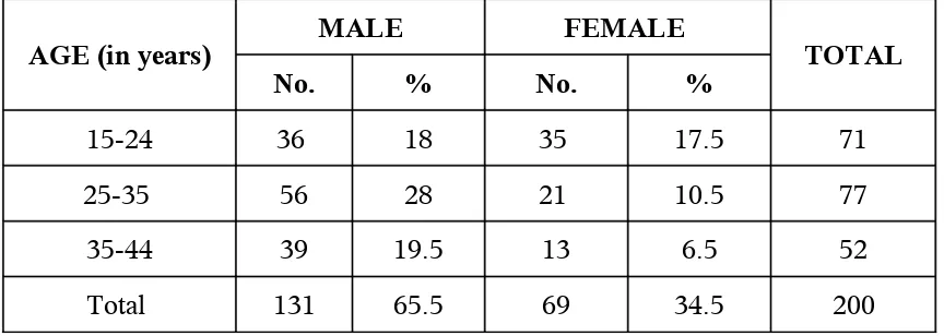
[image:42.612.83.515.217.371.2]A total of 200 patients of TB were studied. The distribution of cases in relation to ageand gender is furnished in table 3.
TABLE 3: DISTRIBUTION OF SUBJECTS
ACCORDING TO AGE & GENDER
AGE (in years) MALE FEMALE TOTAL
No. % No. %
15-24 36 18 35 17.5 71
25-35 56 28 21 10.5 77
35-44 39 19.5 13 6.5 52
Total 131 65.5 69 34.5 200
The mean age is 29.5.
Using chi-square test, it was found the difference between age and sex was not significant. Thus, the distribution of age and sex are independent in this study. Almost 2/3 of patients are male.
The details are given in figure 4.
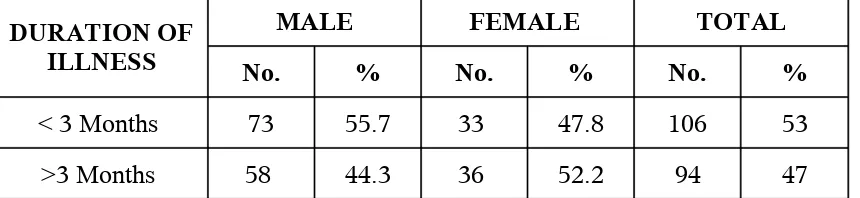
TABLE – 4 DURATION OF ILLNESS AND GENDER.
DURATION OF ILLNESS
MALE FEMALE TOTAL
No. % No. % No. %
< 3 Months 73 55.7 33 47.8 106 53
>3 Months 58 44.3 36 52.2 94 47
This table number 4 shows that majority of the male
subjects (55.7%) presented with duration of illness
for < 3 months, followed by 44.3% for > 3 months.
In contrast, about 47.8% female subjects had the
illness for < 3 months, 52.9% for >3 months before
they sought treatment. The distribution of cases in
those with symptoms less than 3 is slightly higher in
male and more than 3 months was high in females
irrespective of age.
The details are provided in
pictorial manner in figure 5.
The distribution of subjects according to clinical
symptoms and gender is furnished in table 5 given
[image:43.612.87.517.112.211.2]TABLE – 5 CLINICAL SYMPTOMS AND GENDER
SYMPTOMS MALE FEMALE TOTAL
No. % No. % No. %
Cough 131 100 69 100 200 100
Productive 121 92.4 67 97.1 188 94
Fever 104 79.4 59 85.5 163 81.5
Hemoptysis 59 45 32 46.4 91 45.5
Breathlessness 69 52.7 36 52.2 105 52.5
Chest Pain 101 77.2 52 75.3 153 76.5
Weight Loss 98 74.8 52 75.3 150 75
Anorexia 95 72.5 50 72.5 145 72.5
Other symptoms 19 14.5 10 14.4 29 14.5
Numbers does not tally with total due to more than
one symptom.
In this study, cough was the most common
symptom observed in males and females (100%)
productive sputum is present in 94% of patients.
Fever present in 76.5% (males 104 cases and females
[image:44.612.85.513.123.406.2]males the next common symptom was cough with
sputum (92.4%), followed by fever (79.4%). Almost
more than 75% of the study subjects complained of
weight loss.
The difference between the observed and
expected values was not significant and hence
clinical symptoms are independent of each other.
Also the calculated probability value of chi-square
was much less than the observed value of the
chi-square at 5% level of significance with which it
could be concluded that clinical symptoms among
the subjects were independent of gender.
The details are provided in figure
6.
TABLE – 6 SMOKING STATUS OF THE STUDY SUBJECTS
SMOKING STATUS NO. OF CASES PERCENTAGE
Current Smokers 86 43
Ex-Smokers 25 12.5
Non-Smokers 89* 44.5
In this study population all the female subjects were
non-smokers as smoking tobacco is considered a
taboo in this part of the country. Among the study
population 43% were current smokers, 12.5% were
Ex-smokers and 44.5% were non smokers.
*
Including female patients.
[image:46.612.90.514.118.218.2]These adult patients have one or more co morbid
illness. The distribution of cases in relation to co
morbid illness is given below in table 7.
In this study of 200 population, diabetes was the
most commonly observed co morbid illness affecting
about 11% of the study population. Hypertension
was found in 9% of patients followed by chronic
obstructive pulmonary disease in 1% and ischemic
heart disease in 2% of study subjects. Chronic renal
failure noted in 1.5% of subject studies. No subject
presented with malignancy.
.
TABLE – 7 COMORBID ILLNESSESS AMONG THE STUDY SUBJECTS
COMORBID ILLNESS NO. OF CASES PERCENTAGE
Diabetes 22 11
Hypertension 18 9
Ischemic Heart Disease 4 2
Chronic obstructive
pulmonary disease 2 1
Malignancy 0 0
[image:47.612.88.509.498.691.2]Details of wasting, anemia, clubbing and
lymphadenopathy observed in the study population
are provided in the table 8 given below. Wasting was
the most common sign 68.5%, followed by anemia
39% in our subjects. clubbing 12.5% and
lymphadenopathy 11% were infrequently noted and
very few had hepatomegaly(2%), spleenomegaly
TABLE – 8 GENERAL EXAMINATION FINDINGS IN STUDY SUBJECTS
GENERAL EXAMINATION
FINDINGS NO. OF CASES PERCENTAGE
Wasting 137 68.5
Anemia 78 39
Clubbing 25 12.5
Lymphadenopathy 22 11
Hepatomegaly 4 2
Spleenomegaly 2 1
Oedema 3 1.5
Wasting is the commonest finding present in
68.5% followed by anemia is the next finding
present in about 39% cases followed by clubbing in
12.5% of subjects. Only 11% of the study subjects
presented with Lymphadenopathy. 2% present with
hepatomegaly and 1% with spleenomegaly. Oedema
present in 1.5% of study subjects.
Details are provided in
figure 8.
AUSCULTATION.
[image:49.612.85.512.118.327.2]finding, most commonly heard in the upper and
lower zones. In Ethiopia and Gambian study lower
zone crepitations are more. Bronchial breath sound
heard more in upper zone most commonly in right
side. Dullness, absence of breath sounds and wheeze
were infrequently detected. In this study upper zone
means supra clavicular, infra clavicular and supra
scalpular. Middle Zone means mammary and
axillary area. Lower zone means infra scapular,
infra axillary and inter scapular area
TABLE-18 LUNG EXAMINATION FINDINGS
CREPITATION BY LUNG FEILD
LUNG
UPPER
ZONE
MIDDLE
ZONE
LOWER
ZONE
RIGHT
54.5%
18.5%
42.5%
LEFT
34.5%
8%
48.5%
54.5% of right side upper zone crepitations
were recorded among the lesions in the right upper
zone lesion. In middle zone 18.5% and lower zone
42.5%. Left lung upper zone 34.5%, middle zone
8% and lower zone 48.5%.
Bronchial breath
TABLE-19 LUNG EXAMINATION
FINDINGS –BRONCHIAL BREATH SOUNDS BY
LUNG FIELD—NUMBER OF SUBJECTS.
LUNG
UPPER
ZONE
MIDDLE
ZONE
LOWER
ZONE
RIGHT
43
4
2
LEFT
18
7
2
Bronchial breath sounds are heard more in
right upper zone (43), left upper zone it is 18 in
number. Crepitations were common auscultatory
finding recorded and auscultatory findings were not
correlated with chest x-ray.
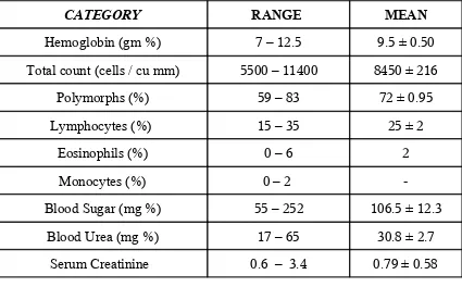
TABLE – 9 BIOCHEMICAL & HEMATOLOGICAL PROFILE
IN STUDY SUBJECTS
CATEGORY RANGE MEAN
Hemoglobin (gm %) 7 – 12.5 9.5 ± 0.50
Total count (cells / cu mm) 5500 – 11400 8450 ± 216
Polymorphs (%) 59 – 83 72 ± 0.95
Lymphocytes (%) 15 – 35 25 ± 2
Eosinophils (%) 0 – 6 2
Monocytes (%) 0 – 2
-Blood Sugar (mg %) 55 – 252 106.5 ± 12.3
Blood Urea (mg %) 17 – 65 30.8 ± 2.7
Serum Creatinine 0.6 – 3.4 0.79 ± 0.58
RADIOLOGICAL PROFILE OF STUDY SUBJECTS
Majority of the subjects (58%) had involvement of both
lungs followed by 23.5% with involvement of the right lung
and only about 9.5% had involvement of the left lung. The
details are furnished in 10provided below as well as in figure
9.
TABLE – 10 LUNG INVOLVEMENT AMONG STUDY
SUBJECTS
X-RAY CHEST No. OF SUBJECTS AFFECTED %
Right Lung 57 23.5
Left Lung 19 9.5
Both Lungs 116 58
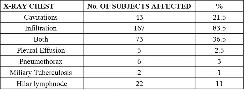
Infiltration was a common observation in the
x-ray chest in 83.5% of the subjects while cavitations
were found only in 21.5% of subjects. In 36.5% of
subjects both cavitations and infiltration was found.
Pleural effusion, pneumothorax was found in 2.5%
and 3% respectively. 11% shows hilar
lymphadenopathy. Miliary pattern seen in 2% of the
study subjects. The details are furnished in table 11
TABLE – 11A PATTERN OF LESIONS IN THE LUNG
AMONG THE STUDY SUBJECTS
X-RAY CHEST No. OF SUBJECTS AFFECTED %
Cavitations 43 21.5
Infiltration 167 83.5
Both 73 36.5
Pleural Effusion 5 2.5
Pneumothorax 6 3
Miliary Tuberculosis 2 1
Hilar lymphnode 22 11
Involvement of upper and lower zone was found to
be equally distributed in 46% of the study subjects where
as middle zone involvement was seen in 39% of the
subjects.34% of the subjects all the zones involved. The
details are furnished in table 11 B.
TABLE – 11B INVOLVEMENT OF ZONES OF LUNG IN
STUDY SUBJECTS
X-RAY CHEST No. OF SUBJECTS
AFFECTED %
Upper zone
134
67
Middle zone
46
23
Lower zone
54
27
All zones
42
21
The following table 12 gives the distribution of radiological
findings according to gender in the study population.
TABLE – 12 RADIOLOGICAL FINDINGS ACCORDING
TO GENDER IN THE STUDY POPULATION
SEX
RADIOLOGICAL FINDINGS
CAVITATI
ONS
INFILTRA
TION
UPPER
ZONE
LOWE
R
ZONE
Male
37
112
82
37
Female
6
49
41
12
In assessing the radiological findings in the
x-ray chest of the study subjects, it was seen that in
[image:55.612.92.521.436.604.2]infiltrations 82 had upper zone involvement and 37
had lower zone involvement. Similar observation
was seen among females as 6 of them had
cavitations, 54 had infiltration, 41 had upper zone
involvement and 12 had lower zone involvement.
As the observed and expected values are almost equal, in
both sexes, it is clear that cavitation ( p = 0.99), infiltration(p
= 0.6) and involvement of zones of the lung were independent
of gender.
[image:56.612.97.531.510.707.2]The table 13 gives the radiological presentation of
tuberculosis in diabetic patients.
TABLE–13
:
THE RADIOLOGICAL PRESENTATION IN
TUBERCULOSIS PATIENTS WITH DIABETIC
STATUS.
STATUS OF DIABETES LUNG INVOLVEMENT Infiltration Cavitatio nUpper
Zon
e
Middle Zone Lower ZoneDiabetic 15 7 8 12 14
Among the diabetic patients in the study
population 15 presented with infiltration and 7 had
cavitations in the lung which was evident from the
x-ray chest. The middle zone involvement was found
in 12 subjects, closely followed by lower zone
involvement (14 subjects) while the upper zone is
less affected among diabetics (8 subjects).Among the
non-diabetic subjects infiltration was seen in 152
subjects, cavitations in 36 subjects, upper zone
involvement in 124 subjects, middle zone
involvement in 29 subjects and lower zone
involvement in 38 subjects.
In the study population, it was observed that 22
diabetic patients had infiltration of the lung while
152 non diabetic subjects had infiltration.
The details are furnished in table 13A
given below.
STATUS OF
DIABETES INFILTRATION NO INFILTRATON TOTAL
Diabetic 15 7 22
Non Diabetic 152 36 188
Total 167 43 200
.
Using Chi-Square test, we observed that the
evaluated chi-square probability value is deviated
far away from the chi-square table value. So, we
conclude that diabetes significantly influences the
presence of infiltration of the lung. (p = 0.0034)
Among the 43 subjects with cavitations in the lung 7
of them had diabetes while 36 subjects were non
diabetic. The details are furnished below in table 13
[image:58.612.87.513.72.177.2]B.
TABLE – 13B INFLUENCE OF DIABETES ON THE
PRESENCE OF CAVITATION IN THE LUNG.
STATUS OF
DIABETES CAVITATION NO CAVITATION TOTAL
Diabetic 7 15 22
Non Diabetic 36 152 188
The observed and expected values almost
coincide with each other and hence the difference is
not significant at 5% level of significance. Thus it
can be concluded that cavitations in the lung was
independent of diabetic status (p = 0.50).
TABLE – 14 SMOKING AND HEMOPTYSIS IN STUDY
SUBJECTS
STATUS OF
SMOKING HEMOPTYSIS NO HEMOPTYSIS TOTAL
Smoker 59 42 101
Non Smoker 32 59 91
Total 91 101 200
.
However, the difference between the expected
and observed values was not significant at 2% level
of significance using chi-square test. Thus it can be
concluded that hemoptysis was independent of
smoking status.
In this study about 96 subjects with the habit of
smoking had infiltration of the lung while 40
subjects with no smoking habit had infiltration (p =
TABLE – 15 : SMOKING AND INFILTRATION OF THE
LUNG IN STUDY SUBJECTS
STATUS OF
SMOKING INFILTRATION NO INFILTRATON TOTAL
Smoker 96 15 111
Non Smoker 40 49 89
Total 136 64 200
In this study about 22 smokers had cavitations
and 18 non smokers had cavitations in the lungs (p =
TABLE – 16
: SMOKING AND CAVITATION OF THE
LUNG IN STUDY SUBJECTS
STATUS OF
SMOKING CAVITATION NO CAVITATION TOTAL
Smoker 22 89 111
Non Smoker 18 71 89
Total 40 160 200
Using chi-square test, it was found that
smoking has no influence on the presence of
infiltrations and cavitations in the lung .
In this study about 99 subjects with the habit of
smoking had upper zone involvement while 47
subjects with no smoking habit had upper zone
involvement of the lung.
TABLE – 17 : SMOKING AND THE INVOLVEMENT OF
THE UPPER ZONE OF THE LUNG
STATUS OF SMOKING INVOLVEMENT OF UPPER ZONE NON INVOLVEMENT
OF UPPER ZONE TOTAL
Smoker 99 12 111
Non Smoker 47 42 89
Total 146 54 200
the study subjects in contrast to the involvement of
the lower zone in diabetic patients in the study
population.
DISCUSSION
In this report the clinical and radiological
presentations of 200 adult patients with
smear-positive tuberculosis discussed. These patients were
predominantly males (131). They present to hospital
with in 3 months of symptoms in 55.7%. Where as
females present late to diagnostic health facility
52.9% > 3 months of symptoms .Patients usually
present to diagnostic health facility with a
productive cough, fever, and frequently, weight loss
and wasting. In population India is currently the
This study was intended to find out the Socio demographic patterns, signs, symptoms and diagnostic aspects of tuberculosis in the adults. 200 people who satisfied the inclusion criteria were subjected for study. After getting history, all the patients were clinically examined and relevant investigations were carried out.
TABLE 15 Comparison of sex differences in the tuberculosis in WHO region
Study population 0.22
SEARO Region 0.3
WPRO Region 0.5
AFRO Region 1
Chan et al (2002) 0.25
According to Chan et al (2002) tuberculosis rate was higher in men than women of all age groups and the sex differences increase with age. The high rate of tuberculosis observed in women of reproductive age in the past had been attributed to the stress of pregnancy. However, studies by Snider et al (1984) and Hamedah et al (1992) failed to support such hypothesis. There was also high degree of unreporting by females in our set up.
was 80.4% and it was higher in women than men especially for those with extra pulmonary disease, when treatment was usually more prolonged.
TABLE 16 Frequency of symptoms in adult Tuberculosis in various studies
Clinical
symptoms Present study Miller-wt Amj et al Teklu.B.Ethiopia et al
COUGH 100 % 85 % 100 %
PRODUCTIVE 94% 79.3 % 97 %
FEVER 81.5% 27.6 % 94.4 %
Chest pain 76.5 % 65.3% 78.8 %
Weight loss 75 % 73.5 % 97.4 %
Anorexia 72.5 % 69.5 % 82.1 %
Hemoptysis 45.5% 27.4% 35.9 %
In our present study, the most common
presenting complaint was cough (100%) which was
also the commonest symptom in the series of
Ethiopia study and Chan et al (1994). According to
Ritasood (1993) presentation of patients with fever
and hemoptysis was significantly low. It was 64%
and 23% respectively in our population. Brande et
al (1990) stated that prevalence of cough, anorexia,
weight loss and wasting was higher in tuberculosis
In our study population Diabetes was the
commonest co morbid illness (11%). Next was
hypertension which was present in 9% of cases.
Comparative analysis of co morbid illness among
the adult tuberculosis is furnished in table 17.
TABLE 17 Comorbid illness in various studies in
Tuberculosis in the adult
Comorbid
illness Present study Vats et al (2003) TEKLU.B.ETHIOPIA et al
Diabetes 11 % 13 % 7.5%
Hypertension 9 % 11 % 11.5%
Ischaemic heart
disease 2 % 0% 5%
COPD / Asthma 2 % 5.5% 3.5%
Malignancy 0% 0% 1%
TABLE 18 Prevalence of Diabetes in tuberculosis among
the adult.
Present study 11%
Claw et al (1995) 14.3%
Vats et al (2003) 14%
Chan et al (1994) 7.5%
Villarino et al (2001) 22%
Yamaguchi et al (2001) 12.7%
TABLE 19 Incidence of Pulmonary tuberculosis in Diabetic
population
Windke et al (1883) 50%
Root et al (1984) 2.8%
Philadelphia Survey (1952) 8.4%
Korean study 8.3%
TABLE 20 Prevalence of Diabetes in Pulmonary
tuberculosis population
Nicholas et al (1957) 5%
Muticentric study in India (1957) 9.7%
TANZANIA study 9%
OG TT Surrey (1990) 4%
Diabetes mellitus is recognized as an independent risk factor for developing lower respiratory tact infections. Tuberculosis occurs with increase frequency in diabetes and causes a significant mortality (Konda et al 1996). Root (1994) postulated that the association between two diseases was one sided i.e. diabetic patients tended to contract tuberculosis but the reverse was rare.
The Philadelphia population survey revealed that 8.4% of 3,106 diabetics had pulmonary tuberculosis as compared to 4.3% of the 71,767 presumably healthy industrial workers. Tuberculosis was present in 17% of the diabetics who had the disease for more than 10 years compared to 5% in the diabetics with less than 10 years of the disease. Diabetes mellitus was present in 8.3% of the cases of reactivation tuberculosis in New York City. (Basach et al 1928). The prevalence of diabetes in pulmonary tuberculosis and pulmonary tuberculosis among diabetes are provided in table 18 & 19 and table 20 respectively.
controlled diabetic, with high levels of glycosylated hemoglobin, tuberculosis follows a more destructive course and associated with higher mortality. (Noziet et al1995). Infection with tubercle bacilli leads to further alteration of cytokines, monocytes, macrophages and CD4 / CD8 T cell population. The balance of the T-lymphocyte subsets CD 4 & CD8 plays a central role in the modulation of host defenses against mycobacteria and has a profound influence on the rate of regression of active Pulmonary Tuberculosis (Wang et al 1995).
TABLE 21 Frequency of smokers in Tuberculosis in the adult patients
Present study (Madurai – Tamilnadu) 84.7% Chennai survey (1995 – 97) (Urban)(Northern Tamilnadu) 72.2% Villupuram survey (1997-98) (Rural)(Tamilnadu) 58.62%
From this table, it became clear there is a high prevalence of smokers in adult tuberculosis population. The mechanisms for the development of TB among smokers are furnished below.
mechanism is further affected by declining mucocilliary clearance. Now the lung becomes the fertile ground for Mycobacterium tuberculosis (Stephen et al 1998). Smokers usually suffer from chronic bronchitis with constant coughing which leads to increase chances of droplet infection. Besides smoke itself may act as a carrier. Smoking has become a risk factor for development of active disease in family contacts of pulmonary tuberculosis cases with a close relationship to the number of cigarettes smoked per day (Al Caide et al 1996).
The mean body mass index in our population was 17.4. Chan et al (1994) showed that the body weight of the adult patients was significantly lower than that of the old age patients (. 48.3 + 8.84 Kg Vs 44.2 + 14.6 Kg, p < 0.05). Teklu.B. STUDY also made out that weight loss was significantly higher in adult tuberculosis patients (97%).
The reasons for the weight loss in adult tuberculosis may be a) Coexisting medical illness
b) Malnutrition
Anemia was diagnosed in about 39% of study population. The mean Hemoglobin observed in our study was 8.1 mg .Chan et. al (1994) observed that 52% of elderly tuberculosis patients were anemic.
The causes for anemia in tuberculosis in the elderly are a) Appetite loss
b) Hemoptysis
c) Anemia of chronic disease
d) Disseminated tuberculosis which have depressive effect on bone marrow
e) Co morbid medical illness and f) Malnutrition
In this study 1% of patients had military tuberculosis. According to MacGreor R R (2000) 1% of adult tuberculosis had miliary pattern whereas it was 0.7 % in the younger patients.
In this study infiltrative pattern was observed
in 83.5% whereas cavitation was made out in 21.5%
of the chest x-rays. Rizvi et al (2003) also made out
extensive infiltrative lesions in their study of
“Clinical presentation of pulmonary tuberculosis in
Perez-Guzman et al (2000) also noted declining frequency
of cavitations with age. Chan et al (1995) stated that
tuberculosis patients had extensive infiltrative
lesions involving both the lungs.
Lower and upper zone involvement was found
to be 67% and 27% distributed among the study
subjects. This result was similar to the study by
Perez- Guzman et al (2000).As age advances there
will be reduced perfusion and increased alveolar
ventilation. This results in ventilation perfusion
mismatch. These changes were more observed in the
lower lung fields. They have higher alveolar oxygen
concentration and ventilation perfusion ratio. In
contrast to the above Brande et al (1989) concluded
that the radiological manifestations of pulmonary
frequency or distribution from those seen in the
elderly adults.
When the radiological manifestations of adult
tuberculosis in diabetic population were studied it
was found that there was a significant involvement
of lower zone and more infiltrative pattern when
compared with non diabetic population. Perez –
Guzman et al (2000) also demonstrated similar type
of findings. Marias (1980) observed lower lung field
tuberculosis in 29% patient with diabetes as
compared to 4.5% in non diabetic
population.Cavitation was less common because
diabetes mellitus itself is an immunodeficiency state
which decreases cell mediated immunity and it
In this study, infiltration and cavitation was
observed in 43 and 23 patients respectively among
smokers. Upper zone and lower zone was almost
equally involved. Cavitation and infiltration was
noted in 35% and 70% in males where as it was
35% and 80% in females respectively. It was found
statistically that smoking and gender has no
influence on the radiological patterns in tuberculosis
in the elderly.
In the study sputum was positive in all
tuberculosis patients in our study. Gaur et al (2004)
had shown previously bacteriologically positive
cases were more in adult people when compared
with old age people (59.6% Vs 63.8%). Cavitation
was more commonly found with a high grade
AREAS OF RESEARCH IN PULMONARY TUBERCULOSIS 1. Response to DOTS schedule.
2. Drug toxicity.
3. Pharmacodynamics status. 4. Compliance pattern.