ASSESSMENT OF THE BIOMECHANICAL
PROPERTIES OF
ANTERIOR CRUCIATE LIGAMENT
RECONSTRUCTION
USING DIFFERENT TECHNIQUES OF
FIXATION IN A BOVINE KNEE MODEL
DISSERTATION SUBMITTED IN PARTIAL
FULFILMENT OF THE REQUIREMENT OF THE
Dr. M G R MEDICAL UNIVERSITY, CHENNAI,
FOR THE
DEGREE OF M.S.
BRANCH -
װ
CERTIFICATE
certified that the accompanying dissertation entitled
‘ASSESSMENT OF THE BIOMECHANICAL PROPERTIES OF
ANTERIOR CRUCIATE LIGAMENT RECONSTRUCTION
USING
DIFFERENT TECHNIQUES OF FIXATION IN BOVINE KNEE
MODEL’
is the bonafide work by
Dr.C.ANBU SURESH RAO
in
partial fulfillment of the requirement for the
DEGREE OF
M.S. Branch -
װ
(
ORTHOPAEDIC SURGERY
) of the Tamil Nadu
Dr. M.G.R Medical University, Chennai, to be held in March 2007.
Dr. RAVI. J. KORULA,
Professor
Department of Orthopaedic surgery Christian Medical College
CERTIFICATE
This is to certify that the dissertation entitled
‘
ASSESSMENT OF
THE BIOMECHANICAL PROPERTIES OF ANTERIOR CRUCIATE
LIGAMENT RECONSTRUCTION USING DIFFERENT
TECHNIQUES OF FIXATION IN BOVINE KNEE MODEL
’
is the
bonafide work by
Dr.C.ANBU SURESH RAO
in the Dept of
Orthopaedics and Accident Emergency surgery , Christian Medical
College ,Vellore, during his two years (2005-2007) for the
DEGREE OF MS BRANCH
-
װ
(ORTHOPAEDIC SURGERY)
in
partial fulfillment of requirements for the award of Master of surgery
in Orthopaedics by Tamilnadu Dr.M.G.R Medical university
This consolidated report presented herein is based on
bonafide study by the candidate himself
Dr. VRISHA MADHURI,
Professor & Head Department of Orthopaedic surgery Christian Medical College
ACKNOWLEDGEMENTS
TABLE OF CONTENTS
Page No.
INTRODUCTION
…
10
AIMS
&
OBJECTIVES
…
13
REVIEW
OF
LITERATURE
…
14
MATERIALS
&
METHODS
…
31
RESULTS
…
44
DISCUSSION
…
60
CONCLUSIONS
…
68
BIBLIOGRAPHY
…
70
APPENDIX –I
INTRODUCTION
The importance of secure graft fixation in ligament reconstruction of the anterior cruciate ligament (ACL) has changed dramatically over the last twenty years. Evolving methods of graft fixation has been paralleled by marked changes in the postoperative rehabilitation program (1). In the past, prolonged non weight bearing was recommended to protect the graft (2). Current rehabilitation protocols after knee ligament surgery stress immediate full range of motion, return of neuromuscular function and early weight bearing. In the early postoperative period, graft fixation is the weakest link within the entire system. This early rehabilitation program demands a strong primary fixation of the graft. Thus, rigid fixation of the bone block in the tunnel is crucial for the initial strength of the graft.
Since in this technique (i.e. with Interference screws) the patellar block is placed exactly flush with the entrance of the femoral tunnel, a relative mismatch between the length of the graft and length of the tibial tunnel may occur leading to a protrusion of tibial block outside of the tunnel (7). To a certain degree this can be avoided by a distal positioning of the tibial tunnel outlet. However a steep tibial tunnel may not allow transtibial drilling of the femoral tunnel. In these cases a standard Interference screw fixation is not feasible at the tibial site. Staples can be used to fix the tibial bone block in a shallow trough outside of the tibial tunnel entrance (8)
AIMS & OBJECTIVES
1. To evaluate the biomechanical properties of bone patellar tendon bone graft fixed with three different techniques in a bovine tibial model:
a) INTERFERENCE SCREW
b) STAPLE WITH STAINLESS STEEL WIRE
c) SCREW FIXATION POST WITH POLYESTER (No.5’
ETHIBOND)
The parameters to be evaluated include
- Ultimate failure load (Pullout strength)
- Stiffness
- Mode of failure
2. To compare the Set force (Initial load given to the graft before fixation) and Residual load (tension in the graft after fixation) in the implanted graft fixed with the three different fixations techniques.
REVIEW OF LITERATURE
The knee joint is a complex hinge joint and has principle motions of flexion and extension. The ligaments and other supporting soft tissue structures (joint capsule, muscles, tendons and menisci) control the stability of the knee (20). The Anterior Cruciate Ligament (ACL) is the primary restraint preventing anterior displacement of the tibia relative to the femur. It also serves as an important secondary restraint to varus-valgus movements, as well as internal-external rotation (21). It is the most commonly injured ligament in the knee.
The tibial origin of the ACL is in the anterior part of the intercondylar area just posterior to the attachment of the medical meniscus and anterior to the lateral meniscal attachment. It is directed superiorly, posteriorly and laterally through the intercondylar notch to attach to the posteromedial aspect of the lateral femoral condyle (22). It has three bundles - anteromedial, intermediate and posterolateral – which are named according to their tibial attachment.
ACL reconstruction
Technique
cleared. The intercondylar notch may require notchplasty to prevent graft impingement. After graft harvesting and preparation, the tibial and femoral tunnels are drilled to the corresponding graft size. The prepared graft is then pulled through the tibial tunnel into the femoral tunnel with the aid of passing sutures and the femoral side of the graft is secured first. The tibial side of the graft is then secured, while tension is applied to the graft.
Graft Placement
Placement of the graft in the tibia and the femur has been considered one of the most critical factors in determining the outcome of ACL reconstruction (23). Incorrect tibial or femoral tunnel placement results in changes in the graft motion and tension, and this may restrict knee motion or result in joint laxity(23).
In the tibia, a tunnel placed too anteriorly can cause impingement. This leads to increased graft tension in full extension and in full flexion. This leads to a higher incidence of early graft failure (24). A tibial tunnel placed too posteriorly causes excessive laxity during flexion. Often the PCL can be injured while drilling the femur in such a situation(25).
reproduce the anteromedial bundle of the ACL. A femoral tunnel placed too posteriorly causes increased graft tension in flexion, while one placed too anterior is associated with increase in graft failure rates (26).
Graft preconditioning
The preconditioning of the graft prior to fixation and the initial graft tension at the time of fixation (also referred to as the initial load) are considered as
important factors determining the long-term function of the reconstructed knee(9). Under a constant load, the graft elongates (i.e. creep) over time.
optimal biomechanical properties of the graft (14,15). Conversely, inadequate graft tension may possibly fail to re-establish the stability of the knee.
The degree of knee flexion during graft fixation has also been shown affect graft tension. It has been shown that in full extension the distance between the femoral tunnel and the tibial tunnel will be longest. As a consequence, a graft fixed at full extension will slacken during flexion. Conversely, a graft fixed at 30 degrees of knee flexion will tighten as the knee is extended. In a cadaveric study, Hoher et at. (27) found that a hamstring graft fixed at 30 degrees of flexion with 67 N posterior tibial load most closely re-established ACL function.
As mentioned above, initial load applied to the graft is considered to be among the important factors that influence the result of anterior cruciate ligament reconstruction (9). This has been assessed in various studies (13-15 ,17,18). However, it has been reported that a discrepancy exists between the initial load and residual load of the graft after fixation (16). Each fixation method
Types of Grafts
The two types of biologic substitutes used in intra-articular reconstruction for ruptured ACL are autografts and allografts. The most commonly used autografts are bone-patellar tendon-bone (BPTB), multiple strand hamstring tendons and quadriceps tendon-bone, whereas the two most commonly used allografts are BPTB and Achilles tendon-bone (28).
Bone patellar bone tendon is the gold standard for ACL reconstruction (9,28). The popularity of BPTB graft is based on its structural properties, quality of fixation, and the fact that it provides bone-to-bone healing (29). The major concern with the use of BPTB graft has been the donor site morbidity, anterior knee pain, kneeling discomfort, loss of motion, and weakness of the quadriceps muscle (28,30). Fractures of the patella have also been reported(31).
The quadriceps tendon-bone graft has also been used for ACL reconstruction (28). It is commonly used for revision ACL reconstructions and for multiple ligament reconstructions (28).
[image:16.612.103.536.426.658.2]Allografts are commonly harvested sterile and preserved by deep freezing, or secondarily sterilized by low-dose gamma irradiation (32). Accordingly the allografts are today most commonly proposed for multiple ligament reconstructions and for revision surgery (28,32). The disadvantages of allograft include the fear of disease transmission, loss of structural properties with freezing and sterilisation, and delayed graft incorporation (28,29).
Table 1.
Biomechanical properties of the normal ACL and commonly used autografts.
Ultimate failure Stiffness Reference
Load (N) (Mean ± SD) (N/mm)
ACL 2160 ± 157 242 (19)
ACL 2195 ± 427 306 (33)
Doubled semitendinosus
and gracilis graft 1709 ± 581 213 (34)
Doubled semitendinosus
and gracilis graft 2428 ± 475 310 (34)
Quadrupled STG graft 2421 ± 538 238 (35)
BPTB (10mm) 1953 ± 325 423 (36)
BPTB (10 mm ) 1784 ± 580 210 (35)
ACL graft fixation
Graft fixation site is the weakest link in the ACL reconstruction during the immediate postoperative period until incorporation occurs within the bone tunnel. (37,38). Graft incorporation can take 6 to 12 weeks to occur after the reconstruction (37). Many methods of graft fixation have been used, including staples, sutures over a screw post, sutures tied to an endobutton, screws and washers, transfixations, and interference screws of various materials. Fixation devices are classified as direct or indirect (37). In indirect fixation, there is a connecting material, like an ethibond suture which is attached to the graft, and this connecting material is anchored to the bone. In direct fixations, the graft is fixed directly to the bone by the device.
Ideal graft fixation
For an ideal ACL graft fixation, there should be sufficient initial strength to avoid fixation failure i.e. the ultimate failure load, or the pull out strength of
the fixation should be high. There should be sufficient resistance to slippage under cyclic loading conditions to avoid gradual loosening in the early postoperative period after ACL reconstruction. In addition, there should be sufficient stiffness - to restore the stability of the knee and to minimize
It has been estimated that the graft is loaded to approximately 150-500 N during normal activities (39,40). Noyes et al.(39) estimated that the ACL is loaded to approximately 454 N (20 % of its strength in biomechanical testing) during normal activities. The fixation of the graft has to be strong enough to withstand these forces i.e. the ultimate failure load or the pull out strength has to be greater than these forces.
To achieve high stiffness, the ideal fixation should be at the tunnel opening (near the articular surface). This minimizes graft motion relative to the bone tunnel and improves stability (43). Therefore, fixation with devices like the interference screw or transfixations devices would have high stiffness.
BPTB graft fixation
Abundant research has shown that for Bone-Patellar Tendon-Bone grafts, the tibial fixation is generally the weak point (41). The pull out strength of the graft depends on the fixation device used. Fixation of the graft closer to the surface of the joint may offer theoretically superior stability of the joint due to an increased stiffness.
Cortical fixation
better fixation - with higher pull out strength. They stated that a suture tied over a post was not suitable for graft fixation under clinical circumstances, as in addition to inferior stiffness of the sutures, the prominent screw post could irritate the patient, and would require removal later. At the femoral side, sutures tied over a screw post would require a lateral thigh incision.
Interference screw fixation
There are several factors that affect the initial graft fixation strength (pull out strength) of the interference screw. They include the quality of the bone, the shape of the bone block, the gap between the screw and the bone block, the divergence of the screw, design, material and size of the screw (45). Posterior wall blow out/ tibial tunnel explosion too precludes the safe use of interference screws.
Interference screw divergence i.e. the angle of the interference screw with respect to the bone block - is a common clinical concern. It has been shown that an increase in the divergence angle decreases fixation strength at angles greater than 20 degrees (46,47).
During endoscopic ACL reconstruction using the BPTB graft, the surgeon may encounter problems associated with graft-tunnel mismatch. The length of the BPTB graft may exceed the combined tunnel and intra-articular distance, with the consequent problem of the bone block protruding out of the tibial tunnel. The incidence of graft-tunnel mismatch has been reported to be as high as 28 % (49). However, this can be decreased by increasing the length of the tibial tunnel, or alternatively, advancing the femoral bone block further within the femoral socket. Also, after the femoral fixation, the BPTB graft can be shortened by flipping the patellar tendon or by recessing the tibial bone block . Other suggested fixation options for the BPTB bone block protruding distally outside the tibial tunnel are sutures tied over a screw post, and staples (50). This situation precludes the use of interference screws.
Soft tissue graft Fixation
Cortical fixations
are currently recommended as a back-up for other fixations of the soft tissue graft(53).
Indirect soft tissue graft fixation options rely on connecting materials (sutures, and polyester tapes), which connect the graft to the point of actual fixation. Tying sutures over a screw post and washer/ button placed just outside the bone tunnel has been used for indirect fixation both on the femoral and tibial side. Yamanaka et al. (52) found that it had ultimate failure load of 458 ± 72 N and the stiffness was only 19.8 ± 1.4 N/mm. The disadvantages of this technique include the lateral thigh incision on the femoral side. The strength and stiffness of the fixation post – graft complex is dependant on the stiffness and ultimate failure load of the suture connecting the graft and the fixation device.
Interference screw fixation
Recent reports comparing the biomechanical properties of interference fixation with that of other fixations in soft tissue graft have been controversial (51). Magen et al. (51) evaluated the fixation properties of different tibial soft tissue fixation methods. Using the human tibia and quadrupled hamstring grafts, the tandem washers and washerLoc provided significantly higher yield load. The yield value provided by interference fixation was only 350 ± 134 N. In addition, 4 of the seven interference fixations failed at 500 N or lower. Although the results from biomechanical studies of hamstring graft interference screw fixation in human bone have not been very encouraging (51), the method has gained wide acceptance, because clinical outcome reports have been promising.
As a result of the relatively poor pull out strength, and slippage of the graft when subjected to cyclical loads, when using the interference screw soft tissue fixation in the tibia, a back-up fixation is often recommended. (43)) Interference screws have the theoretical advantage of lowering the stiffness.
Bio absorbable screws have been shown to have good biomechanical properties, but complications of breakage during insertion, inflammatory synovitis have been reported (56).
Transfixation
Laboratory assessment of the Biomechanical properties of ACL
reconstructions
Tissue source for biomechanical studies:
As the quality of human bone specimen often varies considerably, porcine and bovine knee specimens with more uniform bone quality offer a reasonable alternative to human bone. The increase in bone density mainly affects fixation that rely on interference to secure the graft within the bone tunnel. Therefore, results from animal studies evaluating interference fixation are presumably overly optimistic in comparison with the situation in humans.
ACL graft fixation have been evaluated extensively in numerous laboratory studies. It is somewhat difficult to compare the studies because the experimental methods of the studies have varied so widely.
Assessment of the structural properties of graft fixation complex:
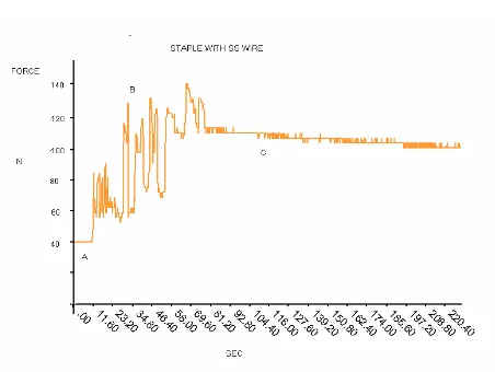
To study the biomechanical properties of the ACL, and it’s substitutes, it is essential to study their response when loaded. The single load-to-failure test is designed to determine the structural properties of a graft fixation construct during a single overload mimicking a traumatic incidence. The response of the specimen to loading is obtained in the form of force-displacement curve (Fig 1). After an initial period of low stiffness, (a small increase in load producing large elongation), further loading produces a nearly linear curve. Since the
stiffness of any loaded construct is calculated as the ratio of force
FIG-1 FORCE –DISPLACEMENT GRAPH
FIG 1 FORCE –DISPLACEMENT GRAPH
The yield / linear load is defined as the force at which the slope of the
force-displacement curve first clearly decreases. The first significant slippage of the ACL graft typically occurs at the yield load point, it thus represents the beginning of abnormal laxity. Beyond the yield point, the force-displacement curve is usually non-linear. The other parameter assessed is the ultimate failure load or the pull out strength – the load at which the graft is pulled off
the tibia. (28)
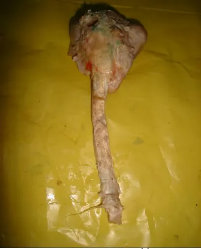
[image:27.612.98.397.97.439.2]FIG –2 TIBIAL SPECIMEN WITH BONE PATELLAR TENDON BONE (BPTB)
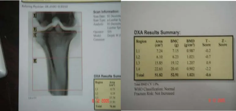
FIG –3 BONE MINERAL DENSITY (BMD) OF TIBIAL SPECIMEN
[image:29.612.92.478.505.687.2]DEXA scan showing BMD at screw insertion site>0.8/cu.cm
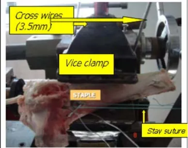
FIG –4 TIBIAL FIXATION
Tibial specimen held by vice grip & cross pins
FIG –4 TIBIAL FIXATION
Tibial specimen held by vice grip & cross pins
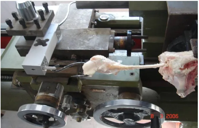
FIG –5 BPTB GRAFT
7mm hole made in patella –to serve as fixation to the load cell
[image:30.612.171.375.499.752.2]A total of 15 fresh bovine knees were obtained from local slaughter house. The ligaments, soft tissues and menisci were dissected off the tibia, and the femur discarded. The patella and the patellar tendon were left attached to the tibia (fig-2). To assess the bone mineral density of the tibia, the tibial specimen was scanned using the DEXA as in fig-3. The purpose of the scan was to ensure that the screw insertion site had a trabecular bone density of greater than 0.8g/cu.cm. After the DEXA scan was performed, a 30mm x 10mm quadrilateral bone plug was harvested from the bovine tibia. The patella was left intact attached to the patellar tendon, which was made to 10mm width.
TIBIAL SPECIMEN PREPARATION:
A 10mm diameter bone tunnel was drilled from the tibial ACL insertion, directed antero-medially in an inside out fashion. The tibia was mounted on the testing apparatus and fixed using a modified vice grip and further stabilized by two 3.5mm cross pins that were passed through the vice grip and in to the tibial specimen (fig-4). During the testing process the specimen was kept moist with normal saline
PATELLAR TENDON GRAFT PREPARATION:
FIG –6 BPTB GRAFT WITH SS WIRE & polyester stay suture
20mmG SS wire placed in proximal hole
FIG –7 BPTB GRAFT WITH No.5 ETHIBOND& polyester stay suture No.5Ethibond placed in proximal hole
[image:32.612.191.363.482.719.2]BPTB graft attached to the load cell by a ‘S’ shaped hook
FIG-10 PRETENSIONING
[image:34.612.96.484.256.561.2]The proximal hole was one cm from the bone tendon junction and the second hole was 1cm from the first. The proximal hole was used for fixation of the graft with 20 gauge SS wire (fig-6) and 5’ETHIBOND suture (fig-7) as explained below. The distal hole was used to pass a stay suture (No.5 polyester) that served to pretension the graft at 40N (explained subsequently).
TESTING APPARATUS
:
FIG –11 Method of fixation - SS WIRE TO STAPLE
After knotting the SS wire on the staple, it was tightened using nose pliers
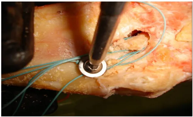
FIG-12 Method of fixation – No.5ETHIBOND WITH SCREW FIXATION POST
[image:36.612.133.451.493.687.2]MEASUREMENT OF INITIAL AND RESIDUAL LOAD
:
The tibial bone plug was fixed by means of three different fixation techniques i.e. 9x30mm Interference screw, No.5’Ethibond tied onto a screw fixation post (6.5mm cancellous screw with washer) and 20 gauge SS wire fixed to a 20x20mm Staple.
A. SS Wire to Staple: The 20G SS wire placed in the bone plug was tied to
the 20 x 20mm staple which was inserted distal to the bone tunnel outlet. After initially tying the SS wire on to the staple it was tightened using nose pliers (fig-11).
B. Polyester suture (No.5’Ethibond) to screw fixation post: The
No.5’Ethibond suture placed in the bone plug was tied to a screw fixation post (55 x 6.5 mm cancellous screw with a 1.5mm washer). The screw was inserted distal to the bone tunnel outlet at 30 degree to the bone surface and tightened after the No.5’Ethibond was tied on to the screw (fig-12).
C. Interference screw fixation: 9 X 30 mm (Smith and Nephew) Titanium
FIG –14 SINGLE LOAD –TO-FAILURE TEST
Followed the fixation, the stay suture was cut.
The forces generated in the graft were recorded throughout the fixation period and for 5min thereafter. The INITIAL LOAD was 40N for all three techniques
i.e. the tension in the graft prior to fixation that was applied through the stay suture. The final RESIDUAL LOAD was the load that was developed in the
tendon at the end of fixation of the graft. The data recorded was used to compare the set initial tension and the residual tension in the implanted graft.
EVALUATION OF PULLOUT STRENGTH, STIFFNESS AND
MODE OF FAILURE
-
In the next part of the study, the graft, after fixation, was again preloaded to 40N. The tendon graft was stretched at the rate of 1.6mm/sec (the mobile platform on which the load cell was attached was moved at the rate of 1.6mm/sec by an electrical motor) (fig-14). The tendon was stretched till failure of the fixation. The ULTIMATE FAILURE LOAD (The PULLOUT STRENGTH)
- i.e. the maximum tension generated in the graft at the time of failure of
fixation- was recorded on the oscilloscope and the MODE OF FAILURE of the
graft was noted. The STIFFNESS of the graft was assessed by calculating the
slope of the linear portion of the force-displacement curve generated during the biomechanical testing.
RESULTS
INITIAL LOAD AND RESIDUAL LOAD:
The Initial graft load that was maintained by the stay suture was 40N for all three fixation techniques. The tension developed in the graft during fixation was recorded by the load cell. The Residual load at the end of the fixation was recorded after the stay suture was cut.
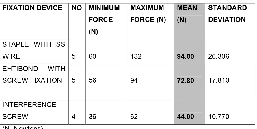
[image:41.612.104.549.396.620.2]The Residual load developed in the graft with three different fixation techniques is shown in the
TABLE- 2 : RESIDUAL LOAD MEASUERMENT
(WITH INITIAL LOAD OF 40N)
FIXATION DEVICE NO MINIMUM FORCE (N) MAXIMUM FORCE (N) MEAN (N) STANDARD DEVIATION
STAPLE WITH SS
WIRE 5 60 132 94.00 26.306
EHTIBOND WITH
SCREW FIXATION 5 56 94 72.80 17.810
INTERFERENCE
SCREW 4 36 62 44.00 10.770
(N- Newtons) Note:
Samples of graphs recorded during and after the fixation of the graft with different techniques are depicted below:
Fig -15
Tendon was stretched at a rate of 1.6mm/second
Description of the graph:
Tendon was stretched at a rate of 1.6mm/second
Fig-16
Tendon was stretched at a rate of 1.6mm/second
Fig-17
Description of the graph:
The Initial load was 40N. While inserting the Interference Screw, the measured load decreased initially, as the tibial bone block was displaced proximally. Subsequently, as the Interference screw engaged the bone block, the tibial bone block was pulled distally and the graft load increased. At the completion of the fixation, the residual load applied to the graft was slightly higher than the initial set force with an average of 45 N.
When Wilcoxon signed ranks test was carried out, a statistically significant difference (p < 0.05) was observed between the initial load and the residual load when the SS wire with Staples and No5 Ethibond suture with screw fixation post was used as the fixation method. There was no statistically significant difference between the initial load and residual load values for the Interference screw fixation method.
Table-3
Statistical Analysis of Initial load and Residual load using three different fixation techniques (Wilcoxon signed ranks test)
Method of fixation
p value ( N Par test)
(between Initial Load & Residual load )
STAPLE WITH SS WIRE
P= 0.043
ETHIBOND WITH POST SCREW
FIXATION
P= 0.040
INTERFERENCE SCREW
EVALUATION OF ULTIMATE LOAD FAILURE ( PULL OUT
STRENGTH)
In the second part of the study, we analyzed the pullout strength and stiffness of the bone patellar tendon bone graft. Load displacement curves were obtained with three different fixation methods.
The Examples of force-displacement graphs with the three techniques are shown below:
X Axis- Time (Displacement was at the rate of 1.6 mm/s)
[image:46.612.119.502.468.673.2]Y Axis- Force Generated
Fig-20
Table-4: PULLOUT STRENGTH MEASUREMENT WITH
THREE FIXATION TECHNIQUES
FIXATION DEVICE NO. MINIMUM FORCE (N) MAXIMUM FORCE (N) MEAN (N) STANDARD DEVIATION
STAPLE WITH SS WIRE
5 640 776 726.40 60.20
ETHIBOND WITH POSTSCREW FIXATION
5 608 776 733.20 72.70
INTERFERENCE SCREW
4 384 752 594.00 173
(N-Newtons)
There was no significant difference between the ultimate failure loads using the three different fixation techniques
Table-5
Statistical Analysis of Pullout Strength of Three Different Fixation Techniques:
(Multiple Comparisons using the Boneferroni Post hoc test was used to analysis statistical significant difference.)
95 % Confidence Interval Groups compared Statistical Significant p value Lower Boundary Upper Boundary
1 2 3
1.000
.277
-197.77 - 70.15
184.17 334.95 2 1
3
1.000 .236
-184.17 - 63.35
197.77 341.75 3 1
2 .277 .236 -334.95 -341.75 70.15 63.35
1- SS wire with staple
EVALUATION OF
STIFFNESS
USING THE THREETECHNIQUES:-The Stiffness was assessed by calculating the slope of the linear portion of the force- displacement curve.
For example: Fig-21
displacement F
o r c e
Table 6 Shows the stiffness of the bone patellar tendon graph using three different fixation techniques.
TABLE-6:
STIFFNESS
WITH THREE DIFFERENT FIXATION
TECHNIQUES
FIXATION DEVICE NO MINIMUM FORCE (N) MAXIMUM FORCE (N) MEAN (N) STANDARD DEVIATION STAPLE WITH SS WIRE5 44 81 61.9 13.12631
ETHIBOND WITH SCREW
FIXATION POST
5 37 63.6 53.22 10.17802
INTERFERENCE SCREW
4 55.5 120 79.50 28.24299
(N-Newtons)
Table -7
Statistical Analysis of Stiffness using Three Different Fixation Techniques:
(Multiple Comparison using the Boneferroni Post hoc test was used to
analysis statistical significant difference.)
95 % Confidence Interval Groups compared Statistical Significant p value Lower Bound Upper Bound
1 2 3 1.000 .507 -23.1187 -51.3276 40.4787 16.1276 2 1
3 1.000 .151 -40.4787 -60.0076 23.1187 7.4476 3 1
2
.507 .151
-16.1276 - 7.4476
51.3276 60.0076
1- SS wire with staple
EVALUATION OF MODE OF FAILURE
Staple with SS wire
Four specimens failed when the SS wire cut through the tibial bone block.
Figure -22
In one specimen the SS wire untwisted as the graft was loaded to failure.
Ethibond with screw post
[image:54.612.107.339.261.548.2]Interference screw
Summary of Results: TABLE- 8
Biomechanical properties
Staple with SS wire (Mean ± SD)
screw fixation post with Ethibond (Mean ± SD)
Interference screw (Mean ± SD)
Residual Load (N) (with initial load of 40N)
94 ( ± 26.306) 72.80 (± 17.81) 44.00 (± 10.77)
Pullout strength N) 726.40(± 60.24) 733.20(± 72.768) 594.00 (± 173.605)
[image:56.612.101.561.114.363.2]Stiffness (N/mm) 61.90 (± 13.126) 53.22 (± 10.178) 79.50 (± 28.242)
Table- 9
Mode of Failure
Mode of failure Staple with SS wire
Ethibond with post screw fixation
Interference screw
Bone Block Pullout
0 0 4
Bone Cut Through 4 0 0
Breakage of Thread
NA 5 NA
SS wire untwisted 1 NA NA
DISCUSSION
The Knee joint has been described as a complex hinge joint (19). The ligaments and other supporting soft tissue structures (joint capsule, muscle, tendons and menisci) control the stability of the knee joint (20). The knee joint is between the long lever arms of the femur and the tibia, and consequently, is extremely vulnerable to injuries. The anterior cruciate ligament injury is the most common ligament injury around the knee joint. The ACL, is the primary restraint preventing anterior displacement of the tibia relative to femur and also serves as an important secondary restraint to varus - valgus rotation, as well as internal- external rotation (21).
Biological substitutes used in the intra-articular reconstruction for ruptured ACL are autografts and allografts. The most commonly used autografts are the bone patellar tendon bone (BPTB) graft, multiple strand hamstring tendon and Quadriceps tendon bone graft. The two commonly used allografts are bone patellar tendon graft and the Achilles tendon - bone graft (28).
The bone patellar tendon bone (BPTB) graft is considered by many as the gold standard for ACL reconstruction (5,28). The popularity of BPTB graft is based on its structural properties, quality of fixation excellent long-term clinical success and the fact that it provides bone-to-bone healing. The major concern with the use of BPTB graft has been the donor site morbidity, anterior knee pain, kneeling discomfort, loss of motion, and weakness of the quadriceps muscle (28,29).
The quadriceps tendon-bone graft and allograft have also been used for ACL reconstruction . They have been shown to have sufficient structural properties compared to BPTB and hamstring grafts. They are commonly used for revision ACL reconstructions and multiple ligament reconstructions (28).
Evolving methods of graft fixation have been paralleled by marked changes in the post operative rehabilitation program. In the past, prolonged non weight bearing was recommended to protect the graft. New techniques of reconstruction and fixation have changed the emphasis towards early weight bearing. Graft fixation site has been found to be the weakest link in the ACL reconstruction during the immediate postoperative period until biologic fixation occurs (10).
Many methods of graft fixation techniques have been described. Fixation devices have been classified as either direct or indirect (22). Indirect fixations rely on connecting materials, that are attached to the graft, whereas in direct fixation, the graft is fixed directly to the bone.
For an ideal graft fixation, there should be sufficient initial strength to avoid failure of fixation (i.e. high pullout strength or ultimate failure load of graft fixation), and sufficient stiffness to restore the stability of the knee to avoid gradual loosening in the early post operative period.
The tension of the graft is considered to be an important factor influencing the result of ACL reconstruction (26).Optimal graft tension should be determined so as to restore physiological kinematics of the knee joint. Studies have shown that a discrepancy exists between the initial load (i.e. the load applied
to the graft during the graft fixation) and the residual load (i.e. tension in the
graft after fixation). The mechanical behavior during and after fixation is specific to the fixation method employed in the procedure (17,18).
The residual load was found to be significantly higher when staple fixation with SS wire and screw fixation post with polyester suture were used to fix the graft. High loads result in difficulty in regaining motion or may lead to articular degeneration from altered joint knee kinematics. Yoshia et al showed in a canine model that over tensioning of the graft resulted in poor graft revascularization and myxoid degeneration within the graft. It would be ideal for the residual load to be as close as possible to the initial set force (initial load).In our study, this was seen to be true for the interference screw fixation. Yokio et al found that the residual load was slightly higher than the initial load when interference screws and screw fixation with ethibond were used – where as the residual load when a button was used for fixation was very much less than the initial load
In the next part of the study the Ultimate failure load, Stiffness and Mode of
failure were evaluated. The graft after fixation was stretched till failure of
The ultimate failure load (or pullout strength) was higher with the use of the staple with SS wire fixation and the screw fixation post with polyester suture - when compared with interference screw fixation. This suggests that the interference screw when used for distal fixation in the tibia does not provide sufficient initial strength to avoid failure of fixation. One specimen was not included in this study, as the screw was inadvertently introduced in an oblique manner - leading to a low ultimate failure load. Interference screw divergence - the angle of the interference screw with respect to the bone block, is a common clinical concern, and consequently, its effect on the strength of fixation of BPTB graft has been evaluated biomechanically in others studies. It has been shown that an increase in the divergence angle decreases fixation strength at angles greater than 20 degree. This study seems to suggest that cortical fixation using indirect techniques have a higher pull out strength (though not statistically significant.)
and the graft. Longitudinal and sagittal graft motions within the bone tunnel are also known as the bungee cord effect and windshield wiper effect, respectively. If the implant for tendon graft fixation is placed closed to the articular cavity, knee stability increases (28). In our study too, interference screw had higher stiffness values than the other fixation methods. However, it was not statistically significant.
CONCLUSION
The biomechanical properties of bone patellar tendon bone graft fixed with three different techniques in a bovine tibial model were evaluated in this study. The following conclusions were made:
1. The Initial load (force applied to the bone patellar tendon graft prior to fixation) was similar to the Residual load in the graft after fixation when Interference screw was used to the fix the graft. There was no statistically difference between the Initial load and the Residual load.
2. The Residual load developed in the bone patellar tendon graft after fixation with SS wire to staple and No.5 Ethibond with screw fixation post was higher than the Initial load that was applied to the graft prior to fixation. There was a significant difference in the Initial load and Residual loads with the use of SS wire with staple and No.5 Ethibond to post screw fixation.
3. The SS wire to staple and No.5 Ethibond with post screw fixation has higher ultimate failure load than the interference screw fixation. (726.40(± 60.24) and 733.20(± 72.76) (Mean ± SD) respectively.
6. There was however no statistical significance between the ultimate failure load using the three fixation techniques.
7. Stiffness of the bone patellar tendon graft was maximum when using the Interference screw, though there was no statistical significant difference when compared to the stiffness of bone patellar tendon graft using other fixation techniques.
8. The bone patellar tendon graft when fixed with SS wire to Staple, failed most commonly by cut out of the SS wire.
9. The bone patellar tendon graft when fixed with No.5Ethibond with screw fixation post, failed by rupture of the thread.
10. The bone patellar tendon graft when fixed with Interference screw, failed by pull out of the bone block.
11. Overall, it appears that the biomechanical properties of the SS wire and staple fixation technique are not much significantly different from that of the Interference screw and it can be used effectively as a substitute fixation method in ACL reconstruction.
BIBILOGRAPHY
1. Kurosaka M, Yoshiya s, Andrish JT (1987) different surgical techniques of graft fixation in anterior crucate ligament reconstruction AMJ sports med 15: 225 – 229
2. Podesta L, Sharma MF, Bonamo JR, Reitea (1990) Rational and protocol for post operative anterior cruciate ligament rehabitation.
3. Paulos LE, Chert J, Rosenberg T P, Beck CL (1991) Anterior curicate ligament reconstruction with auto grafts. Clin sports Med 10: 469-485 4. Lambert KL, (1983) Vascularized patellar tendon graft with rigid internal
fixation for anterior cruciate ligament insufficiently, Clin Orthop 172:85-89 5. Weiler A, Hoffmann R, Stahlin A, et al. (1998) : Hamstring fixation using
interference screws Arthoroscopy ; 14: 32-37
6. Weiler A, Helling HJ, Kirch U, et al. (1996): foreign body reaction and the course of osteolysis after polyglycide implants for fracture fixation. J Bone Joint Surg Br ; 76: 36-376
7. Paschal SO, Seemann MD, Ashman RB, Alland RN, Montgomery JB, (1994) Interference screw fixation versus post fixation of bone-patellar tendon-bone grafts for anterior cruciate ligament reconstruction. A biomechanical comparative study in porcine knees. Clin orthop 300: 281-287
9. Frank CB, Jackson DW (1998) :The science of reconstruction of the anterior cruciate ligament J Bone joint surg BR 79: 1556 – 1576
10. Howell SM, Wallace MP, Hull MC, Deutsch MC ( 1999): Evaluation of the single – incision arthoscopic technique for ACC replacement. A study of tibial tunnel placement, intraoperative graft tension and stability AMJ sports med 27: 284-293
11. Melby A III, Noble JS, Askew MJ, Boom AA, Hurst FW (1991): The effects of graft loading on the laxity and kinematics of the ACC reconstructed knee arthroscopy 7:257-266
12. Nabors ED, Richmond JC, Vannah WM, Mc Conville OR (1995): Anterior Cruciate ligament graft tensioning in full extension AMJ Sport Med 23: 488-492
13. Yasuda K, Tsujino J, Kaneda K (1997): Effects of initial graft tension on clinical outcome after anterior cruciate ligament reconstruction: autogenous doubled hamstring tendons connected in series with polyester tapes. Am J Sports Med 25:99-105
14. Yoshiya S, Andrish Jt, Manley Mt, Bauer TW (1987): Graft tension in anterior cruciate ligament reconstruction: an in vivo study in dogs. Am J sports Med 15: 464-470
16. Beynnon BD, Johnson RJ, Fleming BC, Kannus P, Kaplan M, Samani J and Renstrom P (2000) : Anterior Cruciate ligament replacement: comparison of bone patellar tension-bone grafts with two-strand hamstring grafts. J Bone Joint Surg 84A: 1503-1513
17. Cunningham R, West JR, Greis PE, Burks RT (2000) :A survey of the tension applied to a doubled hamstring tendon graft reconstruction of the anterior cruciate ligament. Arthroscopy 18:983-988
18. Numazaki H, Tohyama H, Nakano H, Kikuchi S and Yasuda K (2002): The effect of initial graft tension in anterior cruciate ligaments reconstruction on the mechanical behaviors of the femur-graft-tibia complex during cyclic loading. Am J Sports Med 30: 800-805.
19. Woo-SL-Y, Debski RE, Withrow JD and Janushek MA (1999): Biomechanics of knee ligaments, current concepts, Am J Sports Med 27:533-543
20. Swenson TM and Harner CD (1995): Knee ligament and meniscal injuries: Current concepts. Orthop Clin North AM 26:529-546
21. Dienst M, Burks RT and Greis PE (2002): Anatomy and biomechanics of the anterior cruciate ligament. Orthop Clin N Am 33:605-620
22. Giragis FG, Marshall JL and Al Monajem ARS (1975): The cruciate ligaments of the knee joint. Anatomical, functional and experimental analysis. Clin Orthop 106: 216-231.
24. Howell SM and Clark JA (1992): Tibial tunnel placement in anterior cruicate ligament reconstructions and graft impingement. Clin Orthop 283: 187-195
25. Jackson DW and Gasser SI ( 1994) :Tibial tunnel placement in ACL reconstruction. Arthroscopy 10 : 124-131.
26. Aglietti P, Buzzi R, giron F, Simeone AJ, Zaccherotti G (1997): Arthroscopic-assisted anterior cruciate ligament reconstruction with the central third patellar tendon. A 5-8 year follow – up. Knee Surg Sports Traumaton Arthrosc 5: 138-144
27. Hoher J, Kanamori A, Zeminski J, Fu FH and Woo SL (2001): The position of the tibia during graft fixation affects knee kinematics and graft forces for anterior cruciate ligament reconstruction. Am J Sports Med 29: 771-776.
28. Fu FH, Bennet CH, Lattermann C and Ma CB (1999): Current Trends in anterior cruciate ligament reconstruction. Part I: Biology and biomechanics of reconstruction. Current concepts. Am J sports Med 27:821-830
29. Miller LS and Gladstone JN (2002): Graft selection in anterior cruciate ligament reconstruction. Orthop Clin N Am33: 675-683
31. Stein DA, Hunt Sa, Rose JE and Sherman OH (2002): The incidence and outcome of patella fractures after anterior cruciate ligament reconstruction. Arthoscopy 18: 578-583
32. Allen CR, Giffin JR and Harner CD (2003): Revision anterior cruciate ligament reconstruction. Orthop Clin N Am 34: 79-98
33. Hamner DL, Brown CH, Jr, Steiner ME, Hecker AT and Hayes WC (1999): Hamstring tendon grafts for reconstruction of the anterior cruciate ligament: Biomechanical evaluation of the use of multiple strands and tensioning techniques. J Bone Joint Surg 81A: 549-557
34. Ferretti A, Conteduca F, Morelli F, Monteleone L (2003): Biomechanics of ACL reconstruction using twisted doubled hamstring tendons. Int Orthop27:22-25
35. Wilson TW, Zafuta MP and Zobitz (1999): A biomechanical analysis of matched bone patellar tendon bone and double looped semitendinosus and gracilis tendon grafts. AM J Sports med 27:202-207
36. Schatzmann L, Brunenr P, and Staubli HU ( 1998): Effect of cyclic preconditioning on the tensile properties of human quadriceps tendons and patellar ligaments. Knee Surg Sports Traumatol Arthrosc 6. (Suppl 1) 56-61.
38. Weiler A, Peine R, Pashmineh- Azar A, Abel C, Sudukamp NP and Hoffman RFG, (2002b) Tendon healing in a bone tunnel. Part I: Biomechanical Results after biodegradable interference fit fixation in a sheep model of anterior cruciate ligament reconstruction in sheep. Arthroscopy 18: 113-123.
39. Noyes FR, Butler DL, Grood ES, Zernick RF Hefzy MS (1984): Biomechanical analysis of human ligment grafts used ion knee-ligament repairs and reconstructions. J Bone joint surg 66A: 334-352
40. Toutoungi DE, Lu TW, Leardini A, Ctani F and O’Connor JJ (2000): Cruciate ligament forces in the human knee during rehabilitation exercises. Clin Biomech 15: 176-187.
41. Morgan CD, Kalman VR and Grawl DM (1995): Isometry testing for anterior cruciate ligament reconstruction revisited. Arthroscopy 11: 647-659.
42. L’Insalata JC, Klatt B, Fu FH and Harner CD (1997): Tunnel expansion following anterior cruciate ligament reconstruction : a comparison of hamstring and patellar tendon autografts. Knee Surg Sports Traumatol Arthrosc 5 : 234-238
44. Brown CH and JH Sklar (1998): Endoscopic anterior cruciate ligament reconstruction using quadrupled hamstring tendons and endobutton femoral fixation. Techniques in Orthopedics 13: 281-298.
45. Weiler A, Hoffman RFG, Stahelin AC, Bail HJ, Siepe CJ and Sudkamp NP (1998a): Hamstring tendon fixation using interference screws: a biomechanical study in calf tibial bone. Arhtroscopy 14: 29-37.
46. Jomha NM, Raso VJ and Leung P (1993): Effect of varying angles on the pullout strength of interference screw fixation. Arthroscopy 9: 580-583. 47. Pierz K, Baltz M and Fulkerson J (1995). The effects of Kurosaka screw
divergence on the holding strength of bone-tendon grafts. Am J Sport Med 23 : 332-335.
48. Butler JC, Branch TP and Hutton WC (1994): Optimal graft fixation – the effect of gap size and screw size on bone plug fixation in ACL reconstruction. Arthroscopy 524-529
49. Shaffer B, Gow W and Tibone JE(1993): Graft-tunnel mismatch in endoscopic anterior cruciate ligament reconstruction. A new technique of intraarticular measurement and modified graft harvesting. Arthroscopy 9: 633-646
51. Magen HE, Howell SM, and Hull ML (1999): Structural properties of six tibial fixation methods for anterior cruciate ligament soft tissue grafts. Am J Sports Med 27: 35-43.
52. Yamanaka M, Yasuda K, Tohyama H, Nakona H and Wada T (1999): The effect of cyclic displacement on the biomechanical characteristics of anterior cruciate ligament reconstructions. Am J Sports Med 27 : 772-777.
53. Martin SD, Martin TL and Brown CH (2002): Anterior cruciate ligament graft fixation Orthop clin N Am 33: 685-696.
54. Weiler A, Hoffman RFG, Stahelin AC, Bail HJ, Siepe CJ and Sudkamp NP (1998a): Hamstring tendon fixation using interference screws: a biomechanical study in calf tibial bone. Arhtroscopy 14: 29-37.
APPENDIX-1
The following are the individual specimen’s biomechanical properties using the three different fixation techniques.
Staple fixation with Stainless Steel wire (Group -1)
Specimen
No.
Initial
load (N)
Residual
load (N)
Pullout
strength (N)
stiffness
(N)
Mode of
failure
1
40
60
640
83.3
SS wire
untwisted
2
40
84
752
66
Bone CutThrough
3
40
92
776
83
Bone CutThrough
4
40
102
776
108
Bone Cut Through5
40
132
688
75
Bone CutThrough
Staple fixation with Stainless wire
Screw fixation post with Polyester suture (Group-2)
Specimen
No.
Initial load (N) Residual
load (N)
Pullout
strength
(N)
stiffness
(N)
Mode of
failure
1
40
56
776
83.3
Breakage of Thread2
40
64
730
75
Breakage ofThread
3
40
60
776
100
Breakage ofThread
4
40
90
608
50
Breakage ofThread
5
40
94
776
100
Breakage ofThread
Screw fixation post with Polyester suture
Residual load (Mean±Sd)
72.80 (± 17.81)N Pullout strength (Mean±Sd
)
733.20(± 72.768)NInterference screw fixation (Group 3)
Specimen No. Initial
load (N)
Residual
load (N)
Pullout
strength
(N)
stiffness
(N)
Mode of
failure
1
40
36
752
60
Bone Block Pullout2
40
38
720
103
Bone Block Pullout3
40
46
384
83
Bone Block Pullout4
40
62
520
120
Bone Block PulloutInterference screw fixation
APPENDIX-
װ
ABSTRACT
Assessment of the biomechanical properties of anterior cruciate
ligament reconstruction using different techniques of fixation in a bovine
knee model.
Aim: To evaluate the biomechanical properties of bone patellar tendon bone
graft ( BPTB) fixation with different techniques in a bovine model.
Introduction: Intra-articular reconstruction with a biologic tendon graft is the
procedure of choice for restoring stability of a knee after rupture of the anterior cruciate ligament (ACL). Rigid fixation of the bone block in the tunnel is crucial for initial strength of the graft. Fixation with the help of an Interference screw is considered as the gold standard. Indirect fixation using polyester suture tied to a screw fixation post, and SS (Stainless Steel) wire tied to staples placed inferior to the bone tunnel outlet are other alternative techniques. This study was performed to evaluate the primary biomechanical parameters of three different fixation techniques –
a) Staple fixation with SS wire
b) Polyester suture tied onto a screw fixation post
Methods: Fifteen fresh bovine knees and bovine patellar tendons were used
for the study. The BPTB graft was fixed to the tibia using the three different fixation techniques mentioned above. The patella was fixed to a load cell, and forces generated in the graft were recorded.
In the first part of the study, the BPTB was pretensioned to an initial tension (or initial load) of 40N. The graft was then fixed using the three different methods, and the changes in tension (load) in the graft during fixation were recorded. The residual load in the graft after fixation was recorded and compared to the initial load (i.e. 40N in all instances).
In the second part of this study, the graft was subjected to a single load-to-failure test, and the following parameters were recorded - ultimate failure load (thepullout strength), stiffness, and mode of failure.
Result: Though the BPTB graft was fixed with an initial load of 40N, the graft tension at completion of fixation (residual load) with staple and SS wire, screw post with polyester suture and Interference screw was 94.00N, 72.80N, and 44.00 N respectively.
In the single load-to-failure biomechanical testing, the ultimate failure load (pullout strength) and stiffness for Staple with SS wire was 726.40N and 61.9N respectively, for the Screw fixation post and polyester suture - 733.20N and 53.22N, and for Interference screw -594.00N and 79.50 N. There
In 4 specimens, the Staple with SS wire fixation failed when the SS wire cutting through the bone. In the fifth specimen, the SS wire untwisted during loading. With the screw fixation post, the polyester suture broke in all 5 specimens. With the interference screw, all failed by bone block pull out.


![Multiple Introductions and Antigenic Mismatch with Vaccines May Contribute to Increased Predominance of G12P[8] Rotaviruses in the United States](data:image/gif;base64,R0lGODlhAQABAIAAAP///wAAACH5BAEAAAAALAAAAAABAAEAAAICRAEAOw==)