A STUDY ON SUBCLINICAL HYPOTHYROIDISM IN
FEMALES OVER FIFTY YEARS OF AGE
submitted to
The Tamil Nadu Dr.M.G.R.Medical University
M.D. DEGREE EXAMINATION
BRANCH – I (GENERAL MEDICINE)
THE TAMIL NADU DR.M.G.R. MEDICAL UNIVERSITY
CHENNAI
BONAFIDE CERTIFICATE
This is to certify that the dissertation titled “A STUDY ON SUBCLINICAL HYPOTHYROIDISM IN FEMALES OVER FIFTY YEARS OF AGE”
is a bonafide work done by Dr.K.S.GOPAKUMAR, post graduate student, Department of General Medicine, Kilpauk Medical College, Chennai-10, under my guidance and supervision in partial fulfillment of regulations of The Tamilnadu Dr.M.G.R.Medical University for the award of
M.D.Degree Branch I (General Medicine) during the academic period from May 2006 to March 2009.
Dr.M.Dhanapal, M.D., D.M.,
Director Of Medical Education- OSD and The Dean Kilpauk Medical College,
Chennai – 10.
Prof.G.Rajendran, M.D., Professor and Head
Department of Internal Medicine
Kilpauk Medical College Chennai-10
Prof.B.Chellam, M.D., Professor
Department of Internal Medicine
ACKNOWLEDGEMENT
I sincerely thank Prof.D.Dhanapal, M.D., D.M., Dean, Kilpauk Medical College, Chennai for permitting me to utilize the facilities needed for this dissertation work.
I am extremely grateful to Prof.Dr.G.Rajendran, M.D., Professor and Head of the Department of Internal Medicine, Kilpauk Medical College and Hospital for permitting me to carry out this study and for his constant encouragement and guidance.
I whole heartedly express my sincere thanks to my Chief
Prof.B.Chellam, M.D Department of Internal Medicine, Kilpauk Medical College for his esteemed guidance and valuable suggestions in all the stages of this dissertation.
I also express my sincere gratitude to Prof.M.D.Selvam, M.D., Prof.A.Joseph Navaseelan,M.D., Prof.Chinnayan M.D.,
I wish to thank Dr. S. Rajasekhar MD, Dr.Jayakumar Jayakrishnan MD., Assistant Professors, Department of Medicine, Kilpauk Medical College
for their valuable suggestions and help rendered throughout this work.
I also thank my parents, colleagues, friends and staff of our hospital, for their support of this work.
CONTENTS
Sl.No. Title Page No.
1. INTRODUCTION 1
2. AIM OF THE STUDY 4
3. REVIEW OF LITERATURE 5 4. MATERIALS AND METHODS 50
5. RESULTS 53
6. DISCUSSION 57
7. CONCLUSION 60
BARS AND CHART PROFOMA
INTRODUCTION
The term subclinical hypothyroidism was originally used to describe the
patient with a low-normal free T4 but a slightly elevated serum TSH level.
Other terms for this condition are mild hypothyroidism early thyroid failure,
preclinical hypothyroidism, and decreased thyroid reserve. The TSH elevation
in such patients is modest, with values typically between 4 and 15 mU/L,
although patients with a TSH above 10 mU/L more often have a reduced free
T4 and may have some hypothyroid symptoms.
The definition of this syndrome depends significantly on the reference range
for a normal TSH concentration.
This syndrome is most often seen in patients with early Hashimoto's disease
and is a common phenomenon, occurring in 7% to 10% of older women.1,2,3
Subclinical hypothyroidism may have endogenous causes (chronic
autoimmune thyroiditis, subacute thyroiditis, postpartum thyroiditis) or
exogenous causes (thyroidectomy, 131I therapy, antithyroid drugs, inadequate
thyroid hormone replacement therapy).The prevalence of subclinical
hypothyroidism is rather high. In the classical population-based study among
adults in the English county of Whickham the prevalence was 75 per 1000
women and 28 per 1000 men1; similar figures have been obtained in other
in males and in older than in younger subjects is in agreement with the higher
prevalence of thyroglobulin and thyroid peroxidase (microsomal) antibodies
in women and in elderly people.
The natural history of subclinical hypothyroidism is reasonably well known.
Spontaneous return of increased TSH values into the normal range occurs in
5.5% after 1. year. 4 Progression to overt hypothyroidism ranges from 7.8% to
17.8% in various studies. 4,5,6
Another report indicates that approximately 30% of patients with subclinical
hypothyroidism had developed overt hypothyroidism after 10 years; the
higher the initial TSH, the greater the risk . 7
Clinical manifestation of subclinical hypothyroidism include abnormal lipid
metabolism 8,9,10,cardiac dysfunction11,12 and neurologic and mental
dysfunction13 and several cross-sectional studies have suggested that it confers
an elevated risk for atherosclerosis and coronary heart disease.14,15However,
neither of these associations have been confirmed by others16,17.This
discrepancy may reflect small size of the study or participation in these study
limited to one sex. Also, few longitudinal studies have been conducted. The
relationship between subclincial hypothyroidism and cardiovascular disease is
therefore controversial and possible outcomes of the conditions remain
autoimmunity is a risk factor for coronary heart disease15,18,19 remain
surrounded by controversies.17,20
Women with subclincal hypothyroidism didn’t differ from controls with
regard to BMI, Hypertension and Diabetes Mellitus in previous studies. 14,21
This study has been performed to estimate the prevalence of subclinical
hypothyroidism and its relation to Hypertension, Diabetes and Ischemic heart
disease among women above the age of 50 years attending medical out
patient clinic at Government Kilpauk Medical College and Hospital.
AIM OF THE STUDY
• To estimate the prevalence of subclinical hypothyroidism among
women above the age of 50 yrs
• To study the relationship of subclinical hypothyroidism to
REVIEW OF LITERATURE
PHYLOGENY, EMBRYOLOGY, AND ONTOGENY
Phylogeny
Thyroid tissue is confined to, and is present in, all vertebrates. The
phylogenetic association of the thyroid gland and the gastrointestinal tract is
evident in several functions. The salivary and gastric glands, like the thyroid,
are capable of concentrating iodide in their secretions.
Structural Embryology
The human thyroid anlage is first recognizable at E 16-17. The primordium
begins as a thickening of epithelium in the pharyngeal floor. The primitive
stalk connecting the primordium with the pharyngeal floor elongates into the
thyroglossal duct , the primordium assumes a bilobate shape, coming into
contact and fusing with the ventral aspect of the fourth pharyngeal pouch
when it reaches its final position at about E50. Cells of the lower portion of
the duct differentiate into thyroid tissue, forming the pyramidal lobe of the
gland. At this time the lobes contact the ultimobranchial glands, leading to the
incorporation of C cells into the thyroid. Concomitantly, histologic alterations
occur throughout the gland, and by 13 to 14 weeks the follicles begin to fill
Functional Ontogeny
Future follicular cells acquire the capacity to form thyroglobulin (Tg) as early
as the 29th day of gestation, whereas the capacities to concentrate iodide and
synthesize thyroxine (T4) are delayed until about the 11th week. As a
consequence of hypothalamic maturation and increasing secretion of
thyrotropin-releasing hormone (TRH), the serum TSH concentration increases
between 18 and 26 weeks.
ANATOMY AND HISTOLOGY
The thyroid is approximately 15 to 20 g in weight .The normal thyroid is
made up of two lobes joined by a thin band of tissue, the isthmus. The
individual lobes normally have a pointed superior pole and a poorly defined
blunt inferior pole that merges medially with the isthmus.
Blood supply is by the superior thyroid artery and the inferior thyroid artery.
Thyroid blood flow range from 4 to 6 mL/min/g.
The gland is composed of follicles .Thyroid tissue appears as closely packed
ring-shaped structures consisting of a single layer of thyroid cells surrounding
inactive.The thyroid also contains parafollicular cells, or C cells, that are the
source of calcitonin.
IODINE AND THE SYNTHESIS AND SECRETION OF THYROID
HORMONES
Dietary Iodine
Formation of normal quantities of thyroid hormone requires the availability of
adequate quantities of exogenous iodine to allow thyroidal uptake of
approximately 60 to 75 µg daily, taking into account the fecal losses of about
10 to 20 µg iodine of iodothyronines as glucuronides and about 100 to 150 µg
as urinary iodine in iodine-sufficient populations. At least 100 µg of iodine
per day is required to eliminate all signs of iodine deficiency . Iodine
deficiency is common in mountainous regions.
Iodide Metabolism by the Thyroid Cell
The process of iodide trapping is accomplished by a membrane protein, the
sodium-iodide symporter (NIS or SLC5A). In addition to the active transport
of iodide from the extracellular fluid, intracellular iodide is also generated by
the action of the iodotyrosine dehalogenase (Dhal) enzymes. The iodide
thereby released is immediately reconjugated to newly synthesized
Iodide Oxidation and Organification
Oxidation of iodide and incorporation of the resulting intermediate into the
hormonally inactive iodotyrosines MIT and DIT is known as organification.
Oxidation of thyroidal iodide is mediated by the heme-containing protein
thyroid peroxidase (TPO) and requires the H2O2 generated by the calcium
dependent Duox1 and 2 enzymes. The rate of organic iodinations is
dependent on the degree of thyroid stimulation by TSH .
Iodothyronine Synthesis
The MIT(mono iodothyronine) and DIT(di iodothyronine) formed via
oxidation and organic binding of iodide are precursors of the hormonally
active iodothyronines T4 and T3. Synthesis of T4 from DIT requires the
TPO-catalyzed fusion of two DIT molecules to yield a structure with two
diiodinated rings linked by an ether bridge (the coupling reaction). Efficient
synthesis of T4 and T3 in the thyroid requires Tg.
Storage and Release of Thyroid Hormone
There are approximately 250 µg T4 per gram of wet weight in normal human
thyroid. This is sufficient to maintain a euthyroid state for at least 50 days.
Thyroglobulin is present in the plasma of normal individuals at concentrations
up to 80 ng/mL. The first step in thyroid hormone release is the endocytosis
pseudopods formed at the apical membrane and micropinocytosis by small
coated vesicles that form at the apical surface. Both processes are stimulated
by TSH. Following endocytosis, endocytotic vesicles fuse with lysosomes,
and proteolysis is catalyzed by cathepsin D and D-like thiol proteases, all of
which are active at the acidic pH of the lysosome.The iodotyrosines released
from Tg are rapidly deiodinated by an NADPH-dependent iodotyrosine
deiodinase, and the released iodine is recycled. The thyroid hormone
transporter MCT8 in the thyroid gland could be involved in the exit of T4
and/or T3 from the phagolysosome or thyroid cell. Stimulation of D1- and
D2- (type 1 and 2 deiodinases ) can catalyze 5′-deiodination of T4. T4 release
from the thyroid cells is inhibited by several agents, the most important of
which is iodide.
Role and Mechanism of Thyrotropin (TSH) Effects
All steps in the formation and release of thyroid hormones are stimulated by
TSH secreted by the pituitary thyrotrophs . Thyroid cells express the TSH
receptor (TSHR), a member of the glycoprotein G protein–coupled receptor
family. Although the TSHR mainly couples to Gs, when activated by high
concentrations of TSH, it couples also to Gq/11, activating the
inositol-phosphate diacylglycerol cascade. The induction of signal via the
efflux, H2O2 production, and Tg iodination while the signal via the protein
kinase A (PKA) pathways mediated by cAMP regulates iodine uptake and
transcription of Tg, TPO, and the sodium-iodide symporter (NIS) mRNAs
leading to thyroid hormone production . The TSHR, in addition to TSH, also
binds thyroid-stimulating antibody (TSAb), thyroid-blocking antibodies
(TBAb), and neutral antibodies to the TSHR .
THYROID HORMONES IN PERIPHERAL TISSUES
Plasma Transport
T4 arises solely from direct secretion by the thyroid gland. In normal humans,
T3 is also released from the thyroid but approximately 80% is derived from
the peripheral tissues by the enzymatic removal of a single 5′ iodine atom
from T4. The remaining iodothyronines and their derivatives are generated in
the peripheral tissues from T4 and T3. Principal among them are 3,3′,5′
-triiodothyronine (reverse T3, or rT3) and 3,3′-diiodo-l-thyronine (3,3′-T2).
The major iodothyronines are poorly soluble in water and thus bind reversibly
to plasma proteins. The plasma proteins with which T4 is mainly associated
are thyroxine-binding globulin (TBG) and transthyretin (TTR) and albumin .
About 75% to 80% of T3 is bound by TBG, and the remainder by TTR and
Thyroxine-Binding Globulin (TBG).
TBG is a glycoprotein SERPIN family of serine antiproteases of with a
molecular mass of about 54 kd. Normal human serum concentration is 270
nmol/L (1.5 µg/dL). The half-life of the protein in plasma is about 5 days.
Free Thyroid Hormones
Because most of the circulating T4 and T3 is bound to TBG, its concentration
and degree of saturation are the major determinants of the free fraction of T4.
Binding of the thyroid hormones to the plasma proteins alters their
metabolism. The negligible urinary excretion of T3 and T4 is due to the
limited filterability of the hormone-binding protein complexes at the
glomerulus. The free fraction of T4 (T4/T4 · TBG) is inversely proportional
to the concentration of unoccupied TBG binding sites.
Estimates of the free T4 concentration in serum can be generated by direct or
indirect assays. It is the free hormone that is available to the tissues for
intracellular transport and feedback regulation, that induces its metabolic
effects, and that undergoes deiodination or degradation. The bound hormone
acts merely as a reservoir. If an increase in TBG occurs, the free T4
if the bound hormone increases. The plasma concentration of T4 is
determined by its rate of entry into, and exit from, the plasma.
For any level of T4 production, be it increased, normal, or decreased, the total
plasma T4 level varies inversely with its MCR (Metabolic Clearance Rate).
Intracellular free T3 and T4 are in equilibrium with the free hormone pool in
plasma. In the steady-state, the rate of T3 and T4 metabolism is rate-limiting
in the exit of hormones from the plasma.
T4 and T3 Transport across Cell Membranes and Intracellular T3 Binding
MCT8 is a T-type amino acid transporter belonging to the monocarboxylate
transporter family and facilitates transport of T3, T4, rT3 across cell
membranes in vitro.
Iodothyronine Deiodination
The most important pathway for T4 metabolism is its outer ring (5′)
monodeiodination to the active thyroid hormone, T3. This reaction is
catalyzed by the type 1 and 2 deiodinases (D1 and D2) and is the source of
more than 80% of the circulating T3. Inner ring deiodination, an inactivating
step, is catalyzed primarily by the type 3 (D3) deiodinase, which inactivates
T3 and prevents activation of T4 by converting it to reverse T3. The
selenocysteine in the active catalytic center . Selenium is thought to be the
iodine acceptor during deiodination reactions.
Quantitative and Qualitative Aspects of Thyroid Hormone Metabolism
Thyroid Hormone Turnover
In the normal adult, T4 has a distribution volume of approximately 10 L .
Because the concentration of total T4 in plasma is approximately 100nmol/L
(8µg/dL), the extrathyroidal T4 pool is approximately 1mmol (800µg). In the
adult, the fractional rate of turnover of T4 in the periphery is about 10% per
day (halflife, 6.7 days). Thus, about 1.1 L of the peripheral T4 distribution
space is cleared of hormone daily, a volume containing approximately 110
nmol (85 µg) of T4.
The kinetics of T3 metabolism differ from those of T4, partly because of its
10- to 15-fold lower affinity for TBG. The volume of distribution of T3 in the
normal adult is about 40 L. At a mean normal serum T3 concentration of
1.8nmol/L (120ng/dL), the daily production of T3 is approximately 50 nmol
(33µg).The rapid metabolic clearance rate of the product of inner ring T4
deiodination, and the low concentration in plasma (0.25nmol/L, 15ng/dL)
of T3 and all of rT3 production can be accounted for by peripheral
deiodination of T4.
Mechanism of Thyroid Hormone Action
Thyroid hormone acts by binding to a specific nuclear thyroid hormone
receptor (TR), which, in turn, binds to DNA usually as a heterodimer with
retinoid X receptor at specific sequences (thyroid hormone response elements,
or TREs) dictated by the DNA binding-site preferences of the RXR-TR
complex .Triiodothyronine has a 15-fold higher binding affinity for TRs than
does T4, explaining its function as the active thyroid hormone. In humans,
there are two TR genes, α and β, found on different chromosomes (TRα,
chromosome 17; TRβ, chromosome 3). There are several alternatively spliced
gene products from each of these genes forming both active and inactive gene
products. The active proteins are TRα1, and TRs β1, β2, and β3.
REGULATION OF THYROID FUNCTION
The Hypothalamic-Pituitary-Thyroid Axis
The thyroid participates with the hypothalamus and pituitary in a classical
feedback control loop . In addition, there is an inverse relationship between
the glandular organic iodine level and the rate of hormone formation.
Autoregulatory mechanisms within the gland, in turn, tend to maintain a
mechanism senses variations in the availability of free thyroid hormones,
however small, and acts to correct them. There is a close relationship between
the hypothalamus, the anterior pituitary, the thyroid gland, and still higher
centers in the brain, the function of the entire complex being modified in a
typical negative-feedback manner by the availability of the thyroid hormones.
Thyrotropin-Releasing Hormone (TRH) Synthesis and Secretion
TRH, a modified tripeptide (pyroglutamyl-histidyl-proline-amide), is derived
from a large prepro-TRH molecule. The parvocellular region of the
paraventricular nuclei (PVN) of the hypothalamus is the source of the TRH
that regulates TSH secretion. TRH travels in the axons of the peptidergic
neurons through the median eminence and is released close to the
hypothalamic-pituitary portal plexus. T3 suppresses the levels of prepro-TRH
mRNA in the hypothalamus, but normal feedback regulation of prepro-TRH
mRNA synthesis by thyroid hormone requires a combination of T3 and T4 in
the circulation. In addition to inhibiting the synthesis of prepro-TRH mRNA,
thyroid hormone also blocks the capacity of TRH to stimulate TSH release
from the thyrotroph.
Thyrotropin (Thyroid-Stimulating Hormone) Synthesis and Secretion
It is a glycoprotein secreted by the thyrotrophs in the anteromedial portion of
acids) and a specific β-subunit synthesized in thyrotrophs, which is a
112-amino-acid protein. The quantity of β-subunit is rate-limiting for TSH
secretion. TRH increases and thyroid hormone suppresses the transcription of
both subunits.
In normal serum, TSH is present at concentrations between 0.4 and 4.2 mU/L.
The level is increased in primary hypothyroidism and reduced in
thyrotoxicosis. The plasma TSH half-life is about 30 minutes, and production
rates in humans are 40 to 150 mU/day. Circulating TSH displays both
pulsatile and circadian variations. Both T4 and T3 mediate the feedback
regulation of TSH secretion, and TRH determines its set-point. There is a
linear inverse relationship between the serum free T4 concentration and the
log of the TSH, making the serum TSH concentration an exquisitely sensitive
indicator of the thyroid state of patients with an intact hypothalamic-pituitary
axis.
Somatostatin decreases TSH secretion in vitro and in vivo, but prolonged
treatment with a somatostatin analogue does not cause hypothyroidism.
Dopamine is a regulator of TSH secretion, but chronic administration of
dopamine agonists do not cause central hypothyroidism, indicating that
compensatory mechanisms negate these acute effects. Glucocorticoids in
not associated with central hypothyroidism. Bexarotene, a retinoid X receptor
agonist used for treatment of T-cell lymphoma, suppresses TSH sufficiently
to cause central hypothyroidism, presumably by reducing TSH-β gene
transcription.
Iodine Deficiency
In humans compensatory alterations in thyroid function come into operation
when total iodine intake falls below 75 µg/d.
Effects of Increased Iodine Intake on Thyroid Hormone Synthesis
The quantity of iodine organified in thyroglobulin which includes T4 and T3
displays a biphasic response to increasing doses of iodide, at first increasing
and then decreasing as a result of a relative blockade of organic binding. This
decreasing yield of organic iodine from increasing doses of iodide, termed the
Wolff-Chaikoff effect, results from a high concentration of inorganic iodide
within the thyroid cell. The mechanism for organification inhibition may
involve inhibitory effects of high iodide concentrations on TPO and THOX2.
Effects on Thyroid Hormone Release
An important practical effect of pharmacologic doses of iodine is the prompt
inhibition of thyroid hormone release. The mechanism is unknown, but the
effect is mediated at the thyroid cell level, rather than through an action on
Aging and the Thyroid
In the healthy elderly patient, there is a normal free T4 but a relatively lower
serum TSH than in younger individuals. The requirement for complete
levothyroxine replacement is reduced about 20% by the eighth decade.
LABORATORY ASSESSMENT OF THYROID STATUS
Tests of the Hypothalamic-Pituitary-Thyroid Axis
The rate of TSH secretion is exquisitely sensitive to the plasma concentrations
of free thyroid hormones. The normal range of the serum TSH concentration
by immunometric assay varies slightly in different laboratories but is most
commonly 0.4 to 4.2mU/L. The free α-subunit common to TSH, FSH, LH,
and hCG is generally detectable in serum with a normal range of 1 to 5µg/L,
but the TSH β-subunit is not.
TSH in Patients with Thyroid Dysfunction
Patients with hyperthyroidism (excess thyroid hormone secretion) and/or
thyrotoxicosis (excess thyroid hormone from any cause) will virtually always
have a subnormal TSH. Patients with hypothalamic or pituitary
hypothyroidism often have normal or even slightly elevated serum TSH. The
circulating TSH generally has reduced biologic activity due to abnormal
general, the degree of TSH elevation correlates with the clinical severity of
the hypothyroidism. Patients with serum TSH values in the range of 5 to 15
mU/L have few if any symptoms23, and the serum free T4 is typically
low-normal while serum free T3 concentrations are low-normal. 22 24 Such individuals
with modest TSH elevation are said to have subclinical hypothyroidism if the
serum free T4 is in the normal range. An elevation in both serum TSH and
free T4 is unusual and indicates either autonomous TSH production as with a
TSH-secreting pituitary tumor, resistance to thyroid hormone (RTH), or
hyperthyroidism with an artifactual elevation in TSH.
QUANTITATION OF SERUM THYROID HORMONE
CONCENTRATIONS
Total T4 and T3
The normal range for total T4 is 64 to 142 nmol/L (5 to 11 µg/dL). Normal
serum T3 concentrations are 1.1 to 2.9 nmol/L (70 to 190 ng/dL).
Concentrations of Free T4 and T3.
The absolute concentration of free hormone is the product of the total
hormone concentration and the fraction that is dialyzable or ultrafiltrable .
for free T4 and T3 are 9 to 30 pmol/L (0.7 to 2.5 ng/dL) and for free T3, 3 to
8 pmol/L (0.2 to 0.5 ng/dL).
There are two general categories of methods for estimating free thyroid
hormones: Comparative free T4 methods and so-called free T4 index
methods. Three general approaches are used: (1) two-step labeled hormone
methods, (2) one-step labeled analogue methods, and (3) labeled antibody
approaches .In general, two-step labeled hormone back titration methods are
less subject to artifacts due to abnormal binding proteins, changes in albumin,
TBG, or increases in free fatty acids than are one-step hormone analogue
methods.25 26 28 All general approaches are subject to artifacts from
endogenous antibodies to T4, abnormal binding proteins, or illness. 27 29
The Free T4Index (FT4I).
Particularly useful in estimating the free T4 in severely ill patients is the
determination of the thyroid hormone–binding ratio (THBR), multiplying this
result by the total T4 (or T3) to obtain a free hormone index (FT4I or FT3I).
Causes of a Suppressed TSH.
The most common cause of a reduction in serum TSH is an excess supply of
thyroid hormone due to either increased endogenous thyroid hormone
production or excessive exogenous thyroid hormone. Because the
hormone excess, patients with clinical symptoms almost invariably have
serum TSH concentrations below 0.1 mU/L. Such patients nearly always have
an increase in the serum free T4. The hypothalamic-pituitary axis may remain
suppressed for up to 3 months after complete resolution of the thyrotoxic
state. 30 31 The best test for assessing the physiologic state in such patients is
the free T4 or FT4I. In severe illnesses, with or without dopamine infusion or
excess glucocorticoid, TSH is suppressed, making assessment of thyroid
functional status difficult . Because the FT4I may also be reduced in such
patients, astute clinical judgment is required to assign thyroid status.
Causes of an Elevated TSH
Elevations in TSH nearly always imply a reduction in the supply of T4 or T3,
which may be permanent or transient. Primary hypothyroidism is far and
away the usual explanation. Other causes include acutely ill patients as in
renal insufficiency32 or the asynchronous return of the hypothalamic-pituitary
and thyroid axes to normal as critically ill patients recover. 33 Iodine
deficiency is the most common cause of an elevation in TSH worldwide.
Patients with hypothalamic-pituitary dysfunction may have clinical and
chemical hypothyroidism, but low, normal, or even elevated serum TSH
effectiveness of the circulating TSH is impaired due to abnormal
glycosylation secondary to reduced TRH stimulation of the thyrotrophs.
In adrenal insufficiency, TSH may be modestly elevated but returns to normal
with glucocorticoid replacement
Tests That Assess the Metabolic Impact of Thyroid Hormones
Basal Metabolic Rate
Basal metabolic rate (BMR) measures oxygen consumption under specified
conditions of fasting, rest, and tranquil surroundings. Under these conditions,
the energy equivalent of 1 L of oxygen is 4.83 kcal. Values in patients,
calculated as a percentage of established normal means for gender and age,
normally range from -15% to +5%. In severely hypothyroid patients, values
may be as low as -40%, and in thyrotoxic patients, these may reach +25% to
+50%.
Biochemical Markers of Altered Thyroid Status
Occasionally markedly elevated creatine kinase MM isoenzyme or
low-density lipoprotein (LDL) cholesterol lead to the recognition of
Serum Thyroglobulin
The mean normal values vary with the assay used but are on the order of 30
pmol/L (20 ng/mL). Values are elevated in both endemic and sporadic
nontoxic goiter, and the degree of elevation correlates with the thyroid size.
Tests for Thyroid Autoantibodies
Autoantibodies to Thyroid Peroxidase and Thyroglobulin
Tg and TPO autoantibodies appear to be a secondary response to thyroid
injury and are not thought to cause disease themselves.TPOAb autoantibodies
correlate with thyroidal damage and lymphocytic infiltration. The disease
widely most associated with TgAb and TPOAb is autoimmune thyroiditis, or
Hashimoto's disease. Both TgAb and TPOAb are found in almost 100% of
such patients. Antibodies to Tg and TPO are also detectable in 50% to 90% of
patients with Graves' disease.
Thyroid Autoantibodies in Nonautoimmune Thyroid Disorders
Antibodies to Tg and TPO are more common in patients with sporadic goiter,
multinodular goiter, and isolated thyroid nodules and cancer and in other
autoimmune diseases, particularly insulin-dependent diabetes mellitus
Radioiodine Uptake
Several factors make this test less frequently used than in the past. The first is
the improvement in indirect methods for assessing thyroid status. The second
is the decrease in normal values for thyroid RAIU consequent to the
widespread increase in daily dietary iodine intake. 34
131I (half-life 8.1 days) and 123I (half-life 0.55 day) both emit gamma
radiation, which permits their external detection and quantitation at sites of
accumulation, such as the thyroid.
Measurements of RAIU are generally made at 24 hours. The RAIU usually
indicates the rate of thyroid hormone synthesis and the rate of thyroid
hormone release into the blood.
States Associated with Increased RAIU
Hyperthyroidism, aberrant hormone synthesis, abnormal thyroglobulin
synthesis, iodine deficiency, rebound increases in RAIU are seen after
withdrawal of antithyroid therapy, after subsidence of transient or subacute
thyroiditis, excessive hormone losses as in nephrotic syndrome and chronic
States Associated with Decreased RAIU.
Exogenous thyroid hormone(thyrotoxicosis factitia), disorders of hormone
storage,the early phase of subacute thyroiditis and in chronic thyroiditis with
transient hyperthyroidism, exposure to excessive iodine.
HYPOTHYROIDISM
CLINICAL PRESENTATION
Hypothyroidism can affect all organ systems. The term myxedema, formerly
used as a synonym for hypothyroidism, refers to the appearance of the skin
and subcutaneous tissues in the patient in a severely hypothyroid state.
Skin and Appendages
Hypothyroidism causes an accumulation of hyaluronic acid in the dermis and
other tissues. 35 Myxedematous tissue is characteristically boggy and non
pitting and is apparent around the eyes, on the dorsa of the hands and feet, and
in the supraclavicular fossae. The skin is pale and cool as a result of
cutaneous vasoconstriction. The secretions of the sweat glands and sebaceous
glands are reduced, leading to dryness and coarseness of the skin.
Cardiovascular System
The cardiac output at rest is decreased because of reduction in both stroke
of thyroid hormones. Peripheral vascular resistance at rest is increased, and
blood volume is reduced. 12 36 37 38
In severe primary hypothyroidism the cardiac silhouette is enlarged, and the
heart sounds are diminished in intensity.39 These findings are the result largely
of effusion into the pericardial sac of fluid rich in protein and
glycosaminoglycans.
Electrocardiographic changes include sinus bradycardia, prolongation of the
PR interval, low amplitude of the P wave and QRS complex, alterations of the
ST segment, and flattened or inverted T waves. Pericardial effusion is
probably responsible for the low amplitude in severe hypothyroidism.
Systolic time intervals are altered; the preejection period is prolonged, and the
ratio of preejection period to left ventricular ejection time is increased.
Echocardiographic studies have revealed resting left ventricular diastolic
dysfunction in overt, and in some studies, subclinical hypothyroidism.38 The
combination of large heart, hemodynamic and electrocardiographic
alterations, and the serum enzyme changes has been termed myxedema heart
Hypothyroidism has been shown to be a risk factor for atherosclerosis and
cardiovascular disease by several studies, although others have not shown this
Respiratory System
Pleural effusion is usually evident only in radiological examination.Lung
volumes are usually normal, but maximal breathing capacity and diffusing
capacity are reduced. Obstructive sleep apnea is common but is reversible
with restoration of a euthyroid state.
Alimentary System
Although most patients experience a modest gain in weight, appetite is
usually reduced. Peristaltic activity is decreased and together with the
decreased food intake, is responsible for constipation. The latter may lead to
fecal impaction (myxedema megacolon). Gaseous distention of the abdomen
(myxedema ileus), if accompanied by colicky pain and vomiting, may mimic
mechanical ileus. 41
Central and Peripheral Nervous Systems
All intellectual functions, including speech, are slowed. Dementia is seen in
elderly patients. Psychiatric disorders are common and are usually of the
paranoid or depressive type and may induce agitation (myxedema madness).42
The tendon reflexes are slow, especially during the relaxation phase and is
Skeletal System: Calcium and Phosphorus Metabolism
Deficiency of thyroid hormone in early life leads to both a delay in the
development of, and an abnormal, stippled appearance of the epiphyseal
centers of ossification (epiphyseal dysgenesis). Impairment of linear growth
leads to dwarfism in which the limbs are disproportionately short in relation
to the trunk but cartilage growth is unaffected.
Renal Function
Renal blood flow, glomerular filtration rate, and tubular reabsorptive and
secretory maxima are reduced. Blood urea nitrogen and serum creatinine
levels are normal, but uric acid levels may be increased. There is impaired
renal excretion of water and the retention of water by the hydrophilic deposits
in the tissues result in an increase in total body water, even though plasma
volume is reduced.
Hematopoietic System
In response to the diminished oxygen requirements and decreased production
of erythropoietin, the red blood cell mass is decreased; this is evident in the
mild normocytic, normochromic anemia that often occurs.
Reproductive Function
In adult women, severe hypothyroidism may be associated with diminished
endometrial proliferation persists, resulting in excessive and irregular
breakthrough menstrual bleeding.
Hypothyroidism in men may cause diminished libido, impotence, and
oligospermia.
Energy Metabolism: Protein, Carbohydrate, and Lipid Metabolism
Both the synthesis and the degradation of protein are decreased. The
decrease in protein synthesis is reflected in retardation of both skeletal and
soft tissue growth.
Hypothyroidism is associated with a reduction in glucose disposal to skeletal
muscle and adipose tissue.43 Hypothyroidism is also, however, associated
with reduced gluconeogenesis.43 The net effect of these influences is usually a
minimal effect of hypothyroidism on serum glucose levels. Degradation of
insulin is slowed and the sensitivity to exogenous insulin may be increased.
An elevation in serum LDL cholesterol has been associated, in most studies,
with overt and subclinical hypothyroidism.40 According to most studies,
serum HDL and triglycerides levels, are not influenced by
CLASSIFICATION OF HYPOTHYROIDISM
Acquired
Hashimoto's thyroiditis
Iodine deficiency (endemic goiter)
Drugs blocking synthesis or release of T4 (e.g., lithium, ethionamide,
sulfonamides, iodide)
Goitrogens in foodstuffs or as endemic substances or pollutants
Thyroid infiltration (amyloidosis, hemochromatosis, sarcoidosis, Riedel's
struma, cystinosis, scleroderma)
Postablative due to 131I, surgery, or therapeutic irradiation for nonthyroidal
malignancy
Congenital
Iodide transport or utilization defect (NIS or pendrin mutations)
Iodotyrosine dehalogenase deficiency
Organification disorders (TPO* deficiency or dysfunction)
Defects in thyroglobulin synthesis or processing
Thyroid agenesis or dysplasia
TSH receptor* defects
Thyroidal Gs protein abnormalities (pseudohypoparathyroidism type 1a)
Transient (Post-thyroiditis) Hypothyroidism
Following subacute, painless, or postpartum thyroiditis
Consumptive Hypothyroidism
Rapid destruction of thyroid hormone due to D3 expression in large
hemangiomas or hemangioendotheliomas
Defects of Thyroxine to Triiodothyronine Conversion
Selenocysteine insertion sequence–binding protein (SECIS-BP2) defect
Drug-Induced Thyroid Destruction
Tyrosine kinase inhibitor (sunitinib)
Central Hypothyroidism
Acquired
Pituitary origin (secondary)
Hypothalamic disorders (tertiary)
Bexarotene (retinaid X receptor agonist)
Dopamine and/or severe illness
Congenital
TSH deficiency or structural abnormality
TSH receptor defect
Resistance to Thyroid Hormone
TREATMENT
Treatment is nearly always with levothyroxine, and the proper use of this
medication has been reviewed extensively.45 46 47 A primary advantage of
levothyroxine therapy is that the peripheral deiodination mechanisms can
continue to produce the amount of T3 required in tissues under the normal
physiologic control.
Pharmacologic and Physiologic Considerations
Levothyroxine has a 7-day half-life; the typical dose of levothyroxine,
approximately 1.6 to 1.8µg/kg ideal body weight per day (0.7 to
0.8µg/pound). Because of the 7-day half-life, approximately 6 weeks is
required before there is complete equilibration of the free T4 and the biologic
effects of levothyroxine. Return of the serum TSH level to normal is the goal
of levothyroxine therapy in the patient with primary hypothyroidism.
Institution of Replacement Therapy
The initial dose of levothyroxine prescribed depends on the degree of
hypothyroidism and the age and general health of the patient. Patients who are
young or middle-aged and otherwise healthy with no associated
cardiovascular or other abnormalities and mild to moderate hypothyroidism
replacement dose of about 1.7 µg/kg of ideal body weight. The resulting
increase in serum T4 concentration to normal requires 5 to 6 weeks, and the
biologic effects of T3 are sufficiently delayed that these patients do not
experience adverse effects.
At the other extreme, the elderly patient with heart disease, particularly
angina pectoris, without reversible coronary lesions, should be given a small
initial dose of levothyroxine (25 µg/day), and the dosage should be increased
in 12.5-µg increments at 2- to 3-month intervals with careful clinical and
laboratory evaluation. The serum TSH should be evaluated 6 weeks after a
theoretically complete replacement dose has been instituted to allow minor
adjustments to optimize the individual dose.48 In patients with central
hypothyroidism, serum TSH is not a reliable index of adequate replacement
and the serum free T4 should be restored to a concentration in the upper half
of the normal range. Such patients should also be evaluated and treated for
glucocorticoid deficiency before institution of thyroid replacement .
The interval between the initiation of treatment and the first evidence of
improvement depends on the strength of dose given and the degree of the
deficit. An early clinical response in moderate to severe hypothyroidism is a
diuresis of 2 to 4 kg. Thereafter, pulse rate and pulse pressure increase,
activity increases and the delay in the deep tendon reflex disappears.
Hoarseness abates slowly, and changes in skin and hair do not disappear for
several months. In individuals started on a complete replacement dose, the
serum free T4 level should normalize after 6 weeks; a somewhat longer
period may be necessary for serum TSH levels to return to normal, perhaps up
to 3 months.
Monitoring Replacement Therapy
Monitoring the adequacy of, and compliance with, thyroid hormone therapy
in patients with primary hypothyroidism is easily done by measurement of
serum TSH. Based on analysis of the NHANES III reference group,2 a
reference TSH range with an upper limit of 2.5 mU/L(The normal serum TSH
concentration varies between 0.5 and 4.0 mU/L) has been suggested.
After the first 6 months of therapy, the dose should be reassessed because
restoration of euthyroidism increases the metabolic clearance of T4. A dose
that was adequate during the early phases of therapy may not be adequate
when the same patient is euthyroid owing to an acceleration in the clearance
of thyroid hormone.The finding of a normal serum TSH level on an annual
basis is adequate to ensure that the proper levothyroxine dose is being taken
by the patient. If the serum TSH level is above the normal range and
increments, can be made with reassessment of TSH concentrations after the 6
weeks required for full equilibration have passed.
CONDITIONS THAT ALTER LEVOTHYROXINE REQUIREMENTS
Increased Levothyroxine Requirements
Pregnancy
Gastrointestinal Disorders
Mucosal diseases of the small bowel (e.g., sprue), After jejunoileal bypass
and small bowel resection, Impaired gastric acid secretion (e.g., atrophic
gastritis).
Therapy with Certain Pharmacologic Agents
Drugs That Interfere with Levothyroxine Absorption
Cholestyramine, Sucralfate, Aluminum hydroxide, Calcium carbonate,
Ferrous sulfate.
Drugs That Increase the Cytochrome P450 Enzyme (CYP3A4)
Rifampin, Phenytoin, Carbamezapine, Estrogen, Sertalin
Drugs That Block T4 to T3 Conversion
Amiodarone
Conditions That May Block Deiodinase Synthesis
Decreased Levothyroxine Requirements
Aging (65 years and older),Androgen therapy in women
Adverse Effects of Levothyroxine Therapy
Excessive doses of levothyroxine causes accelerated bone loss in
postmenopausal patient49 50 Administration of excessive doses also increases
cardiac wall thickness and contractility and, in elderly patients,
increases the risk of atrial fibrillation. 36 38
HEART DISEASE AND THYROID HORMONE THERAPY
Patients with coronary artery disease and primary hypothyroidism cardiac
function is improved in response to levothyroxine therapy because of a
decrease in peripheral vascular resistance and improvement in myocardial
function.36 37 However, patients with preexisting angina pectoris should be
evaluated for correctable lesions of the coronary arteries and treated
appropriately before levothyroxine is administered. 51 52 53
MYXEDEMA COMA
This state, which almost invariably affects older patients and is associated
with a high mortality rate. It is usually accompanied by a subnormal
temperature. The external manifestations of severe myxedema, bradycardia,
comatose state. Factors that predispose to its development include exposure to
cold, infection, trauma, and central nervous system depressants or anesthetics.
Alveolar hypoventilation, leading to carbon dioxide retention and narcosis,
and dilutional hyponatremia resembling that seen with inappropriate secretion
of arginine vasopressin (AVP) may also contribute to the clinical state.The
importance of the difficulty in diagnosing myxedema coma is that a delay in
therapy worsens the prognosis. Consequently, the diagnosis should be made
on clinical grounds, and, after sending serum for thyroid function tests,
therapy should be initiated without awaiting the results of confirmatory tests
because mortality may be 20% or higher. Treatment consists of administration
of thyroid hormone and correction of the associated physiologic
disturbances.54 55 56
Administration of levothyroxine as a single intravenous dose of 500 to 800µg
serves to replete the peripheral hormone pool and may cause improvement
within hours. Daily doses of intravenous levothyroxine, 100 µg, are given
thereafter. Hydrocortisone (5 to 10 mg/hr) should also be given because of the
possibility of relative adrenocortical insufficiency as the metabolic rate
increases. Hypertonic saline and glucose may be required to alleviate severe
A critical element in therapy is support of respiratory function by means of
assisted ventilation and controlled oxygen administration. Internal warming
by gastric perfusion may be useful. Further heat loss can be prevented with
blankets. An increase in temperature may be seen within 24 hours in response
to levothyroxine. Finally, the physician should assess the patient for the
presence of coexisting disease, such as infection and cardiac or
cerebrovascular disease. In particular, the myxedematous patient may be
afebrile despite a significant infection. As soon as the patient is able to take
medication by mouth, treatment with oral levothyroxine should be instituted.
SUBCLINICAL HYPOTHYROIDISM
Subclinical hypothyroidism is defined as an isolated elevated serum
thyrotropin level in the setting of normal serum thyroid hormone levels, in the
presence or absence of symptoms. 57
The findings of slightly elevated TSH and normal thyroid hormone levels do
not necessarily imply the presence of subclinical hypothyroidism. Several
medications and conditions are known to cause an elevation in TSH. Some
drugs such as lithium, sufonylureas, amiodarone, ethionamide,
phenylbutazone, aminoglutethemide and iodine can interfere with thyroid
TSH. In addition, dopamine antagonists such as metoclopromide and
domperidone may cause exaggerated TSH response to TRH stimulation by
altering the inhibitory effect of dopamine on TSH secretion. Furosemide has
also been shown to increase levels of TSH, especially in recovering critically
ill patients. Other conditions that cause elevated TSH include thyroid
hormone resistance, thyroid hormone secreting tumors(both should be
associated with high free thyroxine levels),psychiatric illness, adrenal
insufficiency, renal failure, hyperprolactinemias and systemic illness. 60
PREVALENCE AND NATURAL HISTORY
Hypothyroidism is much higher in women than men and increases with age.
In the Whickham survey TSH levels above 6mIU/L were approximately three
times more common in females (7.5%) than in males (2.8%) and occurred
more frequently in females over 45 years of age . TSH levels also showed
progressive increase with age in women but not in men. 61
The overall prevalence have been reported to range from 4-10% in large
population screening surveys and from 7-26% in studies of elderly. Most
studies have shown that subclinical hypothyroidism is more frequent in
female sex. Another study demonstrated a prevalence of elevated TSH in 16%
In patients found to have elevated TSH levels approximately 75% have values
lower than 10mIU/L. 62 Of patients with subclinical hypothyroidism
approximately 2%to5% per year will progress to overt hypothyroidism. The
rate of progression is proportional to the baseline serum TSH concentration
and is higher in individuals with antithyroid antibodies.58 There is also a
strong association between positive antithyroid antibodies and elevated TSH.
Generally the prevalence of elevated TSH levels parallels that of antibody
positivity. 61 A high prevalence of antibodies was found in a UK study where
antibodies was present in 81% of those with TSH concentration over
10mU/L, 46% of those with TSH over 5 mU/L and less than or equal to
10mU/L and only in 5.7% of those whose TSH concentration was less than
5mU/L.4 Interestingly the NHANES 111 survey found a significant
association between anti-thyroid peroxidase antibody with hypo- or
hyperthyroidism but not with thyroglobulin antibody.
After 20 years of follow up of subjects in the Whickham Survey, the risk of
overt hypothyroidism was found to be 4.3% per year in women with elevated
TSH and antithyroid antibodies at baseline. This is a 38 times increased risk
over normal women. Moreover an isolated elevation in TSH or presence of
antithyroid antibodies alone at baseline conferred an increased risk of overt
to hypothyroidism was noted to be more common in those with initial TSH
value greater than 10mU/L and in those with positive anti-thyroid antibodies.4
Huber et al found that basal TSH , thyroid reserve( increase in T3 after TRH
stimulation) and the presence of antimicrosomal antibodies are important
prognostic factors in the development of overt hypothyroidism .Interestingly
antibodies against thyroglobulin did not have a predictive value
Effects on serum lipid levels
Several cross sectional studies show that serum cholesterol levels are elevated
in individuals with mild thyroid failure when compared with euthyroid
controls.64 In other studies however the observed differences between
euthyroid and mild hypothyroid individuals have not been significant. 65
The Colorado study which screened 25862 subjects found that mean total
cholesterol and low density lipoprotein cholesterol progressively increased
with increasing TSH levels.62 A reanalysis by Tanis et al in 1996 found that
subclinical hypothyroidism was 2 to 3 times more frequent in people with
elevated total plasma cholesterol levels.
Thyroid substitution therapy replacing the TSH levels to normal decreased
total cholesterol by 0.2 to 0.4 mmol/L and mean LDL cholesterol by 0.26
mmol/L and increased HDL cholesterol by 0.08 mmol/L while triglyceride
cholesterol and LDL cholesterol level decreased only in patients with
pretreatment TSH values >10mU/L.66 The decrease in total cholesterol and
LDL levels with pretreatment values greater than 40mU/L was greater than
those between 10mU/L and 40 mU/L.
CARDIAC EFFECTS
Cardiac changes are evident in subclinical hypothyroidism. These include
impairment of left ventricular diastolic function at rest (affecting the
relaxation of left ventricle and hence filling), reduced left ventricular systolic
function, prolongation of pre- ejection time and lastly impaired intrinsic
myocardial contractility. There is evidence that these abnormalities improve
with levothyroxine, demonstrating that adequate thyroid replacement
improves cardiac output accompanied by substantial decrease in systemic
vascular resistance, a reversal of diastolic function, and importantly an
improvement in left ventricular ejection fraction during exercise. It has been
demonstrated in Rotterdam study that subclinical hypothyroidism is a strong
indicator risk for atherosclerosis and myocardial infarction. 14
Inadequately treated hypothyroidism has also been demonstrated to have
angiographic evidence of coronary atherosclerosis progression. Impairment of
been detected in patients with subclinical hypothyroidism which can be
reversed by levothyroxine supplementation.
SOMATIC AND NEUROMUSCULAR EFFECTS
Patients with subclinical hypothyroidism can have subtle clinical
manifestations and non-specific symptomatlogy such as dry skin, cold
intolerance, constipation and easy fatigability. In addition, patients with
muscular symptoms have mitochondrial oxidative dysfunction with
significant lactate increment during exercise. Misuna et al also demonstrated
the presence of subclinical polyneuropathy of probable axonal orgin in
patients with subclinical hypothyroidism. 67
Subclinical hypothyroidism patients reported significantly more total
symptoms than euthyroid individuals in Colarado study 62 and
these symptoms do improve with Levothyroxine therapy. The greatest
improvement is seen of patients with baseline TSH of > 12 mU/L. Kong et al
observed no improvement in symptoms score after trial of thyroxine for six
months in patients with TSH level between 5 and 10mU/L. 67
Prospective studies suggest that patients with mild thyroid failure have a
higher prevalence of somatic symptoms, mood disorders, cognitive
interventions.57 Subclinical hypothyroidism lowers the threshold of
depression.
TREATMENT
Risk and benefits of treatment
Among patients with untreated subclinical hypothyroidism there is no single
level of serum TSH at which clinical action is always either indicated or
contraindicated. As the serum TSH concentration increases above 10mU/L
however, the basis for initiating treatment is more compelling. Clinical
context is particularly important. This opinion reflects clinical experience and
judgment as well as literature that suggest improvement in symptoms and
possible lowering of LDL cholesterol. There are no studies that demonstrate
decreased morbidity or mortality with treatment. The potential risk of therapy
are limited to the development of subclinical hyperthyroidism, which may
occur in 14% to 21% of individuals treated with levothyroxine.
Subclinical hypothyroidism with serum TSH of 4.5-10mU/L
Although some studies suggest an association between subclinical
hypothyroidism and systemic hypothyroid symptoms62 or cardiac
benefits for early therapy compared with treatment when symptoms of overt
hypothyroidism develop. 67
Therefore routine levothyroxine treatment for patients with TSH levels
between 4.5 and 10 mU/L is not recommended but thyroid function tests
should be repeated at 6-12 month intervals to monitor improvement or
worsening in TSH level
Subclinical hypothyroidism with serum TSH higher than 10 mU/L
Levothroxine therapy is reasonable for patients with subclinical
hypothyroidism and serum TSH higher than 10 mU/L. The rate of
progression is 5% in comparison with lower levels of TSH and treatment may
potentially prevent the manifestations and consequences of hypothyroidism in
those patients who do progress. Still, the evidence that therapy will reduce
total and LDL cholesterol levels and improve symptoms in these patients is
inconclusive.
Subclinical hypothyroidism during pregnancy
Pregnant women or women of child bearing potential planning to become
pregnant who are found to have elevated TSH should be treated with
This recommendation is based on the possible association between high TSH
and either increased fetal wastage or subsequent neuropsychological
complications occurring in the offspring due to thyroid insufficiency.
Although there are no published intervention trials assessing the benefits of
thyroid hormone replacement in this special population, the potential benefit-
risk ratio of levothyroxine therapy justifies its use .It is important to note that
the requirement for levothyroxine in treated hypothyroid women frequently
increases in pregnancy, therefore serum TSH concentration should be
monitored every 6 to 8 weeks during pregnancy and the levothyroxine dose
modified as needed. The risks of appropriately managed levothyroxine
therapy are minimal.
Subclinical hypothyroidism in treated overt hypothyroid individuals
When subclinical hypothyroidism is noted in levothyroxine treated patients
with overt hypothyroidism, the dosage of levothyroxine should be adjusted to
bring serum TSH into reference range. Whether the target TSH level should
be in the lower half of reference range is controversial because there are no
data demonstrating improved clinical outcomes with this strategy.
Nevertheless , when serum TSH is in the upper half of reference range and
levothyroxine treated patients continue to note symptoms suggestive of
serum TSH into lower portion of reference range. The rapidity of dosage
adjustment depends on patient’s age and medical comorbidities. Minimal
TSH elevations may not require dosage adjustment in patients who feel well,
particularly those with arrhythmias or other cardiac disorders.
To conclude, the patients with TSH levels greater than 10mU/L should be
treated with thyroxine .The AACE has recommended treatment in patients
with TSH levels between 5 and 10 m U/L in conjunction with goiter or
positive anti-thyroid, peroxidase antibodies or both and also in the presence of
symptoms. If the patients are antibody negative and TSH levels are between 5
and 10 mU/L, then an annual check of serum TSH is recommended with
commencement of T4 once serum TSH rises above 10mU/L
Arguments against treatment
The arguments against treatment are its expense and the likelihood that some
or even most patients will not benefit. There is also a danger of over
treatment, which could cause iatrogenic hypethyroidism and ultimately lead
to more serious abnormalities ( eg; osteopenia and atrial fibrillation) than
leaving the subclinical hypothyroidism untreated. Indeed in one large study,
suppressed serum TSH consistent with the occurrence of over treatment were
THYROXINE THERAPY
Given the high rate of conversion of subclinical hypothyroidism to overt
hypothyroidism in the presence of circulating antithyroid antibodies, it makes
sense to treat asymptomatic patients with positive antibody tests even if they
have normal serum lipid levels. However , because an elevated serum
thyrotropin level is associated with an increased risk of overt hypothyroidsm
even in the absence of antithyroid antibodies, positive antithyroid antibody
titers should not be the sole criterion for therapy. It is also reasonable to treat
subclinical hypothyroidism in pregnant women and women who have
ovulatory dysfunction with infertility.
A therapeutic trial for subclinical hypothyroidism is warranted if patients
have symptoms consistent with the presence of mild hypothyroidism,
hypercholesterolemia , or goiter . Although the overlap in symptoms between
patients with subclinical hypothyroidism and euthyroid patients make it
difficult to predict who will have response to treatment, some patients have a
remarkable improvement in their symptoms with thyroxine therapy. The
positive findings in some small clinical trials 3 68 also support the use of
therapy in symptomatic patients and thyroid replacement can always be
An initial dose of thyroxine of 0.05 to 0.075 mg per day is usually sufficient
to normalize the serum TSH level. Patients with coronary artery disease
should receive lower initial doses (eg: 0.0125 to 0.025 mg). Serum TSH level
should be measured 4 to 6 weeks after therapy is begun and after any change
in dose and then annually once levels becomes stable. Without treatment only
5% of elevated serum TSH levels will return to normal values in one year in
older patients.
The evidence supports the use of treatment for most patients, as long as
MATERIALS AND METHODS
Case selection
Women above the age of 50 years attending Medical out patient clinic of
Kilpauk Medical College and Hospital , Chennai from January 2008 to June
2008 were studied. A sample of 100 women was randomly selected. Informed
consent was obtained from all participants. All the participants were
examined for thyroid function. Women with subclinical hypothyroidism
(defined as TSH > 5.5µIU/ml with normal free T4 and free T3 ) were
considered as cases, and women without subclinical hypothyroidism were
considered as controls. Laboratory measurements and clinical assessment was
carried out in all the participants.
Exclusion criteria
Those with
Known thyroid disease
History of neck irradiation
Chronic renal failure
Severe illness ( such as infections , recent myocardial infarctions, severe heart
failure or recent intensive care admission)
Taking drugs such as beta-blockers, amiodarone, interferon –α
Measurements
Thyroid function test- Free T4, Free T3 and TSH levels were measured.
Thyroid function test is done using the electro chemiluminence method. The
normal range for TSH is 0.30-5.50 µIU/ml, for Free T4 the normal range is
0.70-1.80 ng/dl, and for Free T3 it is 1.70-4.20 pg/ml.
Clinical Assessments
Participants were examined for the presence of goiter and symptoms of
hypothyroidism.
Analytical methods
The following data were collected from the entire study group
• Age
• Presence of Hypertension ( defined as BP > 140/90 mm Hg on more
than one occasion or the patient is known to be hypertensive)
• Diabetes mellitus ( defined as fasting blood sugar >126 mg% on two
consecutive readings one month apart or the patient is known to be
diabetic)
• Ischemic heart disease ( defined as angina or myocardial infarction by
self report or by analysis of standard 12 lead ECG for ischemic heart
• Comparison between cases (subclinical hypothyroidism) and normal
control subjects of similar age and ethnic group was done with regard
to presence of IHD ,Hypertension and Diabetes mellitus
Statistical Analysis
Statistical analysis was done using the statistical package for social sciences
(SPSS).Different statistical methods were used as appropriate. Mean ± SD
was determined for quantitative data and frequency for categorical variables.
The independent t- test was performed on all continuous variables. The
normal distribution data was checked before any t-test. The Chi-Square test
was used to analyze group difference for categorical variables. In logistic
regression models, age was adjusted for estimation of each or all the
independent effects of hypertension, ischemic heart disease and diabetes
RESULTS
100 women above the age of 50 years who visited the medical outpatient
clinic during the study period were studied
23 women were found to have the criteria set for the definition of subclinical
hypothyroidism which meant a rate of 23%. Patients with subclinical
hypothyroidism were regarded as cases and remaining 77 patients were the
control group.
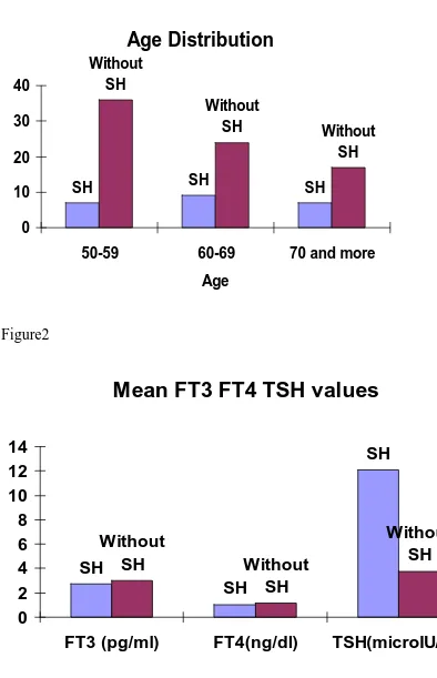
There were differences in the mean age distribution among cases and controls
are shown in table 1 and fig 1
Table 1
Age in years Patients with SH Patients without SH
50-59 7 36
60-69 9 24
70 and above 7 17
There were 43 patients in the age group 50-59 of which 7 (19.44%) were
having subclinical hypothyroidism .
In the 60-69 age group there were 33 patients of which 9 (27.27%) were
In the 70 and above age group there were 24 patients of which 7 (29.17%)
were having subclinical hypothyroidism.
The mean TSH level in patients with subclinical hypothyroidism was
12.11µIU/mL. For FT4 it was 1.03ng/dl and for FT3 it was 2.76pg/ml . There
were differences in FT4, FT3, TSH distribution in cases and control as shown
in Table 2
Table 2
Mean Patients with SH Patients without TSH
FT3 (pg/ml) 2.76 3.01
FT4 (ng/dl) 1.03 1.14
TSH (µIU/ml) 12.11 3.75
There were 23 patients with TSH level more than 5.5 µIU/ml, the upper level
of normal range ( 0.30-5.5 µIU/ml). They are the subclinical hypothyroid
patients in this study. Of those 23 patients 15(65.2%) had TSH level between
5.5 to 10 µIU/ml. The remaining 8 (34.8%) patients had TSH levels more than
10 µIU/ml as shown in the following Table 3
Table 3
TSH levels in patients with SH
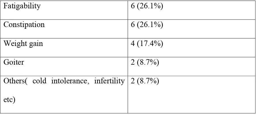
Hypothyroid symptoms were reported in 7 out of 23 (30.43%) patients with
subclincal hypothyroidism. Fatigability and constipation were the most
common complaint, followed by weight gain .The frequency of hypothyroid
symptoms in the subclinical hypothyroid patients are as shown in the table 4
Table 4
Frequency of hypothyroid symptoms in patients with SH
Fatigability 6 (26.1%)
Constipation 6 (26.1%)
Weight gain 4 (17.4%)
Goiter 2 (8.7%)
Others( cold intolerance, infertility
etc)
2 (8.7%)
TSH level in µIU/ml No. of patients(%)
Total no = 23
5.5 – 10 15 (65.2%)
Goiter was present in 2 out of 23 patients with subclinical hypothyroidism
(8.7%) and 5 out of 77 patients without subclinical hypothyroidism (6.5%).
Other symptoms like cold intolerance infertility were present in 2 of the 23
patients (8.7%) with subclinical hypothyroidism and 1 of the 77 patients
(1.3%) without subclinical hypothyroidism
The incidence of risk factors like hypertension diabetes and ischemic heart
disease were compared between patients with subclinical hypothyroidism and
control.
They were analyzed independently with Chi-Square test. The p- value
showed that patients with subclinical hypothyroidism were significantly
associated with ischemic heart disease compared to controls. The p- value is
not significant for hypertension and diabetes. This is shown in table 5
Table 5
Patients with
SH
Patients without
SH
p- value
IHD 5 (21.71%) 5 (6.5%) 0.047
DM 4 (17.4%) 16 (20.1%) 0.490
DISCUSSION
Sub clinical hypothyroidism is highly prevalent in elderly women. A
prevalence of 11 – 26 % had been reported in previous studies, 4,8,23,62 our
study shows a prevalence of 23 % in concordance with the other studies.
Surveys that stratified TSH levels indicate a predominance of TSH <
10µIU/ml , which accounts for about 55-85% of cases. 5,58,69 Almost 65% of
our patients with subclinical hypothyroidism had TSH levels < 10µIU/ml.
Studies that have reported thyroid antibody test on subjects with elevated
TSH demonstrated seropositivity rates from 20-78%.1,4,59,70
Several studies have suggested that mild symptoms of hypothyroidism are
more prevalent in patients with subclinical hypothyroidism than in age
matched controls.62,70 Fatigability and weight gain were the most frequent
symptoms, 67 but not all studies have found his to be true.71 30% of our
patients with subclinical hypothyroidism had symptoms of which fatigability
(26%) and constipation (26%) were the most common
There have been three published randomized prospective placebo controlled
trials for the therapy of symptoms in patients with subclinical
hypothyroidism. 21,68,72
Two trials reported significant improvement in symptoms of hypothyroidism,
was related to TSH level, being more in those who have mean TSH level
greater than or equal to 12.7µIU/ml at baseline.71 In women with SH and
ovulatory dysfunction , thyroxine therapy may restore fertility.73