Copyright © 1998, American Society for Microbiology
Detection of Viral Proteins after Infection of Cultured
Hepatocytes with Rabbit Hemorrhagic Disease Virus
MATTHIAS KO¨ NIG,† HEINZ-JU¨RGEN THIEL,†ANDGREGOR MEYERS*
Department of Clinical Virology, Federal Research Centre for Virus Diseases of Animals, D-72001 Tu¨bingen, Germany
Received 15 October 1997/Accepted 28 January 1998
The calicivirus rabbit hemorrhagic disease virus (RHDV), which replicates predominantly in the livers of infected rabbits, cannot be propagated in tissue culture. To enable the performance of in vitro studies, rabbit hepatocytes were isolated by liver perfusion and gradient centrifugation. After inoculation with purified RHDV, more than 50% of the cells proved to be infected. Protein analyses led to the detection of 13 RHDV-specific polypeptides within the infected cells. These proteins were assigned to defined regions of the viral genome, resulting in a refined model of RHDV genome organization.
Rabbit hemorrhagic disease (RHD) is a contagious disease often associated with liver necrosis, hemorrhages, and high mor-tality. The causative agent of RHD is a positive-stranded RNA virus which belongs to the family Caliciviridae, a group of non-enveloped animal viruses (8). The RHD virus (RHDV) ge-nome consists of a polyadenylated RNA molecule of 7,437 nucleotides with a virus-encoded protein (VPg) covalently at-tached to its 59end (12, 13). The genomic RNA contains one long open reading frame (ORF1) encoding a hypothetical pri-mary translation product of 257 kDa, which gives rise to ma-ture viral proteins by proteolytic processing. Most, if not all, cleavages are executed by a virus-encoded trypsin-like cysteine protease showing significant similarity to the 3C proteases of picornaviruses (4). So far, viral protein expression has only been studied by in vitro translation of viral RNA and de-tection of RHDV-encoded proteins with specific antibodies (2, 24). Together with data obtained after bacterial expres-sion of RHDV proteins, these studies led to the first compre-hensive model of the organization of a calicivirus genome (23, 24). Accordingly, the identified viral gene products are ar-ranged in the ORF1-encoded polyprotein in the order NH2-p16-p23-p37-p41-p69-VP60-COOH. A second ORF (ORF2) is located at the extreme 39end of the genomic RNA; expression of ORF2 via a not-yet-identified mechanism leads to VP10, a component of RHDV virions (24). In RHDV-infected cells, a 2.2-kb subgenomic mRNA which is colinear with the 39 one-third of the genomic RNA is transcribed (13). This mRNA apparently represents the major source of the RHDV capsid protein VP60; the latter is also generated via cleavage of the ORF1-encoded polyprotein (15, 23).
Like the human caliciviruses, e.g., Norwalk virus or South-ampton virus, RHDV so far cannot be propagated in tissue culture cells. To enable the investigation of protein synthesis and other aspects of the RHDV life cycle in infected cells, we devised a system for in vitro propagation of RHDV based on primary rabbit liver cells. Infected hepatocytes were used for analysis of RHDV protein expression, resulting in a refined model of the organization of the calicivirus genome.
Isolation and cultivation of rabbit hepatocytes.Infected an-imals usually contain large amounts of RHDV virions in the liver and the spleen. By immunocytochemical methods, viral antigen was detected in hepatocytes and reticuloendothelial cells of the liver (14). As a first step toward in vitro propagation of RHDV, rabbit hepatocytes were isolated and maintained in vitro. Hepatocytes have to be released carefully from their tissue environment by enzyme digestion since mechanical mo-bilization will unequivocally result in severe cell damage (20). Perfusion techniques using collagenase have been successfully applied for the isolation of hepatocytes from rabbits (22).
We used an extracorporeal two-step perfusion technique to obtain large quantities of vital hepatocytes for subsequent studies. The first perfusion step included removal of remaining blood cells and Ca21by using preperfusion buffer [140 mM
NaCl, 7 mM KCl, 10 mM HEPES, 8 mMD-(1)-glucose, 0.1
mM EGTA, pH 7.4]. Ca21is believed to stabilize
intercel-lular hepatic adhesion factors. Therefore, deprival of Ca21
is regarded as a prerequisite for optimal results in collagenase digestion (20). In the second step, the liver lobes were perfused with a solution consisting of collagenase (500 mg/liter; Sigma, Deisenhofen, Germany) in perfusion buffer [67 mM NaCl, 7 mM KCl, 100 mM HEPES, 8 mMD-(1)-glucose, 6 mM CaCl2,
pH 7.6]. Finally, after removal of the liver capsule, further collagenase digestion was performed in suspension, thereby mobilizing the parenchymal cells, with a yield of about 109 viable cells per liver as determined by trypan blue exclusion (16).
Freshly prepared cells plated on tissue culture vessels pre-coated with collagen (type 1; Sigma) dissolved in 0.2% acetic acid were rapidly adsorbed and formed a confluent mono-layer. After 24 h, the majority of the cells showed a polygonal shape resembling that of hepatocytes and had assembled in trabecular structures. Hepatocytes containing two or more nu-clei were consistently observed. Electron microscopic analysis of cells from a 5-day culture revealed an ultrastructure similar to that of hepatocytes (data not shown).
Since addition of fetal bovine serum proved to be toxic to the parenchymal cells, cultures were kept in a chemically defined medium (Williams medium E [Gibco] buffered with 10 mM HEPES and supplemented with 5mg of insulin per ml, 2mg of glucagon per ml, 0.1mg of epidermal growth factor per ml, 0.25
mg of hydrocortisone per ml, 5 ng of H2SeO3 per ml, 2 mM
L-glutamine, and 100 mg of gentamycin per ml); the culture
medium was exchanged every other day. Hepatocyte cultures * Corresponding author. Mailing address: Department of Clinical
Virology, Federal Research Centre for Virus Diseases of Animals, P.O. Box 1149, D-72001 Tu¨bingen, Germany. Phone: 49 7071-967207. Fax: 49 7071-967303. E-mail: gregor.meyers@tue.bfav.de.
† Present address: Institut fu¨r Virologie, FB Veterina¨rmedizin, Jus-tus-Liebig-Universita¨t Giessen, D-35392 Giessen, Germany.
4492
on November 9, 2019 by guest
http://jvi.asm.org/
were maintained for 2 to 3 weeks under the conditions de-scribed above. After this period, hepatocytes were increasingly overgrown by nonparenchymal cells.
To obtain cell preparations of higher purity, low-speed iso-density Percoll gradient centrifugation was employed (11). More than 90% of the gradient-purified liver cells were viable. The proportion of contaminating nonparenchymal cells was negligible, since hepatocyte cultures could be maintained for more than 4 weeks without visible signs of overgrowth (data not shown).
Infection of cultured rabbit hepatocytes with RHDV. To study the ability of RHDV to infect cultured rabbit hepato-cytes, the cells were inoculated with CsCl gradient-purified virus. Infection was monitored by the detection of the RHDV major capsid protein (VP60) in an immunoperoxidase assay using a mixture of three monoclonal antibodies (1H8, 5G3, and 6G2 [6]).
Examination of hepatocyte cultures 24 h after infection with RHDV revealed cells expressing VP60 (Fig. 1). The percent-age of infected cells increased significantly in cultures main-tained for 48 or 72 h after infection (Fig. 1). However, even after prolonged incubation, a considerable proportion of cells remained negative in the immunostaining assay. Virus-infected cells revealed signs of degeneration, including condensed nu-clei considerably smaller than the ones from noninfected cells; excessive lysis of cells was not observed (Fig. 1).
In infected cultured hepatocytes, VP60 was mostly found in the cytoplasm; only weak staining occurred in the nuclei of the
cells. This finding stands in marked contrast to the picture observed in liver sections from diseased rabbits, where the nu-clei of hepatocytes were intensively stained by anti-VP60 an-tibodies (1). Transport of VP60 into the nucleus has not been thoroughly investigated. Heterologous expression of VP60 in different eukaryotic cell lines did not result in the detection of VP60 within nuclei. In situ hybridization studies with liver sections from infected rabbits revealed that viral RNA is not located in the nucleus (data not shown).
The presence of RHDV VP60 in cultured hepatocytes dem-onstrated that these cells can be infected in vitro. Furthermore, the strong signals observed in infected cells suggested that replication of the viral genome and/or transcription of the subgenomic mRNA takes place in cultured hepatocytes. Neg-ative cells may represent populations of hepatocytes that ei-ther cannot be infected by RHDV or, alternatively, do not support translation and/or replication/transcription of viral RNA. The question of whether infectious virus is actually produced and released by cultured hepatocytes remains un-answered.
Analysis of viral proteins produced in infected hepatocytes.
[image:2.612.54.548.67.375.2]Characterization of calicivirus genomes has rapidly progressed in the past few years (for a review, see reference 7). In contrast, little is known about the proteins expressed by caliciviruses. This is mainly due to the fact that tissue culture systems and/or antibodies specific for individual viral proteins are not avail-able. The only authentic calicivirus proteins demonstrated so far are the major capsid protein (with a molecular weight of 58 FIG. 1. Infection of cultured hepatocytes with RHDV. Overnight cultures were either mock infected (A) or inoculated with gradient-purified virus and subsequently incubated for 24 h (B), 48 h (C, E, and F), or 72 h (D). Immunostaining was performed with monoclonal antibodies directed against RHDV VP60 major capsid protein and anti-mouse immunoglobulin G conjugated to horseradish peroxidase. Cells were counterstained with hematoxylin. (E and F) Higher magnifications of a culture 48 h after infection. Note the noninfected cells (E, bottom region), apparently newly infected cells (E, center), and cells showing a pronounced cytopathic effect (E, left and right margins; F [note condensation and size of nuclei]). Instrumental magnification,3100 (A to D) or31,000 (E and F). The dark staining of the condensed nuclei in panel F resulted from the blue counterstain, which cannot be distinguished from the antibody staining evident on a black and white picture.
on November 9, 2019 by guest
to 76 kDa) (reviewed in reference 7), VP10 (24), and VPg (3, 5, 13, 19), which are all found in virions. By in vitro translation of viral RNA and precipitation of translation products with a set of region-specific antisera raised against bacterial fusion proteins, the putative organization of RHDV ORF1 has been determined (24). To check whether the data obtained in vitro reflect the in vivo situation, the expression of RHDV proteins in infected rabbit hepatocytes was analyzed by metabolic la-beling and immunoprecipitation. Freshly prepared hepato-cytes (106) were seeded in a 3.5-cm-diameter culture dish and then infected with gradient-purified RHDV 24 h later. After 1 h, the virus suspension was removed; the cells were then washed twice with labeling medium (18) and subse-quently incubated in this medium for 1 h. Afterward, the supernatant was removed and the cells were incubated for 4 h in 0.5 ml of labeling medium containing 125mCi of Tran35 S-label (ICN Biochemicals, Meckenheim, Germany). The la-beled cells were washed twice with phosphate-buffered sa-line and processed for immunoprecipitation as described previously (18).
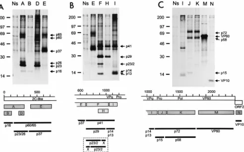
[image:3.612.56.548.68.376.2]After in vitro translation of the viral RNA, the amino-ter-minal one-third of the polyprotein encoded by ORF1 gave rise to three processing products of 16, 23, and 37 kDa (24). In addition, a band of about 60 kDa, which presumably repre-sented a precursor composed of the latter two cleavage prod-ucts, was sometimes detected (24). The analogous proteins were also found in RHDV-infected hepatocytes. Polypeptides of 16, 23, and 60 kDa were identified after precipitation with antiserum A, which is directed against the first 300 amino acids encoded by ORF1 (Fig. 2A). The 16-kDa protein also reacted with antiserum B, which was raised against amino acids 9 to 112 of the protein encoded in ORF1. Thus, p16 represents the amino-terminal cleavage product of the polyprotein. The amount of p16 precipitated with either antiserum is very small, possibly due to instability of this protein in the infected cells. The polypeptides of 23 and 60 kDa were recognized not only by antiserum A but also by antiserum D, which is directed against amino acids 232 to 303. p60 was also precipitated with an antiserum covering the region from amino acids 393 to 702 of the polyprotein (antiserum E). Antiserum E precipitated a FIG. 2. Immunoprecipitation of proteins from RHDV-infected hepatocytes with a set of antisera raised against bacterial fusion proteins containing defined regions of the polypeptides encoded by the RHDV genome (24). The upper portion of each panel shows the precipitated proteins separated by sodium dodecyl sulfate-polyacrylamide gel electrophoresis. Each lane is labeled with a letter indicating the antiserum used for precipitation. On the right side of each gel, bands discussed in the text are marked with arrows and the designations of the precipitated proteins. The positions of size marker proteins (14C-labeled molecular mass markers;
Amersham, Braunschweig, Germany) are indicated on the left (in kilodaltons). A scheme below each gel indicates the reactivities of the different antisera. The ORF1-encoded polyprotein is symbolized by a white bar marked by a scale (in 100-amino-acid increments). Within the polyprotein, the locations of VPg and the RHDV protease (Pro) as well as the positions of amino acid motifs common for RNA virus polymerases (Pol) and helicases (2C-like) are shown. The protein encoded by ORF2 is shown below the ORF1 product. Gray bars labeled with letters indicate the protein segments contained in the different fusion proteins used for generation of the antisera. The exact features of the antisera have been described previously (24). In the lower part of each panel, the putative locations of the precipitated proteins (black bars) with respect to the polyprotein are shown. Please note that the gels of all three panels have been mounted from one protein gel. In panels B and C, the last lane of panel A and the last lane of B, respectively, are included to facilitate comparison of the electrophoretic mobilities of the individual proteins. (A) Proteins precipitated with antisera directed against regions within the amino-terminal one-third of the ORF1-encoded polyprotein. (B) Precipitation products obtained with antisera recognizing different segments of the central region of the polyprotein. Further processing of p29 into p23/2 and X is hypothetical and could occur in different ways, as indicated in the box at the bottom. (C) Proteins precipitated with antisera directed against the carboxy-terminal half of the ORF1-encoded polyprotein and the ORF2 product. Ns, rabbit preimmune serum.
on November 9, 2019 by guest
http://jvi.asm.org/
After coupled in vitro transcription-translation of RHDV cDNA constructs, Alonso et al. detected a band of 80 kDa, derived from the region corresponding to the 59 part of the genome, which was apparently not further processed (2). This finding stands in marked contrast to our data; the reason for this discrepancy is not known.
Antisera F, H, and I are directed against bacterial fusion proteins encompassing different parts of the central part of the ORF1-encoded polyprotein; the region covered by antiserum H (amino acids 875 to 1023) represents the C-terminal part of the larger region F (amino acids 704 to 1023), whereas the region recognized by antiserum I is located further down-stream (amino acids 1023 to 1170) (24). After in vitro transla-tion of viral RNA, a product of 41 kDa was shown to react with these antisera. A product of similar size (43 kDa) was detected after coupled in vitro transcription-translation of cDNA con-structs (2). Using extracts of infected hepatocytes, precipita-tion with antiserum F resulted in the detecprecipita-tion of four bands of 41, 29, 23, and 13 kDa. The proteins of 41, 29, and 23 kDa were also recognized by antiserum H. In addition, after prolonged exposure, bands corresponding to proteins of 13 and 14 kDa were identified with this antiserum (data not shown). The latter two proteins were also precipitated with antiserum I, together with p41 (Fig. 2B). Because of its size and reaction pattern, p41 is likely to represent a fusion of p29 and a second protein of about 12 to 14 kDa. This second protein could be p13 or p14. It is not clear whether p13 and p14 represent different processing products or one of these polypeptides is a posttranslationally modified version of the other. With regard to size and location in the polyprotein, p13/p14 represents VPg (13, 23, 24). Analogously to VPg-pU and VPg-pUpU of picor-naviruses (21), RHDV VPg could be covalently linked to nu-cleotides in the course of RNA replication. Accordingly, one of the detected bands may represent the original cleavage prod-uct whereas the other is modified by addition of one or more nucleotides. Further experiments are needed to verify this hy-pothesis. It is not clear at present why antiserum F precipitates p13 but not p14.
According to the processing scheme indicated above, the polypeptide of 23 kDa (p23/2) should represent a product generated by cleavage of p29 (Fig. 2B). The hypothetical sec-ond cleavage product of about 6 kDa could not be detected, possibly due to a lack of specific antibodies in our sera or instability of the cleavage product. Alternatively, p23/2 could represent the product of a distinct route of p41 processing. This hypothesis would also imply the existence of at least one additional protein. However, the reaction pattern of p23/2 with the different antisera and the failure to detect further cleavage products make the latter alternative unlikely.
In addition to the products described above, a band of 37 kDa was visible with antisera F, H, and I, as well as with
apparently not observed for the polypeptides derived from in vitro translation, since products with molecular masses below 69 kDa were not found in the in vitro experiments (2, 24). Interestingly, RHDV-infected hepatocytes contained not only a protein of about 72 kDa but also polypeptides of 58 and 15 kDa (Fig. 2C). This was shown by analyses using antisera J, which is directed against amino acids 1172 to 1332, and anti-serum K, which is directed against residues 1332 to 1727. While p72 is recognized by antisera J and K, p15 is precipitated with antiserum J but not with antiserum K. After longer exposure times, a weak band comigrating with p15 was also visible after precipitation with antiserum I (data not shown). Taken to-gether, the data imply that p15 represents the RHDV protease, which is known to consist of 143 amino acids located in the ORF1 polyprotein between residues 1109 and 1251 (24). Con-sequently, p58 must be regarded as the RHDV polymerase and p72 must represent the uncleaved protease-polymerase precur-sor. The C-terminal cleavage product encoded by ORF1 is VP60, which is detected by antiserum M.
The genome of every calicivirus contains at its 39end a small ORF with the capacity to encode a protein of 100 to 200 amino acids (reviewed in reference 7). It was shown for RHDV that the product of this ORF represents a structural protein of 10 kDa which was termed VP10 (24). After precipitation with antiserum N, which is directed against part of the ORF2-en-coded protein, VP10 could be demonstrated in the extract from the infected hepatocytes (Fig. 2C). The generation of VP10 was also observed after in vitro translation (data not shown); the mechanism responsible for expression of this protein has not yet been elucidated.
Genetic map of RHDV.Analysis of the proteins present in RHDV-infected primary hepatocytes allowed the establish-ment of a genetic map for RHDV (Fig. 3). According to this proposed map, the translation product of ORF1 is initially cleaved into products p16, p60/65, p41, p72, and VP60. Further processing of the p60/65 region leads to p23/26 and p37, while products of 15 and 58 kDa can be derived from the part of the polyprotein comprising p72. It is not known whether p60/65 and p72 represent incompletely processed precursors or sta-ble proteins with specific functions in virus replication, as described, e.g., for 3AB or 3CD of picornaviruses (17). Also, p41 is further processed into several polypeptides (see be-low).
The conclusions drawn from the in vitro translation experi-ments and the results obtained by analysis of the infected hepatocytes led to the establishment of similar genetic maps. However, the existence of several important differences must be stressed. Expression of p15 and p58 as well as the putative modification of p60 and p23, leading to p65 and p26, respec-tively, could only be demonstrated in vivo. Moreover, there were obvious differences between the in vitro and the in vivo
on November 9, 2019 by guest
results with regard to the processing of p41. After in vitro translation, only p41 was detected. Since it was known that the VPg gene is located in the part of the genome coding for p41, further processing of p41 had to be postulated. Nevertheless, the detection of four different polypeptides in vivo, namely p13, p14, p23/2, and p29, with a hypothetical fifth cleavage product of about 6 kDa was unexpected. Even though the scheme presented in Fig. 2 and 3 represents a consistent model for the processing of this part of the polyprotein, a consider-able effort that includes the generation of additional serolog-ical reagents, heterologous expression, and pulse-chase label-ing of RHDV proteins will be necessary to elucidate the exact cleavage cascade. Such approaches will also help to elucidate the nature of some bands visible in Fig. 2 that to date cannot be explained.
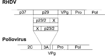
With regard to the arrangement of the genes coding for nonstructural proteins, the genomic organization of RHDV ex-hibits obvious similarities to that of picornaviruses. In a sche-matic alignment of their polyproteins, p16 would correspond to poliovirus 2A, p23 would correspond to 2B, and p37 would correspond to 2C (24). The arrangement of the protease and putative RNA-dependent RNA polymerase at the end of the nonstructural region of the polyprotein fits with the organiza-tion of all members of the picornavirus superfamily (10). In-teresting differences are found for the regions containing p41 of RHDV and the similarly positioned 3AB of picorna-viruses. With the exception of foot-and-mouth disease virus, only two processing products of 3AB are known (9, 17); in contrast, p41 apparently gives rise to at least four different polypeptides (Fig. 4). Since the carboxy-terminal processing product in both cases is VPg, the differences concern 3A and the amino-terminal two-thirds of p41, which gives rise to RHDV p29. 3A of poliovirus is a small hydrophobic protein of about 10 kDa. RHDV polypeptide p29 contains a highly hydrophobic region at its carboxy terminus, whereas the rest of the protein is moderately hydrophilic. Further processing of p29 could result in a hydrophobic carboxy-terminal prod-uct, exhibiting similarities to poliovirus 3A, and an amino-terminal polypeptide, for which a counterpart is lacking in picornaviruses. The polypeptide p23/2 most likely represents a processing product of p29; a corresponding second cleavage product could not be identified. Elucidation of the processing
of p41 will also be interesting with regard to the relationship between caliciviruses and picornaviruses.
[image:5.612.117.489.72.229.2]The method for isolation and cultivation of primary rabbit hepatocytes described here allowed for the first time the study of RHDV after infection of cells in vitro. It is assumed that transcription and replication of viral RNA take place within the infected hepatocytes even though we have not been able to prove that viral replication in the cultured cells is completed by the release of infectious virus. The amount of viral protein detected in the infected cells is most likely not solely attribut-able to translation of RNA derived from the input virus. Fur-thermore, detection of efficient processing at sites which are not cleaved in vitro and of products which are either not generated or not stable after in vitro translation is also indic-ative of viral replication. In any case, the infection of cultured hepatocytes represents a means of analyzing RHDV gene ex-pression on the basis of infected cells. Even though the method described here is laborious and expensive, it has opened a door to work on selected topics of RHDV biology. As outlined above, this approach has provided the most authentic view of the organization of a calicivirus genome available thus far. This is remarkable since some other family members, e.g., feline
FIG. 3. Schematic representation of the RHDV-encoded proteins. The upper bars indicate the hypothetical primary translation products of ORF1 and ORF2; the scale represents amino acid numbers. Regions which contain known sequence motifs or already-identified viral proteins are indicated. EG and ET, processing sites determined so far (2, 23, 24). The gray-shaded bars below represent VP10 and the products resulting from processing of the ORF1-encoded polyprotein. The designations of proteins which could not be demonstrated after in vitro translation are written in boldfaced letters. The polypeptides generated by the hypothetical cleavage of p29 are enclosed in a box. The hypothesized further processing of p29 has not been proven; two alternative ways for this processing to occur are indicated. Protein “X” has not been demonstrated.
FIG. 4. Comparison of the genomic regions encoding part of the nonstruc-tural proteins of RHDV and poliovirus. The genomes are shown as bars, and the locations of individual genes are indicated. The regions shown in gray symbolize those parts of the genomes for which the experiments indicate the existence of a major difference between RHDV and picornaviruses (see also the text and the legend to Fig. 3).
on November 9, 2019 by guest
http://jvi.asm.org/
[image:5.612.324.526.568.676.2]4. Boniotti, B., C. Wirblich, M. Sibilia, G. Meyers, H.-J. Thiel, and C. Rossi. 1994. Identification and characterization of a 3C-like protease from rabbit hemorrhagic disease virus, a calicivirus. J. Virol. 68:6487–6495.
5. Burroughs, J. N., and F. Brown. 1978. Presence of a covalently linked protein on caliciviral RNA. J. Gen. Virol. 41:443–446.
6. Capucci, L., G. Frigoli, L. Rønshold, A. Lavazza, E. Brocchi, and C. Rossi. 1995. Antigenicity of rabbit haemorrhagic disease virus studied by its reac-tivity with monoclonal antibodies. Virus Res. 37:221–238.
7. Clarke, I. N., and P. R. Lambden. 1997. The molecular biology of calicivi-ruses. J. Gen. Virol. 78:291–301.
8. Cubbitt, D., D. W. Bradley, M. J. Carter, S. Chiba, M. K. Estes, L. J. Saif,
F. L. Schaffer, A. W. Smith, M. J. Studdert, and H.-J. Thiel.1995. Family Caliciviridae. Arch. Virol. 10(Suppl.):359–363.
9. Forss, S., and H. Schaller. 1982. A tandem repeat gene in a picornavirus. Nucleic Acids Res. 10:6441–6450.
10. Goldbach, R., and J. Wellink. 1988. Evolution of plus-strand RNA viruses. Intervirology 29:260–267.
11. Kreamer, B. L., J. L. Staecker, N. Sawada, G. L. Sattler, M. T. S. Hsia, and
H. C. Pitot. 1985. Use of a low-speed, iso-density Percoll centrifugation
clones. Virology 171:18–27.
19. Schaffer, F. L., D. W. Ehresmann, M. K. Fretz, and M. E. Soergel. 1980. A protein, VPg, covalently linked to 36S calicivirus RNA. J. Gen. Virol. 47: 215–220.
20. Seglen, P. O. 1976. Preparation of isolated rat hepatocytes. Methods Cell Biol. 13:30–83.
21. Takeda, N., R. J. Kuhn, C.-F. Yang, T. Takegami, and E. Wimmer. 1986. Initiation of poliovirus plus-strand RNA synthesis in a membrane complex of infected HeLa cells. J. Virol. 60:43–53.
22. Walpole, H. E., W. M. Lee, T. Walle, U. K. Walle, M. J. Wilson, and J. W.
Kennedy.1990. Rabbit hepatocytes in primary culture: preparation, viability and use in studies of propanolol metabolism. Hepatology 11:394–400. 23. Wirblich, C., M. Sibilia, M. B. Boniotti, C. Rossi, H.-J. Thiel, and G. Meyers.
1995. 3C-like protease of rabbit hemorrhagic disease virus: identification of cleavage sites in the ORF1 polyprotein and analysis of cleavage specificity. J. Virol. 69:7159–7168.
24. Wirblich, C., H.-J. Thiel, and G. Meyers. 1996. Genetic map of the calicivirus rabbit hemorrhagic disease virus as deduced from in vitro translation studies. J. Virol. 70:7974–7983.