0022-538X/96/$04.0010
Copyrightq1996, American Society for Microbiology
Previously Unsuspected cis-Acting Sequences for DNA Replication
Revealed by Characterization of a Chimeric
Heron/Duck Hepatitis B Virus
KARLYN MUELLER-HILLANDDANIEL D. LOEB*
McArdle Laboratory for Cancer Research, University of Wisconsin Medical School, Madison, Wisconsin 53706
Received 28 May 1996/Accepted 19 August 1996
Heron hepatitis B virus (HHBV) is an avian hepadnavirus that is closely related to duck hepatitis B virus (DHBV). To learn more about the mechanism of hepadnavirus replication, we characterized a clone of HHBV that contains a substitution of DHBV sequence from nucleotide coordinates 403 to 1364. This clone, named HDE1, expresses a chimeric pregenomic RNA, a chimeric polymerase (P) protein, and a core (C) protein with a one-amino-acid substitution at its carboxy terminus. We have shown that HDE1 is defective for minus-strand DNA synthesis, resulting in an overall reduction of viral DNA. HDE1 was also defective for plus-strand DNA synthesis, resulting in aberrant ratios of replication intermediates. Genetic complementation assays indicated that HDE1 replication proteins, C and P, are functional for replication and wild-type HHBV proteins do not rescue either defect. These findings indicate that the HDE1 substitution mutation acts primarily incis. By restoring nucleotides 403 to 902 to the HHBV sequence, we showed thatcis-acting sequences for plus-strand DNA synthesis are located in the 5*half of the HDE1 chimeric region. These data indicate the presence of one or more formerly unrecognizedcis-acting sequences for DNA synthesis within the chimeric region (nucleotides 403 to 1364). Thesecis-acting sequences in the middle of the genome might interact directly or indirectly with known cis elements that are located near the ends of the genome. Our findings suggest that a specific higher-order template structure is involved in the mechanism of hepadnavirus DNA replication.
Hepatitis B viruses infect the livers of a number of mamma-lian and avian species, resulting in acute and chronic liver diseases such as hepatitis, cirrhosis, and hepatocellular carci-noma (for a review, see reference 9). Hepadnaviruses are small, enveloped, circular double-stranded DNA viruses that replicate through an RNA intermediate via reverse transcrip-tion (28). After viral entry into the cell, the relaxed circular (RC) DNA genome (3.0 kb) of the hepatitis B virus is depos-ited into the nucleus and is converted to the covalently closed circular template (30) for transcription of the subgenomic and pregenomic RNAs by the host RNA polymerase II (2). The polymerase (P) and core (C) proteins are translated separately from the pregenomic RNA (5, 25). In the cytoplasm, the core protein encapsidates P protein-associated pregenomic RNA (1, 11), and complete replicative DNA synthesis takes place within the viral capsid (28). DNA synthesis begins when minus-strand DNA synthesis is primed by the P protein binding on an
RNA stem-loop near the 59 end of the pregenome (epsilon)
(Fig. 1a and b) (29, 31). A template switch, from epsilon to the
39copy of direct repeat 1 (DR1), precedes continued
minus-strand DNA synthesis (Fig. 1c) (29, 31). Minus-minus-strand elonga-tion and compleelonga-tion are accompanied by degradaelonga-tion of the pregenomic RNA up to the last 18 ribonucleotides (Fig. 1d to f), which serve as the primer for plus-strand DNA synthesis (17–19). The plus-strand primer translocates to an acceptor
site (DR2) near the 59end of minus-strand DNA (Fig. 1h) and
initiates synthesis of plus-strand DNA (Fig. 1g) (17). A tem-plate switch allows continuation of plus-strand DNA synthesis (Fig. 1i), ultimately forming complete, RC, genomic DNA
(Fig. 1j). A small fraction of the plus-strand primers do not translocate and are elongated in situ, forming a duplex linear
(DL) species (Fig. 1g9and h9) (27). Cytoplasmic nucleocapsids
that contain mature viral DNA can either deposit their genomic DNA in the nucleus, increasing the copy number of covalently closed circular DNA, or associate with viral surface proteins, acquire an envelope, and exit the cell via an endo-plasmic reticulum-Golgi pathway.
Heron hepatitis B virus (HHBV) is related to duck hepatitis B virus (DHBV). In addition to having viral particles of similar size and morphology, HHBV has a genetic organization nearly identical to that of DHBV (79% identity at the nucleotide level) (26). The terminally redundant pregenomic RNA, DR1 and DR2, and the overlapping C and P genes are conserved (26). Southern blot analysis of a molecular clone of wild-type HHBV reveals a pattern of replication intermediates that is identical to the DHBV pattern.
In this report, we describe a chimeric heron/duck hepatitis B virus that is defective in its ability to synthesize DNA. The chimera, HDE1, is a molecular clone of HHBV with an ap-proximately 1-kbp substitution of DHBV sequence near the middle of the pregenome (from nucleotides [nt] 403 to 1364) (Fig. 2). With the goal of understanding the mechanism of hepadnavirus DNA replication, we have examined why HDE1 is defective for DNA synthesis. In this report we show that the chimeric virus is defective for both minus- and plus-strand DNA synthesis and that both defects are manifest primarily in
cis. By restoring nt 403 to 902 to the HHBV sequence, we
determined that cis-acting sequences for plus-strand DNA
syn-thesis are located in the 59half of the HDE1 chimeric region.
We interpret our results to indicate the presence of one or more formerly unrecognized acting elements. These cis-acting sequences may be involved in formation of a
higher-* Corresponding author. Mailing address: McArdle Laboratory for Cancer Research, University of Wisconsin Medical School, 1400 University Ave., Madison, WI 53706. Phone: (608) 262-1260. Fax: (608) 262-2824. Electronic mail address: [email protected].
8310
on November 9, 2019 by guest
http://jvi.asm.org/
order template structure that could be part of the mechanism of DNA synthesis.
MATERIALS AND METHODS
Materials.Enzymes were purchased from New England BioLabs and Boehr-inger Mannheim and were used as instructed by the manufacturers. Radionu-clides were purchased from Amersham and NEN.
Construction of mutant HHBV genomes.Plasmid 413-2, previously referred to as H1.4G (13), contains 1.4 tandem copies of HHBV-4 DNA inserted into an EcoRI site in the vector pIBI21. Plasmid HDE1 (derived from 412-3) contains a substitution of DHBV type 16 DNA from the AvrII site (nt 403 in the numbering system for DHBV) (21) to the XbaI site (nt 1358 for DHBV, nt 1364 for HHBV). A frameshift mutation resulting from a 4-bp deletion was introduced into plas-mid 413-2 at the BstXI site (nt 1439) to create a mutant null for P protein function (HHBVP2). A frameshift mutation resulting from a 4-bp insertion was introduced into plasmid 413-2 at the HindIII site (nt 38) to create a mutant null for C protein function (HHBVC2). Both frameshift mutations were combined to create a mutant null for replication proteins (HHBVP2C2). The same frame-shift mutations were constructed into HDE1 to create single and double mutants null for proteins (HDE1P2and HDE1P2C2).
Additional DNA plasmid mutants expressing encapsidation-negative pre-genomic RNA of HHBV (ENHHBV) and HDE1 (ENHDE1) were constructed by oligonucleotide-directed mutagenesis (15) to have a 7-nt deletion from nt 2574 to 2580 (59-GGTATCTTTACGTC-39to 59-GGACGTC-39). These muta-tions were first introduced into a subcloned fragment of HHBV. After confir-mation of the sequence, monomers of HHBV or HDE1 sequence were inserted 39of the deletion to create plasmids that express the pregenomic RNAs with the deletion in epsilon.
Plasmid 530-1 (a clone of HHBV with DHBV sequence from nt 902 to 1358) was derived from HDE1 as follows. First, a StuI site (59-GGGCCC-39to 59-AG GCCT-39) was constructed into the HHBV sequence at nt 894. Then, a fragment of HHBV sequence from AvrII (nt 403) to the new StuI site was cloned into HDE1 at the AvrII site (403) and a filled-in Tth111 (nt 902 in DHBV type 16) site. The resulting 39junction sequence was 59-GAGGtggtc-39, where capital letters represent HHBV sequence and lowercase letters represent DHBV se-quence. Plasmid 530-1P2 also contained the BstXI frameshift mutation de-scribed above. All mutant constructs dede-scribed here were verified by restriction digest and sequence analysis.
Cell culture and DNA transfections.The chicken hepatoma cell line LMH (8, 14) was used in all cell culture experiments and grown in a 1:1 mixture of Dulbecco’s modified Eagle’s medium and nutrient F-12 medium (Gibco BRL
catalog no. 12500-070) supplemented with 5% fetal bovine serum. Transfections were performed by the calcium phosphate method (6). Typically, 3 to 10mg of plasmid DNA was transfected into LMH cells that were at 50 to 70% of conflu-ency on a 60- or 100-mm-diameter plate. In cotransfections, the ratio of protein donor plasmid to recipient plasmid was typically 1:1.
Isolation of viral nucleic acid.Isolation of viral DNA from cytoplasmic core particles was performed 3 days posttransfection as described by Staprans et al. (27).
Southern blot analysis.DNA electrophoresis (1.25% agarose in Tris-borate-EDTA buffer) and transfer to Hybond-N (Amersham) were carried out by using standard methods (22). Typically, 1/5 to 1/10 of a single transfection was ana-lyzed. Hybridizations were carried out by the methods of Church and Gilbert (7). Radiolabeled, minus-strand-specific, subgenomic RNA probes [nt 2583-(3027/ 1)-403 in HHBV] were in vitro transcribed from a HHBV plasmid with T7 polymerase and32
P-labeled UTP (Amersham).
Restriction digest analysis of replication intermediates from cotransfections.
Viral DNAs isolated from cotransfections were digested with HindIII or PstI. HindIII cleaves HHBV once and HDE1 three times. PstI cleaves HHBV once. To evaluate the extent of the PstI or HindIII restriction digest of viral DNA, bacteriophage lambda DNA was added to each digest, and subsequent to elec-trophoresis, the lambda DNA was visualized by ethidium bromide staining. Southern blot analysis was performed as described above except that the radio-labeled RNA probe used to detect viral DNA was plus-strand specific and subgenomic (nt 1364 to 2583).
Primer extension analysis.Primer extension analysis of minus-strand DNA was performed with 1/25 to 1/10 of the viral DNA isolated from a single trans-fection. An end-labeled oligonucleotide derived from nt 2431 to 2453 of HHBV was used. Primer extension analysis was performed as described by Loeb and Tian (20) except that the primer extension reaction mixture contained 0.5 U of Vent exonuclease minus polymerase and 0.5 pmol of the end-labeled oligonu-cleotide.
RNA preparation and RNA analysis.Three days posttransfection, polyadeny-lated RNA was isopolyadeny-lated (10) from LMH cells and examined by Northern (RNA) blot analysis using standard methods (22).
Assay for nucleocapsid particles and for encapsidation of viral nucleic acid (3).Three days posttransfection, cells were rinsed once with phosphate-buffered saline and lysed by incubation in 0.5 ml of lysis buffer (10 mM Tris [pH 8], 1 mM EDTA, 0.2% Nonidet P-40) per 60-mm-diameter plate for 10 min at 378C. The lysate was centrifuged in a microcentrifuge for 3 min at 48C to remove nuclei and cell debris. The supernatant was adjusted to 6 mM magnesium acetate and 100
mg of DNase I per ml and incubated for 30 min at 378C. Two to 10ml of lysate was electrophoresed through a 0.8% agarose gel in Tris-acetate-EDTA buffer. FIG. 1. Schematic representation of a model for DHBV replication. Dashed lines and shaded boxes indicate RNA. Solid lines and unshaded boxes indicate DNA. Boxes labeled 1 and 2 represent the 12-nt direct repeats, DR1 and DR2. 19and 29indicate minus polarity of sequence. (a) 3.3-kb pregenomic RNA and polymerase (P) protein. (b) The P protein primes minus-strand DNA synthesis on the 59stem-loop (epsilon). (c) The 4-nt minus-strand DNA that is covalently linked to P protein switches templates, transferring from epsilon to the 39copy of DR1. (d) Elongation of minus-strand DNA is accompanied by (e) degradation of the pregenomic RNA. (f) The last 18 ribonucleotides of the pregenomic RNA are not degraded and serve as the primer for plus-strand DNA synthesis. (g) The plus-strand primer translocates to DR2. (g9) Less than 10% of the time, the plus-strand primer fails to transfer and (h9) in situ priming results in a DL species. (h) Plus-strand DNA synthesis initiates, and approximately 50 nt of plus-strand DNA is synthesized. (i) A template switch precedes continued plus-strand DNA synthesis. (j) Completion of plus-strand DNA results in the RC genomic form of DHBV.
on November 9, 2019 by guest
http://jvi.asm.org/
Core particles were transferred to a nitrocellulose membrane by capillary action in TNE (10 mM Tris-HCl [pH 7.5], 150 mM NaCl, 1 mM EDTA). Membranes were rinsed in distilled water and air dried under a heat lamp. Capsids were detected by immunostaining with rabbit core antibody prepared against DHBV cores produced in Escherichia coli (kind gift of Jesse Summers) followed by binding with35
S-labeled donkey anti-rabbit immunoglobulin G (Amersham). Duplicate samples of the same lysates were electrophoresed as described above and transferred to a nylon membrane for detection of either viral plus-strand nucleic acid or viral minus-plus-strand DNA. After transfer, the membranes were air dried under a heat lamp. Encapsidated nucleic acid was released from core particles in situ by treatment for 30 s with 0.2 N NaOH–1.5 M NaCl. The membrane was neutralized for 5 min in 0.2 N Tris-HCl–1.5 M NaCl. Nucleic acid was fixed to the membrane by UV light and then air dried under a heat lamp. For detection of total encapsidated plus-strand nucleic acid, the membranes were hybridized with a32
P-labeled, subgenomic, HHBV-specific RNA probe (nt 1364 to 2583). Minus-strand DNA was detected with a32
P-labeled RNA probe specific for HHBV minus-strand DNA. Quantitation of radioactivity (autoradiography) was performed with a Molecular Dynamics PhosphorImager 445SI.
RESULTS
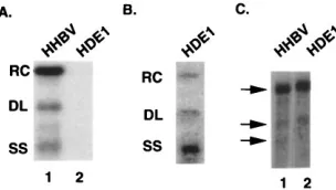
There are two defects in HDE1 replication.The character-istic pattern of HHBV DNA replication intermediates (ex-pressed from plasmid 413-2) seen by Southern blot analysis consists of RC, DL, and a 3-kb minus-strand (single-strand [SS]) species (Fig. 3A, lane 1). Initial characterization of HDE1 replication intermediates by Southern blot analysis in-dicated a substantial decrease in the amount of viral DNA (Fig. 3A, lane 2). The decrease in the amount of DNA synthe-sized by HDE1 relative to HHBV was not a result of decreased transfection efficiency of the HDE1 plasmid because capsid production from HDE1 was comparable to that from HHBV (see below; also data not shown). A longer autoradiographic exposure of the Southern blot indicated that the relative ratios of the RC, DL, and SS forms were aberrant (Fig. 3B). While the wild-type HHBV forms, RC, DL, and SS, were present at relative levels of approximately 7:1:1 (Fig. 3A, lane 1), the relative levels of these forms were approximately 1:1:7 for HDE1 (Fig. 3B). From these data we concluded that there are two defects in HDE1 DNA synthesis. One defect occurs prior to or during minus-strand DNA synthesis, resulting in the decreased amount of DNA. A second defect occurs during plus-strand DNA synthesis, resulting in aberrant ratios of
rep-lication intermediates. Intrigued by the initial observation of these defects, we set out to determine at which step during hepadnavirus replication each defect can be detected and whether each defect reflects failures in cis or in trans.
The level of HDE1 pregenomic RNA is equivalent to the level of wild-type HHBV.Since pregenomic RNA is the tem-plate for reverse transcription and for translation of the pro-teins required for replication, a decrease in the levels of HDE1 pregenomic RNA could account for the decrease in HDE1 DNA detected by Southern blot analysis. To determine the level of HDE1 pregenomic RNA, we transfected parallel plates of LMH cells with plasmids expressing HDE1 and HHBV. Polyadenylated RNA was isolated 3 days posttransfec-tion. Northern blot analysis showed that the level of HDE1 poly(A) RNA (Fig. 3C, lane 2) was equivalent to the level of wild-type HHBV RNA (Fig. 3C, lane 1). Therefore, the de-crease in HDE1 DNA cannot be explained by a dede-creased level of HDE1 pregenomic RNA.
HDE1 pregenomic RNA is encapsidated efficiently.One of the first steps in DNA synthesis is encapsidation of the pre-genomic RNA and P protein by the C protein. It seemed
FIG. 2. (A) Schematic representation of (1) the terminally redundant, HDE1 pregenomic RNA, (2) the HDE1 P protein gene, and (3) the HDE1 core protein gene. T.P., terminal protein domain; R.T., reverse transcription domain; RNase H, RNase H domain; R, terminally redundant region. Rectangles on pregenomic RNA represent the 12-nt direct repeats, DR1 and DR2.o, region of the HHBV sequence that is substituted with DHBV sequence. (B) Schematic representation of pregenomic RNA from mutant 530-1 that has the 59half (nt 403 to 902) of the chimeric region restored to wild-type HHBV sequence.
FIG. 3. (A) Southern blot analysis of viral DNA from cytoplasmic capsids. Lane 1, wild-type HHBV; lane 2, HDE1. (B) Longer exposure of the autoradio-gram of HDE1. Indicated at the left are the positions of the RC, DL, and SS DNA forms of viral DNA. (C) Northern blot analysis of HHBV and HDE1 poly(A) RNA isolated from transfected LMH cells. Viral RNA was detected by hybridization with a32P-labeled RNA probe specific for HHBV plus-strand
nucleic acid. Arrows at the left indicate the 3.3-kb pregenomic RNA and the two subgenomic RNAs, 2.1 and 1.8 kb.
on November 9, 2019 by guest
http://jvi.asm.org/
[image:3.612.152.465.75.276.2] [image:3.612.360.512.571.657.2]possible that the chimeric HDE1 proteins and/or RNA might not interact appropriately for efficient encapsidation, which could account for the decrease in HDE1 DNA. Although the pregenomic RNA was found to be present at wild-type levels, the level or stability of the chimeric replication proteins, P and C, could have been reduced. To assay the ability of the HDE1 proteins and pregenomic RNA to interact during encapsida-tion, we isolated intact core particles (capsids) from trans-fected LMH cells and ran duplicate sets of HDE1 and wild-type HHBV capsids on a nondenaturing agarose gel (Fig. 4) (3). Because the P protein is required for encapsidation of the pregenomic RNA (1, 11), HHBV and HDE1 mutants null for
P protein function (HHBVP2and HDE1P2) were included as
negative controls for encapsidation. One set of samples was transferred to a nylon membrane, capsids were disrupted, and encapsidated, plus-strand nucleic acid was detected by an HHBV, subgenomic RNA probe (Fig. 4A). A second set of samples was transferred to a nitrocellulose membrane, and capsids were detected by immunostaining with anti-DHBV core antibody (Fig. 4B). The ratio of total nucleic acid to core protein was interpreted as a measure of encapsidation. The results from multiple independent repetitions of this assay indicated that HDE1 pregenomic RNA was encapsidated (Fig. 4A and B, lanes 3) as well, or nearly as well, as was HHBV pregenomic RNA (Fig. 4A and B, lanes 1). Capsids composed of HHBV protein migrated faster than capsids composed of HDE1 proteins (Fig. 4B). The difference in mobility of the HDE1 and HHBV capsids appears to be a function of the capsid subunit protein (data not shown). Consistent with the data from the above-described encapsidation assay, an RNase protection assay for encapsidation showed that the ratio of HDE1 poly(A) RNA to core RNA was comparable to the wild-type HHBV ratio (data not shown). Therefore, an inabil-ity to encapsidate the HDE1 pregenomic RNA did not account for the decrease in HDE1 DNA synthesis. Since the HDE1 pregenomic RNA and trans factors were present in sufficient quantity for the encapsidation reaction, it is reasonable to assume that they are present in sufficient quantity for other steps in replication as well. From the Northern and encapsi-dation analyses, we conclude that the decrease in HDE1 DNA occurs during minus-strand DNA synthesis, after or indepen-dent of encapsidation.
The decrease in HDE1 minus-strand DNA synthesis occurs prior to synthesis of 111 nt of minus-strand DNA.We wanted to identify the step during HDE1 minus-strand DNA synthesis that was defective. Toward that end, we performed primer
extension analysis of the 59end of minus-strand DNA to
de-termine if the position of the 59 terminus of HDE1
minus-strand DNA was normal and if the amount of 59 termini was
comparable to that of HHBV. The 59end of the
oligonucleo-tide used for the primer extension analysis annealed 111 nt
away from the 59end of minus-strand DNA. Using this
oligo-nucleotide, we mapped the 59 end of wild-type HHBV to nt
2542, which corresponds to nucleotide C in the UUAC se-quence overlapping DR1 (Fig. 5A, lane 2). Primer extension
analysis showed that the 59end of HDE1 minus-strand DNA
mapped to the same position as determined for wild-type
HHBV (Fig. 5A, lane 1). In addition, no other 59termini of
minus-strand DNA were detected between the normal
termi-nus and the 39 end of the RNA template (data not shown).
Normalized to encapsidated plus-strand nucleic acid (Fig. 5B), synthesis of the first 111 nt of minus-strand DNA was less than 10% of that for wild-type HHBV. Therefore, the decrease in total HDE1 DNA can be explained as the result of an early defect in minus-strand DNA synthesis.
To corroborate the magnitude of the defect in HDE1 DNA synthesis, we performed a variation of the encapsidation assay. Duplicate sets of capsids were electrophoresed through an agarose gel, transferred to two nylon membranes, and hybrid-ized with a probe specific for HHBV minus- or plus-strand DNA (data not shown). We found for HDE1 that the level of minus-strand DNA relative to plus-strand DNA from dis-rupted capsids was reduced to less than 10% of the level for wild-type HHBV. This finding was consistent with the results from Southern blot analysis (Fig. 3A and data not shown). The results from the Southern, Northern, encapsidation, and primer extension analyses indicate that there is a defect during HDE1 DNA synthesis that occurs prior to synthesis of 111 nt of minus-strand DNA and results in a substantial reduction in the level of DNA synthesis. These data also indicate that there is a defect during HDE1 plus-strand DNA synthesis that re-sults in an altered ratio of RC DNA to DL DNA and less total plus-strand DNA relative to minus-strand DNA.
Are the defects in HDE1 DNA synthesis manifest incis, in
trans, or both? The HDE1 C protein has one amino acid
substitution, while the P protein and pregenomic RNA are chimeric (Fig. 2). Any of these participants in DNA synthesis
[image:4.612.379.500.491.643.2]FIG. 4. HDE1 pregenomic RNA is encapsidated at a level that is comparable to that of wild-type HHBV. (A) HHBV, HDE1, HHBVP2, and HDE1P2were transfected separately into LMH cells. Three days posttransfection, viral capsids were isolated and subjected to nondenaturing agarose gel electrophoresis. Cap-sids were transferred to a nylon membrane, lysed in situ, and probed for plus-strand nucleic acid. Lane 1, HHBV; lane 2, HHBVP2; lane 3, HDE1; lane 4, HDE1P2. (B) Duplicate samples were transferred to a nitrocellulose membrane, and capsids were detected by immunostaining with anti-DHBV core antibody. Lane 1, HHBV; lane 2, HHBVP2; lane 3, HDE1; lane 4, HDE1P2.
FIG. 5. Synthesis of the first 111 nt of HDE1 minus-strand DNA is less than 10% of the level for wild-type HHBV. (A) Primer extension analysis of viral DNA isolated from transfected LMH cells to determine the 59end of HDE1 minus-strand DNA. Lane 1, HDE1; lane 2, HHBV; lanes 3 to 6, sequencing ladder of cloned wild-type HHBV DNA. The 59 end of the32P-end-labeled
oligonucleotide used for primer extension analysis annealed 111 nt from the 59
end of HHBV minus-strand DNA. The arrow indicates the position of the 59end of minus-strand DNAs at nt 2542. (B) Intact viral capsids from transfected LMH cells were subjected to nondenaturing agarose gel electrophoresis, transferred to a nylon membrane, and probed for plus-strand nucleic acid.
on November 9, 2019 by guest
http://jvi.asm.org/
could contribute to the defects in DNA synthesis. To deter-mine whether the HDE1 defects are manifest in cis or in trans, we performed a series of genetic complementation assays. In each assay, we examined the level of minus-strand DNA and the ratios of replication intermediates. If the defect in minus-or plus-strand HDE1 DNA synthesis could be cminus-orrected by adding wild-type HHBV protein, then we would conclude that the HDE1 proteins were responsible for the defects in minus-strand DNA synthesis. If the HDE1 defect is not comple-mented by wild-type HHBV protein, assuming that the possi-bility of a dominant negative has been eliminated, then we would conclude that the defects during HDE1 DNA synthesis were manifest in cis.
The HDE1 C protein functions as wild-type HHBV C pro-tein.The HDE1 C protein has a glutamic acid-to-lysine sub-stitution at its carboxy-terminal residue relative to HHBV C protein. To determine the possible role of HDE1 C protein in either of the two defects, HDE1 was cotransfected with a
mutant of HHBV null for C protein (HHBVC2). Viral
repli-cation intermediates from cytoplasmic capsid particles were digested with restriction enzymes to distinguish the two poten-tially replicating genomes (HDE1 and HHBV) and examined by Southern blot analysis using a plus-strand-specific RNA probe. An aliquot of viral DNA from a cotransfection of
HHBV and HHBVC2was digested with HindIII. The
restric-tion enzyme HindIII uniquely cleaves HHBV DNA and not
HHBVC2DNA. Southern blot analysis of an undigested
ali-quot from the cotransfection of HHBV and HHBVC2showed
total DNA synthesis from both genomes (Fig. 6A, lane 1). Southern blot analysis of the aliquot that was digested with
HindIII showed that the RC DNA from the wild-type HHBV
genome was converted to a linear conformation that comi-grated with the DL species (Fig. 6A, lane 2). The remaining uncut RC DNA represented viral DNA produced from the
HHBVC2genome (Fig. 6A, lane 2). To distinguish replication
products from the cotransfection of HDE1 and HHBVC2, we
took advantage of the unique PstI site in HHBVC2and the
presence of three HindIII sites in HDE1. PstI digestion of viral
DNA from the cotransfection of HDE1 and HHBVC2showed
only a linear 3.0-kb molecule (Fig. 6A, lane 4), indicating that the HDE1 C protein had successfully complemented the
HH-BVC2 virus. HindIII digestion of viral DNA from the same
cotransfection showed a 3.0-kb relaxed circular molecule (Fig. 6A, lane 5), also indicating that HDE1 C protein had
success-fully complemented the HHBVC2 virus. Notice that the
amount of RC DNA in lane 3 (Fig. 6A) is comparable to the amount of 3.0-kb linear DNA in lane 4 and to the amount of RC DNA in lane 5. These data indicated that most or all of the plus-strand DNA species from the cotransfection of HDE1 and
HHBVC2were from the HHBVC2genome. From these
find-ings, we conclude that the HDE1 C protein is functional for DNA synthesis.
We also examined the overall level and pattern of
replica-tion intermediates from the cotransfecreplica-tion of HHBVC2and
HHBV or HHBVC2and HDE1 by Southern analysis using a
minus-strand specific probe (Fig. 6B). Southern blot analysis indicated that HDE1 C protein supports normal HHBV DNA replication compared with HHBV C protein (Fig. 6). Both the level and the ratio of replication intermediates made by HDE1 C protein were similar to those of the wild-type HHBV co-transfection (Fig. 6B). Thus, the HDE1 C protein does not contribute to the defects in HDE1 DNA synthesis.
HDE1 replication proteins, P and C, complement HHBV pregenomic RNA. The chimeric region of HDE1 is partially contiguous with a portion of the P open reading frame often referred to as a spacer region because of its ability to tolerate substitutions and deletions (4, 16, 24) and because the nucle-otide sequence within the region is less well conserved than are other parts of the genome. The amino acid sequence motifs predicted to code for reverse transcriptase and RNase H ac-tivities are not included within the chimeric region. However, by analogy to DHBV P protein, the chimeric region encodes the tyrosine residue that is linked to minus-strand DNA (33, 34). In light of these observations, it seemed possible that the HDE1 P protein could contribute to the defects in HDE1 DNA synthesis.
Our assay to determine the role of the HDE1 P protein in the defects that occur during HDE1 DNA synthesis was as follows. We constructed encapsidation-negative variants of both HDE1 (ENHDE1) and HHBV (ENHHBV) that were competent for donation of their respective C and P proteins but could not encapsidate their pregenomic RNAs and thus could not replicate themselves. We also constructed a variant of HHBV that was defective for the expression of the C and P
proteins (HHBVP2C2). The HHBVP2C2pregenomic RNA
is wild-type for the cis-acting sequences for DNA synthesis and is competent for DNA replication. Each of these encapsida-tion-negative mutants, ENHDE1 or ENHHBV, was
cotrans-fected separately into LMH cells with HHBVP2C2. Three
days posttransfection, viral replication intermediates were iso-lated from cytoplasmic capsid particles and analyzed. The lev-els and ratios of replication intermediates were determined by Southern blot analysis, using HHBV, strand-specific, sub-genomic probes (Fig. 7A, lanes 1 and 2), and the levels of minus-strand DNA were confirmed by primer extension anal-ysis (data not shown).
[image:5.612.121.239.70.156.2]The relative quantities of HHBV DNA produced by the HDE1 proteins and by the HHBV proteins from the wild-type pregenomic RNA were determined by normalization to the level of encapsidated plus-strand nucleic acid as determined by the encapsidation assay described above (Fig. 7B, lanes 1 and 2). While the ratios of RC, DL, and SS DNA forms synthesized by HDE1 P and C protein were similar to those of the HHBV P and C proteins, the level of DNA synthesis by HDE1 P and C proteins was approximately 50% of the level for the wild-type trans complementation. Clearly, the reduction in DNA synthesis by HDE1 C and P protein seen in this complemen-tation experiment can account for only a fraction of the orig-inally observed defect that occurs during HDE1 minus-strand DNA synthesis. Because the ratios of the replication interme-diates made by the HDE1 P and C proteins were similar to
FIG. 6. Southern blot and restriction enzyme analysis shows that HDE1 core protein supports normal HHBV DNA replication. (A) Lane 1, viral DNA iso-lated from a cotransfection of HHBV and HHBVC2plasmid DNA. Lane 2, HindIII digest of viral DNA isolated from a cotransfection of HHBV and HH-BVC2plasmid DNA. HindIII cleaves wild-type HHBV DNA but not HHBVC2 DNA. Lanes 3 to 5, viral DNA from cotransfection of HDE1 and HHBVC2. Lane 4, viral DNA is cleaved with PstI. PstI cleaves wild-type HHBV DNA and not HDE1 DNA. Lane 5, viral DNA is cleaved with HindIII. HindIII cleaves only HDE1 DNA. Viral DNAs detected with a plus-strand specific probe. (B) Lane 1, viral DNA from cotransfection of HDE1 and HHBVC2; lane 2, viral DNA from cotransfection of HHBV and HHBVC2. Viral DNAs were detected with a minus-strand specific probe.
on November 9, 2019 by guest
http://jvi.asm.org/
those made by HHBV P and C proteins, these data indicate that the HDE1 trans factors are not responsible for the defect that occurs during plus-strand DNA synthesis. Additional re-sults (data not shown) from Southern blot analysis of restric-tion digests of viral DNA from cotransfecrestric-tions of HDE1 with
HHBVP2or HHBVP2C2are consistent with the data shown
here.
Wild-type HHBV proteins do not complement HDE1 pre-genomic RNA.The ability of the HDE1 trans factors to com-plement HHBV pregenomic RNA indicated the possibility of one or more novel cis-acting elements involved in minus- and plus-strand DNA synthesis. To confirm the cis-acting nature of the two defects in HDE1 DNA synthesis, wild-type HHBV proteins were provided in trans from the expression construct ENHHBV to a variant of HDE1 that is defective for
expres-sion of functional C and P protein (HDE1P2C2). Southern
blot analysis of viral DNA from cotransfections of ENHHBV
plus plasmids that express HDE1P2C2or HHBVP2C2
pre-genomic RNA showed that the wild-type HHBV proteins did not significantly rescue either of the two defects in HDE1 DNA synthesis (Fig. 7C, lanes 1 and 2). Normalized to the level of encapsidated plus-strand nucleic acid (Fig. 7D, lanes 1 and 2), the level of minus-strand DNA from HDE1 pregeno-mic RNA was not significantly improved by wild-type HHBV
trans factors.
When HDE1 was cotransfected with HHBV, no reduction in HHBV replication was observed, indicating that the HDE1 replication proteins were not acting in a dominant negative manner (data not shown). The results of all the genetic com-plementation assays described here lead us to conclude that the defects in HDE1 minus- and plus-strand DNA synthesis result from the substitution of the nucleic acid sequence and are manifest primarily in cis.
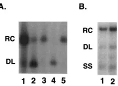
The defect during HDE1 plus-strand DNA synthesis can be rescued by restoring nt 403 to 902 to HHBV sequence.In an effort to locate the cis-acting sequences involved, and if possi-ble to separate the minus- and plus-strand defects, we
con-structed a variant of HDE1, named 530-1, that has the 59half
of the chimeric region (nt 403 to 902) restored to wild-type HHBV sequence (Fig. 2B). Characterization of cytoplasmic replication intermediates from 530-1 by Southern blot analysis showed that the ratios of replication intermediates were re-stored (Fig. 8A and B, lanes 1) to the wild-type HHBV ratios
(Fig. 8A, lane 3). However, the level of 530-1 minus-strand DNA synthesis (Fig. 8A, lane 1) relative to encapsidated plus-strand nucleic acid (Fig. 8C, lane 1) was not improved from the level seen with HDE1 (Fig. 8A and C, lanes 2).
Because the defects during HDE1 DNA synthesis were man-ifest primarily in cis, we constructed a variant of 530-1
(530-1P2) that did not express full-length P protein. To look for
res-cue of the defects during DNA synthesis by wild-type HHBV
proteins, 530-1P2was cotransfected with ENHHBV.
Cotrans-fections of ENHHBV with either HHBVP2or HDE1P2were
included as controls. Replication intermediates were examined by Southern blot analysis and the encapsidation assay. As ex-pected, the level and ratios of replication intermediates from
530-1P2remained unchanged by the addition of HHBV
pro-teins (data not shown). From our analyses of 530-1 (Fig. 8 and data not shown), we concluded that cis-acting sequences for
plus-strand DNA synthesis are contained within the 59half (nt
403 to 902) of the HDE1 chimeric region. And, the cis-acting sequences that would be sufficient to restore minus-strand
DNA synthesis are not contained within the 59 half of the
chimeric region.
DISCUSSION
We have examined hepadnavirus DNA replication by char-acterization of a chimeric heron/duck hepatitis B virus. Our results indicate that the chimera, HDE1, has two defects in DNA synthesis. One defect occurs early during minus-strand DNA synthesis (Fig. 1a to d) and results in less than 10% of wild-type HHBV DNA synthesis. The second defect occurs during plus-strand DNA synthesis (Fig. 1f to j) and results in
aberrant ratios of replication intermediates (Fig. 1h9 and j).
Our results also indicate that both the minus- and plus-strand HDE1 DNA synthesis defects are manifest primarily in cis. We interpret these data to indicate the presence of one or more formerly unrecognized cis-acting elements for minus- and plus-strand DNA synthesis.
By restoring the 59 half of the HDE1 chimeric region to
HHBV sequence (Fig. 2B, mutant 530-1), we determined that
cis-acting sequence for plus-strand DNA synthesis is located
within the 59half of the chimeric region (nt 403 to 902). The
inability to restore minus-strand DNA synthesis by restoration
of the 59 half of the chimeric region could indicate that the
[image:6.612.99.261.70.158.2]cis-acting sequence for minus-strand DNA synthesis is not
FIG. 7. HDE1 P and C proteins trans complement HHBVP2C2. (A) South-ern blot analysis of viral DNA from cotransfection of HHBVP2C2with eith-er ENHDE1 (lane 1) or ENHHBV (lane 2). The positions of the RC, DL, and SS replication intermediates are indicated on the left. (B) Intact capsids were lysed and probed for encapsidated HHBV plus-strand nucleic acid as described in Materials and Methods. Lane 1, viral nucleic acid from cotransfection of HHBVP2C2and ENHDE1; lane 2, viral nucleic acid from cotransfection of HHBVP2C2and ENHHBV. (C) HHBV C and P protein do not rescue the defects in HDE1 minus- and plus-strand DNA synthesis. Southern blot analysis of viral DNA from cotransfection of ENHHBV with either HHBVP2C2(lane 1) or HDE1P2C2(lane 2). (D) Intact capsids were lysed and probe for encapsi-dated HDE1 or HHBV plus-strand nucleic acid. Lane 1, viral nucleic acid from cotransfection of HDE1P2C2and ENHHBV; lane 2, viral nucleic acid from cotransfection of HHBVP2C2and ENHHBV.
FIG. 8. Restoration of HHBV sequence from nt 403 to 902 restores ratios of replication intermediates. (A) Southern blot analysis of viral DNA from core particles shows that the ratios of 530-1 replication intermediates are similar to the HHBV ratios. The positions of the RC, DL, and SS replication intermediates are indicated on the left. (B) Longer exposure of the autoradiogram shown in panel A shows ratios of 530-1 replication intermediates. (C) Levels of DNA synthesis shown in A were normalized to the level of encapsidated plus-strand nucleic acid shown in panel C.
on November 9, 2019 by guest
http://jvi.asm.org/
[image:6.612.337.529.535.658.2]located in the 59half of the chimeric region, or the cis-acting sequence may be located in both halves of the chimeric region, such that partial restoration was not sufficient to restore DNA synthesis. Alternatively, restoration of only half of the chimeric region could have resulted in a different defect that obscured any restoration of the original defect in DNA synthesis. Con-struction of variants with different portions of the chimeric region restored should aid in determining the location of the
cis element(s) involved in the defect that occurs during
minus-strand DNA synthesis.
As the pregenomic RNA (Fig. 2A) is the template for trans-lation of the replication proteins, C and P, and for reverse transcription (Fig. 1a to e), a decrease in this molecule could have accounted for the observed decrease in HDE1 DNA. In our first set of experiments, we showed that the observed decrease in HDE1 DNA does not result from a decrease in the level or stability of the chimeric pregenomic RNA (Fig. 3). We also showed that the decrease in HDE1 DNA does not result from a decrease in the level of the HDE1 C and P proteins (Fig. 4). Previously, two regions were shown to be required in
cis for DHBV encapsidation (3, 12). The first region, region I,
is within nt 2562 to 2652 and contains the RNA stem-loop (epsilon) that also was shown to be required for initiation of minus-strand DNA synthesis (23, 29, 31, 32) as well as RNA encapsidation (3, 12, 23). The second region, region II, is within nt 551 to 719. HDE1 has an HHBV region I, a DHBV region II, and a chimeric P protein, yet we found no significant reduction in encapsidation efficiency (Fig. 4). These findings could indicate that the encapsidation components are inter-changeable and that the mechanism of encapsidation is con-served among avian hepadnaviruses.
One potential caveat of equating plus-strand nucleic acid to total encapsidated nucleic acid would be the potential for un-derestimating the extent of encapsidation of a mutant that accumulates minus-strand DNA. If we underestimated the ex-tent of encapsidation, then we might have underestimated the severity of the DNA synthesis defects when we normalized the level of minus-strand DNA from Southern and primer exten-sion analyses to the level of encapsidated plus-strand nucleic acid.
In our second set of experiments, we examined the cis/trans nature of the two defects that occur during HDE1 DNA syn-thesis. We determined that the HDE1 C protein supports replication of HHBV pregenomic RNA at a level that is com-parable to that of HHBV C protein (Fig. 6), while the HDE1 P protein functions less well during minus-strand DNA syn-thesis than does the wild-type HHBV P protein (Fig. 7). We also showed that the majority of the HDE1 minus-strand de-fect is manifest in cis (Fig. 7), occurring prior to synthesis of 111 nt of minus-strand DNA (Fig. 5 and 1a to d). The HDE1 minus-strand defect could occur during priming on epsilon (Fig. 1a and b). However, encapsidation of HDE1 pregenomic RNA by HDE1 P protein is not significantly reduced, and previous work has shown that encapsidation and priming gen-erally correlate (24). The majority of the HDE1 minus-strand defect is manifest in cis, yet the stem-loop (epsilon) used for priming of minus-strand on HDE1 pregenomic RNA is derived from HHBV. A defect in priming of HDE1 minus-strand DNA would suggest that cis-acting sequences within nt 403 to 1364 are involved in some sort of tertiary or protein-RNA interac-tion important for this process. We do not favor this nointerac-tion, because previous studies (32) suggest that epsilon is sufficient in cis for the initiation of minus-strand DNA synthesis. An-other possible explanation for the HDE1 minus-strand defect is that there is a defect in template switching of the minus-strand DNA, and its attached P protein, from epsilon to the
acceptor site overlapping the 39copy of DR1 (Fig. 1b and c).
A template-switching defect could result from incompatible elements that are involved in proteRNA or RNA-RNA in-teractions. A third possible defect could occur at the time of posttransfer elongation of minus-strand DNA (Fig. 1c to f). A fourth possible explanation for the decrease in HDE1 DNA synthesis would be a defect in RNase H activity (Fig. 1c to f). However, we think that this explanation is unlikely because the majority of the defect is manifest in cis.
The defect during HDE1 plus-strand DNA synthesis could occur during plus-strand primer translocation from DR1 to DR2 (Fig. 1f and g), posttranslocation elongation (Fig. 1g and h), the intrastrand template switch (circularization) (Fig. 1h and i), or postcircularization elongation (Fig. 1j). Currently, we cannot distinguish between these possibilities. However, we think that the HDE1 plus-strand DNA synthesis defect occurs at an early point during plus-strand DNA synthesis (Fig. 1f to i), resulting in aberrant ratios of replication intermediates. Because the primer translocation step (27) (Fig. 1f and g) and the intramolecular template switch (Fig. 1h and j) are pro-cesses that involve cis-acting sequences, it is interesting to speculate that HDE1 is defective for one or both of these events. Additional experimentation will allow us to determine more precisely the nature of the defect in HDE1 plus-strand DNA synthesis.
The experiments shown here do not distinguish whether the HDE1 defects are a result of incompatibility of DHBV and HHBV cis elements, or if the HDE1 defects result from incom-patibility of HDE1’s DHBV cis element(s) with HHBV pro-teins. If the cis-acting sequences within the HHBV sequence of HDE1 interact with other (HHBV) cis-acting sequences that are outside of nt 403 to 1358 to effect minus- and/or plus-strand DNA synthesis, then by substituting additional regions of HHBV sequence in HDE1 with DHBV sequence and looking for restoration of function, we might identify other potential
cis-acting partners. It may be that by substituting additional
DHBV sequence into HDE1, we restore wild-type function. This finding would suggest that the HDE1 defect lies solely in interspecies nucleic acid incompatibilities. Alternatively, if the
cis-acting sequence within the DHBV sequence of HDE1
in-teracts with the C and/or P protein, then by providing the DHBV versions of C and/or P, we might be able to restore func-tion. Of course, by using wild-type DHBV proteins in an at-tempt to affect cis element-protein compatibility, we may also create a new incompatibility of HHBV sequence or protein with DHBV protein. Finally, it may be that additional chimer-ization of HDE1 protein or nucleic acid alone is insufficient and that a particular combination of chimerized protein and nucleic acid will be required to restore wild-type function to HDE1. This finding would indicate that a specific protein-nucleic acid interaction was involved.
One possible explanation for the defects in HDE1 synthesis is that an aberrant stabilization of higher-order RNA or DNA structure could interfere with DNA synthesis. The results shown here do not allow us to say whether the cis-acting HDE1 de-fects result from unmet species-specific sequence requirements or whether the chimeric sequence is unable to form a partic-ular secondary structure. The answer to this question awaits a much finer mutational analysis of the nucleotide sequence.
During hepadnavirus DNA synthesis, there are three tem-plate switches or strand transfers: the minus-strand transfer (Fig. 1b and c), the plus-strand primer translocation (Fig. 1f and g), and the plus-strand template switch (circularization) (Fig. 1h to j). It is reasonable to posit a juxtaposition of the donor and acceptor templates as part of the mechanism of these three processes. Specific cis-acting sequence might be
on November 9, 2019 by guest
http://jvi.asm.org/
involved in this juxtaposition. Thus, a higher-order RNA tem-plate structure could be involved in the minus-strand transfer and a specific minus-strand template structure could be in-volved in the two plus-strand template switches. It is even conceivable that the structure of the minus-strand DNA tem-plate could be influenced by a previous RNA temtem-plate struc-ture. The cis-acting sequences that are described in this report could be involved in determining the functional structure of the templates (either RNA or DNA) for the template switches. The discovery of cis-acting sequence for plus-strand DNA synthesis in the middle of the genome was unexpected and indicates that plus-strand DNA synthesis is a more complex process than originally conceived. cis-acting mutations that are so far from the known cis-acting sites of action for minus- and plus-strand DNA synthesis suggest that topology is important for both these processes.
ACKNOWLEDGMENTS
We thank Rey Carabeo, Terri Chiaverotti, Nicole Miller-Rank, and Ru Tian for contributions to preliminary studies related to this work. We thank Amy Qualey, Sandra Suh, and Karolyn Gulya for help in the laboratory. We thank Hans Will for HHBV plasmid DNA and Takashi Ishikawa and Don Ganem for HDE1. We are grateful to Mike Havert, Haiyan Jiang, Lou Mansky, Janet Mertz, Antonito Panganiban, and Bill Sugden for critical review of the manuscript, and we thank Ru Tian for especially excellent technical assistance.
This work was supported by NIH grants GM50263, CA09135, and CA07175.
REFERENCES
1. Bartenschlager, R., M. Junker-Neipmann, and H. Schaller. 1990. The P gene product of hepatitis B virus is required as a structural component for genomic RNA encapsidation. J. Virol. 64:5324–5332.
2. Buscher, M., W. Reiser, H. Will, and H. Schaller. 1985. Transcripts and the putative RNA pregenome of duck hepatitis B virus: implications for reverse transcription. Cell 40:717–724.
3. Calvert, J., and J. Summers. 1994. Two regions of an avian hepadnavirus RNA pregenome are required in cis for encapsidation. J. Virol. 68:2084– 2090.
4. Chang, L., R. Hirsch, D. Ganem, and H. Varmus. 1990. Effects of insertional and point mutations on the functions of the duck hepatitis B virus polymer-ase. J. Virol. 64:5553–5558.
5. Chang, L.-J., P. Pryciak, D. Ganem, and H. E. Varmus. 1989. Biosynthesis of the reverse transcriptase of hepatitis B viruses involves de novo translational initiation not ribosomal frameshifting. Nature (London) 337:364–368. 6. Chen, C., and H. Okayama. 1987. High-efficiency transformation of
mam-malian cells by plasmid DNA. Mol. Cell. Biol. 7:2745–2752.
7. Church, G. M., and W. Gilbert. 1984. Genomic sequencing. Proc. Natl. Acad. Sci. USA 81:1991–1995.
8. Condreay, L., C. Aldrich, L. Coates, W. Mason, and T. Wu. 1990. Efficient duck hepatitis B virus production by an avian liver tumor cell line. J. Virol.
64:3249–3258.
9. Ganem, D., and H. Varmus. 1987. The molecular biology of the hepatitis B viruses. Annu. Rev. Biochem. 56:651–693.
10. Hirsch, R., R. Colgrove, and D. Ganem. 1988. Replication of duck hepatitis B virus in two differentiated human hepatoma cell lines after transfection with cloned viral DNA. Virology 167:136–142.
11. Hirsch, R., J. Lavine, L. Chang, H. Varmus, and D. Ganem. 1990. Polymer-ase gene products of hepatitis B viruses are required for genomic RNA packaging as well as for reverse transcription. Nature (London) 344:552–555. 12. Hirsch, R., D. Loeb, J. Pollack, and D. Ganem. 1991. cis-acting sequences
required for encapsidation of duck hepatitis B virus pregenomic RNA. J. Vi-rol. 65:3309–3316.
13. Ishikawa, T., and D. Ganem. 1995. The pre-S domain of the large viral envelope protein determines host range in avian hepatitis B viruses. Proc. Natl. Acad. Sci. USA 92:6259–6263.
14. Kawaguchi, T., K. Nomura, Y. Hirayama, and T. Kitagawa. 1987. Establish-ment and characterization of chicken hepatocellular carcinoma cell line, LMH. Cancer Res. 47:4460–4464.
15. Kunkel, T. A., J. D. Roberts, and R. A. Zabour. 1987. Rapid and efficient site-specific mutagenesis without phenotypic selection. Methods Enzymol.
154:367–382.
16. Li, J., L. Cova, R. Buckland, V. Lambert, G. Deleage, and C. Trepo. 1989. Duck hepatitis B virus can tolerate insertion, deletion, and partial frameshift mutation in the distal pre-S region. J. Virol. 63:4965–4968.
17. Lien, J., C. Aldrich, and W. Mason. 1986. Evidence that a capped oligori-bonucleotide is the primer for duck hepatitis B virus plus-strand DNA synthesis. J. Virol. 57:229–236.
18. Lien, J., D. Petcu, C. Aldrich, and W. Mason. 1987. Initiation and termina-tion of duck hepatitis B virus DNA synthesis during virus maturatermina-tion. J. Vi-rol. 61:3832–3840.
19. Loeb, D., R. Hirsch, and D. Ganem. 1991. Sequence-independent RNA cleavages generate the primers for plus strand DNA synthesis in hepatitis B viruses: implications for other reverse transcribing elements. EMBO J. 10: 3533–3540.
20. Loeb, D., and R. Tian. 1995. Transfer of the minus strand of DNA during hepadnavirus replication is not invariable but prefers a specific location. J. Virol. 69:6886–6891.
21. Mandart, E., A. Kay, and F. Galibert. 1984. Nucleotide sequence of a cloned duck hepatitis B virus genome: comparison with woodchuck and human hepatitis B virus sequences. J. Virol. 49:782–792.
22. Maniatis, T., E. F. Fritsch, and J. Sambrook. 1989. Molecular cloning: a laboratory manual, 2nd ed. Cold Spring Harbor Laboratory, Cold Spring Harbor, N.Y.
23. Pollack, J., and D. Ganem. 1994. Site-specific RNA binding by a hepatitis B virus reverse transcriptase initiates two distinct reactions: RNA packaging and DNA synthesis. J. Virol. 68:5579–5587.
24. Radziwill, G., W. Tucker, and H. Schaller. 1990. Mutational analysis of the hepatitis B virus P gene product: domain structure and RNase H activity. J. Virol. 64:613–620.
25. Schlicht, J., R. Bartenschlager, and H. Schaller. 1989. The duck hepatitis B virus core protein contains a highly phosphorylated C terminus that is es-sential for replication but not for RNA packaging. J. Virol. 63:2995–3000. 26. Sprengel, R., E. Kaleta, and H. Will. 1988. Isolation and characterization of
a hepatitis B virus endemic in herons. J. Virol. 62:3832–3839.
27. Staprans, S., D. Loeb, and D. Ganem. 1991. Mutations affecting hepadnavi-rus plus-strand DNA synthesis dissociate primer cleavage from translocation and reveal the origin of linear viral DNA. J. Virol. 65:1255–1262. 28. Summers, J., and W. Mason. 1982. Replication of the genome of a hepatitis
B-like virus by reverse transcription of an RNA intermediate. Cell 29:403– 415.
29. Tavis, J. T., S. Perri, and D. Ganem. 1994. Hepadnavirus reverse transcrip-tion initiates within the stem-loop of the RNA packaging signal and employs a novel strand transfer. J. Virol. 68:3536–3543.
30. Tuttleman, J., C. Pourcel, and J. Summers. 1986. Formation of the pool of covalently closed circular viral DNA in hepadnavirus-infected cells. Cell
47:451–460.
31. Wang, G., and C. Seeger. 1993. Novel mechanism for reverse transcription in hepatitis B viruses. J. Virol. 67:6507–6512.
32. Wang, G., F. Zoulim, E. Leber, J. Kitson, and C. Seeger. 1994. Role of RNA in enzymatic activity of the reverse transcriptase of hepatitis B viruses. J. Virol. 68:8437–8442.
33. Weber, M., V. Bronsema, H. Bartos, A. Bosserhoff, R. Bartenschlager, and
H. Schaller.1994. Hepadnavirus P protein utilizes a tyrosine residue in the TP domain to prime reverse transcription. J. Virol. 68:2994–2999. 34. Zoulim, F., and C. Seeger. 1994. Reverse transcription in hepatitis B viruses
is primed by a tyrosine residue of the polymerase. J. Virol. 68:6–13.