0022-538X/96/$04.0010
Copyrightq1996, American Society for Microbiology
cis-Acting Elements Involved in Transcriptional Regulation of
the Herpes Simplex Virus Type 1 Latency-Associated
Promoter 1 (LAP1) In Vitro and In Vivo
KARINA SOARES,
1DONG-YOUN HWANG,
1RAMESH RAMAKRISHNAN,
2MARTIN C. SCHMIDT,
1DAVID J. FINK,
2ANDJOSEPH C. GLORIOSO
1*
Department of Molecular Genetics and Biochemistry, University of Pittsburgh School of Medicine,
1and Department of
Neurology and VA Medical Center, University of Pittsburgh,
2Pittsburgh, Pennsylvania 15261
Received 28 February 1996/Accepted 25 April 1996
Latency-associated promoter 1 (LAP1) of herpes simplex virus type 1 is required to generate a series of
latency-associated transcripts (LATs) in sensory neurons of latently infected animals. Sequence analysis and
DNA binding studies have suggested the existence of several
cis
-acting elements within LAP1 that are
poten-tially important for promoter function, although their role in LAT gene expression during latency is largely
unexplored. In this report, we present evidence that the LAP1 TATA box is essential for transcription initiation
in vitro. A reduction in LAT synthesis measured by in situ hybridization and reverse transcription-PCR
(RT-PCR) of rat brain tissue latently infected with a LAP1 TATA substitution virus demonstrated that this
sequence was required for full LAP1 activity in vivo. Analysis of additional site-directed and 5
*
-deletion
mutants of LAP1 by in vitro transcription-primer extension assays showed that upstream elements including
the USF and cyclic AMP response element (CRE) site specifically contributed to LAP1 function and that
sequences beginning at position
2
620 relative to the transcription start site were essential for full promoter
activity. The combination of deleting USF, CRE, and TATA completely abolished LAT expression in the brain,
identifying these as essential elements for the neuron-specific functioning of LAP1 during latency. Mutation of
the transcription start site did not abolish transcription, suggesting the absence of an initiator element.
However, one of the most exciting findings from this study is that the region downstream of the TATA box
appears to contain a true enhancer that is not only essential for transcription, but also functional when
positioned 1.6 kb downstream of the start site of transcription. It was concluded that (i) the TATA box was
essential for full transcriptional activity from LAP1 both in vitro and in vivo, (ii) the USF element and CRE
contribute to LAP1 function during latency in combination with the TATA element, (iii) multiple
trans
-acting
factors besides the USF- and CRE-binding proteins were required for full promoter activity in vitro, and (iv)
sequences downstream of the TATA box enhanced promoter activity in vitro.
One of the distinctive features of herpes simplex virus type
1 (HSV-1) is its ability to persist in a select population of host
neurons in a latent state (14, 43). During latency, multiple
copies of the viral genome are maintained within the nucleus
as nucleosome-bound episomes (9, 31, 44). Upon the
estab-lishment of latency within neurons of sympathetic and sensory
ganglia, a unique set of HSV-1 transcripts is encoded from
sequences within the repeat elements that flank the unique
long region of the viral genome (7, 33, 45, 52, 56). Since these
overlapping transcripts represent the only HSV-1 genes
ex-pressed during latency, they have been referred to as the
la-tency-associated transcripts (LATs).
Although the transcriptional control regions and origins of
the different LAT RNAs have not been fully elucidated, the
colinear 2-, 1.5-, and 1.45-kb LAT species are believed to be
derived via splicing and/or processing of an unstable primary
8.3-kb RNA transcribed from a TATA box almost 700 bp
upstream of the 5
9
end of the 2-kb LAT (for reviews, see
references 16, 54, and 55). Several lines of evidence identify
the sequences that span this TATA box, designated
latency-associated promoter 1 (LAP1), as being the promoter region
responsible for most of the LAT transcription during latency:
(i) KOS/29, a virus with a deletion of LAP1 at both loci, does
not express LAT during latency (6, 10, 11, 13); (ii) variant 1704,
which is similar to KOS/29 but retains some downstream
se-quence in one LAT locus, not only fails to express the 2-kb
LAT but is also negative for the weak in situ hybridization
signals specific for the large precursor transcript (33, 34); and
(iii) in the recombinant virus KOS/72, in which the genomic
copy of the rabbit
b
-globin gene was cloned downstream of the
TATA box, accurate initiation of the
b
-globin message
oc-curred 28 bp downstream of the TATA sequence with
concur-rent abolition of LAT expression in latently infected murine
trigeminal ganglia (13). However, the inability of recombinant
viruses such as KOS/62-3 and KOS/67-7 to express the lacZ or
NGF gene cassettes that were inserted at position
1
41 of
LAP1 suggests that sequences beyond the boundaries of LAP1
that are deleted in these two viruses may also be necessary for
expression (28). It was previously shown that the sequences
that lie between LAP1 and the 5
9
end of the 2-kb LAT,
des-ignated LAP2, can express the
b
-galactosidase reporter gene in
trigeminal ganglion neurons even when moved to an ectopic
locus in the viral genome (18). Recent studies suggest that
LAP1 and LAP2 may function differentially during lytic versus
latent infections (6). Accumulation of the 2-kb LAT during
lytic infection depends on LAP2 and is independent of LAP1
sequences, while during latency the reverse is true (6, 35).
The LAT loci in HSV-1 and HSV-2 share more than 80%
* Corresponding author. Mailing address: Department of Molecular Genetics and Biochemistry, E1240 Biomedical Science Tower, Univer-sity of Pittsburgh School of Medicine, Pittsburgh, PA 15261. Phone: (412) 648-8105. Fax: (412) 624-8997. Electronic mail address: Michele @server1.mgn.pitt.edu.
5384
on November 9, 2019 by guest
http://jvi.asm.org/
sequence identity and contain similarly organized groups of
putative cis-acting elements (25, 30). Although much
informa-tion has been inferred from transient-transfecinforma-tion and gel
re-tardation assays using neuronal and nonneuronal cell lines (2,
3), the relevance of most of these findings to LAT expression
during latency has yet to be evaluated in the context of viruses
with mutations or deletions in the LAT loci. Besides the TATA
box, the LAP1 region contains a CCAAT box homology and
potential recognition sequences for several transcription
fac-tors, including Sp1, AP-2, YY1, CREB, LPBF/MLTF/USF,
Egr-1, a POU-domain protein, and the HSV-1
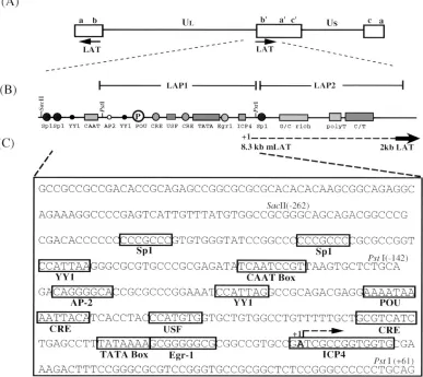
immediate-early transactivator, ICP4 (3, 4, 15, 24, 42, 58–60, 62) (see Fig.
1). In this study, we have used an in vitro transcription system
to characterize cis-acting elements of LAP1 required for
pro-moter activity. Mutations that affect LAP1 function in vitro
have also been tested for their contribution to LAT expression
during latency.
MATERIALS AND METHODS
Plasmid constructions.The sequences and positions of known and putative cis-acting elements in the LAP1 promoter of KOS strain of HSV-1 are as shown
in Fig. 1 (57). A series of chloramphenicol acetyltransferase (CAT) reporter gene constructs containing progressive 59deletions (see Fig. 4A) and site-directed mutations of putative cis-acting elements in the LAP1 promoter (see Fig. 2, 3, and 5) were constructed by using standard cloning techniques and oligonucle-otide-directed mutagenesis (48).
pUC.CAT and pGem.CAT, promoterless control templates, were constructed by cloning a 1.6-kb BglII-BamHI fragment containing the CAT gene and simian virus 40 early polyadenylation site from pSVOCAT into BamHI-linearized pUC19 and pGem7Zf(2), respectively. The simian virus 40 promoter in pSV2CAT was replaced by the adenovirus type 5 major late promoter (positions
2270 to133 relative to the transcription start site) to create pML.CAT, the positive-control plasmid. pLAP1[2142/161]CAT and pGemLAP1CAT were constructed by cloning the 203-bp PstI restriction fragment (nucleotides 118664 to 118866) (30) into pUC.CAT and pGemCAT, respectively. pLAP-R contains this sequence in reverse orientation relative to the CAT gene in pUC.CAT.
The HincII-SphI LAP fragment (nucleotides 117012 to 119290) subcloned into pTZ18U (United States Biochemical, Cleveland, Ohio) was subjected to oligo-nucleotide-directed in vitro mutagenesis using the Muta-Gene phagemid kit (Bio-Rad, Hercules, Calif.). The sequences of the oligonucleotides used are as follows: rT, (22) 59-CGGCACGGCCGCGCCCCCGCGGTACCGGCTGAGA TGACGCAGCAAA-39(250);DUC, (218) 59-CCGCTTTTATAAAGGCTGA GGGTACCAGGTGATGTAATTTTATTTT-39 (294); andDUCT, (22) 59
-CGGCACGGCCGCGCCCCCGCGGTACCAGGTGATGTAATTTTATTTT-39
[image:2.612.113.500.78.423.2](294). The positions of the 59and 39ends of the oligonucleotides relative to position11 of LAP1 (Fig. 1C) are indicated in parentheses. The unique restric-tion sites created for screening purposes are underlined. The prefix “r” indicates FIG. 1. Schematic representation of the LAT region of HSV-1. (A) Diagram of the prototypic arrangement of the HSV-1 genome indicating the location and direction of transcription of the LAT loci within the repeats (open boxes) that flank the unique long (UL) segment of the genome. The LAT region in the internal
repeats is expanded below to show the LAT promoter region, which is structurally and functionally divided into the TATA-containing LAP1 and TATA-less LAP2 regions (6, 17). US, unique short region. (B) Line drawing of the LAT promoter region depicting the structural organization of putative cis-acting transcriptional
elements of LAP1 and LAP2. The location of the predicted start site of the primary 8.3-kb LAT species (dashed line) and the 59end of the 2-kb LAT (boldface arrow) are as shown. (C) Nucleotide sequence of LAP1 in the KOS strain of HSV-1 (57) showing the spatial relationship of the putative cis-acting elements relative to the TATA box. The consensus binding sites of potential trans-acting factors of neuronal and nonneuronal origin (boxes), predicted start site of the 8.3-kb primary LAT (11), and the direction of transcription from LAP1 (bent arrow) are indicated. All restriction enzyme sites relevant to this study are numbered with respect to the A residue at11 of LAP1.
VOL. 70, 1996 TRANSCRIPTIONAL REGULATION OF HSV-1 LAP1 5385
on November 9, 2019 by guest
http://jvi.asm.org/
replacement of a specific transcription element with a unique restriction site such that spacing is maintained, while theD’s inDUC andDUCT indicate deletions of 43 and 53 bp, respectively. The site-directed mutant plasmids pLAP1[rT]CAT, pLAP1[DUC]CAT, and pLAP1[DUCT]CAT were then derived from the corre-sponding mutagenized HincII-SphI subclones by cloning the respective 203-bp PstI restriction fragment (positions2142 to161) into pUC.CAT. The presence of the mutation was confirmed by DNA sequencing. pLAP1[rU]CAT, pLAP1[rC]CAT, pLAP1[rINR]CAT, and pLAP1[rINRKO]CAT were derived by oligonucleotide-directed mutagenesis of single-stranded DNA derived from pGEM.LAP1CATpA. The sequences of the oligonucleotides used are as follows: rU, (288) 59-AATTACATCACCTACGAAGCTTGTGCTGTGGCCTGTT-39 (264); rC, (260) 59-GTGGCCTGTTTTTGCGAAGCTTGCTCAGCCTTTATA AAAGC-39(220); rINR, (217) 59-GGCGCGGCCGTGCCCATTCCGGATG GTGCG-39(114); and rINRKO, (217) 59-GGCGCGGCCGTGGAGGCCTGG GATGGTGCG-39(114). The HindIII site in the multiple cloning site of the parent vector was used to derive plasmids pLAP1[242/161]CAT and pLAP1[272/161]CAT from pLAP1[rU]CAT and pLAP1[rC]CAT, respectively, by removing the intervening upstream LAP1 sequences by HindIII digestion and religation.
The EagI site at position211 of LAP1 was converted to a BglII site by filling in with Klenow fragment and ligation of BglII linkers to produce pLAP1[2142/ Bg*/161]. pLAP1[2142/Bg*CAT/161] was constructed by inserting the CAT gene at the newly created BglII site at position211 (denoted Bg*) rather than at the downstream BamHI site of the vector. This strategy effectively places the 59
end of the CAT gene at211 and places LAP1 sequences from211 to161 downstream of the entire CAT gene cassette. pLAP1[2142/Bg*]CAT was con-structed from pLAP1[2142/Bg*CAT/161] by replacing the Bg*-BamHI frag-ment spanning LAP1[211/161]CAT sequences with the 1.6-kb BglII-BamHI CAT gene fragment used before. pLAP1[211/161]CAT was created by subclon-ing the Bg*-BamHI restriction fragment from pLAP1[2142/Bg*/161]CAT into BamHI-linearized pUC19 vector. pLAP1[279/161]CAT was constructed by in-sertion of a T4 DNA polymerase blunt-ended 140-bp HphI-PstI restriction frag-ment into the EcoRV site of pBSIISK2followed by insertion of the CAT reporter gene at the downstream BamHI site. This cloning strategy is expected to add an extra 3 bases to the size of the primer extension product. pLAP1[2262/
161]CAT was constructed in two steps: (i) the Bg*-BamHI fragment from pLAP1[2142/Bg*/161]CAT containing LAP1[211/161]CAT was inserted into BamHI-linearized pBSIISK2, and (ii) sequences between the SacII site in the multiple cloning site of the vector and the SacII site at position141 of the LAP1 sequence were replaced with a 303-bp SacII fragment of LAP1 corresponding to positions2262 to141. Similarly, pLAP1[2620/161]CAT was constructed as follows: the Bg*/BamHI fragment containing LAP1[211/161]CAT sequences from pLAP1[2142/Bg*/161]CAT was inserted into BamHI-linearized pUC19. Next, sequences between the BanII site in the vector and the BanII site at position153 of LAP1 in pBanII/Bg*CAT were replaced with a 673-bp BanII fragment of LAP1 corresponding to positions2620 to153 to give pLAP1[2620/
161]CAT.
Cell culture and nuclear extract preparation.Spinner cultures of HeLa cell strain S3 (ATCC CCL 2.2), grown in Joklik’s minimal essential medium (ICN Biomedicals, Aurora, Ohio) supplemented with 5% fetal calf serum (Gibco-BRL), were harvested in the exponential growth phase at a density of 2.83105
cells per ml, and nuclear extract was prepared as described by Dignam et al. (12).
In vitro transcription-primer extension assay.In vitro transcription reaction mixtures (50ml) contained 24ml of HeLa nuclear extract (75mg of protein), 6ml of D buffer (20 mM HEPES [N-2-hydroxyethylpiperazine-N9-2-ethanesulfonic acid], K1[pH 7.9], 20% [vol/vol] glycerol, 0.1 M KCl, 0.2 mM EDTA, 0.5 mM phenylmethylsulfonyl fluoride, and 0.5 mM dithiothreitol), 2mg of supercoiled plasmid template, 6.25 mM MgCl2, 0.5 mM ribonucleoside triphosphates, and 40
U of RNasin (Promega). Where indicated,a-amanitin was added to a final concentration of 8mg/ml, a concentration low enough to selectively inhibit RNA polymerase II transcription (27). After 1 h of incubation at 308C, the reactions were halted by addition of 150ml of stop solution (20 mM EDTA [pH 8.0], 0.2 M NaCl, 5ml of 2.5-mg/ml proteinase K, 1% sodium dodecyl sulfate [SDS]). The reaction products were extracted once with 200ml of phenol-chloroform-isoamyl alcohol (25:24:1) and then with an equal volume of chloroform. A 300-ml volume of 0.3 M sodium acetate was added to the resulting aqueous phase, and the reaction products were precipitated with 1 ml of 95% ethanol. The pellet ob-tained was washed with 70% ethanol, vacuum dried, and dissolved in 10ml of hybridization solution (10 mM Tris [pH 7.9], 1 mM EDTA [pH 7.9], 0.25 M KCl, and 3 ng of an end-labelled oligonucleotide primer that is complementary to sequences in the CAT gene). Two different CAT primers were employed, de-pending on the length of the expected extension product from the specific promoter template. The 18-mer CAT primer (59 -AGCTCCTGAAAATCTCGC-39), complementary to CAT sequence between positions16 and 124 with respect to the 59end of the CAT reporter gene, was used for promoter constructs with LAP leader up to 161. The 24-mer CAT primer (59-GCCATTGGGA TATATCAACGGTGG-39), complementary to CAT sequences between161 and185, was used to assay transcription activity specifically from constructs pLAP1[2142/Bg*]CAT and pLAP1[2142/Bg*CAT/161], in which the CAT gene was placed at position211 relative to the normal LAP1 initiation site (see Fig. 5C). Following hybridization at the appropriate temperature (558C for the 18-mer CAT primer or 658C for the 24-mer CAT primer) for 1 h, 25ml of reverse
transcription mix (20 mM Tris [pH 8.7], 10 mM MgCl2, 5 mM dithiothreitol, 0.33
mM (each) deoxynucleoside triphosphate, 10 U of avian myeloblastosis virus reverse transcriptase [RT], and 100mg of actinomycin D per ml) was added, and the primer was extended at 428C for 1 h. The products were ethanol precipitated and dissolved in 9ml of formamide loading buffer–0.1 M NaOH (2:1, vol/vol). The solution was heated at 908C for 3 min and analyzed on a 6% polyacryl-amide–7 M urea sequencing gel. The gel was dried and exposed to X-ray film at
2708C, and the intensity of the signals was measured in terms of relative optical density with a Molecular Dynamics Optical Imaging system.
Construction of mutant viruses.To create recombinant viruses with some of these site-directed mutations, we used the double mutant 4:27, which is a cross between tsY62, an ICP27 temperature-sensitive mutant virus (47), and d120, an ICP4 deletion virus (8). The point mutation in the ICP27 coding sequence of tsY62, which renders it unable to produce a functionally active ICP27 protein at the nonpermissive temperature of 398C, can be rescued by homologous recom-bination with the BamHI B fragment of HSV-1, which contains a functional copy of ICP27. Rescue of the temperature-sensitive phenotype in the double mutant thus provides selection for introduction of mutations into the adjacent LAT region, yielding a virus lacking only ICP4.
The 203-bp PstI fragments from the respective pTZ18U-based site-directed mutant plasmids were cloned in place of the analogous fragment from the wild-type plasmid containing the HSV-1 strain KOS BamHI-B sequence and cotransfected with DNA prepared from the 4:27 double mutant virus into E5 cells, an ICP4-complementing cell line (8). Each virus was subjected to three rounds of limiting dilution in 96-well trays of E5 cells and characterized by Southern blot analysis (51) to verify that each mutant virus was homozygous for the site-directed mutation in both LAT loci.
In situ hybridization studies.Male Sprague-Dawley rats (250 to 300 g) (Har-lan, Indianapolis, Ind.) were anesthetized with ketamine and rompun, and 250 to 500 nl (23109PFU/ml) of virus was stereotactically injected into the
hippocam-pus with a glass micropipette by using coordinates from the atlas of Paxinos and Watson (36). At 2 weeks postinjection, the animals were sacrificed by perfusion of the heart with 4% paraformaldehyde in phosphate-buffered saline (PBS) (pH 7.4). The brains were postfixed for 2 h and cryoprotected with 30% sucrose, and 50-mm sections were cut on a sliding microtome. The sections were rinsed three times in PBS and treated with 1% HCl in PBS for 5 min and then with acetic anhydride for 20 min. After dehydration through a graded series of ethanol concentrations, the sections were prehybridized in 50% formamide–10% dex-tran–23SSC (13SSC is 0.15 M NaCl plus 0.015 M sodium citrate)–0.01% salmon sperm DNA–0.01% tRNA–0.02% SDS at 568C for 4 h and hybridized with a digoxigenin-labelled probe complementary to LAT at 568C overnight. The probe was synthesized from a pGEM-4Z subclone of the LAT region spanning the PstI to MluI sites (nucleotides 118862 to 121447) (53). After hybridization, the sections were rinsed three times in 50% formamide–13SSC at 568C for 15 min, two times in 13SSC at room temperature for 15 min, and three times in Tris-buffered saline for 5 min at room temperature. The bound digoxigenin-labelled probe was localized with an alkaline phosphatase-conjugated antidigoxi-genin antibody (1:250 dilution; Boehringer Mannheim) by using BCIP-NBT (5-bromo-4-chloro-3-indolylphosphate toluidinium–nitroblue tetrazolium) de-tection (Vector Laboratories, Inc., Burlingame, Calif.).
PCR analysis of DNA and RNA.Following sacrifice of each rat by decapita-tion, the hippocampus was removed and homogenized immediately in 100ml of TRI reagent (Total RNA Isolation reagent; Molecular Research Center, Inc.). DNA and RNA were extracted from each sample according to the manufactur-er’s instructions and dissolved in 200ml of diethylpyrocarbonate-treated distilled water.
(i) DNA PCR.A 2-ml volume of DNA extracted from each sample was am-plified with PCR primers for the HSV-1 glycoprotein B (gB) and cellular glyc-eraldehy3-phosphate dehydrogenase (GAPDH) genes, as previously de-scribed (41), in the presence of 0.5 ml of [a-32P]dCTP (800 Ci/mmol;
Amersham), in a DNA thermal cycler 480 (Perkin-Elmer). For gB, the viral DNA control, the PCR conditions were 30 cycles of 958C for 1 min and 608C for 1 min followed by a single extension at 728C for 10 min. For GAPDH, the cellular DNA control, PCR conditions included 30 cycles of 958C for 1 min and 588C for 1 min followed by a single extension at 728C for 10 min. A 20-ml sample of PCR product was then subjected to electrophoresis on a 1% agarose gel in 13
Tris-borate-EDTA buffer. The gel was dried and exposed to X-ray film at room temperature, and the signal was quantitated by using a Molecular Dynamics Phosphorimaging system.
(ii) RT-PCR.A 2-ml volume of the RNA extracted from each sample was digested with DNase I at 378C for 1 h and then reverse transcribed in a final volume of 20ml, containing 1ml of SuperScript II (Gibco-BRL), 4ml of 53 SuperScript buffer, 1ml of either 39LAT primer or the 39GAPDH primer, and 1ml of RNasin inhibitor, at 428C for 1 h. Following heat inactivation at 958C for 5 min, the template RNA was digested with 2 U of RNase H (Boehringer Mannheim) at 378C for 45 min. The RNase H was heat inactivated at 958C for 5 min, and then the entire 20ml was amplified as described previously (41) with either LAT or GAPDH primers and 0.5 ml of [a-32
P]dCTP (800 Ci/mmol; Amersham) in a final volume of 100ml under the same conditions described above. A 20-ml sample was then electrophoresed on a 1% agarose gel. The gels were dried and processed as described above for DNA PCR.
on November 9, 2019 by guest
http://jvi.asm.org/
RESULTS
Establishment of an in vitro transcription-primer extension
assay to identify functional
cis
-acting elements in LAP1.
We
have used an in vitro transcription-primer extension system to
localize cis-acting elements within LAP1 (Fig. 1). Our
tran-scription reaction mixtures were standardized by using the
pML.CAT template, which contains the adenovirus type 5
ma-jor late promoter (positions
2
279 to
1
33) fused to the
bacte-rial CAT gene. Accurate transcription initiation at the major
late promoter was inferred by the detection of a 76-base primer
extension product using the 18-mer CAT primer (Fig. 2, lane
3). The size of this extension product is consistent with
accu-rate initiation 30 bp downstream of the major late TATA box
element. To test for LAP1 promoter activity in vitro, we used
the pLAP1[
2
142/
1
61]CAT template, which contains
se-quences from positions
2
142 to
1
61 relative to the putative
start site of the 8.3-kb primary LAT (13). Primer extension of
transcripts from the pLAP1[
2
142/
1
61]CAT template using
the 18-mer CAT primer yielded a 104-nucleotide extension
product (Fig. 2, lane 8), the 5
9
end of which mapped to an A
residue 28 nucleotides downstream of the LAP1 TATA box
(Fig. 2, lanes 4 to 7). We also observed a smaller extension
product which could represent an additional start site or an
incomplete primer extension product of the full-length species.
The 104-base extension product was not detected in the
pres-ence of low concentrations of
a
-amanitin in the transcription
reaction mixture (Fig. 2, lane 9), indicating that the
104-nucle-otide band is an extension product of an RNA polymerase II
transcript. Furthermore, the 104-base extension product is not
detected when pLAP-R, in which LAP1 sequences are in
re-verse orientation (Fig. 2, lane 12), or the promoterless pUC.CAT
template (lane 11) is used or when HeLa nuclear extract was
omitted from the transcription reaction mixture (lane 2). We
conclude that the 104-base extension product is LAP1 derived
and orientation dependent. The 104-base extension product
maps the 5
9
end of the LAP1 transcript to the same start site
as that of the 8.3-kb LAT in vivo, establishing that the HeLa
nuclear extract could be used to reproduce accurate
transcrip-tion initiatranscrip-tion from LAP1 in vitro. This assay was used to
identify cis-acting elements that mediate accurate LAP1
tran-scription in vitro.
TATA box dependence of the LAP1 promoter in vitro.
To
investigate whether transcription initiation at LAP1 is
depen-dent on the TATA box element, we used site-directed
mu-tagenesis to replace the TATAAA sequence with CCGGTA,
resulting in a plasmid template (pLAP1[rT]CAT) otherwise
identical to the wild-type template (pLAP1[
2
142/
1
61]CAT).
The substitution mutation of the TATA sequence completely
eliminated the 104-nucleotide extension product (Fig. 2,
com-pare lanes 8 and 10). We conclude that the TATA box in LAP1
is essential for in vitro promoter function.
Contribution of USF, CRE, and the TATA box to
transcrip-tion in vitro.
A number of proposed cis-acting elements are
contained within the pLAP1[
2
142/
1
61]CAT construct,
in-cluding putative binding sites for several transcription factors
such as CREB, USF, AP1, YY1, and octamer-binding
pro-teins. In this experiment, we tested the contributions made by
the elements closest to the TATA box, a USF binding site and
the TATA-proximal CRE (Fig. 3A). In vitro mutagenesis was
used to make precise replacements of these elements without
FIG. 2. In vitro transcription of LAP1 of HSV-1, performed with supercoiled template DNA using HeLa nuclear extract. Wild-type or mutant LAP1 fragments used in this experiment correspond to the 203-bp PstI restriction fragment (nucleotides 118664 to 118866) of HSV-1 KOS strain fused to the CAT reporter gene. The level and position of transcription initiation were monitored by primer extension with end-labelled 18-mer oligonucleotide CAT primer (see Materials and Methods). Lane 1, end-labelled HinfI digest of pSV325 as a size standard; lane 2, transcription reaction with Hela nuclear extract excluded; lane 3, in vitro transcription-primer extension of the positive-control template, pML.CAT; lanes 4 to 7, sequencing reactions of the pLAP1[2142/161]CAT template; lane 8, in vitro transcription-primer extension of the pLAP1[2142/161]CAT template; lane 9, in vitro transcription of the pLAP1[2142/161]CAT template in the presence ofa-amanitin; lane 10, in vitro transcription-primer extension of the TATA substitution mutant, pLAP1[rT]CAT; lane 11, promoterless CAT construct (pUC.CAT) as a negative control; lane 12, pLAP-R. The extension products from the adenovirus type 5 major late promoter (ML) and LAP1 templates are indicated on the right. The smaller extension product referred to in the text is also shown (asterisk). nt, nucleotide.
VOL. 70, 1996 TRANSCRIPTIONAL REGULATION OF HSV-1 LAP1 5387
on November 9, 2019 by guest
http://jvi.asm.org/
altering the spacing between the remaining promoter
ele-ments (see Materials and Methods for details). Each mutant
template was then tested for promoter activity in our in vitro
transcription assay. As shown in Fig. 3B, mutation of either
the USF binding site (lane 3) or the TATA-proximal CRE
(lane 4) appreciably reduced the yield of the 104-base
ex-tension product. The apparent reduction in promoter
activ-ity was not due to variation in the transcription reaction
mix-tures, since the level of transcription from the control template
(pML.CAT) was equivalent in each reaction mixture.
There-fore, both elements appear to contribute to LAP1 promoter
function in vitro. Two deletion mutants with either USF and
CRE (
D
UC) or USF, CRE, and the TATA box (
D
UCT)
com-pletely removed were also constructed. Transcription with
these templates indicated that these deletions are deleterious
for LAP1 function in vitro. The finding that the
D
UC template
is more defective (Fig. 3B, lane 5) than the replacement of
either element individually suggests that these elements may
function in concert, although effects due to changes in
pro-moter spacing cannot be ruled out. The
D
UCT template is
completely inactive (Fig. 3B, lane 6), consistent with the
find-ing that the TATA element is essential for LAP1 function in
vitro.
Contribution of upstream sequence elements to
transcrip-tional activity in vitro.
Up to this point in this study, LAP1
[image:5.612.57.293.59.369.2]pro-moter activity has been analyzed by using sequences between
positions
1
61 and
2
142. To determine the upstream boundary
of the sequences that contribute to LAP1 activity in vitro, a
series of templates were generated so that each contained
variable lengths of upstream LAP1 sequences (ranging from
positions
2
620 to
2
11 relative to the transcription start site)
with a common 3
9
end corresponding to
1
61 of LAP1 cloned
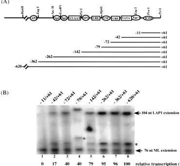
upstream of the CAT reporter gene cassette (Fig. 4A).
Tran-scription levels normalized to that of the pMLCAT internal
control have been expressed in terms of relative transcription
(percent) with respect to that of the largest LAP1 fragment
tested (
2
620/
1
61) (Fig. 4B). Transcription assays with this set
of LAP1 templates defined the sequences critical for LAP1
promoter function in vitro as being between positions
1
61 and
2
262. Deletion of sequences upstream of
2
262 had a minimal
effect on promoter activity in vitro (Fig. 4B, lanes 6 to 8).
However, removal of sequences between position
2
262 and
the start site resulted in progressively lower promoter activities
(Fig. 4B, lanes 1 to 6). The largest changes in LAP1 function
occurred when sequences between positions
2
262 and
2
79
(Fig. 4B, lanes 4 to 6) and those between
2
72 and
2
42 (lanes
2 and 3) were deleted. Further deletion to position
2
11 (Fig.
4B, lane 1) deletes the essential TATA box (Fig. 2, lane 10, and
Fig. 3B, lane 2) and thus completely abolished transcription
activity. Sequences between positions
2
142 and
2
262 include
two potential Sp1 binding sites and the CCAAT box homology
(Fig. 4A). Since both Sp1 and CTF are abundant in HeLa
nuclear extracts, it is likely that these proteins contribute to
LAP1 activity in vitro. Together, these results confirmed that
the LAP1 TATA box and sequences downstream to position
1
61 constitute the minimum basal promoter in vitro and that
other upstream elements contribute to LAP1 function in vitro.
Importance of sequences at the initiation site of LAP1 to
promoter function in vitro.
Transcription reactions with a
tem-plate that lacked LAP1 sequences downstream of position
2
11
(not shown) suggested that sequences in the region from
2
11
to
1
61 contained an essential promoter element(s). Our initial
suspicion was that the activity detected in this region was due
to sequences in the immediate vicinity of the transcription start
site. Initiator elements have been shown to promoter
transcrip-tion in numerous promoters. The sequence around the LAP1
start site (CCGATCGC, where the start site is underlined)
shows some similarity to the consensus sequence for initiator
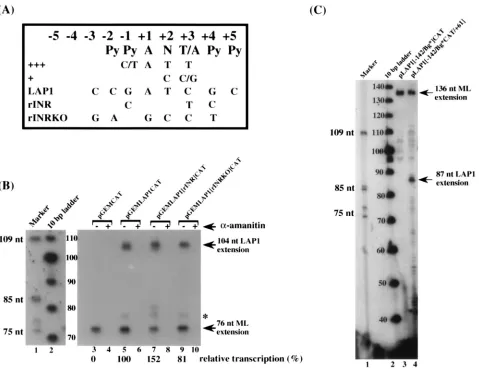
elements [YYAN(T/A)YY] (21). To test whether the LAP1
promoter contained a functional initiator element, we used
site-directed mutagenesis to change the sequences at the
tran-scription start site such that they either increased (rINR) or
decreased (rINRKO) their match with functional initiator
el-ements (Fig. 5A). Transcription analysis with these constructs
indicates that the initiator is not an important functional
ele-ment in the LAP1 promoter, since mutation of the potential
initiator element did not significantly reduce LAP1 function
(Fig. 5B, compare lanes 5 and 9). However, the introduction of
changes to match the consensus initiator (rINR) caused an
increase in LAP1 activity when normalized to the major late
internal-control template (Fig. 5B, compare lanes 5 and 7).
These results demonstrate that the native LAP1 does not
con-tain a functional initiator element.
Contribution of sequences downstream of the LAP1 start
site to promoter function in vitro.
Since the initiator element
was not contributing to LAP1 function, we considered the
possibility that there were as yet unidentified promoter
ele-ments in the region downstream of the transcription start site.
To test this hypothesis, we generated two templates that
con-FIG. 3. Functional synergism between the TATA box, USF, and CRE of LAP1 in vitro. (A) Schematic diagram of the putative TATA-proximal cis-acting elements of LAP1. Wild-type and mutant templates contain sequences from positions2142 to161 of LAP1 fused to the CAT gene. rT, rU, and rC are site-directed substitution mutants of the TATA, USF, and CRE binding sites, respectively.DUC andDUCT are actual deletions that remove sequences span-ning the USF element and CRE or the USF element, CRE, and the TATA box, respectively. (B) In vitro transcription-primer extension analysis of wild-type and mutant LAP1 templates with the pML.CAT template included in each transcrip-tion reactranscrip-tion mixture as an internal control. Positranscrip-tions of extension products corresponding to accurately initiated transcripts from LAP1 and the adenovirus type 5 major late promoter are shown on the right.
on November 9, 2019 by guest
http://jvi.asm.org/
tained LAP1 sequences from positions
2
142 to
2
11 fused to
the CAT gene. To one template, the LAP1 sequences from
positions
2
11 to
1
61 were returned to the template but
down-stream of the CAT gene. The 24-mer CAT primer used in this
experiment hybridizes to CAT gene sequences further
down-stream than did the 18-mer CAT primer used in previous
experiments and detects the major late transcript as a 136-base
extension product (Fig. 5C). Transcription analysis of these
LAP1 templates demonstrates the presence of an essential
promoter element within the region from positions
2
11 to
1
61
(Fig. 5C, compare lanes 3 and 4). The critical sequence
ele-ment is not the start site of transcription itself, since LAP1
promoter activity is restored when the region from
2
11 to
1
61
is returned to the plasmid downstream of the CAT gene. The
87-base primer extension product in Fig. 5C, lane 4, maps to 27
bp downstream from the LAP1 TATA box. Thus, we conclude
that the region from
2
11 to
1
61 contains at least one element
that is essential for LAP1 function in vitro. Furthermore, this
element is capable of promoting LAP1 activity even when
present 1.6 kb downstream of the transcription start site.
Se-quence analysis of this region has not identified any specific
elements within this region.
Contribution of the USF, CRE, and TATA box to LAT
ex-pression in vivo.
To determine whether the cis-acting elements
[image:6.612.115.485.75.416.2]that were found to be important for transcription in vitro were
also essential for LAT expression in vivo, we constructed
vi-ruses with mutations in the LAP1 promoter of both LAT loci.
To prevent virus replication and encephalitis, the mutations
were recombined into d120, a replication-defective ICP4
2vi-rus backbone (8). Each vivi-rus was stereotactically injected
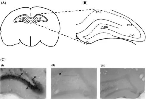
di-rectly into the rat hippocampus (Fig. 6A). At 2 weeks
postin-jection, a time consistent with latency in the central nervous
system, brains were sectioned and subjected to in situ
hybrid-ization using a probe specific for the 2-kb LAT (Fig. 6C). The
2-kb LAT is readily detected when the virus contains a
wild-type LAP1 promoter (Fig. 6C, panel i). LAT expression is
observed predominantly in granule cells of the dentate gyrus
layer of the hippocampus. In contrast, LAT expression is
greatly reduced or undetectable in sections of rat brains
in-jected with viruses containing either a substitution mutation of
FIG. 4. 59-deletion analysis: contribution of upstream sequences to LAP1 transcriptional activity. (A) Schematic representation of the putative cis-acting elements in the distal and proximal sequences of LAP1. Aligned below this schematic is the set of LAP1-CAT templates that have progressive 59deletions from positions2620 to211. The 39limit of LAP1 sequence in all these templates is at161. The double slash on the line drawing indicates that it is not to scale. (B) In vitro transcription-primer extension analysis of the 59-deletion templates of LAP1, with pML.CAT included in each transcription reaction mixture as an internal control. The 59and 39extents of the respective LAP1 fragments are indicated above each lane. The transcription activity of each construct normalized to that of the internal control is expressed as a percentage of transcription relative to that of the2620/161 construct at the bottom of each lane. The positions of extension products corresponding to accurately initiated transcripts from LAP1 and the adenovirus type 5 major late promoter are indicated on the right. The slight difference in size of the extension product from the279/161 LAP1 template (lane 4) is caused by Klenow end filling of a restriction terminus (see Materials and Methods). The position of the smaller extension product referred to in the text (asterisk) is also shown.
VOL. 70, 1996 TRANSCRIPTIONAL REGULATION OF HSV-1 LAP1 5389
on November 9, 2019 by guest
http://jvi.asm.org/
the LAP1 TATA box (Fig. 6C, panel ii) or a 53-bp deletion
spanning the USF, CRE, and TATA box (panel iii). These
results confirm that the same cis-acting elements critical for
LAP1 function in vitro are also required for the accumulation
of the 2-kb LAT during latency. To ensure that each virus had
established equivalent copies of latent viral genomes in each
animal, DNA extracted from the hippocampus was analyzed by
PCR. To normalize for the amount of total DNA harvested per
tissue sample, PCR was first performed using primers specific
to the cellular gene for GAPDH (data not shown).
Amplifica-tion of HSV-1 DNA sequences with primers specific for the gB
locus indicated that each animal contained equivalent amounts
of latent viral genomes (Fig. 7A). Similarly, to normalize for
the amount of RNA in each sample, RT-PCR was first
per-formed, using primers specific to the cellular transcript for
GAPDH (data not shown). Subsequently, RT-PCR using
[image:7.612.72.551.69.438.2]primers specific for the 2-kb species detected LAT RNA in
animals injected with the wild-type and TATA mutant viruses
(Fig. 7B, lanes 1 and 3) but not in animals injected with the
virus containing a deletion of the USF, CRE, and TATA box
of LAP1 (Fig. 7B, lane 5). The difference in LAT expression
from the rT mutant virus (Fig. 6 and 7) is consistent with (i) the
lower level of sensitivity of in situ hybridization itself and (ii)
the fact that the section shown is one representative of 100
sections through the hippocampus region with LAT-expressing
cells distributed across the plane of sectioning. On the other
hand, RT-PCR was performed on total RNA extracted from
the entire hippocampus and is capable of amplifying much
lower levels of LAT RNA than could be detected by in situ
hybridization. Finally, the RT-PCR establishes that even with
the degree of amplification afforded by PCR, there is
abso-lutely no LAT RNA expressed by deleting the sequence that
FIG. 5. Contribution of sequences downstream of the TATA box to LAP1 activity in vitro. (A) Consensus sequence of an initiator element.111, sequence preferences in a strong initiator;1, alternate nucleotides at specific positions relative to11 that permit accurate, albeit weaker, initiation levels (21). LAP1, nucleotide sequence surrounding the putative start site of the 8.3-kb primary LAT species in strain KOS; rINR and rINRKO, site-directed substitution mutations of sequences around the LAP1 start site which strengthen or weaken the match to the consensus initiator, respectively. (B) In vitro transcription-primer extension analysis of wild-type and initiator mutants of LAP1 with pML.CAT included in each reaction mixture as an internal control. The transcription reactions have been performed in the presence (1) or absence (2) ofa-amanitin. Transcription activity of pLAP1[2142/rINR/161]CAT and pLAP1[2142/rINRKO/161]CAT templates normalized to that of the adenovirus type 5 major late promoter (ML) has been expressed in terms of percent transcription relative to that of wild-type LAP1 at the bottom of the lanes. The position of the small extension product referred to in the text (asterisk) is shown on the right. (C) Identification of enhancer function associated with sequences between positions211 and161 using an in vitro transcription-primer extension assay. Lane 1, end-labelled HinfI digest of pSV325 as a size standard; lane 2, end-labelled 10-bp DNA size standard (Bethesda Research Laboratories); lane 3, in vitro transcription-primer extension of a template containing the CAT gene at position211 relative to the start site of LAP1; lane 4, in vitro transcription-primer extension of pLAP[2142/Bg*CAT/161], a template in which the sequence between
211 and161 is returned downstream of the CAT gene. The positions of extension products obtained with the 24-mer CAT gene primer that correspond to accurately initiated transcripts from LAP1 and ML promoters are indicated on the right (arrows). nt, nucleotide.
on November 9, 2019 by guest
http://jvi.asm.org/
spans the USF element, CRE, and the TATA box. This result
refines the limits of sequence elements required for LAT
ex-pression by narrowing the 203-bp PstI fragment deleted in
KOS/29 to a mere 53-bp sequence encompassing USF, CRE,
and TATA box.
DISCUSSION
The goal of this study was to identify and characterize the
role of several cis-acting elements in transcriptional activity of
LAP1 of HSV-1. Our strategy was to study the effects of
sub-stitution mutations and 5
9
deletions on transcriptional activity,
using in vitro transcription-primer extension assays, and
sub-sequently assess the effect of mutation of critical elements on
latency-specific expression of the LAT transcript in vivo.
Results from in vitro transcription-primer extension assays
provided evidence that the TATA box in LAP1 was essential
for accurate transcription initiation. The start site 28 bases
downstream of the TATA box was in agreement with previous
primer extension data that mapped the 5
9
end of CAT mRNA
28 nucleotides downstream of the TATA box (63) and with
previous predictions of the start site of the 8.3-kb minor LAT
RNA from latently infected rabbit trigeminal ganglia (13, 63).
Substitution mutation of the TATA box in LAP1 resulted in
loss of transcriptional activity in vitro. Furthermore, in contrast
to the TATA box in the adenovirus type 2 major late promoter
(5, 49), our results showed that the LAP1 TATA box required
other additional downstream sequences to support basal levels
of transcription.
[image:8.612.62.552.69.403.2]One of the most exciting findings of this study is the
possi-bility of an as-yet-uncharacterized downstream enhancer
ele-ment(s) in LAP1 that not only is essential for promoter
func-tion but also is funcfunc-tional when posifunc-tioned downstream of the
reporter gene cassette. That this critical element is not a strong
initiator element at the start site of transcription that
collabo-rates with the LAP1 TATA box for transcription initiation is
implied by (i) accurate and efficient transcription from a
site-directed substitution mutant of the sequence at the start site
and (ii) the ability of sequences between positions
2
11 and
1
61 to restore accurate transcription initiation even when
dis-tanced from the TATA box and upstream elements by as much
as 1.6 kb of intervening CAT gene sequence. The latter
obser-vation also rules out the possibility that this sequence is
re-quired to promote contacts made by TFIID at the TATA box
that sometimes extend into the leader (23, 37, 38). The region
between positions
2
11 and
1
61 displays weak homology to
leader sequences of other HSV-1 late genes, such as the UL19
(VP5) and UL44 (gC) genes, which have been shown to play a
FIG. 6. In situ hybridization to LAT RNA in rat brain injected with site-directed mutant virus. (A) Schematic diagram of the cross section of a rat brain indicating the hippocampal region into which the replication-defective LAP1 mutant viruses are delivered by stereotactic injection. (B) Anatomical organization of the hippocampus. DG, dentate gyrus; PoDG, polymorph layer of the dentate gyrus; CA1 to CA3, different layers of Ammon’s horn. (C) Representative tissue sections of rat brain at 2 weeks postinjection, showing in situ hybridization to a LAT-specific riboprobe. The photographs show the hybridization profiles of brains infected with d120 (ICP42virus with wild-type LAT loci) (i), d120:LAP1rT (LAP1 TATA substitution mutant in an ICP42virus background) (ii), and d120:LAP1DUCT (ICP42 virus with a 53-bp deletion that removes the USF, CRE, and TATA elements in LAP1) (iii) in neurons of the dentate gyrus layer of the hippocampus. Positive hybridization signals, predominantly in the dentate gyrus layer of the hippocampus, are indicated (arrows).
VOL. 70, 1996 TRANSCRIPTIONAL REGULATION OF HSV-1 LAP1 5391
on November 9, 2019 by guest
http://jvi.asm.org/
role in transcriptional regulation (20, 61). It is possible that
there are cis-acting elements within this sequence that
collab-orate with upstream activating sequences to promote
preini-tiation complex assembly. The location of this sequence
juxta-posed in between the two LAT promoter regions invites
speculation as to whether it may be shared between LAP1 and
LAP2. A similar situation has been seen with the USF binding
site that is located between the divergent IVa2 promoter and
the major late promoter in adenovirus (1). The latter is an
example not only of a promoter element that is shared between
two different promoter motifs, the IVa2 promoter being a
TATA-less and the major late a classical TATA-containing
promoter, but, more interestingly, of a shared element being a
decisive factor in the switch of expression from one promoter
to the other.
In vitro transcription-primer extension assays to assess the
importance of the USF element, CRE, and the TATA element
for LAP1 activity confirmed the predominant role of the LAP1
TATA box and suggested that both the USF element and CRE
contribute to the level of in vitro transcription. Deletion of the
TATA box in addition to the USF element and CRE (
D
UCT)
abolished transcription. These results are in agreement with
those of previous studies by Batchelor and O’Hare (3), who
have reported that deletion of 40 bp surrounding and including
the TATA box reduced expression in transient-transfection
assays in HeLa cells to
,
5% of that of the wild-type promoter
construct. In addition, there is evidence that the USF site
contributes to LAP1 activity, since addition of a fraction of
HeLa cell nuclear extract enriched in USF activity to a
recon-stituted in vitro transcription system greatly stimulated basal
levels of transcription from LAP1 (42). Furthermore, an 8- to
30-fold reduction in promoter activity was observed in a
mu-tant with abolished binding of USF in transient-transfection
assays in both neuronal and nonneuronal cells (62). The
con-tribution of USF to LAT expression during latency remains to
be investigated in vivo. However, the significance of any effect
seen in vitro and its role in determining the level of LAT
expression or effects on reactivation can be fully appreciated
only in the context of viral latency in the nervous system. For
example, although mutation of CRE at positions
2
79 to
2
83
resulted in a fourfold reduction in reporter gene activity in
transient-transfection assays, and although addition of this
el-ement to the HSV-1 immediate-early (IE110K) gene promoter
enhanced expression levels in vitro, a virus with a deletion of
CRE showed delayed reactivation kinetics but normal LAT
expression by in situ hybridization to latently infected
trigem-inal ganglia (26, 39).
In this study, the predominant role of the TATA box in
expression from the LAP1 promoter was also seen in vivo,
where substitution of the TATA box resulted in a reduction in
LAT expression from the d120:LAP1rT virus compared with
d120, while LAT expression was altogether abolished upon
deletion of USF, CRE, and the TATA box. Previous results
have shown that although deletion of the TATA-containing
PstI fragment in KOS/29 was deleterious to LAT expression in
latently infected murine trigeminal ganglia (10, 11, 13),
muta-tion of the TATA box (LATBcl virus) only resulted in weaker
in situ hybridization signals in one-third the number of neurons
compared with those for the wild-type virus (39). The effect of
mutation of the TATA box on LAT expression in latently
infected trigeminal ganglia (39) was not as dramatic as the
severely reduced expression in in situ hybridization of latently
infected rat brain tissues reported in this study. This difference
may be a function of the virus backbone used to construct these
mutant viruses or the specific neuronal cell populations in
which latency was established. Besides phenotypic differences
that have been used to distinguish neuronal cell
subpopula-tions from one another, the fact that not all neurons are
equiv-alent with respect to latency is exemplified by the finding that
only a fraction of neurons harboring HSV-1 genomes express
LATs (29, 40, 46).
To further identify the upstream boundary of sequences that
contribute to the transcriptional activity of LAP1 in vitro, a
series of progressive 5
9
LAP1 deletion mutants were studied.
pLAP[
2
620/
1
61]CAT exhibited maximum in vitro
transcrip-tional activity. Progressively lower transcriptranscrip-tional levels were
observed with more truncated promoter constructs. This result
appears to reflect the contribution of multiple promoter
ele-ments to LAP1 activity. Basal transcription was first detected
in a template containing the TATA box and downstream
se-quence up to position
1
61. While only slight enhancement of
transcription was observed when an additional USF binding
FIG. 7. PCR of DNA and RNA from homogenates of latently infected rat brain tissues. (A) Amplification of viral DNA using primers specific for the gB gene of HSV-1 from rat brains infected with the d120 virus (lane 1), d120:LAP1rT virus (lane 2), and d120:LAP1DUCT virus (lane 3). A 191-bp fragment corresponding to the amplified gB product is seen at equivalent levels in all three animals, showing that each of the LAP1 mutant viruses had established an equivalent number of latent genomes. (B) Amplification of viral RNA by RT-PCR using LAT-specific primers. Lanes 1, 3, and 5, RT-PCRs performed on RNA extracted from rat brains injected with the d120, d120:LAP1rT, and d120:LAP1DUCT viruses, respectively. A band corresponding to the amplified LAT product (195 bp) is seen only in lanes 1 and 3. Lanes 2, 4, and 6, control reactions of the same RNA extracts without reverse transcriptase added to the reaction mixture to show that the 195-bp PCR product is derived from LAT RNA and not genome contamination in the RNA preparation.
on November 9, 2019 by guest
http://jvi.asm.org/
site, two CREs, and an AP-2 site were present in the promoter,
whether this apparently minimal contribution to LAP1 in vitro
reflected their relative importance in vivo remains to be
deter-mined. Although several of these cellular factors are
ubiqui-tous, the unique ability of the LAT promoter to remain
tran-scriptionally active in particular populations of neurons that
support latency-specific transcription may be due to the
exis-tence of neuron-specific homologs of these transcription
fac-tors or the relative abundance of these facfac-tors in different cell
types resulting in a difference in LAP1 activity (19, 22, 32, 50).
It has also been suggested that the level of LAP1 activity in
Vero, L929, and C1300 cells upon deletion of the USF site may
also result from differential regulation of USF function in these
different cell types (62).
In conclusion, we have shown that the TATA box in LAP1
directs accurate transcription initiation in vitro and is
abso-lutely essential for transcription from LAP1 as determined by
in vitro transcription and in situ hybridization studies during
latency in rat brain hippocampal neurons. LAP1 is a complex
promoter having multiple elements which control its function,
most interesting are those which activate LAP1 during latency
in vivo. LAP1 functions as a late gene during lytic infection and
does not function in the absence of viral DNA synthesis. In
vivo, however, LAP1 is constitutively active in at least a portion
of latently infected neurons. Future experiments will be
di-rected at identifying both the cis-acting elements of LAP1 and
the relevant transcription factors which control its activity
un-der these different circumstances. Finally, the phenotype of the
subpopulation of neurons that harbor viral genomes expressing
LAT must be determined in order to fully understand the
cis-trans interactions which control LAP1 function.
ACKNOWLEDGMENTS
We thank L. Brunet for the pML.CAT plasmid, N. A. DeLuca for the d120 virus and E5 cell line, S. Person for the 4:27 virus, G. Jiang and W. A. Wojnar for technical assistance, J. G. Spivack for the
PstI-MluI LAT subclone, and W. F. Goins, S. W. French, C. A.
Meaney, and S. Rasty for helpful suggestions in preparation of the manuscript.
This work was supported by Public Health Service grants NIH GM34534, NIH AG0947001, NIH NINCOS NS 19608, and NIH1 RC1 DK49095-01.
REFERENCES
1. Adami, G., and L. E. Babiss. 1992. Evidence that USF can interact with only a single general transcription complex at one time. Mol. Cell. Biol. 12:1630– 1638.
2. Batchelor, A. H., and P. O’Hare. 1990. Regulation and cell-type-specific activity of a promoter located upstream of the latency-associated transcript of herpes simplex virus type 1. J. Virol. 64:3269–3279.
3. Batchelor, A. H., and P. O’Hare. 1992. Localization of cis-acting sequence requirements in the promoter of the latency-associated transcript of herpes simplex virus type 1 required for cell-type-specific activity. J. Virol. 66:3573– 3582.
4. Batchelor, A. H., K. W. Wilcox, and P. O’Hare. 1994. Binding and repression of the latency-associated promoter of herpes simplex virus by the immediate early 175K protein. J. Gen. Virol. 75:753–767.
5. Breathnach, R., and P. Chambon. 1981. Organization and expression of eucaryotic split genes coding for proteins. Annu. Rev. Biochem. 50:349–383. 6. Chen, X., M. C. Schmidt, W. F. Goins, and J. C. Glorioso. 1995. Two herpes simplex virus type 1 latency-active promoters differ in their contribution to latency-associated transcript expression during lytic and latent infection. J. Virol. 69:7899–7908.
7. Deatly, A. M., J. G. Spivack, E. Lavi, D. R. O’Boyle II, and N. W. Fraser. 1988. Latent herpes simplex virus type 1 transcripts in peripheral and central nervous system tissues of mice map to similar regions of the viral genome. J. Virol. 62:749–756.
8. DeLuca, N. A., A. McCarthy, and P. A. Schaffer. 1985. Isolation and char-acterization of deletion mutants of herpes simplex virus type 1 in the gene encoding immediate-early regulatory protein ICP4. J. Virol. 56:558–570.
9. Deshmane, S. L., and N. W. Fraser. 1989. During latency, herpes simplex virus type 1 DNA is associated with nucleosomes in a chromatin struc-ture. J. Virol. 63:943–947.
10. Deshmane, S. L., M. Nicosia, T. Valyi-Nagy, L. T. Feldman, A. Dillner, and
N. W. Fraser.1993. An HSV-1 mutant lacking the LAT TATA element reactivates normally in explant cocultivation. Virology 196:868–872. 11. Devi-Rao, G. B., S. A. Goodart, L. Hecht, R. Rochford, M. K. Rice, and E. K.
Wagner.1991. Relationship between polyadenylated and nonpolyadenylated herpes simplex virus type 1 latency-associated transcripts. J. Virol.65:2179–2190. 12. Dignam, J. D., R. M. Lebovitz, and R. G. Roeder. 1983. Accurate transcrip-tion initiatranscrip-tion by RNA polymerase II in a soluble extract from isolated mammalian nuclei. Nucleic Acids Res. 11:1475–1489.
13. Dobson, A. T., F. Sedarati, G. Devi-Rao, W. M. Flanagan, M. J. Farrell, J. G.
Stevens, E. K. Wagner, and L. T. Feldman. 1989. Identification of the latency-associated transcript promoter by expression of rabbit beta-globin mRNA in mouse sensory nerve ganglia latently infected with a recombinant herpes simplex virus. J. Virol. 63:3844–3851.
14. Efstathiou, S., A. C. Minson, H. J. Field, J. R. Anderson, and P. Wildly. 1986. Detection of herpes simplex virus-specific DNA sequences in latently in-fected mice and humans. J. Virol. 57:446–455.
15. Farrell, M. J., T. P. Margolis, W. A. Gomes, and L. T. Feldman. 1994. Effect of the transcription start region of the herpes simplex virus type 1 latency-associated transcript promoter on expression of productively infected neu-rons in vivo. J. Virol. 68:5337–5343.
16. Fraser, N. W., T. Block, and J. G. Spivack. 1992. The latency-associated transcripts of herpes simplex virus: RNA in search of a function. Virology
191:1–8.
17. French, S., W. F. Goins, M. C. Schmidt, D. Montesanti, A. J. Kinniburgh,
and J. C. Glorioso.In vitro studies of the TATA-less latency active promoter (LAP2) of herpes simplex virus type-1 identify essential promoter elements which form non-B-DNA structures. Submitted for publication.
18. Goins, W. F., L. R. Sternberg, K. D. Croen, P. R. Krause, R. L. Hendricks,
D. J. Fink, S. E. Straus, M. Levine, and J. C. Glorioso.1994. A novel latency-active promoter is contained within the herpes simplex virus type 1 UL flanking repeats. J. Virol. 68:2239–2252.
19. He, X., M. N. Treacy, D. M. Simmons, H. A. Ingraham, L. W. Swanson, and
M. G. Rosenfeld.1989. Expression of a large family of POU-domain regu-latory genes in mammalian brain development. Nature (London) 340:35–42. 20. Huang, C.-J., S. A. Goodart, M. K. Rice, J. F. Guzowski, and E. K. Wagner. 1993. Mutational analysis of sequences downstream of the TATA box of the herpes simplex virus type 1 major capsid protein (VP5/UL19) promoter. J. Virol. 67:5109–5116.
21. Javahery, R., A. Khachi, K. Lo, B. Zenzie-Gregory, and S. T. Smale. 1994. DNA sequence requirements for a transcription initiator activity in mam-malian cells. Mol. Cell. Biol. 14:116–127.
22. Johnson, W. A., and J. Hirsh. 1990. Binding of a Drosophila POU-domain protein to a sequence element regulating gene expression in specific dopa-minergic neurons. Nature (London) 343:467–470.
23. Kaufmann, J., and S. T. Smale. 1994. Direct recognition of initiator elements by a component of the transcription factor IID complex. Genes Dev. 8:821–829. 24. Kenny, J. J., F. C. Krebs, H. T. Hartle, A. E. Gartner, B. Chatton, J. M. Leiden,
J. P. Hoeffler, P. C. Weber, and B. Wigdahl.1994. Identification of a second ATF/CREB-like element in the herpes simplex virus type 1 (HSV-1) latency-associated transcript (LAT) promoter. Virology 200:220–235.
25. Krause, P. R., J. M. Ostrove, and S. E. Straus. 1991. The nucleotide se-quence, 59end, promoter domain, and kinetics of expression of the gene encoding the herpes simplex virus type 2 latency-associated transcript. J. Vi-rol. 65:5619–5623.
26. Leib, D. A., K. C. Nadeau, S. A. Rundle, and P. A. Schaffer. 1991. The promoter of the latency-associated transcripts of herpes simplex virus type 1 contains a functional cAMP-response element: role of the latency-associated transcripts and cAMP in reactivation of viral latency. Proc. Natl. Acad. Sci. USA 88:48–52.
27. Lindell, T. J., F. Weinberg, P. W. Morris, R. G. Roeder, and W. J. Rutter. 1970. Specific inhibition of RNA polymerase II by alpha-amanitin. Science
170:447–449.
28. Margolis, T. P., D. C. Bloom, A. T. Dobson, L. T. Feldman, and J. G. Stevens. 1993. Decreased reporter gene expression during latent infection with HSV LAT promoter constructs. Virology 197:585–592.
29. Margolis, T. P., C. R. Dawson, and J. LaVail. 1992. Herpes simplex viral infection of the mouse trigeminal ganglion: immunochemical analysis of cell populations. Invest. Ophthalmol. Visual Sci. 33:259–267.
30. McGeoch, D. J., C. Cunningham, G. McIntyre, and A. Dolan. 1991. Com-parative sequence analysis of the long repeat regions and adjoining parts of the long unique regions in the genomes of herpes simplex viruses types 1 and 2. J. Gen. Virol. 72:3057–3075.
31. Mellerick, D. M., and N. W. Fraser. 1987. Physical state of the latent herpes simplex virus genome in a mouse model system: evidence suggesting an episomal state. Virology 158:265–275.
32. Mitchell, P. J., P. M. Timmons, J. M. Hebert, P. W. J. Rigby, and R. Tjian. 1991. Transcription factor AP-2 is expressed in neural crest cell lineages during mouse embryogenesis. Genes Dev. 5:105–119.
VOL. 70, 1996 TRANSCRIPTIONAL REGULATION OF HSV-1 LAP1 5393
on November 9, 2019 by guest
http://jvi.asm.org/
33. Mitchell, W. J., R. P. Lirette, and N. W. Fraser. 1990. Mapping of low abundance latency-associated RNA in the trigeminal ganglia of mice latently infected with herpes simplex virus type 1. J. Gen. Virol. 71:125– 132.
34. Mitchell, W. J., I. Steiner, S. M. Brown, A. R. MacLean, J. H. Subak-Sharpe,
and N. W. Fraser.1990. A herpes simplex virus type 1 variant, deleted in the promoter region of the latency-associated transcripts, does not produce any detectable minor RNA species during latency in the mouse trigeminal gan-glion. J. Gen. Virol. 71:953–957.
35. Nicosia, M., S. L. Deshmane, J. M. Zabolotny, T. Valyi-Nagi, and N. W.
Fraser. 1993. Herpes simplex virus type 1 latency-associated transcript (LAT) promoter deletion mutants can express a 2-kilobase transcript map-ping to the LAT region. J. Virol. 67:7276–7283.
36. Paxinos, G., and C. Watson. 1986. The rat brain in stereotaxic coordinates, 2nd ed. Academic Press, San Diego, Calif.
37. Purnell, B. A., P. A. Emanuel, and D. S. Gilmour. 1994. TFIID sequence recognition of the initiator and sequences farther downstream in Drosophila class II genes. Genes Dev. 8:830–842.
38. Purnell, B. A., and D. S. Gilmour. 1993. Contribution of sequences down-stream of the TATA element to a protein-DNA complex containing the TATA-binding protein. Mol. Cell. Biol. 13:2593–2603.
39. Rader, K. A., C. E. Ackland-Berglund, J. K. Miller, J. S. Pepose, and D. A.
Leib.1993. In vivo characterization of site-directed mutants in the promoter of the herpes simplex virus type 1 latency-associated transcripts. J. Gen. Virol. 74:1859–1869.
40. Ramakrishnan, R., D. J. Fink, G. Jiang, P. Desai, J. C. Glorioso, and M.
Levine.1994. Competitive quantitative PCR analysis of herpes simplex virus type 1 DNA and latency-associated transcript RNA in latently infected cells of the rat brain. J. Virol. 68:1864–1873.
41. Ramakrishnan, R., M. Levine, and D. J. Fink. 1994. PCR-based analysis of herpes simplex virus type 1 latency in the rat trigeminal ganglion established with a ribonucleotide reductase-deficient mutant. J. Virol.
68:7083–7091.
42. Rivera-Gonzalez, R., A. N. Imbalzano, B. Gu, and N. A. Deluca. 1994. The role of ICP4 repressor activity in temporal expression of the IE-3 and latency-associated transcript promoters during HSV-1 infection. Virology
202:550–564.
43. Rock, D. L., and N. W. Fraser. 1983. Detection of HSV-1 genome in central nervous system of latently infected mice. Nature (London) 302:523–525. 44. Rock, D. L., and N. W. Fraser. 1985. Latent herpes simplex virus type 1 DNA
contains two copies of the virion DNA joint region. J. Virol. 63:943–947. 45. Rock, D. L., A. B. Nesburn, H. Ghiasi, J. Ong, T. L. Lewis, J. R. Lokensgard,
and S. L. Wechsler.1987. Detection of latency-related viral RNAs in tri-geminal ganglia of rabbits latently infected with herpes simplex virus type 1. J. Virol. 61:3820–3826.
46. Rødahl, E., and J. G. Stevens. 1992. Differential accumulation of herpes simplex virus type 1 latency-associated transcripts in sensory and autonomic ganglia. Virology 189:385–388.
47. Sacks, W. R., C. C. Greene, D. P. Aschman, and P. A. Schaffer. 1985. Herpes simplex virus type 1 ICP27 is an essential regulatory protein. J. Virol. 55: 796–805.
48. Sambrook, J., E. F. Fritsch, and T. Maniatis. 1989. Molecular cloning: a
laboratory manual, 2nd ed. Cold Spring Harbor Laboratory Press, Cold Spring Harbor, N.Y.
49. Sassone-Corsi, P., J. Corden, C. Kedinger, and P. Chambon. 1981. Promo-tion of specific in vitro transcripPromo-tion by “excised” TATA box sequences inserted in a foreign nucleotide environment. Nucleic Acids Res. 9:3941– 3958.
50. Schreiber, E., A. Tobler, U. Malipiero, W. Schaffner, and A. Fontana. 1993. cDNA cloning of human N-Oct 3, a nervous system-specific POU-domain transcription factor binding to the octamer DNA motif. Nucleic Acids Res.
21:253–258.
51. Southern, E. M. 1975. Detection of specific sequences among DNA frag-ments separated by gel electrophoresis. J. Mol. Biol. 98:503–517. 52. Spivack, J. G., and N. W. Fraser. 1987. Detection of herpes simplex virus
type 1 transcripts during latent infection in mice. J. Virol. 61:3841–3847. 53. Spivack, J. G., G. M. Woods, and N. W. Fraser. 1991. Identification of a
novel latency-specific splice donor signal within the herpes simplex virus type 1 2.0-kilobase latency-associated transcript (LAT): translation inhibition of LAT open reading frames by the intron within the 2.0-kilobase LAT. J. Virol.
65:6800–6810.
54. Steiner, I., and P. G. E. Kennedy. 1995. Herpes simplex virus latent infection in the nervous system. J. Neurovirol. 1:19–29.
55. Stevens, J. G. 1994. Overview of herpes virus latency. Semin. Virol. 5:191– 196.
56. Stevens, J. G., E. K. Wagner, G. B. Devi-Rao, M. L. Cook, and L. T. Feldman. 1987. RNA complementary to a herpesvirusagene mRNA is prominent in latently infected neurons. Science 235:1056–1059.
57. Strelow, L. I., K. A. Laycock, P. Y. Jun, K. A. Rader, R. H. Brady, J. K.
Miller, J. S. Pepose, and D. A. Leib.1994. A structural and functional comparison of the latency-associated transcript promoters of herpes simplex virus type 1 strains KOS and McKrae. J. Gen. Virol. 75:2475–2480. 58. Wechsler, S. L., A. B. Nesburn, R. Watson, S. M. Slanina, and H. Ghiasi.
1988. Fine mapping of the latency-related RNA of herpes simplex virus type 1 in humans. J. Gen. Virol. 64:3269–3279.
59. Wechsler, S. L., A. B. Nesburn, R. Watson, S. M. Slanina, and H. Ghiasi. 1988. Fine mapping of the latency-related gene of herpes simplex virus type 1: alternative splicing produces distinct latency-related RNAs containing open reading frames. J. Virol. 62:4051–4058.
60. Wechsler, S. L., A. B. Nesburn, J. Zwaagstra, and H. Ghiasi. 1989. Sequence of the latency-related gene of herpes simplex virus type 1. Virology 168:168– 172.
61. Weir, J. P., and P. R. Narayanan. 1990. Expression of the herpes simplex virus type 1 glycoprotein C gene requires sequences in the 59noncoding region of the gene. J. Virol. 64:445–449.
62. Zwaagstra, J. C., H. Ghiasi, A. B. Nesburn, and S. L. Wechsler. 1991. Identification of a major regulatory sequence in the latency associated tran-script (LAT) promoter of herpes simplex virus type 1 (HSV-1). Virology
182:287–297.
63. Zwaagstra, J. C., H. Ghiasi, S. M. Slanina, A. B. Nesburn, S. C. Wheatley, K.
Lillycrop, J. Wood, D. S. Latchman, K. Patel, and S. L. Wechsler.1990. Activity of herpes simplex virus type 1 latency-associated transcript (LAT) promoter in neuron-derived cells: evidence for neuron specificity and for a large LAT transcript. J. Virol. 64:5019–5028.