0022-538X/96/$04.0010
Copyrightq1996, American Society for Microbiology
The RGG Box Motif of the Herpes Simplex Virus ICP27 Protein
Mediates an RNA-Binding Activity and Determines
In Vivo Methylation
WENDY E. MEARSANDSTEPHEN A. RICE*
Department of Biochemistry, University of Alberta, Edmonton, Alberta, Canada T6G 2H7
Received 31 May 1996/Accepted 23 July 1996
ICP27 is an essential herpes simplex virus type 1 nuclear regulatory protein that is required for efficient viral gene expression. Although the mechanism by which ICP27 regulates genes is unknown, a variety of evidence suggests that it functions posttranscriptionally, and recent studies indicate that it is an RNA-binding protein. Previously, we noted that a short arginine- and glycine-rich sequence in ICP27 (residues 138 to 152) is similar to an RGG box motif, a putative RNA-binding determinant found in a number of cellular proteins (W. Mears, V. Lam, and S. Rice, J. Virol. 69:935–947, 1995). In the present study, we have further investigated ICP27’s association with RNA and examined the role of the RGG box in RNA binding. We find that ICP27 binds efficiently to RNA homopolymers composed of poly(G) and weakly to poly(U) RNA homopolymers. Poly(G) binding activity maps to the N-terminal 189 residues of ICP27 and requires the RGG box sequence. Using a northwestern blotting assay, we demonstrate that the RGG box alone (residues 140 to 152) can mediate RNA binding when attached to a heterologous protein. As many cellular RGG box proteins are methylated on arginine residues, we also investigated the in vivo methylation status of ICP27. Our results demonstrate that ICP27 is methylated in herpes simplex virus-infected cells. Methylation is dependent on the presence of the RGG box, suggesting that one or more arginine residues in the RGG box sequence are modified. These data demonstrate that ICP27 displays the characteristics of an RGG box-type RNA-binding protein.
Herpes simplex virus type 1 (HSV-1) is a common human alphaherpesvirus which establishes life-long infections in af-fected individuals. Its effects range from asymptomatic infec-tions to serious, life-threatening illnesses. HSV-1 is also an important research tool, serving as a model system with which to study the fundamental pathways used by herpesviruses to replicate in their host cells. Our laboratory is particularly in-terested in using HSV-1 to characterize the mechanisms by which herpesviruses regulate the expression of their genes dur-ing lytic infections.
Much is already known about the pattern of gene expression in cells lytically infected with HSV-1 (reviewed in reference 35). After the virus enters the cell, its double-stranded DNA genome is transported to the cell nucleus, where it is tran-scribed by the host RNA polymerase II. The approximately 80 viral genes are similar in structure to cellular genes, with the exception that most do not possess intron sequences. During infection, the genes are expressed in a highly regulated fashion and have been grouped into three general categories based on the temporal order of, and requirements for, their expression.
Immediate-early (IE, also calleda) genes are the first to be
expressed and encode several regulatory proteins, including ICP0, ICP4, ICP22, and ICP27. The transcription of IE genes can occur in the absence of viral protein synthesis and is stim-ulated by VP16, a virus-encoded virion protein. The next genes
to be expressed are the delayed-early (DE, also called b)
genes. Transcription of these genes requires the prior synthesis of IE proteins, in particular ICP4. For the most part, the DE genes encode proteins involved in viral DNA replication. The
last genes to be expressed are the late (L, also calledg) genes.
Like DE gene expression, L gene expression requires prior viral protein synthesis and, in addition, is stimulated by viral DNA replication. The majority of L genes encode viral struc-tural proteins. Concomitant with the activation of viral genes in HSV-1-infected cells, shutoff of cellular gene expression also occurs. Host gene shutoff (reviewed in reference 41) is a com-plex phenomenon involving the down-regulation of transcrip-tion, the inhibition of mRNA splicing (10), and the induced degradation of cellular mRNAs.
The mechanisms controlling viral and cellular gene expres-sion during HSV-1 infection have been the subject of numer-ous investigations. One important fact which has emerged from these studies is that the HSV-1 IE proteins play critical roles in gene regulation. Our laboratory is particularly inter-ested in the IE protein ICP27. This polypeptide is composed of 512 residues, migrates on sodium dodecyl sulfate (SDS)-poly-acrylamide gel electrophoresis (PAGE) as a 63-kDa protein, and is localized predominantly to the cell nucleus (1, 15, 22). ICP27 is posttranslationally modified by phosphorylation (1, 46), although the function and sites of phosphorylation are unknown. Genetic studies indicate that ICP27 is absolutely essential for virus growth (21, 36). One phenotype of HSV-1 ICP27 null mutants is that they are deficient in the expression of many L proteins and their corresponding mRNAs (21, 23, 31, 36). A second phenotype is that they show a 5- to 20-fold reduction in the level of viral DNA synthesis (31). ICP27’s effects on late gene activation and DNA replication may be due to distinct functions of the protein, as they can be differentially affected by mutations in the ICP27 gene (31–33). Recent work suggests that ICP27 stimulates DNA replication indirectly, by enhancing the expression of some of the DE mRNAs which encode DNA replication proteins (23, 44). Thus, ICP27 ap-pears to mediate at least two separable functions which to-gether enhance the production of both DE and L mRNAs. In addition, ICP27 contributes to the shutoff of host gene
expres-* Corresponding author. Mailing address: Department of Biochem-istry, University of Alberta, 474 Medical Sciences Building, Edmonton, Alberta, Canada T6G 2H7. Phone: (403) 2717. Fax: (403) 492-0886. Electronic mail address: [email protected].
7445
on November 9, 2019 by guest
http://jvi.asm.org/
sion, as cellular protein synthesis (36) and mRNA levels (9) are greater in ICP27 mutant infections than in wild-type (WT) HSV-1 infections.
It is not yet known if ICP27’s effects on viral and cellular genes occur at the transcriptional level, the posttranscriptional level, or both. However, a variety of evidence suggests that ICP27 mediates at least some of its effects through changes to the cell’s pre-mRNA processing machinery. One effect appears to be an inhibition of pre-mRNA splicing. This was first noted in transfection assays, in which it was found that ICP27 can inhibit the expression of cotransfected target genes which pos-sess introns (39). Furthermore, ICP27 can partially inhibit in vitro splicing in nuclear extracts (10) and can alter the intranu-clear distribution of splicing factors in vivo (29, 38). In addition to affecting splicing, ICP27 may also modulate pre-mRNA polyadenylation. The specificity of in vitro poly(A) site utiliza-tion is altered in HSV-1-infected cells (25), and this effect is dependent on a functional ICP27 (24). Moreover, ICP27’s effects on polyadenylation may be responsible for its ability to stimulate certain target genes in cotransfection assays, as the
responsive sequences map to the 39 segment of the target
genes, which includes their poly(A) sites (5, 39). Recent data reported by McGregor et al. (23) support the hypothesis (39) that ICP27 increases the efficiency with which weak poly(A) sites are utilized.
The evidence for posttranscriptional regulation by ICP27 suggests the possibility that the protein interacts with RNA in infected cells. Consistent with this possibility, two recent stud-ies have demonstrated that ICP27 can interact with RNA mol-ecules in vitro (3, 13). Previously (26), we noted that ICP27 possesses a sequence which resembles an RGG box, a putative RNA-binding motif found in a number of cellular nuclear proteins involved in mRNA and rRNA metabolism (4, 14, 20). ICP27’s RGG box sequence is composed of 15 consecutive arginine and glycine residues and maps to residues 138 to 152. It is required for ICP27’s nucleolar localization, possibly re-flecting an in vivo RNA-binding activity (26). It is also essential for its biological function, as a recombinant HSV-1 which en-codes an RGG box-minus ICP27 fails to replicate efficiently in cultured cells (26). The goal of this study was to further inves-tigate ICP27’s in vitro interaction with RNA and to explore the role of the RGG box motif in RNA binding.
MATERIALS AND METHODS
Plasmids.Plasmid constructs for in vitro transcription-translation of ICP27 were constructed by using the vector pCITE-1 (Novagen). To clone the WT ICP27 gene, pCITE-1 was first modified by the introduction of anSstI site into its polylinker sequence. This was done by cleaving pCITE-1 withAccI, filling in the DNA ends with the Klenow fragment ofE. coliDNA polymerase, and inserting anSstI linker. This plasmid was termed pCITE-SstI. To generate pCITE-27, encoding WT ICP27, the following steps were performed. A plasmid containing the ICP27 gene, pM27 (32), was digested withDrdI, which cuts just upstream of ICP27’s initiation codon. The resulting ends were made blunt by using T4 DNA polymerase. The DNA was then digested withSstI, which cleaves downstream of ICP27’s termination codon. The 2.0-kbDrdI-SstI fragment con-taining the ICP27 gene was then cloned intoBalI-SstI-digested pCITE-SstI. An analogous procedure was used to engineer pCITE-d4-5, in this case using plas-mid pMd4-5 (26) instead of pM27. pCITE-d4-6 was constructed by ligation of the 1.5-kbXhoI-SstI ICP27 gene-containing fragment from pM6 (32) to the 4.4-kb
XhoI-SstI vector fragment of pCITE-d4-5. pCITE-d5-6 was constructed by ligat-ing the 1.6-kbDraIII-SstI fragment of pMd5-6 (16) to the 4.0-kb vector fragment of pCITE-d4-6 which is obtained after completeSstI digestion and partialDraIII digestion. pCITE-M11, pCITE-M15, and pCITE-M16 were constructed by sub-stitution of the 1.2-kbSalI-SstI ICP27 gene fragments of pM11, pM15, and pM16 (32) into pCITE-27 in place of the WT ICP27 gene fragment. The ICP27 molecules encoded by pCITE-27 and the derivatives described above have three additional N-terminal residues (Met-Ala-Val) compared with WT ICP27. To construct pCITE-262C, pBS27 (33) was digested withSalI, and the DNA ends were made blunt by using the Klenow enzyme. After cleavage withSstI, the 1.2-kb fragment was cloned intoBalI-SstI-cleaved pCITE-SstI. pCITE-262C en-codes a protein having an N-terminal methionine residue followed by residues
262 to 512 of ICP27. Heterogeneous nuclear ribonucleoprotein particle (hnRNP) protein C1 was produced by in vitro transcription-translation using plasmid pHC12 (43), obtained from G. Dreyfuss (University of Pennsylvania).
Plasmids for bacterial expression of glutathioneS-transferase (GST)–ICP27 fusion proteins were constructed by using the vectors SstI and BglII, which are modified forms of 5X-3 (Pharmacia). To create pGEX-SstI, theNotI site in pGEX-5X-3 was converted to anSstI site by digesting pGEX-5X-3 withNotI, filling in the DNA ends with the Klenow fragment, and inserting an SstI oligonucleotide linker. pGEX-BglII was made in a similar fashion except that aBglII linker was used. To generate pGEX-27, the 2.0-kb
DrdI-SstI ICP27 gene fragment obtained from pM27 was cloned into the 4.9-kb
SmaI-SstI vector fragment of pGEX-SstI. This plasmid encodes a GST-ICP27 fusion protein consisting of GST, 12 linker residues, and all 512 residues of WT ICP27. pGEX-d4-5 was constructed by ligating the 1.7-kbDraIII-SstI fragment of pMd4-5 to the 5.2-kbDraIII-SstI vector fragment of 27. Plasmid pGEX-RGG was constructed as follows. First, oligonucleotide-directed mutagenesis (Altered Sites system; Promega) was used to engineer aBglII site in plasmid pM4 (32). This resulted in plasmid derivative pM4-5B, which contains anXhoI site at codons 138 and 139 of the ICP27 gene and aBglII site at codons 153 and 154. pM4-5B was cleaved withXhoI andBglII, and the smallXhoI-BglII fragment was cloned into the 4.9-kbSalI-BglII vector fragment of pGEX-BglII to produce pGEX-RGG. The polypeptides encoded by pGEX-5X-3 and pGEX-RGG differ only at their C termini: GST contains the sequence -SSGRIVTD, which is replaced by -GRRGRRRGRGRGGPDLPAAS in GST-RGG (the underlined residues correspond to residues 140 to 152 of ICP27).
The plasmids used to generate RNA probes for the northwestern blotting assays, pBSSV40PA and pBSIFNPA (3), were provided by Charles Brown and L. P. Perera (National Institutes of Health).
Expression of in vitro-translated ICP27.For in vitro transcription-translation, plasmids were first linearized at appropriate restriction sites. pHC12 was cleaved with NsiI, whereas pCITE-27, pCITE-d4-5, pCITE-d4-6, pCITE-d5-6, and pCITE-262C were cleaved withEcl136II. C-terminally truncated forms of ICP27 were produced by linearization of plasmids as follows: pCITE-d4-5 was digested withXhoI to generate the N137 template; pCITE-27 was digested withNcoI to generate the N189 template; pCITE-27 was digested withSalI to generate the N261 template; pCITE-M11 was digested withXhoI to generate the N339 tem-plate; pCITE-27 was digested withStuI to generate the N405 template; and pCITE-M16 was digested withXhoI to generate the N487 template. RNA tran-scripts were produced by in vitro transcription of the linearized plasmids by using the RIBOMAX system (Promega) according to the manufacturer’s directions. Transcription was carried out with T7 RNA polymerase (Bethesda Research Laboratories), with the exception of pHC12, which was transcribed by using SP6 RNA polymerase (Promega). After transcription, RNAs were precipitated by the addition of ammonium acetate to 2.5 M and resuspended in water. In vitro translation was carried out in a rabbit reticulocyte lysate (Promega) in the presence of 800mCi of [35S]methionine-cysteine (DuPont NEN) per ml as
recommended by the manufacturer.
RNA homopolymer binding assays.Binding of in vitro-translated protein to ribonucleotide homopolymers or single-stranded DNA (ssDNA) was carried out as described by Kiledjian and Dreyfuss (14). Briefly, 105cpm of trichloroacetic
acid-precipitatable translated protein was made up to a final volume of 0.5 ml with binding buffer (10 mM Tris [pH 7.6], 2.5 mM MgCl2, 0.5% Triton X-100,
NaCl of the appropriate concentration). To this, 50 to 60ml of washed agarose beads containing attached ribonucleotide homopolymer (Pharmacia or Sigma) or ssDNA (Pharmacia) was added. The mixture was incubated for 60 min at 48C on a rocking platform. The beads were pelleted by centrifugation for 1 min in a microcentrifuge and washed five times with binding buffer. Protein was eluted from the beads by boiling in PAGE sample buffer and analyzed by SDS-PAGE on 12.5 or 15% gels. Radioactive bands were visualized and quantitated by autoradiography and phosphor image analysis (Fuji).
Expression and purification of GST fusion proteins.GST fusion proteins were expressed inEscherichia coliBL21. Individual transformants were grown over-night at 378C in medium containing 100mg of ampicillin per ml. The overnight cultures were diluted 1:10 in a total volume of 100 ml and grown at 378C until the optical density (A600) reached 1.1 to 1.4 units. Isopropyl-b-D-thiogalactoside (IPTG) was added to a final concentration of 0.1 mM, and induction was allowed to proceed for 2 h. The cells were pelleted at 48C and resuspended in 5 ml of phosphate-buffered saline (PBS), and lysozyme (Sigma) was added to 1 mg/ml. Lysis was allowed to proceed on ice for 60 min. The cell lysate was spun at 5,0003gfor 10 min at 48C, and the resulting pellet was quick-frozen in a dry ice-isopropanol bath. After thawing, the pellet was resuspended in 5 ml of PBS containing 0.1% sodium deoxycholate (Sigma). After a 10-min incubation on ice, MgCl2, Triton X-100, and DNase I were added to final concentrations of 8 mM,
1%, and 10mg/ml, respectively. Incubation was continued for 30 min on ice with gentle agitation. The mixture was then spun at 10,0003gfor 10 min at 48C, and the supernatant was retained. To purify GST and GST fusion proteins, 5 ml of supernatant was incubated with 100ml of prewashed glutathione-Sepharose 4B beads (50% slurry; Pharmacia) for 30 min at 48C. The beads were then washed three to four times in PBS. Bound protein was eluted at room temperature with three to four 50-ml washes of elution buffer (50 mM Tris-HCl [pH 8.0], 10 mM glutathione). Purified protein preparations were stored at2708C until needed. The protein concentration of each preparation was measured by the Bradford
on November 9, 2019 by guest
http://jvi.asm.org/
assay (Bio-Rad). The preparations were also analyzed by Western blotting (im-munoblotting) using monoclonal antibodies directed against GST (Pharmacia) or ICP27 (H1113; Goodwin Institute for Cancer Research, Plantation, Fla.).
Northwestern blotting assays.32
P-labeled riboprobes for the northwestern blotting experiments were generated by in vitro transcription. To generate tem-plates for the beta interferon (IFN-b) and simian virus 40 (SV40) RNA probes, pBSIFNPA and pBSSV40PA were linearized withEcoRI. To generate a tem-plate for the IFN-bantisense probe, pBSIFNPA was linearized withHindIII. One microgram of each DNA template was transcribed in vitro, using T3 RNA polymerase (for the IFN-band SV40 probes; Promega) or T7 RNA polymerase (for the IFN-bantisense probe) in the presence of 50mCi of [a-32
P]CTP (400 Ci/mmol; DuPont NEN). After transcription, the samples were phenol-chloro-form extracted and ethanol precipitated twice, using 2.5 M ammonium acetate as the precipitating salt. The final RNA pellet was resuspended in RNase-free TE (10 mM Tris-HCl [pH 8.0], 1 mM EDTA). Approximately 83105
cpm of each probe was analyzed by electrophoresis in a 5% denaturing acrylamide-urea gel. The northwestern blotting assays were carried out as described by Brown et al. (3). Briefly, 10 or 40mg of purified GST (expressed fromE. colicells harboring pGEX-5X-3) or GST fusion proteins was electrophoresed on SDS–15% acryl-amide gels and electrophoretically transferred to nitrocellulose filters. The trans-ferred proteins were renatured in situ for 12 h at 48C in binding buffer (10 mM Tris-HCl [pH 7.6], 50 mM NaCl, 1 mM EDTA, 13Denhardt’s solution). The filters were incubated at room temperature for 8 h in 15 ml of binding buffer containing 1.73107
cpm of32
P-labeled RNA probe. The blots were individually washed three times in binding buffer for 10 min at room temperature and analyzed by autoradiography.
Cells, viruses, and infections.Vero (African green monkey kidney) cells were propagated in Dulbecco modified Eagle medium containing 5% heat-inactivated fetal bovine serum (GIBCO). HSV infections were carried out at a multiplicity of infection of 10 PFU per cell as described previously (34). Strain KOS1.1 (12) was the WT strain of HSV-1 used. HSV-2 (strain G) was obtained from the American Type Culture Collection (Rockville, Md.). The HSV-1 ICP27 mutants
d27-1,n263R,n406R,d1-2,d3-4, andd4-5 have been described elsewhere (26, 31, 33). The HSV-1 mutant allelesd5-6 andd1-5 were constructed by using engi-neered XhoI sites in the HSV-1 ICP27 gene (32), and recombinant viruses bearing these alleles were isolated (16).d1-5 andd5-6 encode ICP27 molecules missing amino acid residues 13 to 153 and 154 to 173, respectively.
In vivo methylation assay.Metabolic labeling of methylated proteins was performed as described by Liu and Dreyfuss (19). At 5 h postinfection (hpi), medium was removed from the infected cells and replaced with medium con-taining 100mg of cycloheximide per ml and 40mg of chloramphenicol per ml. After 30 min, the cells were rinsed twice with methionine-free Dulbecco modi-fied Eagle medium (GIBCO) and then incubated for 3 h in methionine-free Dulbecco modified Eagle medium containing 100mg of cycloheximide per ml, 40 mg of chloramphenicol per ml, and 45mCi ofL-[methyl-3
H]methionine (DuPont NEN) per ml. In some experiments, 100mM anisomycin was used in place of cycloheximide. After labeling, the cells were rinsed in PBS containing 50mg of
Na-p-tosyl-L-lysine chloromethyl ketone (TLCK) per ml and 100mg of phenyl-methylsulfonyl fluoride per ml and lysed in modified radioimmunoprecipitation assay (RIPA) buffer (150 mM NaCl, 1% Nonidet P-40, 0.5% sodium deoxy-cholate, 0.1% SDS, 50 mM Tris-HCl [pH 8.0], 50mg of TLCK per ml, 100mg of phenylmethylsulfonyl fluoride per ml) for 30 min on ice. The cell lysate was transferred to a microcentrifuge tube, and insoluble material was pelleted at 10,0003gfor 10 min at 48C. The supernatant was transferred to a new tube and analyzed immediately or stored at2708C.
Immunoprecipitations.Immunoprecipitations were carried out with a mixture of H1113 and H1119, two mouse monoclonal antibodies specific for ICP27 (Goodwin Institute for Cancer Research). To carry out the immunoprecipita-tions, 0.8ml of each antibody was added to 400ml of cell lysate, and the samples were incubated at 48C on a rocking platform. After 30 min, 80ml of a 1:40 dilution of rabbit anti-mouse immunoglobulin G (heavy and light chains; Jackson ImmunoResearch Laboratories Inc.) was added as a bridging antibody. After a further 30-min incubation, 200 ml of prewashed Staphylococcus aureuscells (Pansorbin; Calbiochem), resuspended in modified RIPA buffer (10% [wt/vol]), was added, and the incubation was continued for another 30 min. The cells were pelleted and washed four times in modified RIPA buffer. Bound proteins were eluted by heating the cells for 10 min at 858C in SDS-PAGE sample buffer. Immunoprecipitates were analyzed by electrophoresis on SDS–12.5% acrylamide gels. For fluorographic enhancement, gels were impregnated with 1.0 M sodium salicylate before drying. Immunoprecipitates were also analyzed by immunoblot-ting using a 1:1,000 dilution of the H1119 antibody. Immunoreactive proteins were detected by enhanced chemiluminescence (ECL detection kit; Amersham).
RESULTS
ICP27 binds to poly(G) RNA homopolymers.Recent studies by Brown et al. (3) and Ingram et al. (13) have demonstrated that ICP27 can bind to RNA molecules in vitro. However, it is not known what sequence or structural determinants in RNA are recognized by ICP27. To gain insight into ICP27’s
RNA-binding specificity, we tested its ability to interact with various RNA homopolymers in vitro (42). Such homopolymer binding assays have been used by many investigators to characterize the binding potential and target specificity of putative RNA-binding proteins (2, 8, 14, 28, 40). For the RNA-binding studies, full-length ICP27 was expressed by in vitro translation in a
rabbit reticulocyte lysate in the presence of [35
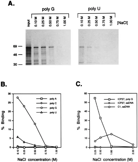
S]methionine-cysteine. The radiolabeled protein was incubated with agarose beads to which RNA homopolymers [poly(A), poly(C), poly(G), or poly(U)] were covalently attached. The binding reactions and subsequent washing steps were carried out at several NaCl concentrations ranging between 0.1 and 1.0 M. After washing, protein was eluted from the beads and analyzed by SDS-PAGE and phosphor image analysis. ICP27 bound to poly(G) ribopolymers, with significant binding of the input material occurring up to an NaCl concentration of 0.5 M (Fig. 1A and B). ICP27 also bound weakly to poly(U) homopoly-meric RNA at 0.1 and 0.25 M NaCl. In contrast, ICP27 did not bind to poly(A) or poly(C). In control experiments, in
vitro-FIG. 1. Binding of in vitro-translated ICP27 to RNA homopolymers. (A and B) Binding of ICP27 to RNA homopolymers. ICP27, produced by in vitro translation in the presence of [35
S]methionine-cysteine, was incubated with RNA homopolymers [poly(A), poly(C), poly(G), or poly(U)] attached to agarose beads. Binding and washing were carried out at various NaCl concentrations ranging from 0.1 to 1.0 M. Bound protein was eluted and analyzed by SDS-PAGE, autoradiography, and phosphor image analysis. (A) Binding of ICP27 to poly(G) and poly(U) ribopolymers. The lane labeled “input” contains an aliquot of the translation mixture used in the binding reaction. The other lanes show protein bound at various salt concentrations. Equivalent fractions of the input and bound samples were loaded on the gel to facilitate the determination of percent binding. The positions of molecular weight markers are indicated at the left (molecular masses in kilodaltons). (B) Quantitation of ICP27 binding to RNA homopolymers at different NaCl concentrations. The percentage of input ICP27 bound at each NaCl concentration was determined by phosphor image analysis. (C) Comparison of ICP27’s binding to poly(G) RNA and ssDNA. In vitro-translated ICP27 or hnRNP C1 were bound at various NaCl concentrations to agarose beads containing either poly(G) RNA or ssDNA. Analysis and quan-titation of binding were carried out as for panels A and B.
on November 9, 2019 by guest
http://jvi.asm.org/
[image:3.612.316.558.71.354.2]translated hnRNP C1 bound avidly to poly(U) agarose beads up to an NaCl concentration of 1.0 M (data not shown), con-sistent with previous results (42). From the results of these experiments, we conclude that ICP27 binds with moderate avidity to poly(G) RNA homopolymers and weakly to poly(U) homopolymers.
Previously, Vaughn et al. demonstrated that ICP27 purified from infected cells can bind to a ssDNA-agarose column and be eluted at relatively high salt (45). We therefore examined ICP27’s interaction with ssDNA in our binding assay. To do this, in vitro-translated ICP27 was incubated with ssDNA- or poly(G)-agarose beads at NaCl concentrations ranging from 0.25 to 2.0 M. As a control, in vitro-translated hnRNP C1, previously demonstrated to interact with ssDNA (30), was also tested for ssDNA binding. After binding and washing, protein was eluted from the beads and analyzed by SDS-PAGE and phosphor imaging (Fig. 1C). ICP27 bound to poly(G) as before but demonstrated little if any binding to ssDNA. In contrast, hnRNP C1 bound to the ssDNA up to an NaCl concentration of 1.0 M. We conclude that under the conditions of our binding assay, in vitro-translated ICP27 binds to poly(G) RNA but not to ssDNA.
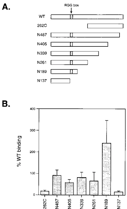
Role of the RGG box in poly(G) binding.To identify the sequences in ICP27 which are responsible for poly(G) binding, several N- or C-terminally truncated forms of ICP27 were expressed by in vitro translation and tested in the binding assay at an NaCl concentration of 0.25 M NaCl (Fig. 2). A fragment corresponding to the C-terminal half of ICP27, 262C, did not
bind efficiently to poly(G) (,10% of WT ICP27 binding). This
result suggests that the N-terminal half of ICP27 is required for strong binding. Several C-terminally truncated forms of ICP27 (N487 to N137) were also studied. Significant poly(G) binding
(.50% of WT binding) was observed for the mutants N487,
N405, N339, N261, and N189. Interestingly, the N189 protein showed enhanced binding compared with the WT protein. The only mutant in this series which did not bind efficiently to poly(G) was N137, which was similar to 262C in its low binding
(;10% of the WT level). Overall, these data demonstrate that
the major determinant of poly(G) binding maps to the N-terminal 189 residues of ICP27. Additionally, the data suggest that sequences important for RNA binding are located be-tween residues 137 and 189, since N189 but not N137 bound strongly to poly(G). This region of ICP27 contains the RGG box motif, which maps to residues 138 to 152.
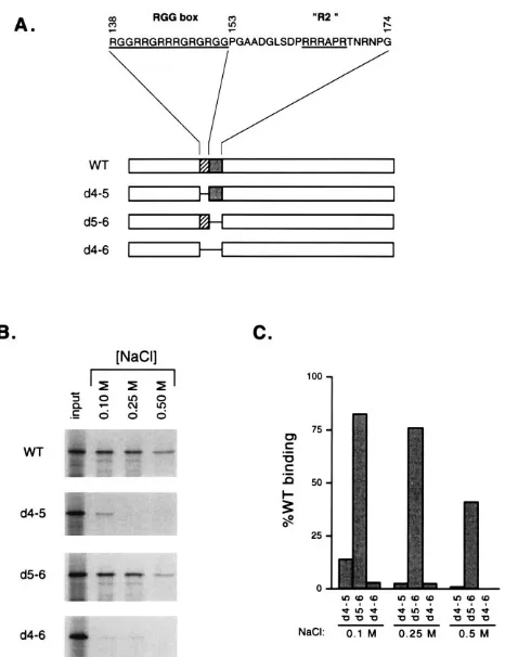
To determine whether the RGG box or nearby sequences are required for ICP27’s interaction with poly(G), three ICP27 deletion proteins (Fig. 3A) were expressed by in vitro
transla-tion and tested in the poly(G) binding assay. Thed4-5 mutant
lacks residues 138 to 152 and thus contains a precise deletion
of the RGG box sequence. Thed5-6 mutant lacks residues 153
to 174; this deletion removes a second arginine-rich sequence designated R2 by Hibbard and Sandri-Goldin (11). Mutant
d4-6 lacks both the RGG box and R2 sequences. The
poly(G)-binding activities of these mutants, as well as WT ICP27, were measured at 0.1, 0.25, and 0.5 M NaCl (Fig. 3B and C). Both
thed4-5 andd4-6 proteins were defective in poly(G) binding,
showing less than 5% of the WT level of binding at 0.25 and 0.5 M NaCl. These two mutants differed slightly at 0.1 M NaCl, in
thatd4-5 mutant bound weakly whereasd4-6 did not bind to an
appreciable extent. In contrast tod4-5 andd4-6, thed5-6
mu-tant bound to poly(G) almost as efficiently as the WT protein, although its binding was reduced approximately twofold com-pared with that of the WT protein at 0.5 M NaCl. Together, these data indicate that the RGG box motif, but not the argi-nine-rich sequence immediately C-terminal to it (R2), is re-quired for efficient poly(G) binding.
Direct interaction of ICP27’s RGG box with RNA.Brown et al. (3) recently used a northwestern blotting assay to demon-strate that ICP27 is an RNA-binding protein. These investiga-tors showed that a bacterially expressed GST-ICP27 fusion
protein can bind to.600-nucleotide-long RNA probes
corre-sponding to sequences from the 39ends of the c-mycor IFN-b
gene. The binding appeared to be target specific, as GST-ICP27 did not bind to a similar-size RNA corresponding to
sequences from the 39end of the SV40 early region. To
deter-mine whether ICP27’s RGG box plays a role in this RNA interaction, we tested several GST fusion proteins for the
abil-ity to bind to theb-RNA probe used by Brown et al. (3). The
proteins tested were (i) GST; (ii) GST-ICP27, a fusion having
all of ICP27 tagged to GST; (iii) GST–d4-5, a fusion identical
to GST-ICP27 but lacking the RGG box; and (iv) GST-RGG, a fusion containing only the RGG box sequence (residues 140
to 152) joined to GST. The proteins were expressed inE. coli,
[image:4.612.328.541.67.418.2]purified by glutathione-Sepharose affinity chromatography, and analyzed by SDS-PAGE and Coomassie blue staining (Fig. 4A). Although the GST and GST-RGG preparations yielded major bands of the expected molecular sizes (lanes 2 and 5,
FIG. 2. Poly(G) binding by ICP27 truncation mutants. (A) Schematic dia-gram of ICP27 N- and C-terminal truncation mutants produced by in vitro translation. The RGG box sequence is indicated. (B) Binding of truncation mutants to poly(G). The truncation mutants were tested in the poly(G) binding assay at an NaCl concentration of 0.25 M. The values given are the percent binding relative to a WT ICP27 control. The values for each mutant are means of four to nine independent binding assays; the error bars indicate standard deviations. The mean value of WT ICP27 binding to poly(G) in these experi-ments was 37.5%.
on November 9, 2019 by guest
http://jvi.asm.org/
respectively), much of the protein in the GST-ICP27 and GST–
d4-5 preparations (lanes 3 and 4) migrated as bands that were
smaller than expected, possibly as a result of proteolysis during expression and/or purification. However, there was some
ap-parently full-length GST-ICP27 and GST–d4-5 in each
prepa-ration, migrating at the expected molecular masses of;95 and
;93 kDa, respectively. Western blot analysis of the
GST-ICP27 and GST–d4-5 preparations demonstrated that the
pu-tative full-length molecules as well as most of the smaller species reacted with both an anti-GST monoclonal antibody and H1113, a monoclonal antibody directed against an N-terminal epitope in ICP27 (data not shown).
For the northwestern blotting assay, GST or the GST fusion proteins were subjected to SDS-PAGE, electrophoretically transferred to nitrocellulose filters, and renatured in situ.
Iden-tical filters were incubated with three different 32P-labeled
RNA probes. One probe was the;650-nucleotide IFN-bgene
probe previously shown to form a complex with GST-ICP27 (Fig. 4B, lane 2). As controls for specificity, two other probes
were used: an antisense version of the IFN-b probe (lane 1)
and an;840-nucleotide RNA corresponding to the 39end of
the SV40 early region (lane 3), previously shown not to form a
complex with GST-ICP27 (3). After binding, the northwestern filters were washed and analyzed by autoradiography. The re-sults (Fig. 4C) indicate that the RGG box is both required and sufficient for interaction with RNA, as both GST-ICP27 and
GST-RGG, but not GST or GST–d4-5, efficiently bound to
RNA molecules. However, we did not observe the specificity reported by Brown et al. (3), as all three probes bound
com-parably to GST-ICP27 and GST-RGG. Although the IFN-b
probe appeared to bind slightly more efficiently to GST-ICP27 in this experiment than did the SV40 probe, this was not observed in a repeat experiment (data not shown).
[image:5.612.63.296.67.370.2]Two major bands in the GST-ICP27 preparation formed complexes with RNA (Fig. 4C, lane 2). These corresponded not to full-length GST-ICP27 but to the two truncated forms of GST-ICP27 indicated by the squares in Fig. 4A. However, a
FIG. 3. Poly(G) binding by ICP27 in-frame deletion mutants. (A) Schematic diagram of ICP27 in-frame deletion mutants. The sequence of residues 138 to 174 of ICP27 is shown at the top. Below, the open bars represent ICP27, while the cross-hatched and gray bars represent the RGG box and R2 regions, respec-tively. The lines connecting bars indicate in-frame deletions. (B) Poly(G) binding by in-frame deletion mutants. WT ICP27 or in-frame deletion mutants were expressed by in vitro translation and tested in the poly(G) binding assay at the NaCl concentrations indicated. Equivalent fractions of the input and bound fractions were analyzed by SDS-PAGE and autoradiography. (C) Quantitation of poly(G) binding by ICP27 deletion mutants. The data from panel B were quantitated by phosphor image analysis. Poly(G) binding of each deletion mu-tant is presented as a percentage of WT ICP27 binding at the same NaCl concentration.
FIG. 4. RNA binding by GST-ICP27 fusion proteins. (A) Purification of GST fusion proteins. GST or GST-ICP27 fusion proteins were expressed inE. coliand purified by affinity chromatography on glutathione-Sepharose. Ten micrograms of each preparation was analyzed by SDS-PAGE and Coomassie blue staining. The molecular masses of the molecular weight (MW) marker proteins are indicated at the left in kilodaltons. The circles to the right of lanes 3 and 4 indicate the posi-tions of full-length GST-ICP27 and GST–d4-5. The squares to the right of lane 3 indicate truncated GST-ICP27 products which interact strongly with RNA. (B)
32P-labeled RNA probes. RNA probes for the northwestern assay were generated
by in vitro transcription in the presence of [a-32P]CTP. An aliquot of each probe
was analyzed by 5% urea-acrylamide gel electrophoresis and autoradiography. Lane 1, antisense version of the IFN-bgene 39-end probe; lane 2, IFN-bgene 39-end probe; lane 3, SV40 early region 39-end probe. The positions and nucle-otide sizes of markers are indicated at the right. The expected size of the IFN-b probes is 650 nucleotides; that of the SV40 probe is 840 nucleotides (3). (C) Binding of GST-ICP27 and GST-RGG to RNA. Ten-microgram aliquots of GST fusion preparations were electrophoresed on an SDS–15% polyacrylamide gel, blotted to nitrocellulose, and incubated with32P-labeled RNA probes as
de-scribed by Brown et al. (3). An autoradiograph of the washed blot is shown. The molecular masses of protein standards are indicated at the left in kilodaltons.
on November 9, 2019 by guest
http://jvi.asm.org/
repeat experiment in which 40mg of protein, rather than 10mg, was analyzed on the northwestern blot demonstrated that the
full-length GST-ICP27, but not full-length GST–d4-5, could
also bind to the IFN-b and SV40 RNA probes (data not
shown).
Two important conclusions can be drawn from these exper-iments regarding the role of ICP27’s RGG box in RNA bind-ing. First, the RGG box is required, in the context of both intact and truncated GST fusion proteins, for interaction with RNA molecules. Second, the RGG box sequence alone can mediate RNA binding, since GST-RGG efficiently binds to RNA.
In vivo methylation of ICP27. The experiments described above suggest that ICP27 is an RGG box-type RNA-binding protein, since its RGG box sequence can mediate RNA bind-ing. A characteristic of many cellular RGG box proteins is that they are targets for posttranslational modification by protein arginine methyltransferases, which dimethylate arginine
resi-dues to produce NG-NG-dimethylarginine (19, 27). The
func-tion of this unusual modificafunc-tion is not known. To determine whether ICP27 is methylated in vivo, we carried out a meta-bolic labeling experiment to specifically identify methylated proteins (19). The assay is based on the fact that the methyl
donor for protein methylation,S-adenosylmethionine, acquires
its methyl group from free methionine in the cell. Thus, if translation is blocked, methylated proteins can be specifically
labeled by incubating cells in L-[methyl-3H]methionine. To
carry out the methylation assay, Vero cells were mock infected,
infected with WT HSV-1, or infected with d27-1, an ICP27
deletion mutant (31). At 5 hpi, the cells were treated with cycloheximide and chloramphenicol to inhibit protein
synthe-sis. Thirty minutes later, the cells were labeled withL-[methyl
-3H]methionine for 3 h in the presence of the translation
in-hibitors. The labeled proteins were examined by SDS-PAGE and fluorography (Fig. 5). Three lines of evidence indicate that the majority of labeling is due to in vivo methylation, not incorporation of methionine via protein synthesis: (i) the pat-tern of labeled proteins is dramatically different from that
obtained when cells are labeled with [35S]methionine in the
absence of translation inhibitors (31, 33); (ii) control experi-ments demonstrated that the cycloheximide-choramphenicol treatment inhibited overall translation levels approximately 20-fold (not shown); and (iii) treatment with a more stringent inhibitor of protein synthesis, anisomycin, led to a nearly iden-tical pattern of labeled proteins (not shown).
The pattern of protein methylation in WT HSV-1-infected cells was quite distinct from that of the mock-infected cells (Fig. 5, lanes 1 and 2). First, the labeling of several cellular proteins was markedly reduced after HSV-1 infection. In par-ticular, the labeling of histones was significantly inhibited. Moreover, this effect requires ICP27, as histone methylation
was not inhibited in thed27-1 infection (lane 3). Second,
sev-eral methylated proteins were either induced or enhanced in WT-infected cells. We noted two prominent bands, labeled a and b in Fig. 5. These proteins may correspond either to viral proteins which are methylated or to cellular proteins whose methylation is induced upon infection.
Although the identity of band b was not investigated further, we suspected that band a might correspond to ICP27, since it
has approximately the same apparent molecular mass (;63
kDa) and is absent from the ICP27 null mutant infection (Fig. 5; compare lanes 2 and 3). To determine whether band a corresponds to ICP27, a second experiment was performed. Vero cells were mock infected or infected with WT HSV-1,
WT HSV-2, d27-1, or d4-5 (26). d4-5 is a defective HSV-1
ICP27 mutant which contains the same RGG box deletion
shown in Fig. 3A. The infected cells were labeled withL-[
meth-yl-3H]methionine as before, and the cell lysates were subjected
to immunoprecipitation using a mixture of two anti-ICP27 monoclonal antibodies. Analysis of the fluorograph (Fig. 6A) indicated that both HSV-1 and HSV-2 ICP27 are methylated
proteins, since they efficiently label with L-[methyl-3
H]methi-onine. Furthermore, comparison of the immunoprecipitated
FIG. 5. Protein methylation in 1-infected Vero cells. Mock- or HSV-1-infected Vero cells were treated with translation inhibitors (100mg of cyclo-heximide per ml and 40mg of chloramphenicol per ml) at 5 hpi. Thirty minutes later, the cells were labeled for 3 h with 45mCi ofL-[methyl-3
H]methionine per ml in the presence of the same translation inhibitors. The cells were scraped and lysed in RIPA buffer, and the lysates were electrophoresed on an SDS–15% polyacrylamide gel. A fluorograph of the gel is shown. Lane M, protein stan-dards; lane 1, mock-infected cells; lane 2, WT HSV-1-infected cells; lane 3,
d27-1-infected cells. At the left, the molecular masses of the protein standards are indicated in kilodaltons. At the right, bands corresponding to the two major methylated proteins in WT HSV-infected cells (a and b) and the positions of cellular histones are indicated.
FIG. 6. In vivo methylation of ICP27. Vero cells were mock infected (lane 1) or infected with WT HSV-1, WT HSV-2,d27-1, ord4-5 (lanes 2 to 5, respec-tively). The cells were labeled from 5.5 to 8.5 hpi withL-[methyl-3H]methionine as described in the text. Cell lysates were subjected to immunoprecipitation using a mixture of two monoclonal antibodies specific for ICP27 (H1113 and H1119). (A) Labeling of ICP27 withL-[methyl-3H]methionine. Aliquots of the immuno-precipitates were subjected to SDS-PAGE (15% gel) and analyzed by fluorog-raphy. A 40-h exposure is shown. Molecular weight markers are shown in lane M (sizes in kilodaltons). (B) Presence of ICP27 in the immunoprecipitates. Aliquots of the immunoprecipitates were subjected to SDS-PAGE (15% gel), and the proteins were electrophoretically transferred to nitrocellulose. The filter was subjected to Western blot analysis using H1119 antibody. Antigen detection was performed with an enhanced chemiluminescence detection system; a 1-s expo-sure is shown.
on November 9, 2019 by guest
http://jvi.asm.org/
HSV-1 ICP27 with the total labeled lysate (not shown) dem-onstrated that ICP27 corresponds to band a seen in Fig. 5.
Interestingly, no labeled ICP27 was present in thed4-5
im-munoprecipitate (Fig. 6A, lane 5), suggesting that the RGG box is required for in vivo methylation of ICP27. To
demon-strate that thed4-5 protein was present in the
immunoprecipi-tate, we subjected the immunoprecipitates to Western blot
analysis (Fig. 6B). The d4-5 protein was indeed present, in
amounts comparable to those of the WT protein (compare lanes 2 and 5). Therefore, the RGG box is required for ICP27’s in vivo methylation. Moreover, this experiment confirms the
validity of the methylation assay, as the WT and d4-5 ICP27
proteins contain equal numbers of methionine residues yet
label distinctly withL-[methyl-3H]methionine.
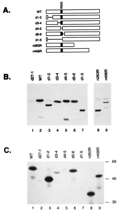
Requirement for the RGG box in in vivo methylation.The foregoing experiment demonstrates that the RGG box se-quence is required for ICP27’s methylation but does not ex-clude the possibility that other regions also are involved. To systematically determine which portions of ICP27 are required for in vivo methylation, we used several HSV-1 ICP27 mutants which contain nonsense or in-frame deletion mutations in the ICP27 gene (Fig. 7A). Infected Vero cells were labeled with
L-[methyl-3H]methionine, and ICP27 proteins were subjected
to immunoprecipitation. To confirm that the ICP27 mutant proteins were expressed and immunoprecipitated, the samples were first subjected to Western blot analysis (Fig. 7B). With one exception, all of the mutant proteins were present in
quan-tities similar to that of WT ICP27. The exception was then406
protein, which was present in somewhat lesser amounts. To determine whether the proteins were methylated, the immu-noprecipitates were analyzed by SDS-PAGE and fluorography
(Fig. 7C). Although the n406 protein was present in lower
amounts, it was clearly methylated (lane 9), as was then263R
protein (lane 8). This result indicates that methylation occurs predominantly or exclusively on the N-terminal half of ICP27. Examination of several N-terminal in-frame deletion mutant proteins gave further information about the sequences in
ICP27 required for methylation. Thed1-2 and d5-6 proteins
were efficiently methylated (lanes 3 and 6, respectively), indi-cating that residues 12 to 63 and 154 to 173 are not required.
Thed3-4 protein (lane 4) was weakly labeled, indicating that
the engineered deletion in d3-4 lowers the efficiency of, but
does not completely prevent, ICP27 methylation. The only two mutant proteins which did not appear to be methylated to any
appreciable extent were those encoded byd4-5 andd1-5, both
of which lack the RGG box motif. These data demonstrate that the RGG box is a critically important determinant of ICP27’s in vivo methylation.
DISCUSSION
ICP27’s RGG box and RNA binding.In this work, we show that ICP27 binds to RNA homopolymers composed of poly(G). This finding is consistent with two recent studies which have also demonstrated that ICP27 can interact with RNA in vitro (3, 13). We used both the poly(G) binding assay and the northwestern blotting assay described by Brown et al. (3) to identify the sequences in ICP27 which mediate RNA binding. Both sets of experiments implicate ICP27’s RGG box motif, which maps to residues 138 to 152. In the poly(G) binding assay, there is a strict correlation between the presence of the RGG box and RNA binding. In addition, the RGG box is required in the context of an otherwise intact ICP27 mole-cule for association with poly(G). In the northwestern assay, the RGG box is required for RNA interaction and alone can mediate RNA binding when attached to a heterologous
pro-tein. Together, our data demonstrate that ICP27’s RGG box is an RNA-binding domain that can mediate interaction with both poly(G) RNA homopolymers and more complex RNA sequences.
Our results differ from those of Brown et al. (3) in that we do not observe the same RNA target specificity in the northwest-ern blotting assay. Brown et al. found that GST-ICP27 could
associate with RNAs derived from the 39 ends of the IFN-b
and c-mycgenes but not with an RNA derived from the 39end
of the SV40 early region. In contrast, we find that GST-ICP27
fusion binds efficiently to both the IFN-band SV40 probes as
well as to an antisense version of the IFN-bprobe. We note
[image:7.612.336.532.69.419.2]that Ingram et al. also failed to find evidence for sequence-specific RNA-binding by ICP27 (13). A complicating factor in our experiments is that the major RNA-binding polypeptides in our GST-ICP27 preparation are approximately 25 to 30 kDa smaller than the full-length protein. However, truncation of
FIG. 7. The RGG box is required for ICP27 methylation. (A) ICP27 proteins expressed by HSV-1 ICP27 mutants. Bars represent ICP27 coding segments; lines connecting bars indicate in-frame deletions. The RGG box is indicated by a stippled box. (B and C) In vivo methylation assay. Cells were infected with WT HSV-1,d27-1, or the ICP27 mutants shown in panel A. Cells were labeled with L-[methyl-3H]methionine, and anti-ICP27 immunoprecipitations were performed
as described in the text. (B) Western blot detection of ICP27 molecules in immunoprecipitates. Aliquots of the immunoprecipitates were subjected to Western analysis using ICP27-specific monoclonal antibodies. Antigen detection was by enhanced chemiluminescence; a 5-s exposure is shown. (C) Labeling of ICP27 withL-[methyl-3H]methionine. Aliquots of the immunoprecipitates were
subjected to SDS-PAGE and fluorography. A 6-day exposure is shown. The positions of molecular weight standards are shown at the right in kilodaltons.
on November 9, 2019 by guest
http://jvi.asm.org/
our GST-ICP27 is unlikely to explain the conflicting results, as
the GST-ICP27 fusion protein of Brown et al. (3) is also;30
kDa smaller than expected and thus likely corresponds to a similar proteolytic fragment. The discrepancies in binding re-sults thus leave open the question of ICP27’s RNA target specificity. However, the fact that ICP27 binds efficiently to poly(G) RNA raises the possibility that G-rich sequences play a role in the recognition of natural RNA targets by ICP27.
The RGG box motif, found in a number of cellular RNA-binding proteins, is one of several known RNA-RNA-binding do-mains (4, 14, 20). It is defined as an arginine- and glycine-rich sequence that usually contains closely spaced RGG repeats interspersed with other, often aromatic amino acids (4). The minimal size of a functional RGG box RNA-binding sequence is unknown, but most RGG boxes are larger than the 15 resi-dues which comprise ICP27’s RGG box. In this work, we have shown that 13 residues of ICP27’s RGG box, GRRGRRRG RGRGG, are sufficient to mediate RNA binding when at-tached to a heterologous protein, GST. To our knowledge, this is the smallest RGG-like sequence which has been shown to bind to RNA. The GST-RGG molecule may therefore be a useful tool for defining the specific amino acid residues which interact with RNA.
Two observations suggest that the C terminus of ICP27 may inhibit RNA binding by the RGG box. First, the C-terminally truncated N189 polypeptide displays significantly enhanced poly(G) binding compared with the WT protein. Second, C-terminally truncated forms of GST-ICP27 appear to be more efficient RNA-binding proteins than full-length GST-ICP27, although the full-length molecule binds RNA to some extent. These observations raise the possibility that the C-terminal region of ICP27 negatively regulates the RNA-binding activity of the RGG box. However, it is also possible that enhanced RNA binding by truncated ICP27 molecules represents an artifactual effect of mutagenesis.
ICP27’s RGG box and protein methylation.Several cellular proteins involved in nuclear RNA metabolism, including fibril-larin (18), nucleolin (17), and hnRNP A1 (47), are methylated on arginine residues. The predominant modified residue in these proteins is the asymmetrically dimethylated derivative
NG-NG-dimethylarginine (DMA). The function of this protein
modification is unknown. Recently, Liu and Dreyfuss found that in addition to hnRNP A1, many other hnRNP proteins are methylated in vivo (19). In addition, these investigators
par-tially purified a protein arginine N-methyltransferase from
HeLa cells, using unmodified recombinant hnRNP A1 as a substrate. The enzyme activity appeared to be specific for RGG box sequences and modified arginine residues to DMA
and NG-monomethylarginine. Moreover, in vitro, the
enzy-matic activity methylated the same spectrum of hnRNP pro-teins that are methylated in vivo. These results suggest that many hnRNP proteins, and possibly other RGG box proteins, are methylated on their RGG box motifs by a common cellular enzyme.
On the basis of the foregoing information, we investigated the methylation status of ICP27 in HSV-1-infected cells. Our results demonstrate that ICP27 is one of the major methylated proteins in infected cells, at least under the conditions of our labeling assay. Furthermore, ICP27’s methylation correlates with and requires the presence of the RGG box sequence. Residues 109 to 137, just N terminal to the RGG box, may also
play a role in methylation, as thed3-4 protein was only weakly
labeled in the in vivo methylation assay. However, it is worth
noting that thed3-4 mutation, in addition to deleting codons
109 to 137, also alters the first two codons of the RGG box, changing them from RG to LE. This modification to the RGG
box could also explain the reduced methylation of the d3-4
protein. Overall, our results indicate that the RGG box is critical for methylation and suggest that it is the RGG box motif itself which is the target of methylation. If so, then the modified residue is arginine, as this is the only residue in the RGG box sequence known to accept methylation (6). How-ever, further experiments will be required to directly map the site(s) of methylation and to identify the modified residue(s). What role, if any, could RGG box methylation play in the biological function of ICP27? An intriguing possibility is that methylation regulates ICP27’s association with RNA. Methyl-ation of arginine’s guanidinium group would not alter the strong positive charge of the arginine side chain, but the bulky methyl groups could sterically affect RNA binding. In addition, arginine methylation might alter hydrogen bonding interac-tions between arginine residues and RNA. It is thus conceiv-able that arginine methylation could alter the overall level, or target specificity, of ICP27’s binding to RNA. It is noteworthy that the enzymatic activity thought to be responsible for RGG
box methylation is not found inE. coli(19). Since bacterially
expressed GST-ICP27 and GST-RGG bind RNA, methylation is probably not required for RNA binding. It is also of interest that the RGG box methyltransferase is present in a rabbit reticulocyte lysate (19). Thus, it is possible that the ICP27 used in our poly(G) binding assays is methylated to some extent. Since there are specific inhibitors available for protein meth-yltransferases, it may be possible to produce methylated and unmethylated ICP27 by in vitro translation for side-by-side comparison in RNA-binding experiments.
Possible in vivo RNA binding by ICP27’s RGG box.We have previously shown that the RGG box is important for ICP27’s biological functions, as an HSV-1 mutant which encodes an RGG box-minus ICP27 fails to replicate in Vero cells (26). The studies described herein demonstrate that ICP27’s RGG box motif can function in vitro as an RNA-binding domain. It is tempting to speculate that ICP27’s RGG box also mediates RNA binding activity in vivo. Previously, we suggested that the nucleolar localization of ICP27, which depends on the RGG box, reflects an in vivo association with abundant RNAs in the nucleolus (26). However, the physiologically relevant RNA target, or targets, of ICP27 are not necessarily nucleolar. Given ICP27’s effects on pre-mRNA splicing, possible targets might be one or more of the small nuclear RNAs (snRNAs) associ-ated with spliceosomal snRNPs. In this regard, it is interesting that at least a small fraction of ICP27 interacts with U snRNPs during infection (37). Another possibility is that ICP27 func-tions as an hnRNP protein, binding to nascent pre-mRNA in the nucleus (7). Indeed, our work shows that ICP27 shares characteristics with several cellular hnRNP proteins, in that it contains an RGG box RNA-binding domain and is methylated. In this scenario, ICP27’s association with pre-mRNAs, perhaps in combination with cellular hnRNP proteins, could lead to modified hnRNP complexes on viral or cellular transcripts. Such novel complexes might determine alternate pathways of mRNA polyadenylation, splicing, or export from the nucleus. This model could explain how a single viral protein can affect multiple and seemingly diverse aspects of pre-mRNA metab-olism.
ACKNOWLEDGMENTS
We are grateful to Charles Brown, Gideon Dreyfuss, Matthew chael, and L. P. Perera for providing plasmids and to Matthew Mi-chael, Leslie Schiff, and Deborah Shepard, for helpful discussions and technical advice. We also thank Scott Bunnell, Leslie Schiff, and Char-lotte Spencer for thoughtful reviews of the manuscript.
on November 9, 2019 by guest
http://jvi.asm.org/
This research was supported by the National Cancer Institute of Canada with funds from the Canadian Cancer Society. S.A.R. is a Senior Scholar of the Alberta Heritage Foundation for Medical Re-search (AHFMR). W.E.M. is supported by a studentship award from the AHFMR.
REFERENCES
1.Ackermann, M., D. K. Braun, L. Pereira, and B. Roizman.1984. Character-ization of herpes simplex virus type 1aproteins 0, 4, and 27 with monoclonal antibodies. J. Virol.52:108–118.
2.Anderson, J. T., S. M. Wilson, K. V. Datar, and M. S. Swanson.1993. NAB2: a yeast nuclear polyadenylated RNA-binding protein essential for cell via-bility. Mol. Cell. Biol.13:2730–2741.
3.Brown, C. R., M. S. Nakamura, J. D. Mosca, G. S. Hayward, S. E. Straus, and L. P. Perera.1995. Herpes simplex virustrans-regulatory protein ICP27 stabilizes and binds to 39ends of labile mRNA. J. Virol.69:7187–7195. 4.Burd, C. G., and G. Dreyfuss.1994. Conserved structures and diversity of
functions of RNA-binding proteins. Science265:615–621.
5.Chapman, C. J., J. D. Harris, M. A. Hardwicke, R. M. Sandri-Goldin, M. K. L. Collins, and D. S. Latchman.1992. Promoter-independent activa-tion of heterologous virus gene expression by the herpes simplex virus im-mediate-early protein ICP27. Virology186:573–578.
6.Clarke, S.1993. Protein methylation. Curr. Opin. Cell Biol.5:977–983. 7.Dreyfuss, G., M. J. Matunis, S. Pin˜ol-Roma, and C. J. Burd.1993. hnRNP
proteins and the biogenesis of mRNA. Annu. Rev. Biochem.62:289–321. 8.Fabre, E., W. C. Boelens, C. Wimmer, I. Mattaj, and E. C. Hurt.1994.
Nup145p is required for nuclear export of mRNA and binds homopolymeric mRNA in vitro via a novel conserved motif. Cell78:275–289.
9.Hardwicke, M. A., and R. M. Sandri-Goldin.1994. The herpes simplex virus regulatory protein ICP27 contributes to the decrease in cellular mRNA levels during infection. J. Virol.68:4797–4810.
10. Hardy, W. R., and R. M. Sandri-Goldin.1994. Herpes simplex virus inhibits host cell splicing, and the regulatory protein ICP27 is required for this effect. J. Virol.68:7790–7799.
11. Hibbard, M., and R. Sandri-Goldin.1995. Arginine-rich regions succeeding the nuclear localization region of herpes simplex virus type 1 regulatory protein ICP27 are required for efficient nuclear localization and late gene expression. J. Virol.69:4656–4667.
12. Hughes, R. G., and W. H. Munyon.1975. Temperature-sensitive mutants of herpes simplex virus type 1 defective in lysis but not in transformation. J. Virol.16:275–283.
13. Ingram, A., A. Phelan, J. Dunlop, and J. B. Clements.1996. Immediate early protein IE63 of herpes simplex virus type 1 binds RNA directly. J. Gen. Virol.77:1847–1851.
14. Kiledjian, M., and G. Dreyfuss.1992. Primary structure and binding activity of the hnRNP U protein: binding RNA through RGG box. EMBO J.11:
2655–2664.
15. Knipe, D. M., D. Senechek, S. A. Rice, and J. L. Smith.1987. Stages in the nuclear association of the herpes simplex virus transcriptional activator pro-tein ICP4. J. Virol.61:276–284.
16. Lam, V., W. Mears, and S. Rice.Unpublished data.
17. Lischwe, M. A., R. L. Ochs, R. Reddy, R. G. Cook, L. C. Yeoman, and H. Busch.1985. Clustering of glycine and NG
,NG
-dimethylarginine in nucleolar protein C23. Biochemistry24:6025–6028.
18. Lischwe, M. A., R. L. Ochs, R. Reddy, R. G. Cook, L. C. Yeoman, E. M. Tan, M. Reichlin, and H. Busch.1985. Purification and partial characterization of a nucleolar scleroderma antigen (Mr 534,000; pI, 8.5) rich in N
G
,NG
-dimethylarginine. J. Biol. Chem.260:14304–14310.
19. Liu, Q., and G. Dreyfuss.1995. In vivo and in vitro arginine methylation of RNA-binding proteins. Mol. Cell. Biol.15:2800–2808.
20. Mattaj, I. W.1993. RNA recognition: a family matter? Cell73:837–840. 21. McCarthy, A. M., L. McMahan, and P. A. Schaffer.1989. Herpes simplex
virus type 1 ICP27 deletion mutants exhibit altered patterns of transcription and are DNA deficient. J. Virol.63:18–27.
22. McGeoch, D. J., M. A. Dalrymple, A. J. Davison, A. Dolan, M. C. Frame, D. McNab, L. J. Perry, J. E. Scott, and P. Taylor.1988. The complete DNA sequence of the long unique region in the genome of herpes simplex virus type 1. J. Gen. Virol.69:1531–1574.
23. McGregor, F., A. Phelan, J. Dunlop, and J. B. Clements.1996. Regulation of herpes simplex virus type 1 poly(A) site usage and the action of the immediate-early protein IE63 in the early-late switch. J. Virol.70:1931– 1940.
24. McLauchlan, J., A. Phelan, C. Loney, R. M. Sandri-Goldin, and J. B. Clem-ents.1992. Herpes simplex virus IE63 acts at the posttranscriptional level to stimulate viral mRNA 39processing. J. Virol.66:6939–6945.
25. McLauchlan, J., S. Simpson, and J. B. Clements.1989. Herpes simplex virus
induces a processing factor that stimulates poly(A) site usage. Cell59:1093– 1105.
26. Mears, W. E., V. Lam, and S. A. Rice.1995. Identification of nuclear and nucleolar localization signals in the herpes simplex virus regulatory protein ICP27. J. Virol.69:935–947.
27. Najbauer, J., B. A. Johnson, A. L. Yound, and D. W. Aswad.1993. Peptides with sequences similar to glycine, arginine-rich motifs in proteins interacting with RNA are efficiently recognized by methyltransferase(s) modifying argi-nine in numerous proteins. J. Biol. Chem.268:10510–10509.
28. Ohno, T., M. Ouchida, L. Lee, Z. Gatalica, V. N. Rao, and E. S. P. Reddy.
1994. The EWS gene, involved in Ewing family of tumors, malignant mela-noma of soft parts and desmoplastic small round cell tumors, codes for an RNA binding protein with novel regulatory domains. Oncogene9:3087–3097. 29. Phelan, A., M. Carmo-Fonseca, J. McLauchlan, A. I. Lamond, and J. B.
Clements.1993. A herpes simplex virus type 1 immediate-early gene product, IE63, regulates small nuclear ribonucleoprotein distribution. Proc. Natl. Acad. Sci. USA90:9056–9060.
30. Pin˜ol-Roma, S., Y. Choi, M. Matunis, and G. Dreyfuss.1988. Immunopuri-fication of heterogenous nuclear ribonucleoprotein particles reveals an as-sortment of RNA-binding proteins. Genes Dev.2:215–227.
31. Rice, S. A., and D. M. Knipe.1990. Genetic evidence for two distinct trans-activation functions of the herpes simplex virusaprotein ICP27. J. Virol.
64:1704–1715.
32. Rice, S. A., and V. Lam.1994. Amino acid substitution mutations in the herpes simplex virus ICP27 protein define an essential gene regulation func-tion. J. Virol.68:823–833.
33. Rice, S. A., V. Lam, and D. M. Knipe.1993. The acidic amino-terminal region of herpes simplex virus type I alpha protein ICP27 is required for an essential lytic function. J. Virol.67:1778–1787.
34. Rice, S. A., M. C. Long, V. Lam, P. A. Schaffer, and C. A. Spencer.1995. Herpes simplex virus immediate-early protein ICP22 is required for viral modification of host RNA polymerase II and establishment of the normal viral transcription program. J. Virol.69:5550–5559.
35. Roizman, B., and A. E. Sears.1990. Herpes simplex viruses and their repli-cation, p. 1795–1842.InB. N. Fields and D. M. Knipe (ed.), Fundamental virology. Raven Press, New York.
36. Sacks, W. R., C. C. Greene, D. P. Aschman, and P. A. Schaffer.1985. Herpes simplex virus type 1 ICP27 is an essential regulatory protein. J. Virol.55:
796–805.
37. Sandri-Goldin, R. M., and M. K. Hibbard.1996. The herpes simplex virus type 1 regulatory protein ICP27 coimmunoprecipitates with Sm anti-serum, and the C terminus appears to be required for this interaction. J. Virol.70:108–118.
38. Sandri-Goldin, R. M., M. K. Hibbard, and M. A. Hardwicke.1995. The C-terminal repressor region of herpes simplex virus type 1 ICP27 is required for redistribution of small nuclear ribonucleoprotein particles and splicing factor SC35; however, these alterations are not sufficient to inhibit host cell splicing. J. Virol.69:6063–6076.
39. Sandri-Goldin, R. M., and G. E. Mendoza.1992. A herpesvirus regulatory protein appears to act post-transcriptionally by affecting mRNA processing. Genes Dev.6:848–863.
40. Siomi, H., M. C. Siomi, R. L. Nussbaum, and G. Dreyfuss.1993. The protein product of the fragile X gene, FMR1, has characteristics of an RNA-binding protein. Cell74:291–298.
41. Smiley, J. R., B. Panning, and C. A. Smibert.1991. Regulation of cellular genes by HSV products, p. 151–179.InE. K. Wagner (ed.), Herpesvirus transcription and its regulation. CRC Press, Inc., Boca Raton, Fla. 42. Swanson, M. S., and G. Dreyfuss.1988. Classification and purification of
proteins of heterogeneous nuclear ribonucleoprotein particles by RNA-bind-ing specificities. Mol. Cell. Biol.8:2237–2241.
43. Swanson, M. S., T. Y. Nakagawa, K. LeVan, and G. Dreyfuss.1987. Primary structure of human nuclear ribonucleoprotein particle C proteins: conserva-tion of sequence and domain structures in heterogeneous nuclear RNA, mRNA, and pre-rRNA-binding proteins. Mol. Cell. Biol.7:1731–1739. 44. Uprichard, S., and D. Knipe.1996. Herpes simplex ICP27 mutant viruses
exhibit reduced expression of specific DNA replication genes. J. Virol.70:
1969–1980.
45. Vaughn, P., K. Thibault, M. Hardwicke, and R. Sandri-Goldin.1992. The herpes simplex virus immediate early protein ICP27 encodes a potential metal binding domain and binds zincin vitro. Virology189:377–384. 46. Wilcox, K. W., A. Kohn, E. Sklyanskaya, and B. Roizman.1980. Herpes
simplex virus phosphoproteins. I. Phosphate cycles on and off some viral polypeptides and can alter their affinity for DNA. J. Virol.33:167–182. 47. Williams, K. R., K. L. Stone, M. B. LoPresiti, B. M. Merrill, and S. R.
Planck.1985. Amino acid sequence of the UP1 calf-thymus helix-destabiliz-ing protein and its homology to an analogous protein from mouse myeloma. Proc. Natl. Acad. Sci. USA82:5666–5670.