0022-538X/96/$04.0010
Copyrightq1996, American Society for Microbiology
Sindbis Virus DNA-Based Expression Vectors: Utility
for In Vitro and In Vivo Gene Transfer
THOMAS W. DUBENSKY, JR.,1* DAVID A. DRIVER,1JOHN M. POLO,1BARBARA A. BELLI,1
EMI M. LATHAM,1CARLOS E. IBANEZ,1SUNIL CHADA,2DUANE BRUMM,2
THERESA A. BANKS,1STEVEN J. MENTO,1,2DOUGLAS J. JOLLY,1,2
ANDSTEPHEN M. W. CHANG1,2
Department of Viral and Genetic Therapeutics1and Department of Immunobiology,2Viagene, Inc., San Diego, California 92121
Received 22 March 1995/Accepted 15 September 1995
Several DNA-based Sindbis virus vectors were constructed to investigate the feasibility and potential applications for initiating the virus life cycle in cells transfected directly with plasmid DNA. These vectors, when transfected into mammalian cells, have been used to produce virus, to express heterologous genes, and to produce infectious vector particles. This approach involved the conversion of a self-replicating vector RNA (replicon) into a layered DNA-based expression system. The first layer includes a eukaryotic RNA polymerase II expression cassette that initiates nuclear transcription of an RNA which corresponds to the Sindbis virus vector replicon. Following transport of this RNA from the nucleus to the cytoplasm, the second layer, autocatalytic amplification of the vector, proceeds according to the Sindbis virus replication cycle and results in expression of the heterologous gene. The Sindbis virus DNA vectors expressed reporter genes in transfected cells at levels that were comparable to those of in vitro-transcribed RNA replicons and were approximately 10-fold higher than the levels produced by conventional RNA polymerase II-dependent plasmids in which the promoter and reporter gene were linked directly. Reporter gene expression was also observed in rodent muscle following injection with Sindbis virus DNA vectors. In a second application, packaged vector particles were produced in cells cotransfected with complementing replicon and defective helper DNAs. The Sindbis virus-derived DNA vectors described here increase the utility of alphavirus-based vector systems in general and also provide a vector with broad potential applications for genetic immunization.
Several members of the Alphavirus genus, first Sindbis virus (4, 45, 53) and later Semliki Forest virus (1, 23, 24) and other alphavirus members (8, 9), have received considerable atten-tion for use as virus-based expression vectors. Many properties of alphavirus vectors make them a desirable alternative to other virus-derived vector systems being developed, including
potential high-level expression of up to 108molecules of
het-erologous protein per cell (53), a broad host range, and infec-tion of nondividing cells (50). In addiinfec-tion, replicainfec-tion occurs entirely in the cytoplasm of the infected cell as an RNA mol-ecule, without a DNA intermediate. This is in contrast to retrovirus and adeno-associated virus vectors, which must en-ter the nucleus and usually integrate into the host genome for initiation of vector activity (18, 27, 43). Thus, retrovirus- and adeno-associated virus-derived vectors have application for long-term expression of foreign proteins, while the alphavirus vectors are likely better suited for short-term high-level expres-sion. Furthermore, although vectors derived from poxviruses, adenoviruses, and herpes simplex viruses all express high levels of foreign proteins, these systems are far more complex than alphaviruses and express many highly antigenic virus-specific gene products, including structural proteins (13, 29, 41). In contrast, current alphavirus vectors express only the four viral replicase proteins (nonstructural proteins nsP1 through nsP4) required for RNA amplification in the transduced cell.
The approximately 12-kb single-stranded positive-sense RNA genome of Sindbis virus and other alphaviruses is
infec-tious upon introduction into the cytoplasm of susceptible cells. During viral replication, the Sindbis virus genomic 49S RNA serves as the template for synthesis of a complementary neg-ative strand by the virus-encoded replicase. The negneg-ative strand in turn serves as the template for additional genomic RNA and for an abundant internally initiated 26S subgenomic RNA. The nonstructural proteins (nsPs) are translated from
the 59 two-thirds of the genomic RNA, while the structural
proteins (sPs) are translated from the subgenomic 26S RNA
that represents the 39one-third of the genome. The nsP and sP
genes are each expressed as polyproteins and are processed posttranslationally into the individual proteins (50). Expres-sion from current alphavirus vectors is based on the same strategy as expression of the sPs of wild-type virus and is initiated by transfection of in vitro-transcribed, self-replicating vector RNA (replicon) molecules (24, 53). The region encod-ing the virus sPs is replaced with a heterologous sequence or gene of interest, and the viral nsP-encoding region and all sequences required in cis for replication and packaging are maintained. Heterologous sequences are synthesized as highly abundant subgenomic mRNA molecules, which in turn serve as the translational template for the heterologous gene. Infec-tious vector particles have been generated by cotransfection and trans complementation of vector RNA replicons with an in vitro-transcribed defective helper (DH) RNA (5). The DH RNA contains the genes encoding the virus sPs and all of the sequences required in cis for replication but is deleted in the viral nsP genes and the virus packaging sequence core (1, 51). Thus, replication of the DH RNA and expression of high levels of the sPs occur in the presence of vector-supplied nsPs and result in the production of particles containing vector ge-nomes.
* Corresponding author. Mailing address: Viagene, Inc., 11055 Ro-selle St., San Diego, CA 92121-1204. Phone: (619) 452-1288. Fax: (619) 623-3428. Electronic mail address: [email protected].
508
on November 9, 2019 by guest
http://jvi.asm.org/
Despite the current interest in alphavirus-derived expression vectors, further development of the system is required for potential human vaccine and therapeutic applications. The preparation and transfection into animals of in vitro-tran-scribed expression vector RNA are rather inefficient, due in part to the length and lability of the vector molecules. This limitation has been circumvented somewhat by the pro-duction of recombinant vector particles through cotransfection of vector replicon and DH RNA molecules. However, this approach results in the generation of replication-competent virus through mechanisms of copackaging of vector and DH RNAs and/or RNA recombination (12, 52). The level of rep-lication-competent virus has been reduced significantly in the Semliki Forest virus system by DH modifications which result in the production of noninfectious packaged vector particles (1). An alternative approach, which has facilitated the appli-cation of Sindbis virus vectors to animal studies, utilizes a duplicated subgenomic promoter within the context of the viral genome (15, 38). However, double subgenomic vectors are propagated as infectious virus, which may be undesirable for applications involving human therapeutics, and the capacity for heterologous genetic material in this system is restricted.
We have been investigating the possibility of using DNA-based plasmid expression vectors to directly initiate the alpha-virus RNA replication cascade in transfected mammalian cells. Such vectors are a necessary step towards a goal of developing Sindbis virus-derived DNA-based therapeutic vectors and vec-tor particle-packaging cell lines. In this report, we describe the development of DNA-based Sindbis virus vector replicons. Transfection of these plasmids into cultured cells or animal muscle resulted in high-level expression of foreign genes. The template for gene expression is a self-replicating vector RNA molecule that resulted in synthesis of reporter proteins at lev-els which were significantly higher than those of conventional expression plasmids in which the promoter and reporter gene were directly linked. Furthermore, we demonstrate that co-transfection of replicon vector and DH plasmid DNAs or in vitro-transcribed RNAs produced similar levels of packaged Sindbis virus vector particles. This work represents the first report detailing the efficient specific initiation of an alphavirus infection in an RNA polymerase II promoter-dependent man-ner, following transfection of plasmid DNA, and represents a novel approach for the use of Sindbis virus-derived vectors. Similar approaches should facilitate the investigations of other cloned RNA viruses of positive polarity, the utility of their derived expression vectors, and their application to animal studies.
MATERIALS AND METHODS
Virus propagation and purification.A virus stock derived from the HR strain (5) of Sindbis virus was obtained from Lee Biomolecular (San Diego, Calif.). Following growth by passage in BHK-21 cells, virus was cloned by five consec-utive rounds of plaque purification as described before (48). A large plaque was selected and expanded by a single low-multiplicity passage in BHK cells to provide a seed stock for subsequent experiments. Virus to be used for RNA isolation was isolated from BHK cell supernatants collected at 18 h postinfection by polyethylene glycol precipitation (48) or by pelleting through a sucrose cush-ion (35).
Construction of genome-length Sindbis virus cDNA clones.RNA was purified from pelleted virus with RNAzol B (Tel-Test, Inc., Friendswood, Tex.) according to the manufacturer’s directions or by poly(A) selection after sodium dodecyl sulfate (SDS) lysis, using a commercially available kit (FastTrack Kit; Invitrogen, San Diego, Calif.). Two rounds of first-strand cDNA synthesis were performed with the purified virion RNA, using a mixture of random primers and primer SIN11703R (Table 1) and SuperScript reverse transcriptase (Gibco-BRL, Gaith-ersburg, Md.), according to the manufacturer’s conditions. The viral cDNA was then amplified in six distinct segments by PCR with six pairs of overlapping primers (sequences may be obtained from the corresponding author). The re-gions of overlap corresponded to unique enzyme recognition sites within the
PCR amplicons and were AgeI (nucleotide [nt] 3172), EcoRI (nt 5870), BamHI (nt 7335), BclI (nt 9356), and BsiWI (nt 10379). PCR amplification of Sindbis virus cDNA and all subsequent amplifications were performed in separate reac-tions with the Thermalase thermostable DNA polymerase (Amresco Inc., Solon, Ohio) buffer containing 1.5 mM MgCl2(provided by the supplier) and the desired primer pair. Additionally, the reactions contained 5% dimethyl sulfoxide and Hot Start Wax beads (Perkin-Elmer, Branchburg, N.J.). The Sindbis virus 59-end forward primer (SINSP61F [Table 1]) contained a 19-nt sequence corre-sponding to the bacteriophage SP6 RNA polymerase promoter and the ApaI recognition sequence. The SP6 RNA promoter was positioned so that transcrip-tion in vitro resulted in the inclusion of only a single nonviral G ribonucleotide linked to the authentic Sindbis virus 59 end. Inclusion of an upstream ApaI recognition sequence facilitated insertion of the PCR amplicon into the plasmid vector (pKSII1; Stratagene, La Jolla, Calif.) polylinker sequence. In addition to sequences that were complementary to the viral RNA 39end, the Sindbis virus 39-end reverse primer (11703R [Table 1]) contained a tract of 25 consecutive thymidylate residues followed by the XbaI recognition sequence. The six PCR amplicon products were first digested with the appropriate enzymes and then inserted stepwise into the pKSII1vector between the ApaI and XbaI sites.
The Sindbis virus genomic cDNA clone sense strand sequence was determined by the dideoxy-chain termination method (44) and revealed several nucleotide differences from the published Sindbis virus HRsp sequence (49). Reverse tran-scription-PCR amplicons derived from the corresponding regions in virion RNA were sequenced directly to determine whether the nucleotide differences were the result of cloning artifacts or strain variation. Artifact analysis of silent mu-tations in the 39wobble position of codons was not performed. Such analysis identified cloning artifacts at viral nt 2245, 6193, and 6730 that resulted in the nonconservative amino acid (aa) changes Gly3Glu, Asp3Gly, and Tyr3Cys, respectively, in the nsP gene coding region. These nucleotide changes were repaired by substitution with reverse transcription-PCR amplicons between bases 1407 and 2289 (nt 2245 change, Eco 47III-BglII fragment) and bases 5870 and 6920 (nt 6193 and nt 6730 changes, EcoRI-HpaI fragment) derived from genomic RNA of fivefold-plaque-purified virus seed stock. Cloning artifacts which resulted in relatively conservative changes at nt 3822 (nsP2 aa 715, Thr3Ala) and at nt 5466 (nsP3 aa 456, Gly3Ser) were not repaired. Artifact analysis was not performed on base changes at nt 1095 (nsP1 aa 346, Ile3Leu) and at nt 5614 (nsP3 aa 505, Val3Ala), which resulted in conservative changes. These base changes were not repaired. Additionally, artifact analysis of the single noncoding region change observed in our laboratory strain at nt 45 (T3C) was not performed. Strain-specific nucleotide differences in the nsP and sP genes of the genomic cDNA clone are presented in the Results. The full-length cDNA clone was designated pRSINg and could be linearized by digestion at a unique
XbaI site downstream from the poly(A) tract for in vitro transcription. Relatively
conservative changes, on which artifact analysis was not performed or which resulted from a cloning artifact, were not repaired because the growth charac-teristics of virus derived from BHK cells transfected with RNA transcribed in vitro from pRSINg were indistinguishable from those of our plaque-purified wild-type Sindbis virus stock.
Construction of genome-length Sindbis virus plasmid DNA vectors.For the construction of plasmid DNA vectors, clones of RNA polymerase II promoters linked to Sindbis virus genomic cDNA were inserted into pCDNA3 (Invitrogen) between the unique BglII and XbaI sites. The Moloney murine leukemia virus (MoMLV) long terminal repeat (LTR) U3 region (36), the simian virus 40 (SV40) early region (39), and the cytomegalovirus (CMV) immediate-early (IE) (2, 30, 47) promoters were positioned at the Sindbis virus genomic 59end by overlapping PCR (16) with the primers shown in Table 1. The templates for promoter amplification were the BAG vector (36), pBR328/SV40 plasmid DNA (ATCC 45019), and human CMV DNA (Towne strain; ATCC VR-977). A BglII site was included at the 59-end forward primer for each promoter to facilitate insertion into the pCDNA3 plasmid. The bovine growth hormone transcription termination/polyadenylation signal was from the pCDNA3 plasmid. These con-structions were designated pDLTRSINg, pDSV40SINg, and pDCMVSINg, re-spectively.
Construction of Sindbis virus DNA and RNA expression vectors and DHs.
Sindbis virus plasmid DNA and RNA replicon expression vectors contained viral nt 1 to 7643, pKSII1polylinker, viral nt 11664 to 11703, and a 25-mer syn-thetic poly(A) tail and were constructed from the pRSINg, pDLTRSINg, and pDCMVSINg plasmids. The RNA expression vector contained the SP6 pro-moter at its 59 end, and the DNA expression vectors contained either the MoMLV LTR, SV40, or CMV IE promoter at their 59ends and the bovine growth hormone transcription termination/polyadenylation signal at their 39
ends. The PCR amplicon product obtained with primer pair SIN3144F and SIN7643R (Table 1) was used to construct a portion of the expression vectors which includes nt 1 to 7643. A unique XhoI site was introduced into the 59end of primer SIN7643R to facilitate insertion of the amplicon between the SfiI site at Sindbis virus nt 5122 and the XhoI site in the pKSII1polylinker. The primer pair SIN11644F and SIN11703R (Table 1) PCR amplicon product was used to assemble the vector 39end between unique NotI and SacI sites at the 39end of the pKSII1polylinker. For insertion into DNA expression plasmids, the 39-end
SacI site of the Sindbis virus vector and the unique XbaI site of pCDNA3 were
digested and blunted with T4 DNA polymerase, and the fragments were ligated. The bacterial lacZ gene, obtained from plasmid pSV-b-galactosidase (Promega,
on November 9, 2019 by guest
http://jvi.asm.org/
Madison, Wis.), and the firefly luciferase gene, obtained from plasmid pT3/T7-luc (Clontech, Palo Alto, Calif.), were inserted into the polylinker of the DNA-and in vitro-transcribed RNA-based expression vectors. These constructions were designated pRSIN-luc and pRSIN-b-gal (in vitro-transcribed RNA expres-sion vectors) and pDLTRSIN-luc, pDLTRSIN-b-gal, pDCMVSIN-luc, and pD-CMVSIN-b-gal (DNA expression vectors). Linearization of pRSIN-luc and pR-SIN-b-gal for in vitro transcription was done with SacI or PmeI, respectively.
The synthetic poly(A) tract in plasmid pDLTRSIN-luc was deleted by over-lapping PCR to fuse the vector 39end, at Sindbis virus nt 11703, with the bovine growth hormone transcription termination/polyadenylation sequence (nt 1132 to 1180, pCDNA3 numbering). Additionally, the vector 39end was fused to the SV40 early-region transcription termination signal (nt 2643 to 2588; number-ing from reference 10). These constructions were designated pDLTRSIN-lucdlA39BGH and pDLTRSIN-lucdlA39SV40.
In other constructions, the hepatitis delta virus (HDV) antigenomic ribozyme (34) was inserted between the synthetic A25tract and the transcription termina-tion/polyadenylation signal of the pDLTRSIN-luc vector. The HDV ribozyme sequence, with SacI sites at each end, was generated by PCR with the primers shown in Table 1. Correct- and reverse-sense HDV insertions were verified by sequence analysis; these constructions were designated pDLTRSIN-lucHDV and pDLTRSIN-lucHDVr, respectively.
Additional plasmids were constructed to compare expression levels with Sind-bis virus DNA expression vectors. In plasmid pDLTRdlnsPSIN-luc, nt 1407 to 6920 of the nsP coding region in vector pDLTRSIN-luc were removed by diges-tion with Eco47III and HpaI, which was followed by blunt-end ligadiges-tion. Plasmid pLTR-luc was constructed by substitution of the MoMLV LTR for the CMV promoter in pCDNA3 by using the BAGB2F and BAGwt441R2 primer pair with the BAG vector template and inserting the luciferase gene from pT3/T7-luc into
the polylinker. The LTR promoter and Sindbis virus nt 1 to 2289 were deleted in plasmid pDLTRSIN-lucdlpro by digestion of pDLTRSIN-luc with BglII and recircularization by ligation.
DNA and RNA DHs for packaging of expression replicons had deleted the nsP coding sequences between nt 422 and 7054 by digestion with BspEI and ligation at a low concentration of DNA. These vectors were designated pRdlnsPSINg, pDLTRdlnsPSINg, and pDCMVdlnsPSINg. Linearization of pRdlnsPSINg for in vitro transcription was done with XbaI.
RNA transcription and DNA and RNA transfections.For in vitro transcrip-tion, Sindbis virus cDNA vector plasmids were linearized as described in their construction detail and transcribed in vitro with a commercially available kit (SP6-mMessage mMachine Kit; Ambion, Austin, Tex.) according to the manu-facturer’s directions. Following in vitro transcription, all RNA reaction mixes were digested with DNase, phenol-chloroform and ether extracted, and precip-itated with LiCl. Nucleoside triphosphates (NTPs) were removed by G-50 spin column chromatography (Boehringer Mannheim, Indianapolis, Ind.), and the transcripts were then quantitated by spectrophotometry, aliquoted, and stored frozen at2808C.
[image:3.612.61.551.81.457.2]Transfection of BHK-21 cells was carried out by either electroporation or cationic lipid-mediated transfer, as described in the figure legends. Electropo-rations were performed with a Bio-Rad Gene Pulser (Richmond, Calif.) as described previously (24). DNA transfections were performed with Lipo-fectamine (Gibco-BRL), with 2mg of plasmid per 10ml of lipid. RNA transfec-tions were performed with Lipofectin (Gibco-BRL), with 2mg of in vitro-syn-thesized transcript per 8 ml of lipid. Cells were incubated with the cationic lipid-nucleic acid inoculum for a minimum of 6 h before addition of Dulbecco’s modified Eagle’s medium (DMEM) plus 10% fetal bovine serum (FBS). Prior to TABLE 1. Primers used to construct Sindbis virus-based vectors
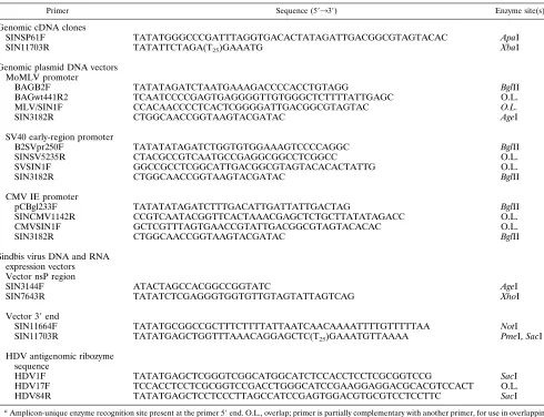
Primer Sequence (59339) Enzyme site(s)a
Genomic cDNA clones
SINSP61F TATATGGGCCCGATTTAGGTGACACTATAGATTGACGGCGTAGTACAC ApaI
SIN11703R TATATTCTAGA(T25)GAAATG XbaI
Genomic plasmid DNA vectors MoMLV promoter
BAGB2F TATATAGATCTAATGAAAGACCCCACCTGTAGG BglII
BAGwt441R2 TCAATCCCCGAGTGAGGGGTTGTGGGCTCTTTTATTGAGC O.L.
MLV/SIN1F CCACAACCCCTCACTCGGGGATTGACGGCGTAGTAC O.L.
SIN3182R CTGGCAACCGGTAAGTACGATAC AgeI
SV40 early-region promoter
B2SVpr250F TATATATAGATCTGGTGTGGAAAGTCCCCAGGC BglII
SINSV5235R CTACGCCGTCAATGCCGAGGCGGCCTCGGCC O.L.
SVSIN1F GGCCGCCTCGGCATTGACGGCGTAGTACACACTATTG O.L.
SIN3182R CTGGCAACCGGTAAGTACGATAC BglII
CMV IE promoter
pCBgl233F TATATATAGATCTTTGACATTGATTATTGACTAG BglII
SINCMV1142R CCGTCAATACGGTTCACTAAACGAGCTCTGCTTATATAGACC O.L.
CMVSIN1F GCTCGTTTAGTGAACCGTATTGACGGCGTAGTACACAC O.L.
SIN3182R CTGGCAACCGGTAAGTACGATAC BglII
Sindbis virus DNA and RNA expression vectors Vector nsP region
SIN3144F ATACTAGCCACGGCCGGTATC AgeI
SIN7643R TATATCTCGAGGGTGGTGTTGTAGTATTAGTCAG XhoI
Vector 39end
SIN11664F TATATGCGGCCGCTTTCTTTTATTAATCAACAAAATTTTGTTTTTAA NotI
SIN11703R TATATGAGCTGGTTTAAACAGGAGCTC(T25)GAAATGTTAAAA PmeI, SacI
HDV antigenomic ribozyme sequence
HDV1F TATATGAGCTCGGGTCGGCATGGCATCTCCACCTCCTCGCGGTCCG SacI
HDV17F TCCACCTCCTCGCGGTCCGACCTGGGCATCCGAAGGAGGACGCACGTCCACT O.L.
HDV84R TATATGAGCTCCTCCCTTAGCCATCCGAGTGGACGTGCGTCCTCCTTC SacI
a
Amplicon-unique enzyme recognition site present at the primer 59end. O.L., overlap; primer is partially complementary with another primer, for use in overlapping PCR.
on November 9, 2019 by guest
http://jvi.asm.org/
cationic lipid-mediated transfection, cells were rinsed twice and preincubated with OPTI-MEM medium (Gibco-BRL).
Transcript 5*-end mapping.Total RNA was isolated from the BHK-21 cells with RNAzol B (Tel-Test) at 6 h postinfection, 24 h post-RNA transfection, or 48 h post-DNA transfection according to the manufacturer’s directions. RNA pellets were incubated at 568C with a 32
P-labeled 20-nt reverse primer (59 -GGCTTCTCCATTGTGATGGT-39) complementary to Sindbis virus genomic RNA bases 70 to 51 and transcribed with MoMLV reverse transcriptase (Pro-mega). The pDCMVSINg plasmid was sequenced by a modified dideoxy chain termination method with the fmol DNA sequencing system (Promega). Reverse transcription and sequencing reaction mixtures were electrophoresed on a 6% denaturing polyacrylamide gel, dried, and exposed to film.
Total RNA and Northern (RNA blot) analysis.Total cellular RNA was iso-lated from transfected or infected BHK cells with Tri-Reagent (Molecular Re-search Center, Inc., Cincinnati, Ohio) as described by the manufacturer. BHK cells infected with wild-type Sindbis virus at a multiplicity of infection (MOI) of 5 were harvested at 8 h postinfection. RNA from BHK cells transfected with in vitro-transcribed pRSINg or pRSIN-luc RNA was isolated at 24 h posttransfec-tion. RNA from BHK cells transfected with pDLTRSINg or pDLTRSIN-luc plasmid DNA was isolated at 48 h posttransfection. Northern blot analysis was performed as described before (42). RNA was electrophoresed through 0.7% formaldehyde agarose gels and transferred to a Zeta-probe (Bio-Rad) mem-brane. The blot was hybridized simultaneously with a mixture of random-primed probes corresponding to the capsid gene and the luciferase gene. RNA load was adjusted to normalize the intensity of the hybridizing species.
Transfection efficiency determination.Transfection efficiency was determined by two different methods. In the first method, expressed luciferase protein in transfected cells was detected in situ. Transfected cells were first incubated with a primary rabbit anti-luciferase antibody (Promega) as recommended by the supplier and then incubated with a secondary goat anti-rabbit antibody conju-gated to horseradish peroxidase (Vector Laboratories, Burlingame, Calif.). De-tection of bound enzyme was done with the colorimetric substrate aminoethyl-carbozole. Transfected cells were counterstained with Mayer’s hematoxylin. In the second method, the number of bacterial CFU present in Hirt extracts (17) of transfected cells was quantitated (19). Plasmid DNA was isolated from trans-fected cells at 48 h postinfection, extracted with phenol-chloroform, and ethanol precipitated. Resuspended DNA was used to transform subcloning efficiency XL1-Blue cells (Stratagene), and Ampr colonies were scored. Plasmid DNA homologous to the test plasmid DNA and treated in parallel was added to Hirt extracts from mock-transfected cells and served as a control to standardize the number of CFU. To compare relative transfection efficiencies among different plasmids, the CFU in transfected cells was determined as a percentage of that in mock-transfected cells.
Virus and DNA and RNA vector assays.The specific infectivity levels of pRSINg in vitro-transcribed RNA and pDLTRSINg plasmid DNA were deter-mined by electroporation of 107BHK cells with 10mg of nucleic acid as de-scribed before (24). Electroporated cells were first diluted into 10 ml of DMEM plus 10% FBS, then serially diluted into 106
fresh BHK cells, and plated into 35-mm wells. The cells were overlaid with agarose 4 h after plating. Plaques were visualized by staining with neutral red at 48 h posttransfection.
Lysates from cells transfected with DNA or RNA or infected with Sindbis virus vector particles were tested for expression of luciferase and b-galactosidase reporter proteins by adding 250ml of reporter lysis buffer (Promega) per 106 cells. Luciferase (Promega) andb-galactosidase (Clontech) expression levels in cell lysates were determined by mixture with commercially available substrate detection systems followed by luminometry (Analytical Luminescence Labora-tory, San Diego, Calif.). Alternatively,b-galactosidase expression in cells trans-fected with DNA or RNA or intrans-fected with Sindbis virus vector particles was determined by staining in situ with X-Gal (5-bromo-4-chloro-3-indolyl-b-D -ga-lactopyranoside) and counting blue cells, as described previously (26). Expres-sion of reporter proteins was determined at 18 or 48 h post-RNA transfection or DNA transfection, respectively, or at 18 h postinfection except as noted in the figure legends.
Mouse and rat DNA injections.Sprague-Dawley rats (Harlan Sprague-Daw-ley, Indianapolis, Ind.) or BALB/c or C3H/HeN (Charles River Laboratories, Wilmington, Mass.) mice were injected in either the gastrocnemius or tibialis anterior muscle with 25mg of pDCMVSIN-b-gal or pDLTRSIN vector contain-ing hepatitis B virus (HBV) core (HBc) and e (HBe) antigen codcontain-ing sequences in a total volume of 100ml of isotonic saline. Mice were injected twice at 2-week intervals with HBc- and HBe-expressing vectors. Fluospheres (fluorescently la-beled latex spheres; Molecular Probes, Eugene, Oreg.) were added to the injec-tate to faciliinjec-tate identification of the injection site. To test for HBV-specific antibody induction, serum was collected 10 days following the second injection and tested for HBV-specific antibodies by an enzyme-linked immunosorbent assay (ELISA; Abbott Laboratories, Chicago, Ill.). For visualization ofb -galac-tosidase expression, the muscles were removed, fixed in toto in 2% formaldehyde in phosphate-buffered saline, and rinsed and stained overnight in X-Gal stain solution (26). The muscles were embedded in paraffin, sectioned, and counter-stained with tartrazine (American Histology, Lodi, Calif.).
RESULTS
Cloning and characterization of full-length genomic infec-tious Sindbis virus cDNA in RNA polymerase II expression cassettes. As a first step towards constructing DNA-based Sindbis virus vectors, a full-length cDNA clone of viral genomic RNA was generated, and the sequence was deter-mined. Sequence analysis revealed nucleotide changes (see Materials and Methods) resulting in 10 amino acid differences between our laboratory strain and the published HRsp strain, located in the nsP2, nsP3, E2, and E1 coding regions. Each of the changes in the nsPs resulted from single differences at nt
2992 (C3T, nsP2 aa 438, Pro3Leu), nt 3544 (T3C, nsP2 aa
622, Val3Ala), nt 3579 (A3G, nsP2 aa 634, Lys3Glu), and
nt 5351 (A3T, nsP3 aa 417, Gln3His). Five of the six changes
in the sPs resulted from single differences at nt 8637 (A3G,
E2 aa 3, Ile3Val), nt 8698 (T3A, E2 aa 23, Val3Glu), nt
9144 (A3G, E2 aa 172, Arg3Gly), nt 9420 (A3G, E2 aa 264,
Ser3Gly), and nt 10773 (T3G, E1 aa 237, Ser3Ala). These
observed differences in the E2 (except E2 aa 264) and E1 glycoproteins of our laboratory strain are locations of variabil-ity among several Sindbis virus strains (25, 35). A fifth change observed in E2 was a 3-nt deletion which eliminated the Glu codon corresponding to aa 160 of E2 and resulted in a genome length for the Sindbis virus strain used in this work of 11,700 nt. The viral genomic cDNA was positioned downstream of the bacteriophage SP6 RNA polymerase promoter, so that in vitro-synthesized transcripts contained a single nonviral G
res-idue at the 59 end. Additionally, a 25-mer poly(A) tract
fol-lowed by a unique XbaI recognition site were placed
down-stream of the viral 39end in the Sindbis virus genomic cDNA
clone designated pRSINg. Transfection of BHK cells with run-off transcripts from XbaI-linearized pRSINg plasmid resulted in cytopathic effect within 18 h, which was due to formation of infectious Sindbis virus.
The Sindbis virus genomic cDNA clone was then inserted into several RNA polymerase II expression cassettes to deter-mine whether the Sindbis virus infection cycle could be initi-ated directly from transfected plasmid DNA. The SV40 early region, MoMLV LTR U3 region, and CMV IE promoters were positioned so that transcription initiation would occur at the precise (or within 1 nt in the case of the LTR promoter)
Sindbis virus 59end. In addition, the bovine growth hormone
transcription termination/polyadenylation signal was positioned
downstream from the viral genomic cDNA 39 end in each
construct to generate plasmids pDSV40SINg, pDLTRSINg,
and pDCMVSINg. The fidelity of the RNA 59ends following
transcription in vitro from linearized cDNA clones is impor-tant to their overall activity compared with wild-type virion RNA (40). This property likely extends to RNAs transcribed in vivo from transfected Sindbis virus plasmid cDNA
con-structs. Thus, the major transcript 59 ends synthesized in
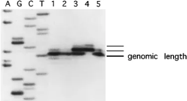
BHK cells transfected with pDCMVSINg or pDSV40SINg plasmid DNAs were determined (Fig. 1). The results of this study demonstrated that the primer extension products of RNAs isolated from cells transfected with pDCMVSINg or pDSV40SINg (lanes 1 and 2) were the same lengths as RNAs isolated from cells infected with wild-type Sindbis virus (lane 5). Although direct evidence for capping was not obtained, the two primer extension products observed in all lanes (except lane 4) most likely correspond to the terminal residue and a partial copying of the cap. The lowest-molecular-weight band, labeled on Fig. 1 as genomic length, corresponds to the
au-thentic Sindbis virus RNA 59 end. Primer extension of the
RNAs isolated from cells transfected with in vitro RNA (lane 3) produced three products. The lowest-molecular-weight
on November 9, 2019 by guest
http://jvi.asm.org/
nome-length band and the middle band comigrated with the two extension products from virus-infected cells and most likely correspond to the authentic terminal residue and a par-tial copying of the cap (which accounts for an undefined frac-tion of the middle band), respectively. The middle band and the highest-molecular-weight band comigrated with the two extension products from the in vitro transcription reaction (lane 4) and correspond to uncapped (which accounts for an undefined fraction of the middle band) and capped transcripts, respectively, each containing an additional nonviral G ribonu-cleotide from the SP6 promoter. A significant proportion of the RNA molecules isolated from cells transfected with in vitro-transcribed RNA appeared to have lost the nonviral res-idue within a single round of viral replication. Also, the addi-tional residue added from the SP6 promoter was not observed in cells infected with a Sindbis virus stock derived from BHK cells transfected with in vitro-transcribed pRSINg RNA. Primer extension products which migrated at a position that was 1 nt shorter than genome length and several lower-molec-ular-weight species were detected after long exposure and ap-peared to be indistinguishable between the various samples tested. RNA transcribed in situ from Sindbis virus plasmid DNA vectors containing the MoMLV LTR are expected to
have a single G residue at the viral 59 end, but this was not
verified experimentally.
The specific infectivities of pDLTRSINg DNA and pRSINg in vitro-transcribed RNA were determined by an infectious center assay to compare the efficiency of initiation of the virus infection cycle in cells electroporated with in vitro-transcribed RNA and plasmid DNA. In an average of three experiments, the specific infectivity of the in vitro-transcribed Sindbis virus
RNA (from pRSINg) was 1.13105PFU/mg (range, 0.63105
to 1.33105), and the specific infectivity of the Sindbis virus
plasmid DNA (pDLTRSINg) was 1.33 104PFU/mg (range,
0.33104to 3.13104), about 10-fold less. These experiments
demonstrated that all of the cis and trans components of Sind-bis virus can be expressed in functional form in vivo from a DNA format.
Production of packaged vector particles in cells cotrans-fected with Sindbis virus expression vector and DH plasmid DNAs. The full-length genomic cDNA clones were used to
construct Sindbis virus-based expression vectors and DH plas-mids for in vitro RNA and DNA transfections. As in previous work (22, 53), the expression vectors contained the entire nsP gene coding region and all sequences required in cis for viral replication and thus should function as replicons in transfected cells. The DHs contained the entire sP gene coding region and all of the sequences required in cis for viral replication but had deleted most of the nsP gene coding region and the Sindbis virus packaging sequence (4). Thus, DH replication and ex-pression of sPs were dependent on nsPs supplied in trans by the vector. Expression vectors and DHs were placed into RNA polymerase II expression cassettes used previously for plasmid DNA-based transfection experiments. Expression vectors and DH DNAs were constructed with either the MoMLV LTR or CMV IE promoter to explore a possible relationship between promoter strength and expression levels or packaging efficien-cies in cells transfected with Sindbis virus-derived plasmids.
We anticipated that the kinetics of reporter expression from DNA-based expression vectors would be slower than that from the corresponding RNA-based vectors. If so, that would affect the time point chosen in subsequent cotransfection experi-ments for harvesting packaged vector particles. Therefore, the kinetics of reporter protein expression in cells transfected with in vitro-transcribed RNA or plasmid DNA vectors were com-pared. As reported previously for in vitro-transcribed RNA vectors containing the chloramphenicol acetyltransferase re-porter gene (53), the peak level of luciferase expression oc-curred at 18 h posttransfection in cells transfected with in vitro-transcribed RNA vectors. In contrast, the kinetics were delayed in cells transfected with pDLTRSIN-luc DNA vectors, and the peak level of expression occurred at approximately 48 h posttransfection (data not shown). On the basis of these results, packaged vector particles were collected at 18 or 48 h posttransfection from cells cotransfected with in vitro-tran-scribed RNA or DNA expression vector and DH molecules, respectively.
The activities of in vitro-transcribed RNA and plasmid DNA
vectors were compared by examining the levels ofb
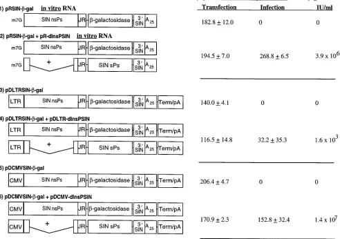
-galactosi-dase expression and the production of packaged vector parti-cles in cells transfected with the expression vectors alone or an expression vector and DH molecules together (Fig. 2). The
level of b-galactosidase expressed in transfected cells was
af-fected somewhat by the RNA polymerase II promoter used in the Sindbis virus DNA vector construct. Reporter enzyme ac-tivity was 1.5-fold higher in cells transfected with DNA vectors containing the CMV IE promoter than with those containing the LTR promoter (samples 5 and 3, respectively). The levels
ofb-galactosidase observed in cells transfected with DNA
vec-tor containing the CMV IE promoter or with in vitro-tran-scribed RNA replicon were similar (samples 5 and 1, respec-tively). The influence of the RNA polymerase II promoter on reporter protein expression was also tested in cells transfected with Sindbis virus DNA vectors containing the luciferase gene. Luciferase activities were about fourfold greater in cells trans-fected with expression vectors containing the CMV promoter
than with the LTR promoter (274,000652,100 relative light
units [RLU] per cell with pDCMVSIN-luc versus 70,600 6
11,400 RLU per cell with pDLTRSIN-luc).
[image:5.612.83.271.72.174.2]A separate experiment designed to compare relative activi-ties between the MoMLV LTR and CMV IE promoters in the absence of subsequent RNA amplification was performed. The level of luciferase activity was determined in BHK cells trans-fected with conventional expression vectors in which the pro-moter and reporter genes were directly linked. The levels of luciferase activities were approximately 18-fold higher in cells transfected with plasmids containing the CMV IE promoter
FIG. 1. Transcript 59-end mapping in cells transfected with Sindbis virus RNA and DNA vectors. Lanes 1 to 5, primer-extended reverse transcription of RNA purified from BHK cells at 48 h posttransfection with pDCMVSINg, BHK cells at 48 h posttransfection with pDSV40SINg, BHK cells at 24 h posttrans-fection with in vitro-transcribed pRSINg RNA or in vitro-transcribed pRSINg transcription reaction, and BHK cells at 6 h postinfection (MOI of 5) with a Sindbis virus stock derived from in vitro-transcribed pRSINg RNA, respectively. The adjacent sequence ladder was derived from the pDCMVSINg construct with the same primer as used for reverse transcription reactions, and the genome-length band is labeled. The reaction products were electrophoresed on a 6% denaturing polyacrylamide gel. The sequence shown is therefore that of the virus minus strand. Thus, the terminal nucleotide in the reverse transcripts shown in the figure comigrates with the T nucleotide and corresponds to the genomic plus strand A residue at nt 1.
on November 9, 2019 by guest
http://jvi.asm.org/
versus the LTR promoter (135,000 6 20,800 RLU per cell
versus 7,690 6 1,660 RLU per cell, respectively). Thus, it
appeared that while the promoter strength influenced the level of reporter gene expression in cells transfected with both Sind-bis virus-derived DNA vectors and conventional vectors, this effect was less pronounced in the context of autocatalytic RNA amplification arising from the Sindbis replicons.
Paradoxically, the titers of packaged vector did not correlate
with the level ofb-galactosidase activity in lysates from cells
infected with 1 ml of medium from cotransfected cells (Fig. 2). The titer of packaged vector in medium from cells cotrans-fected with DNAs that contained the CMV promoter (sample 6) was 3.6-fold higher than the titer from cells cotransfected
with in vitro RNAs (sample 2). In contrast, the level ofb
-ga-lactosidase activity in lysates was 1.8-fold lower in cells infected with 1 ml of medium from cells cotransfected with DNAs containing the CMV promoter (sample 6) than with in vitro-transcribed RNAs (sample 2). This result appeared to be
re-lated to the level of infectious virus produced, presumably by homologous DNA recombination, in cells cotransfected with plasmid DNAs that contained the CMV IE promoter, which
was 23107to 33107PFU/ml, compared with 13104to 5.5
3104PFU/ml in cells cotransfected with in vitro-transcribed
RNA. RNA analysis of PFU produced in cells cotransfected with in vitro-transcribed RNA or DNA to discriminate be-tween copackaging (12) of replicon and DH RNAs and RNA recombination (52) was not performed. However, the infec-tious virus in medium from all cotransfected cells had a large plaque morphology, and infection of cultures with undiluted medium from cells cotransfected with DNAs containing the CMV promoter produced rapid cytopathic effect that was in-distinguishable from that caused by wild-type Sindbis virus.
The titer of packaged vector particles observed in medium from cells cotransfected with plasmid DNAs that contained the LTR promoter was significantly lower than that in all other nucleic acids that were tested (Fig. 2, sample 4). In these
ex-FIG. 2. Expression and packaging of Sindbis virus vectors. BHK cells were transfected with in vitro-transcribed RNA or DNA vectors or cotransfected with in vitro-transcribed RNA or DNA vector and DH molecules. Shown on the left side of the figure are schematic representations of the Sindbis virus RNA and DNA vectors and DH molecules used in the experiment. The vector maps are not drawn to scale. Abbreviations: LTR, MoMLV LTR U3 region promoter; CMV, human CMV IE promoter; JR, Sindbis virus junction region promoter, extending to nt 7643; 39SIN, 39end of Sindbis virus from nt 11664 to 11703; A25, synthetic 25-mer poly(A) tract; Term/pA, bovine growth hormone transcription termination/polyadenylation signal. Shown on the right side of the figure and adjacent to each vector schematic are the levels ofb-galactosidase detected, in RLU per cell, after cotransfection of BHK cells and after infection of fresh BHK cells with 1 ml of clarified culture medium. BHK cells were cotransfected with 2mg of each vector nucleic acid by lipofection, andb-galactosidase activity was measured in cleared cell lysates at 18 h after in vitro-transcribed RNA transfection and 48 h after DNA transfection. Samples 1 and 2 were cotransfected with in vitro-transcribed RNAs and are marked by the m7G cap on the vector schematics. Packaging was determined by transfer of reporter gene expression for clarified cotransfected cell culture medium to fresh BHK monolayers and was measured at 18 h postinfection. The standard deviation represents six samples. The infectious units (IU) per milliliter were determined by infecting dilutions of clarified medium from cotransfected cells onto fresh BHK cells followed by staining with X-Gal. The IU levels shown are an average for three samples.
on November 9, 2019 by guest
http://jvi.asm.org/
[image:6.612.61.553.82.428.2]periments, the level ofb-galactosidase activity in lysates from cells cotransfected with plasmid DNAs that contained the LTR promoter was highly variable and indicated either that the cotransfection frequency of vector and DH was very low or that the frequency of cotransfected cells in which functional replicon and DH RNAs were transported to the cytoplasm was very low.
RNA synthesis in cells transfected with Sindbis virus DNA and RNA vectors.The RNA species synthesized in cells trans-fected with various Sindbis virus DNA and RNA vector con-structs were analyzed by Northern blot and compared with those observed after wild-type virus infection (Fig. 3). The results demonstrated that the genomic and subgenomic RNAs isolated from cells transfected with Sindbis virus genomic in vitro-transcribed RNA or plasmid DNA vectors were comparable to the RNAs synthesized in virus-infected cells. The presence of genomic and subgenomic RNAs in cells
transfected with in vitro-transcribed pRSIN-luc RNA or pDLTRSIN-luc plasmid DNA replicons suggested that these vectors function according to the strategy of wild-type Sindbis virus and the genomic clones and that subgenomic RNA is likely the template for luciferase gene expression. Several bands of various molecular sizes which migrated more rapidly than the subgenomic mRNA were observed. However, there is no direct evidence for splicing of Sindbis virus-derived tran-scripts in plasmid DNA-transfected cells, since these uniden-tified RNAs that migrated more rapidly than 26S mRNA were also observed in wild-type Sindbis virus-infected cells. Further-more, analysis of Sindbis virus genomic RNA revealed no cryptic splice sites (14). Thus, it appears that these species may be due to nonspecific RNA degradation.
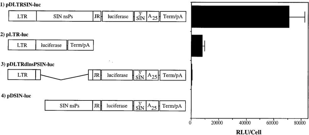
[image:7.612.90.265.70.176.2]Enhancement of gene expression by layered Sindbis virus DNA expression vector.The levels of luciferase synthesized in cells transfected with either Sindbis virus plasmid DNA expres-sion vectors or the analogous promoter-reporter plasmid con-structions were compared in order to determine the relative enhancement from nsP-catalyzed RNA transcript amplifica-tion (Fig. 4). Transfecamplifica-tions of individual test plasmids were performed in quintuplicate. The level of synthesis was about 10-fold higher in cells transfected with the pDLTRSIN-luc Sindbis virus vector DNA than in cells transfected with plasmid pLTR-luc. Two additional control constructs also were tested. Plasmid pDLTRdlnsPSIN-luc had most of the nsP genes de-leted, between viral nt 1407 and 6920, and serves as a baseline control for the level of vector genomic template-derived lucif-erase expression. Plasmid pDSIN-luc was an intermediate in the construction of pDLTRSIN-luc, which lacked the LTR promoter and the first 2,289 nt of Sindbis virus and served as a control for background luciferase expression. Measurable re-porter gene expression was observed in cells transfected with pDLTRdlnsPSIN-luc, although at levels which were more than 100-fold lower than the level in cells transfected with pDL-TRSIN-luc. This low level of activity may be due to alteration
FIG. 3. Sindbis virus-specific RNAs synthesized in BHK cells transfected with various in vitro-transcribed RNA and DNA vectors or infected with wild-type virus. BHK cells were transfected with 5mg of nucleic acid by lipofection or infected with Sindbis virus at an MOI of 5. Total cellular RNA was isolated at 8 h postinfection or 24 h (in vitro-transcribed RNA vectors) or 48 h (DNA vectors) posttransfection and analyzed by formaldehyde gel electrophoresis, transfer to a nylon membrane, and hybridization with capsid gene- and luciferase gene-spe-cific probes. Lanes: 1, Sindbis virus infection; 2, in vitro-transcribed pRSINg RNA; 3, pDLTRSINg; 4, in vitro-transcribed pRSIN-luc RNA; 5, pDLTRSIN-luc.
FIG. 4. Comparison of enhancement of luciferase expression by nsP-catalyzed RNA transcript amplification in BHK cells transfected with Sindbis virus DNA expression vectors or with linked promoter-reporter vectors. Shown on the left side of the figure are the schematic representations of the plasmid DNAs used in the experiment. The vector maps are not drawn to scale. Abbreviations are as described in the legend to Fig. 2. Shown on the right side of the figure and adjacent to each vector schematic are the levels of luciferase activity detected in cleared lysates from BHK cells at 48 h posttransfection with 2mg of plasmid DNA. Error bars represent the standard deviation for five samples.
on November 9, 2019 by guest
http://jvi.asm.org/
[image:7.612.59.552.468.683.2]of some of the input plasmid DNA or, alternatively, translation directly from degraded vector genomic RNA (6).
Further experiments were performed to investigate the re-lationship between transfection and expression efficiency in cells into which Sindbis virus DNA vector or the analogous conventional promoter-reporter vector was introduced. The total amount of luciferase expression in transfected cell lysates was compared with the percentage of cells expressing lucif-erase protein, as determined both by in situ staining with a luciferase-specific antibody and by the total number of trans-fected biologically active DNA molecules, as determined by quantitating the bacterial CFU of plasmid DNA present in Hirt extracts (19) (Table 2). The data indicate that while the fraction of cells expressing reporter protein was about 2-fold less in pDLTRSIN-luc- than in pLTR-luc-transfected samples, the level of luciferase activity was at least 10-fold greater in pDLTRSIN-luc-transfected cell lysates. It appeared that the relative numbers of biologically active plasmid molecules were similar in cells transfected with either pDLTRSIN-luc or pLTR-luc plasmid DNAs. Taken together, these results indi-cated that the relative level of nsP-driven reporter gene ex-pression was at least 10-fold higher than the level from the analogous promoter-reporter vector on the basis of transfected cells expressing reporter protein.
Reporter gene expression in rodents inoculated intramus-cularly with Sindbis virus DNA expression vectors.Previously (54), RNA derived from the Semliki Forest virus replicon was injected intramuscularly into mice to express the nucleoprotein of influenza virus. To examine in vivo gene expression from DNA-based Sindbis virus vectors, BALB/c mice were injected
intramuscularly with 25 mg of pDCMVSIN-b-gal plasmid
DNA. Figure 5 demonstrates the in vivo expression ofb
-ga-lactosidase in mouse muscle stained in toto with X-Gal at 5 days postinjection (Fig. 5A) and a transverse section from a mouse muscle stained in toto with X-Gal at 5 days
postinjec-tion (Fig. 5B). The muscle stained in toto showsb
-galactosi-dase protein expression along the length of the mouse muscle. We routinely observed blue fibers which spanned the length of the muscle. After sectioning, the muscle was counterstained with tartrazine, and more than 30 blue fibers can be observed in a transverse section (Fig. 5B). Of these, almost 90% were strongly stained, although a minority of fibers show variable
weaker staining. Rats injected with pDCMVSIN-b-gal plasmid
DNA also demonstrated positively staining blue muscle fibers (data not shown).
Other experiments were designed to explore further the in vivo expression of clinically relevant genes from Sindbis virus DNA vectors. C3H/HeN mice were injected twice intramuscu-larly with pDLTRSIN vectors containing either the HBc or
HBe gene. With an ELISA detection system, both HBc- and HBe-specific immunoglobulin G antibodies were detected in serum samples collected from the mice 10 days following the second injection with the vectors. Antibody titers of 1:640 were observed in four of five mice inoculated with the HBc vector, and antibody titers of 1:640 or greater were observed in three of five mice inoculated with the HBe vector. Antibodies spe-cific for HBc or HBe were not detected in sera collected from the mice prior to the injection of vector. These experiments demonstrate that Sindbis virus-derived DNA vectors are able to express foreign genes in vivo and that expression levels are sufficient for induction of a humoral immune response. Whether these vectors are also capable of inducing HBV-specific cytotoxic T cells is an area currently under investiga-tion. Indeed, the induction of a vigorous cytotoxic T cell re-sponse is believed to play a major role in the clearance of HBV-infected cells (7).
[image:8.612.317.556.72.436.2]Sindbis virus DNA expression vector modifications.As dis-cussed above, the level of reporter gene expression observed in cells transfected with Sindbis virus plasmid DNA vectors var-ied with the strength of the RNA polymerase II promoter used
[image:8.612.58.298.93.148.2]FIG. 5. Expression ofb-galactosidase in mouse muscle 5 days after injection with 25mg of pDCMVSIN-b-gal plasmid DNA contained in phosphate-buffered saline. (A) Photomicrograph of gastrocnemius muscle stained in toto with X-Gal. Five blue fibers are observed in this micrograph. Magnification,340. (B) Pho-tomicrograph of transverse cryosection of mouse tibialis anterior muscle. Mul-tiple blue-stained transverse fibers are evident. Magnification,3400. TABLE 2. Transfection efficiencies of Sindbis virus plasmid DNA
and conventional plasmid DNA vectors
Vector Mean RLU/ cella6SD
Mean % of cells expressing lucif-eraseb6SD
CFU/mgc(%
of control)
pDLTRSIN-luc 6,31064,496 3.360.8 17
pLTR-luc 200635 6.261.0 21
aLuciferase activity present in a lysate from 106cells at 48 h posttransfection. The standard deviation was determined from three individual transfections.
bStandard deviation was determined from four fields counted (average of 707
total cells) in three individual transfections.
cBacterial CFU present in Hirt extracts of cells at 48 h posttransfection as a
percentage of that in Hirt extracts of cells mock transfected and spiked with test plasmid DNA. Values shown are averages of CFU quantitated from three trans-fections or plasmid-spiked mock transtrans-fections.
on November 9, 2019 by guest
http://jvi.asm.org/
in the vector construct at the time points examined. This level was two- to fourfold greater in Sindbis virus DNA expression vectors that contained the CMV promoter than in those that contained the LTR promoter. In contrast, in cells transfected with conventional vectors in which the promoter and reporter gene were directly linked, the level was about 18-fold greater with plasmids that contained the CMV promoter than with those that contained the LTR promoter. Given the autocata-lytic cytoplasmic amplification of transported RNA replicons, it is expected that the expression levels should not be promoter dependent. Our results, however, indicated that expression was partially related to the promoter and therefore suggested that further optimization of the Sindbis virus DNA expression vec-tors was possible.
Because Sindbis virus plasmid DNA vectors were con-structed from the initial SP6-based vectors, the transcription termination/polyadenylation signal follows a 25-mer
poly(dA-dT) tract. Thus, the 39 end of the primary transcript in the
nucleus most likely consists of 25 consecutive adenylate resi-dues, followed by the transcription termination sequence cRNA, and terminates with a second poly(A) tract. Figure 6
demonstrates the effect on reporter gene expression for two different vector modifications which were designed to generate
a vector RNA 39end that was more similar to that of wild-type
Sindbis virus. In the first configuration (Fig. 6A), the 25-mer
poly(dA-dT) tract was removed, and the viral 39end (nt 11703)
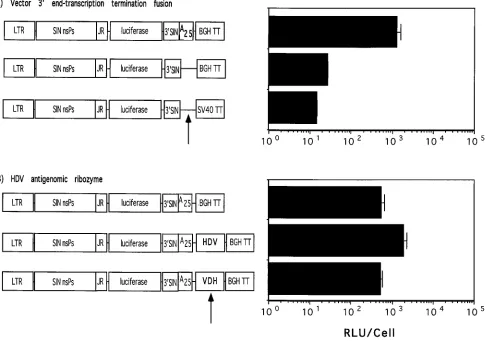
[image:9.612.66.551.75.416.2]was linked directly to the core bovine growth hormone or SV40 RNA processing signals. When transfected into cells, the ac-tivity of these vectors was decreased by more than 100-fold compared with that of the parental pDLTRSIN-luc prototype vector (Fig. 6A). Although measurable expression of luciferase was detected from the poly(dA-dT) tract-deleted plasmids, it was not determined whether any transported RNA actually replicated in transfected cells. In the second configuration (Fig. 6B), the HDV antigenomic ribozyme sequence (34) was inserted immediately downstream of the 25-mer poly(dA-dT) tract to facilitate autocatalytic processing of the primary tran-script and removal of nonviral nucleotides downstream from the vector poly(A) tail. Transfection of cells with DNA vector containing the HDV ribozyme sequence resulted in a three- to fourfold increase in the level of luciferase expression compared with the wild-type vector or an analogous construct with the
FIG. 6. Luciferase expression in BHK cells transfected with 39-end-modified Sindbis virus DNA expression vectors. Shown on the left side of the figure are the schematic representations of the plasmid DNAs used in the experiment. The vector maps are not drawn to scale. Abbreviations: BGH TT, bovine growth hormone transcription termination signal; SV40 TT, SV40 early-region transcription termination signal; HDV, HDV antigenomic ribozyme sequence; VDH, HDV antigenomic ribozyme sequence, reverse orientation. Other abbreviations are as described in the legend to Fig. 2. Shown on the right side of the figure and adjacent to each vector schematic are the levels of luciferase activity detected in cleared lysates from BHK cells transfected by lipofection with 2mg of plasmid DNA in two individual experiments. (A) Deletion of the synthetic poly(A) (A25) tract. The deleted regions of the vectors are denoted by an arrow. Error bars represent the standard deviation for five samples. Luciferase activity was determined at 48 h posttransfection. (B) Insertion of the HDV antigenomic ribozyme sequence immediately downstream of the synthetic poly(A) (A25) tract. Insertions in the correct and reverse orientations are denoted by an arrow. Error bars represent the standard deviation for three samples. Luciferase activity was determined at 36 h posttransfection.
on November 9, 2019 by guest
http://jvi.asm.org/
HDV ribozyme sequence inserted in the reverse orientation. These data were confirmed by testing the specific infectivity of a genome-length construct derived from pDLTRSINg, which also contains the HDV ribozyme sequence insertion. The spe-cific infectivity of this construct, designated pDLTRSINgHDV, was increased fourfold over that of the parental construct pDLTRSINg (data not shown).
DISCUSSION
In this study, we demonstrated the efficient initiation of an alphavirus infectious cycle in vivo from a genomic cDNA clone contained within an RNA polymerase II expression cassette. The ability to express functional alphavirus genes from a DNA format enabled us to develop two new Sindbis virus-based gene expression systems: (i) a plasmid DNA-based vector with po-tential application for genetic immunization, and (ii) the pro-duction of packaged Sindbis virus particles in cells cotrans-fected with vector replicon and DH plasmid DNAs. Although previous investigators (21) have used the DNA-based vaccinia virus-T7 polymerase and T7 promoter system to express func-tional forms of the individual Sindbis virus nsPs, this approach was dependent on infectious vaccinia virus as a helper. Fur-thermore, these experiments were performed to investigate Sindbis virus replication rather than to develop plasmid DNA-based replicon vectors. Prior to this report, molecular ap-proaches to producing infectious Sindbis virus RNA and its derived complementary vectors were restricted primarily to in vitro transcription of cDNA clones from a bacteriophage RNA polymerase promoter, followed by transfection into permissive cells.
The demonstration of efficient RNA polymerase II promot-er-dependent initiation of Sindbis virus infection is in contrast to previous studies (19, 37, 46) with genomic cDNA clones derived from the positive-stranded poliovirus RNA. In these investigations, transfection of cells with the poliovirus cDNA without an RNA polymerase II promoter resulted in the pro-duction of low levels of infectious virus (37). Inclusion of SV40 transcription and replication signals in the poliovirus cDNA constructs dramatically improved the production of poliovirus after transfection with plasmid DNA (19, 46). However, it is not clear whether the high level of poliovirus produced in cells transfected with the modified plasmids was related to in-creased RNA polymerase II-dependent transcription of cDNA amplification, since the increased infectivity of the transfected poliovirus cDNA correlated with the extent of SV40 large-T-antigen-dependent plasmid replication rather than the pro-moter (19). In these investigations with poliovirus, the require-ment for the transfected plasmid to replicate in order to obtain efficient production of virus has several possible explanations, including distant placement of the RNA polymerase II
pro-moter relative to the viral 59 end, resulting in the relatively
poor infectivity of synthesized RNA polymerase II transcripts; cryptic splicing within the viral genome; and poor transport of the viral mRNA. Our current investigation demonstrated that strategic placement of RNA polymerase II promoters in plas-mids not containing eukaryotic replication signals resulted in the synthesis of replication-competent Sindbis virus RNA fol-lowing plasmid DNA transfection of cells. In addition, several examples exist for the RNA polymerase II promoter-depen-dent initiation of plant virus infection in cells transfected with plasmids in which the cauliflower mosaic virus 35S promoter is linked to the virus genomic cDNA (for a review, see reference 3).
Cotransfection experiments with DNA vectors having the CMV IE promoter or with in vitro-transcribed RNA vectors
produced levels of packagedb-galactosidase vector particles
that were similar and comparable to those in previous work with cotransfected in vitro replicon and DH RNAs (4). The level of replication-competent PFU produced in cells cotrans-fected with in vitro-transcribed RNA vectors in this work was
similar to that observed in previous work (4) (13104to 5.53
104PFU/ml versus 23104PFU/ml, respectively), using a DH
deleted of the nsPs gene between nt 502 and 6917. Another DH used in that study, deleted between nt 421 and 7334,
resulted in less than 103PFU/ml produced after cotransfection
with in vitro-transcribed replicon RNA. The DH used in our current study was deleted between nt 422 and 7054. Addition-ally, the vector replicons used in the previous work contained
310 nt from the Sindbis virus 39 nontranscribed region, while
the vector replicons used in this investigation contained only
the 39 39-terminal nucleotides.
The level of PFU produced in cells cotransfected with rep-licon and DH DNAs containing the CMV IE promoter was dramatically higher than that in cells transfected with in
vitro-transcribed RNA vectors (23107to 33107PFU/ml versus 1
3104to 5.53104PFU/ml, respectively). Although it was not
determined whether the PFU produced in cells contained par-ticles which arose from RNA copackaging or recombination between replicon and DH DNAs and/or RNAs, it has been observed that mutations and rearrangements can occur in transfected DNA (20). Thus, it is possible that the culture medium from DNA-cotransfected cells contained significant levels of replication-competent virus generated from homolo-gous recombination. Additionally, the high level of PFU ob-served here after in vitro-transcribed RNA or DNA cotrans-fection may be related partially to the transcotrans-fection efficiencies, which were about 5%. Replication-competent virus produced in cotransfected cells would subsequently be amplified upon infection of naive cells.
The level of reporter gene expression in cells transfected with various Sindbis virus-derived DNA vectors was depen-dent, in part, on the relative RNA polymerase II promoter strength. This result was unexpected, since the level of trans-lational template, which results from the exponential expan-sion of RNA, should not depend on the initial number of functional replicon molecules. However, this correlation of reporter gene expression with promoter strength was signifi-cantly less with Sindbis virus DNA vectors than with conven-tional expression vectors. These results suggested that trans-port of functional replicons from the nucleus to the cytoplasm may be inhibited in some way in cells transfected with Sindbis virus DNA vectors or, alternatively, that transported replicons may be inefficient templates for initiation of replication. Thus, we tested two modifications of the DNA vector to produce
RNAs with 39ends which were more similar to those of
wild-type Sindbis virus. Deletion of the synthetic A25tract by direct
linkage of the vector 39end (nt 11703) and transcription
pro-cessing signals resulted in Sindbis virus plasmid DNA expres-sion vectors that were significantly disabled. This observation
suggests that the 39end and the poly(A) tail of Sindbis virus
RNA must be contiguous in order to be recognized by the viral replicase complex and serve as the template for minus-strand
RNA synthesis. The 19 39-terminal viral nucleotides conserved
among the alphaviruses (32) appear to be insufficient to cata-lyze this process. In contrast, juxtaposition of the cis-acting HDV antigenomic ribozyme sequence with the Sindbis virus
A25tract to generate precise 39termini resulted in DNA
vec-tors which produced comparatively higher levels of luciferase expression in transfected cells. Although direct evidence of autocatalytic cleavage by RNA analysis was not obtained, ex-pression levels from vectors containing the HDV ribozyme
on November 9, 2019 by guest
http://jvi.asm.org/
sequence in the reverse orientation were unchanged, suggest-ing that the ribozyme component in the correct orientation was indeed functional. The HDV ribozyme sequence has been used previously to generate precise termini for the replication in situ of the negative-stranded vesicular stomatitis virus (33).
The relative level of nsP-catalyzed enhancement of reporter gene expression was approximately 10-fold, as determined by comparison of luciferase activity between Sindbis virus plasmid DNA vectors and conventional linked promoter-reporter vec-tors in transfected cells. Current limitations of DNA-based immunization are due, in part, to poor transfection efficiencies and short-lived expression. One approach to mitigate these problems may be to use vectors, such as the Sindbis virus-derived DNA vector described here, which express high levels of the gene of interest through autocatalytic amplification of the vector RNA. We have begun to further modify the Sindbis virus DNA vectors in order to increase the difference in het-erologous gene expression compared with conventional vectors (28). We are continuing our efforts by, among other things, exploring the utility of introns to increase the transport effi-ciency of replicons synthesized from Sindbis virus plasmid DNA vectors. One additional possibility may be to incorporate translation enhancement components, as described previously (11), for Sindbis virus-derived replicons.
The conversion of alphavirus-derived replicon and helper vectors into a plasmid DNA-based expression system is the primary requisite step towards developing alphavirus-based gene transfer systems which parallel the classic retrovirus-based producer cell configurations. The ability to produce packaged vector particles from cotransfected replicon and DH DNAs suggested to us the possibility of developing stable vec-tor-packaging cell lines, in which packaged vector particles are produced following transfection of Sindbis virus-derived plas-mid DNA vectors. This notion is supported further by work describing the generation of a vector particle-titering cell line (31) that constitutively synthesizes a defective RNA, which subsequently replicates and expresses luciferase when induced by Sindbis virus nsPs. We have used the plasmid cDNA-based expression system described here to derive a first-generation Sindbis virus vector-packaging cell line (manuscript in prepa-ration). Further goals include developing inducible producer cell lines in which a burst of packaged Sindbis virus vector particles is produced in response to a particular stimulus.
Accomplishment of our first objective has resulted in the development of a new vector which harnesses the expression potential of alphaviruses and appears to lead to increased expression of heterologous genes compared with conventional expression vector plasmids. Finally, the Sindbis virus DNA vector system was shown to express heterologous genes in vivo when injected into mice and rats and is being developed fur-ther for general physical gene transfer applications.
ACKNOWLEDGMENTS
We thank Sondra Schlesinger for critical review of the manuscript. Sincere thanks to Joanne O’Dea for GenBank searches and computer manipulations.
REFERENCES
1. Berglund, P., M. Sjoberg, G. J. Atkins, B. J. Sheahan, H. Garoff, and P.
Liljestrom.1993. Semliki Forest virus expression system: production of con-ditionally infectious recombinant particles. Bio/Technology 11:916–920. 2. Boshart, M., F. Weber, G. Jahn, K. Dorsch-Hasler, B. Fleckenstein, and W.
Schaffner.1985. A very strong enhancer is located upstream of an immediate early gene of human cytomegalovirus. Cell 41:521–530.
3. Boyer, J.-C., and A. L. Haenni. 1994. Infectious transcripts and cDNA clones of RNA viruses. Virology 198:415–426.
4. Bredenbeek, P. J., I. Frolov, C. M. Rice, and S. Schlesinger. 1993. Sindbis
virus expression vectors: packaging of RNA replicons by using defective helper RNAs. J. Virol. 67:6439–6446.
5. Burge, B. W., and E. R. Pfefferkorn. 1966. Isolation and characterization of conditional-lethal mutants of Sindbis virus. Virology 30:204–213. 6. Cancedda, R., L. Villa-Komaroff, H. F. Lodish, and M. Schlesinger. 1975.
Initiation sites for translation of Sindbis virus 42S and 26S messenger RNAs. Cell 6:215–222.
7. Chisari, F. V., and C. Ferrari. 1995. Hepatitis B virus immunopathogenesis. Annu. Rev. Immunol. 13:29–60.
8. Davis, N. L., K. W. Brown, I. J. Caley, R. I. Swanstrom, and R. E. Johnston. 1995. Protection against influenza in mice by vaccination with a Venezuelan equine encephalitis virus vector expressing the HA protein. J. Cell. Biochem. Suppl. 19A:310.
9. Davis, N. L., L. V. Willis, J. F. Smith, and R. E. Johnston. 1989. In vitro synthesis of infectious Venezuelan equine encephalitis virus RNA from a cDNA clone: analysis of a viable deletion mutant. Virology 171:189–204. 10. Feiers, W., R. Contreras, G. Haegeman, R. Rogiers, A. van der Voorde, H.
van Heuverswyn, J. van Herreweghe, G. Volckaert, and M. Ysebaert.1978. The complete nucleotide sequence of SV40 DNA. Nature (London) 273: 113–117.
11. Frolov, I., and S. Schlesinger. 1994. Translation of Sindbis virus mRNA: effects of sequences downstream of the initiating codon. J. Virol. 68:8111– 8117.
12. Geigenmuller-Gnirke, U., B. Weiss, R. Wright, and S. Schlesinger. 1991. Complementation between Sindbis viral RNAs produces infectious particles with a bipartite genome. Proc. Natl. Acad. Sci. USA 88:3253–3257. 13. Geller, A. I., and X. O. Breakefield. 1988. A defective HSV-1 vector
ex-presses Escherichia colib-galactosidase in cultured peripheral neurons. Sci-ence 24:1667–1669.
14. Green, M. R. 1991. Biochemical mechanisms of constitutive and regulated pre-mRNA splicing. Annu. Rev. Cell Biol. 7:559–599.
15. Hahn, C. S., Y. S. Hahn, T. J. Braciale, and C. M. Rice. 1992. Infectious Sindbis virus transient expression vectors for studying antigen processing and presentation. Proc. Natl. Acad. Sci. USA 89:2679–2683.
16. Heinzinger, N. K., M. I. Bukinsky, S. A. Haggerty, A. M. Ragland, V.
Kewal-ramani, M. A. Lee, H. E. Gendelman, L. Ratner, M. Stevenson, and M. Emerman.1994. The Vpr protein of human immunodeficiency virus type 1 influences nuclear localization of viral nucleic acids in nondividing host cells. Proc. Natl. Acad. Sci. USA 91:7311–7315.
17. Hirt, B. 1967. Selective extraction of polyoma DNA from infected mouse cultures. J. Mol. Biol. 26:365–369.
18. Jolly, D. 1994. Viral vector systems for gene therapy. Cancer Gene Ther.
1:51–64.
19. Kean, K. M., C. Wychowski, H. Kopecka, and M. Girard. 1986. Highly infectious plasmids carrying poliovirus cDNA are capable of replication in transfected simian cells. J. Virol. 59:490–493.
20. Lebkowski, J. S., R. B. Dubridge, E. A. Antell, K. S. Greisen, and M. P.
Calos.1984. Transfected DNA is mutated in monkey, mouse, and human cells. Mol. Cell. Biol. 4:1951–1960.
21. Lemm, J. A., T. Rumenaph, E. G. Strauss, J. H. Strauss, and C. M. Rice. 1994. Polypeptide requirements for assembly of functional Sindbis virus replication complexes: a model for the temporal regulation of minus- and plus-strand RNA synthesis. EMBO J. 13:2925–2934.
22. Levis, R., B. G. Weiss, M. Tsiang, H. Huang, and S. Schlesinger. 1986. Deletion mapping of Sindbis virus DI RNAs derived from cDNAs defines the sequences essential for replication and packaging. Cell 44:137–145. 23. Liljestrom, P. 1994. Alphavirus expression systems. Curr. Opin. Biotechnol.
5:495–500.
24. Liljestrom, P., and H. Garoff. 1991. A new generation of animal cell expres-sion vectors based on the Semliki Forest virus replicon. Bio/Technology
9:1356–1361.
25. Lustig, S., A. C. Jackson, C. S. Hahn, D. E. Griffin, E. G. Strauss, and J. H.
Strauss.1988. Molecular basis of Sindbis virus neurovirulence in mice. J. Virol. 62:2329–2336.
26. MacGregor, G. R., A. E. Mogg, J. F. Burke, and C. T. Caskey. 1987. Histo-chemical staining of clonal mammalian cell lines expressing E. coli beta-galactosidase indicates heterogenous expression of the bacterial gene. So-mat. Cell Mol. Genet. 13:253–265.
27. Miller, A. D., D. G. Miller, J. V. Garcia, and C. M. Lynch. 1993. Use of retroviral vectors for gene transfer and expression. Methods Enzymol. 217: 581–599.
28. Montgomery, D. L., J. W. Shiver, K. R. Leander, H. C. Perry, A. Friedman,
D. Martinez, J. B. Ulmer, J. J. Donnelly, and M. A. Liu.1993. Heterologous and homologous protection against influenza A by DNA vaccination: opti-mization of DNA vectors. DNA Cell Biol. 12:777–783.
29. Moss, B., and C. Flexner. 1987. Vaccinia virus expression vectors. Annu. Rev. Immunol. 5:305–324.
30. Nelson, J. A., J. W. Gnann, and P. Ghazal. 1990. Regulation and tissue-specific expression of human cytomegalovirus. Curr. Top. Microbiol. Immu-nol. 154:77–103.
31. Olivo, P., I. Frolov, and S. Schlesinger. 1994. A cell line that expresses a reporter gene in response to infection by Sindbis virus: a prototype for