0022-538X/96/$04.0010
Copyrightq1996, American Society for Microbiology
The Region of the Herpes Simplex Virus Type 1 LAT Gene
That Is Colinear with the ICP34.5 Gene Is Not
Involved in Spontaneous Reactivation
GUEY-CHUEN PERNG,1K. CHOKEPHAIBULKIT,1RICHARD L. THOMPSON,2NANCY M. SAWTELL,3
SUSAN M. SLANINA,1HOMAYON GHIASI,1,4ANTHONY B. NESBURN,1,4
ANDSTEVEN L. WECHSLER1,4*
Ophthalmology Research Laboratories, Cedars-Sinai Medical Center Research Institute, Los Angeles, California 900481;
Department of Ophthalmology, UCLA School of Medicine, Los Angeles, California 900244; Department of Molecular
Genetics, Biochemistry and Microbiology, University of Cincinnati Medical Center, Cincinnati, Ohio 452672;
and Division of Infectious Disease, Children’s Hospital Medical Center, Cincinnati, Ohio 452293
Received 21 July 1995/Accepted 16 October 1995
The goal of this report was to determine if the region of the LAT gene that is colinear with ICP34.5 (kb 6.2 to 7.1 of LAT) is involved in spontaneous reactivation of herpes simplex virus type 1. We inserted one copy of the ICP34.5 gene into the unique long region of a herpes simplex virus type 1 (strain McKrae) mutant lacking both copies of ICP34.5 (one in each viral long repeat) and the corresponding 917-nucleotide colinear portion of LAT (kb 6.2 to 7.1). Rabbits were ocularly infected with this mutant, and spontaneous reactivation relative to that for the wild-type virus and the original mutant was measured. As we previously reported, the original ICP34.5-deleted virus (d34.5) was significantly impaired for spontaneous reactivation and virulence (G. C. Perng, R. L. Thompson, N. M. Sawtell, W. E. Taylor, S. M. Slanina, H. Ghiasi, R. Kaiwar, A. B. Nesburn, and S. L. Wechsler, J. Virol. 69:3033–3041, 1995). In contrast, we report here that restoration of one copy of ICP34.5 at a distant location completely restored the wild-type level of in vivo spontaneous reactivation, despite retention of the deletion in LAT (spontaneous reactivation rate50.3 to 1.4% for the ICP34.5 deletion mutant, 7.7 to 19.6% for the wild type, and 9 to 16.1% for virus with one copy of ICP34.5). Thus, the 917-nucleotide region of LAT from kb 6.2 to 7.1 was not involved in the LAT function required for wild-type spontaneous reactivation. We also found that restoration of a single ICP34.5 gene in a novel location did not restore wild-type virulence (rabbit death rate50% [0 of 15] for the original ICP34.5 deletion mutant, 8% [2 of 24] for the single-copy IPC34.5 virus, and 52% [14 of 27] for wild-type virus;P< 0.001 for one versus two copies of ICP34.5). It is likely that either two gene doses of ICP34.5 or its location in the long repeat is essential for full functionality of ICP34.5’s virulence function. Furthermore, the ability of the single-copy ICP34.5 virus to reactivate at wild-type levels despite being significantly less virulent than wild-type virus separates the spontaneous reactivation phenotype from the virulence phenotype.
Following peripheral infection, herpes simplex virus (HSV) establishes a lifelong latent infection in host sensory neurons. At various times, the latent virus can reactivate, travel back down the nerve, and produce recurrent disease at the original peripheral site. Recurrent HSV type 1 (HSV-1) corneal infec-tion, which can lead to blindness due to scarring of the cornea, is the most common cause of infectious blindness in the de-veloped world (14).
During latency in sensory neurons, viral RNA transcription appears to be limited to the latency-associated transcript (LAT) RNAs (21, 28). LAT is located in the long repeat and is therefore present in two copies. The primary LAT transcript is 8.3 kb long and completely overlaps the ICP0 and ICP34.5 genes in an antisense direction. In addition to the primary LAT, which is unstable and difficult to detect, two very stable LATs of 2 and 1.5 kb are present in readily detectable amounts during neuronal latency (4, 7, 21, 25, 27, 28, 30–34, 36) (see Fig. 1). The 2-kb LAT appears to be an intron derived from the 8.3-kb LAT (36). The 1.5-kb LAT appears to be derived from the 2-kb LAT (32).
Since its discovery as the only viral gene abundantly
tran-scribed during latency, LAT has been assumed to be an im-portant factor in some aspect of the latency-reactivation cycle. LAT promoter deletion mutants appear to reduce explant or induced reactivation (1, 8–10, 12, 13, 24, 26, 29). Recently, we extended these findings to include spontaneous reactivation. We showed that deleting the LAT promoter and the first 1.6 kb of the 59end of LAT reduced the rate of in vivo spontaneous reactivation in the rabbit ocular model of HSV-1 3- to 10-fold (16). In that study and the studies discussed in this report we were able to detect statistically significant changes in the rate of spontaneous reactivation, because we used the HSV-1 McKrae strain as the parental virus. McKrae has a higher spontaneous reactivation rate than other HSV-1 strains previously used for the construction of mutants. Without this high spontaneous reactivation rate, the range of changes we observed would not have reached statistical significance.
One approach to mapping functional regions of LAT in-volved in spontaneous reactivation is to delete different por-tions of LAT and examine the effect on spontaneous reactiva-tion. However, this approach is complicated by the fact that much of LAT overlaps the important HSV-1 genes ICP0 and ICP34.5. ICP0 is essential for efficient virus replication at a low multiplicity of infection (MOI) (22), while ICP34.5 is essential for efficient replication in neurons (3). Thus, alterations to either gene would be likely to affect apparent reactivation
* Corresponding author. Mailing address: Ophthalmology Research Laboratories, Cedars-Sinai Medical Center, Davis Bldg., Room 5072, 8700 Beverly Blvd., Los Angeles, CA 90048.
282
on November 9, 2019 by guest
http://jvi.asm.org/
rates. This would make LAT deletion mutants that also delete portions of either ICP0 or ICP34.5 difficult to assess for LAT’s role in any observed phenotypic changes.
We recently constructed and studied a 0.9-kb deletion mu-tant in the long repeats in which both copies of ICP34.5 and the colinear regions of LAT from kb 6.2 to 7.1 were deleted (mutant d34.5) (18). Neurovirulence and spontaneous reacti-vation from latency were both dramatically reduced. In this report, we show that the apparent decrease in the spontaneous reactivation rate in d34.5 was due not to the deletion in LAT but rather to the lack of ICP34.5. We inserted a copy of the ICP34.5 gene in the unique long region (UL) of the ICP34.5-LAT kb 6.2 to 7.1 deletion mutant. This produced a virus with the ICP34.5 gene at a novel location and a deletion in both copies of LAT from kb 6.2 to 7.1. This mutant had a normal spontaneous reactivation rate in rabbits (162 of 1,144 [14%] compared with 100 of 668 [15%] for wild-type virus and 7 of 728 [1%] for the ICP34.5 deletion mutant). Thus, the deleted region (kb 6.2 to 7.1) of LAT was not required for the function of LAT involved in spontaneous reactivation.
MATERIALS AND METHODS
Virus and cells.All mutants were derived from HSV-1 strain McKrae. The parental McKrae virus and all mutants were triple plaque purified and passaged no more than two times prior to use. d34.5 has a 0.9-kb deletion in both copies of LAT at the location of the ICP34.5 gene in the viral long repeats (18). d34.5R, marker-rescued d34.5, is identical to wild-type McKrae (18). d34.5A, the virus reported here, is d34.5 with a copy of the ICP34.5 gene added into the UL.
Primary rabbit kidney cells, rabbit skin (RS) cells, and CV-1 cells were grown as monolayers in Eagle’s minimal essential medium supplemented with 5% (RS) or 10% (primary rabbit kidney and CV-1) fetal calf serum.
Construction ofd34.5A containing a single copy of ICP34.5.The parental virus for construct d34.5A was d34.5 (see above) (18). The previously cloned EcoRIA fragment from HSV-1 strain McKrae (17) was digested with BamHI. A 7.5-kb band containing the McKrae genomic region including UL37 and UL38 was isolated and cloned into the BamHI site of plasmid pEV-vrf3 (5, 17) to produce the plasmid pV375. A unique AflII site between the sequences for UL37 and UL38 within pV375 was converted to a unique PacI site to produce pV375Pac. This was amplified by transformation into Escherichia coli RRIlcI857 according to the standard protocol.
The ICP34.5 gene was isolated and prepared for insertion into the PacI site of pV375Pac as follows. The previously cloned BamHI SQ fragment of McKrae (17) was digested with DraI and SphI to produce a 1.5-kb DNA fragment containing the complete ICP34.5 open reading frame (ORF), the ICP34.5 gene poly(A) sequences, and part of the putative ICP34.5 promoter. This was cloned into the pUC19 SmaI-SphI site. The resulting plasmid, designated pM34.5, was amplified, and a 1.5-kb fragment containing ICP34.5 was released from pM34.5 by digestion with EcoRI and BamHI. The ends were filled in with Klenow fragment, PacI linker (New England Biolabs, Beverly, Mass.) was added, and the fragment was digested with PacI to produce PacI ends and ligated into the PacI site of the plasmid pNEB193 (New England Biolabs) to produce the plasmid pNEB193M34.5. The ICP34.5 gene was released from pNEB193M34.5 by diges-tion with PacI and inserted into the PacI site of pV375Pac. The resulting plasmid was designated pV375M34.5. pV375M34.5 contains the complete ORF of the HSV-1 McKrae ICP34.5 gene bounded by UL37 and UL38.
The single-copy ICP34.5 rescued virus was generated by homologous recom-bination as we previously described (16, 18). Briefly, pV375M34.5 was cotrans-fected with infectious d34.5 (ICP34.5 deletion mutant) DNA by the calcium phosphate method. Viruses from the cotransfection were plated, and isolated plaques were picked and screened for insertion of the ICP34.5 gene between UL37 and UL38 by restriction digestion and Southern analysis. Selected plaques were triple plaque purified and reanalyzed by restriction digestion and Southern analysis to ensure that the ICP34.5 gene was present between UL37 and UL38 and that both long repeats retained the original ICP34.5-LAT deletion. A final plaque was purified and designated d34.5A (ICP34.5 deletion with an ICP34.5 addition).
Reverse transcription-PCR (RT-PCR).CV-1 cell monolayers (approximately 53106
cells) were infected with d34.5, d34.5R, or d34.5A at an MOI of 10 PFU per cell. At 8 h postinfection, total RNA was isolated as previously described (19), treated with DNase I (12 U, 378C, 30 min; Stratagene, La Jolla, Calif.), digested with proteinase K (200mg/ml, 558C, 1 h; Sigma), extracted with phenol-chloroform, and precipitated with 100% ethanol. Each RNA pellet was resus-pended in diethyl pyrocarbonate-treated double-distilled water, and 5mg was subjected to first-strand cDNA synthesis by superscript II (Gibco BRL, Grand Island, N.Y.) according to the manufacturer’s protocol. The primer for
first-strand cDNA synthesis from ICP34.5 mRNA was 59-CGGAAGGCGGA
AGGGGCGCGAGGGGGGGTGGGA-39. The cDNA product was then
ampli-fied by PCR with the primers 59-TCGTCGGACGCGGACTCGGGAAC
GGTGGAGC-39and 59-CTCCACGCCCAACTCGGAACCCGCGGTCAG-39.
These primers generate a 134-bp product specific for ICP34.5. The PCRs were done in 100-ml reaction mixtures containing 5ml of the first-strand synthesis mixture, 10ml of 103buffer (Boehringer Mannheim Biochemicals, Indianapolis, Ind.), 2ml of 100 mM deoxynucleoside triphosphate mix (New England Biolabs), 1ml of each of the two primers, 1ml of Taq polymerase (Boehringer Mannheim Biochemicals), and 80ml of double-distilled water. This was overlaid with 100ml of mineral oil. Cycling reactions were performed in a thermal cycler (Appligene, Pleasanton, Calif.) as follows: 1 cycle of denaturation at 958C for 4 min; 30 cycles of denaturation at 948C for 40 s, annealing at 608C for 30 s, and extension at 718C for 2 min; and 1 cycle of extension at 748C for 10 min. The amplified products were fractionated on a 4% NuSieve agarose gel running in Tris-borate-EDTA buffer, transferred to a nylon membrane, and hybridized to a32P-labeled internal
probe.
Replication of virus in tissue culture.Cell monolayers at approximately 70 to 80% confluency were infected with virus at MOIs of 0.01 or 10 PFU per cell. At various times, virus was harvested for titration by two cycles of freeze-thawing the monolayers plus media (2808C to room temperature). PFU per milliliter were determined by standard plaque assays on RS cells.
Rabbits.Eight- to ten-week-old New Zealand White female rabbits were used. The rabbits were treated in accordance with the Association for Research in Vision and Ophthalmology, the American Association for Laboratory Animal Care, and the National Institutes of Health guidelines.
Rabbit model of ocular HSV-1 infection, latency, and spontaneous reactiva-tion.The rabbits were bilaterally infected without scarification or anesthesia by placing 23105PFU of HSV-1 into the conjunctival cul-de-sac per eye, closing
the eye, and rubbing the lid gently against the eye for 30 s (21). At this dose of McKrae virus, virtually all of the surviving rabbits harbor a bilateral latent HSV infection in both trigeminal ganglia (TG), resulting in a high group rate of spontaneous reactivation with this strain of HSV-1 (16). Latency is assumed to have been established by 28 days postinfection. Acute infection of all eyes was confirmed by HSV-1-positive tear film cultures collected on days 3 and 4 postin-fection.
Detection of spontaneous reactivation by ocular shedding.To test for spon-taneously reactivated virus, beginning on day 30 postinfection, tear film speci-mens were collected daily from each eye with a nylon-tipped swab as previously described (15). The swab was then placed in 0.5 ml of tissue culture medium and squeezed, and the inoculated medium was used to infect primary rabbit kidney cell monolayers. These cell monolayers were observed in a masked fashion by phase-light microscopy for up to 30 days for HSV-1 cytopathic effects. All positive monolayers were blind passaged onto fresh cells to confirm the presence of virus. DNA was purified from selected positive cultures derived from latently infected rabbits and analyzed by restriction enzyme digestion and Southern blots to confirm that the cytopathic effects were due to reactivated HSV-1 and that the reactivated virus was identical to the input virus. This was found to be the case in all instances (see Fig. 4).
Acute virus replication in rabbit eyes.Tear films were collected at various times as described above and assayed by plaque assay on RS cells.
Quantitation of latent HSV-1 DNA by competitive PCR.DNA was isolated from individual TG of latently infected rabbits as we previously described (16, 18). The primer pair used for the competitive PCR analyses amplify a 192-bp region of the HSV-1 glycoprotein B (gB) gene as previously reported (20). The competitor DNA corresponds to the same 192-bp region of gB but is missing 35 internal nucleotides. To construct the competitor DNA, the 192-bp wild-type region was amplified by PCR with the primers shown above. The PCR product was digested with BsajI and religated to delete 35 internal nucleotides. The resulting 157-bp product was cloned into the plasmid pGEM-T (Promega, Mad-ison, Wis.) and transformed into E. coli RRIlcI857. The resulting plasmid was designated pT157. When the pT157 plasmid was subjected to PCR with the gB primers mentioned above, a 157-bp product was generated. This was separated from the 192-bp McKrae viral DNA gB PCR product by electrophoresis on 4% NuSieve GTG agarose in 13Tris-borate-EDTA (FMC Bioproducts, Rockland, Maine).
Competitive PCR assays were done as previously described (6, 20) with minor modifications. The DNA extracted from each TG was resuspended in 100ml of double-distilled water, and 5-ml aliquots were placed into PCR tubes containing 10-fold serial dilutions of a known amount of competitor DNA (corresponding to 63103
to 63107
copies of HSV-1 DNA per TG). The competitive PCRs employed an excess of both primers and Taq polymerase. Each PCR was carried out in a final volume of 100ml containing 10ml of 103PCR buffer (Boehringer Mannheim Biochemicals), 1 ml of deoxynucleoside triphosphates (100 mM [each] dCTP, dGTP, dATP, and dTTP) (New England Biolabs), 1ml of the primer pair, and 0.5 ml (2.5 U) of Taq polymerase (Boehringer Mannheim Biochemicals). PCR amplification was carried out in a thermal cycler (Appli-gene) as we previously described (16, 18), with modifications. Briefly, 1 cycle was done at 958C for 3 min; 30 cycles were done at 958C for 15 s, 548C for 15 s, and 718C for 1.5 min; and this was followed by 1 extension at 728C for 10 min. The PCR products were separated by electrophoresis on 4% NuSieve GTG agarose. The DNA gel was denatured in 1.5 M NaCl and 0.5 M NaOH for 20 min, the DNA was transferred to a Hybond-N membrane (Amersham, Arlington, Ill.) and
on November 9, 2019 by guest
http://jvi.asm.org/
probed with a [32
P]dCTP-labeled cloned fragment within the 192-bp gB region and internal to the primers. The amount of radioactivity in each band was determined with a radioanalytic imaging detector (AMBIS Inc., San Diego, Calif.).
For each experimental sample, the number of HSV-1 DNA copies in the original TG was determined from the dilution of the competitor at which the competitor and viral PCR products were present in equal amounts. If the inten-sities of the competitor and latent DNA PCR products did not exactly coincide at any of the competitor DNA dilutions, the competitor DNA dilution at which the PCR products would have been identical was estimated from a linear re-gression plot of the two PCR products with the computer program Graph Pad. The point at which the plots cross indicates the point of equivalence of the competitor and viral PCR products.
Statistical analyses.Statistical analyses were performed with Instat, a personal computer software program. For analyses using either the Student t test, the Mann-Whitney rank sum test, the chi-square test, or the Fisher exact test, results were considered statistically significant when the P value was,0.05.
RESULTS
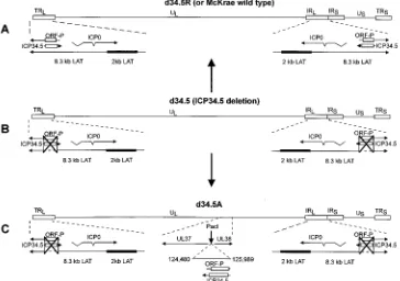
Structure of ICP34.5-deleted and -rescued viruses.McKrae was used as the parental virus for all of the mutants in this study. The McKrae strain of HSV-1 has the highest spontane-ous reactivation rate of any of the commonly used HSV-1 laboratory strains. This previously allowed us to make LAT and ICP34.5 McKrae-based mutants that demonstrated signif-icantly reduced spontaneous reactivation rates. The genomic structures of the three viruses used in this report are shown schematically in Fig. 1. Figure 1A illustrates the wild-type
McKrae and the d34.5R rescued viruses, which are identical. The HSV-1 genome contains a unique long region (UL) and a unique short region (US), both of which are flanked by inverted repeats (terminal and internal long repeats [TRL and IRL, respectively] and terminal and internal short repeats [TRSand IRS, respectively]). Portions of the repeats are expanded under each genome to show the status of the 8.3-kb LAT, ICP34.5, and ORF-P RNAs. ORF-P (11) is a recently described poten-tial ORF with the same sense as LAT whose mRNA may be transcribed independently from the 8.3-kb LAT. The location of the ULcontaining genes UL37 and UL38 is expanded in Fig. 1C (d34.5A) to show the novel location into which a copy of ICP34.5 was inserted in that virus.
[image:3.612.125.489.76.332.2]The ICP34.5-deleted virus, d34.5 (Fig. 1B), is the precursor for both the d34.5R virus (Fig. 1A) and the d34.5A virus (Fig. 1C). We have previously described the construction of d34.5 and d34.5R (18). In d34.5, both copies of the ICP34.5 ORF (one in each long repeat) are deleted (Fig. 1B and C). The colinear kb 6.2 to 7.1 region of LAT in each long repeat and the ORF-P ORF are also deleted in these viruses (Fig. 1B and C). d34.5R (Fig. 1A) was rescued from d34.5 by homologous recombination with a cloned restriction fragment and shown to be restored to the wild-type (McKrae) phenotype (18). The structures of d34.5 and d34.5R were confirmed by Southern analyses (18).
FIG. 1. Schematic representation of d34.5, d34.5R, and d34.5A. (A) Schematic representation of the genome of d34.5R and McKrae. d34.5R was rescued from the ICP34.5 deletion mutant (d34.5) by homologous recombination with a cloned HSV-1 restriction fragment as we previously described (18). This derivation is indicated by the large vertical arrow pointing from d34.5 to d34.5R. The structure of d34.5R is identical to that of the parental McKrae virus genome. The prototypic orientation of HSV-1 shown here contains a ULand a US, each bounded by inverted repeats. The unique regions are indicated by a solid line. The repeats are indicated by open
rectangles. The lines with arrows under the genomes indicate the locations and directions of the ICP34.5, LAT, ORF-P, and ICP0 transcripts. The open rectangles within the ICP34.5 and ORF-P arrows show the relative sizes and locations of these ORFs. (B) ICP34.5 deletion mutant, d34.5, whose construction we previously described (18). Both copies of ICP34.5 (large X’s) and the corresponding location in LAT (dashed line) are deleted. The extent of the deletion is shown by the vertical lines outside the large X. Both the ICP34.5 and the ORF-P ORFs are completely deleted. (C) The large vertical arrow pointing to the schematic for d34.5A signifies that d34.5A was derived from d34.5. d34.5A contains one copy of ICP34.5 inserted into the ULbetween genes UL37 and UL38 (center, expanded portion of UL) at
genomic nucleotide position 84252. This corresponds to an AflII site that we converted to a unique PacI site. This site is 168 nucleotides from the 59end of the UL37 ORF and 279 nucleotides from the 59end of the UL38 ORF. Previous studies have shown that disruption of the genome at this location does not appear to alter any of the biological characteristics of the virus, including establishment and reactivation from latency (unpublished observations). The derivation of the inserted fragment is shown by the nucleotide positions this fragment has in its original position in the IRL(124480 to 125989). d34.5A retains the LAT and ICP34.5 deletions of d34.5,
as indicated by the large X’s.
on November 9, 2019 by guest
http://jvi.asm.org/
The virus shown in Fig. 1C (d34.5A) was constructed from
d34.5 by the addition (hence the ‘‘A’’ in d34.5A) of one copy of
the ICP34.5 ORF into the gap between genes UL37 and UL38 in the viral UL. This insertion was at the location of an AflII site that we had converted to a unique PacI site as described in Materials and Methods (Fig. 1C). The ICP34.5 addition (HSV-1 nucleotides 125989 to 124480) does not include all of the predicted ICP34.5 promoter (23). This is because the 59 portion of the promoter is located within the ‘‘a’’ region of the long repeats, and it is extremely difficult to construct a stable HSV-1 mutant that contains an extra copy of ‘‘a’’ in the UL. This would have been required if we had attempted to include the entire ICP34.5 promoter. We therefore included in the insert only that portion of the ICP34.5 promoter that we ex-pected would not unduly complicate construction of the virus. We then determined that ICP34.5 expression from this partial promoter was sufficient to restore biological function (see be-low). This insert also contains the complete sequence for the postulated ORF-P and its postulated promoter but not the 39 end and poly(A) sequence of the postulated ORF-P mRNA which is the same as the 39end of the 8.3-kb LAT (35). Further details of the construction of d34.5A are given in Materials and Methods. d34.5A is identical to d34.5 except that it contains a copy of ICP34.5 (and ORF-P) introduced into the ULof the virus rather than the normal ICP34.5–ORF-P location. For simplicity (and because ORF-P has not been conclusively shown to be independently transcribed or translated except in ICP4-deleted mutant viruses), in the remainder of this section of this report we will refer only to ICP34.5. The structure of
d34.5A was confirmed by Southern analysis (see Fig. 4, below).
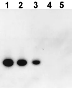
Expression of ICP34.5 mRNA in cells acutely infected with
d34.5A. CV-1 cells were infected with 10 PFU of wild-type McKrae, d34.5, or d34.5A per cell. Total RNA was harvested, digested with DNase I, and analyzed by RT-PCR as described in Materials and Methods with primers that are within the ICP34.5 gene (Fig. 2). The PCR primer pair produces a 134-nucleotide PCR product corresponding to a portion of the ICP34.5 mRNA. The ICP34.5 mRNA-specific RT-PCR prod-ucts were visualized by Southern hybridization with a labeled probe internal to the primers. The RT-PCR product represent-ing ICP34.5 mRNA from d34.5A is clearly visible in the auto-radiogram (Fig. 2, lane 3). This band comigrated with the RT-PCR product representing ICP34.5 mRNA from the
pa-rental wild-type McKrae virus (Fig. 2, lane 2) and with the size marker PCR product generated with the same primer set from purified viral DNA (Fig. 2, lane 1). No ICP34.5-related prod-uct was detected following RT-PCR of RNA from uninfected cells (Fig. 2, lane 5) or of RNA from cells infected with the ICP34.5 deletion mutant d34.5 (lane 4). In addition, PCR analysis confirmed that the original RNA preparations did not contain any contaminating HSV-1 DNA (not shown). Al-though the relative amounts of the RT-PCR products shown here cannot be accurately quantitated, the d34.5A RT-PCR product was readily apparent, and the quantity of this product appeared to be approximately half that of the McKrae RT-PCR product. These results demonstrate that d34.5A, despite not containing the complete ICP34.5 gene promoter (23) and containing only one copy of the ICP34.5 gene, nonetheless appeared to express a significant amount of ICP34.5 mRNA.
Spontaneous reactivation of d34.5A from latently infected rabbits. We recently showed that a LAT deletion mutant (dLAT2903) had significantly reduced spontaneous reactiva-tion in latently infected rabbits (16). We also recently reported that the ICP34.5 deletion mutant d34.5 (18) had significantly reduced spontaneous reactivation under the same conditions. Since the 0.9-kb deletion that produced d34.5 also deleted 0.9 kb of the overlapping LAT gene, it was not clear if the reduc-tion in spontaneous reactivareduc-tion was due to the loss of the ICP34.5 gene, the kb 6.2 to 7.1 colinear deletion in LAT, or a combination of the two. We therefore constructed d34.5A and examined its ability to reactivate spontaneously. As described above, d34.5A retains the 0.9-kb deletion in both copies of LAT and contains a functional copy of ICP34.5 in the ULof the viral genome (Fig. 1C).
Rabbit eyes were infected with 2 3 105 PFU of d34.5A,
d34.5, or d34.5R. Beginning 30 days postinfection (at which
time latency had already been established), all eyes were swabbed once a day to collect tear films for analysis of spon-taneously reactivated virus as described in Materials and Meth-ods. Two independent experiments were done. The numbers of rabbits per group examined for spontaneous reactivation in experiments 1 and 2 were 17 and 7 for d34.5A, 8 and 7 for
d34.5, and 17 and 10 for d34.5R, respectively.
The cumulative number of virus-positive tear film cultures during 26 days in each experiment is shown in Fig. 3. Because of the different numbers of rabbits (and eyes) in the different groups, the data were standardized to represent cumulative positive cultures per eye. In both experiments, the ICP34.5 deletion mutant d34.5 had a very low level of spontaneous reactivation (Fig. 3). This was in agreement with our previous findings (18). In each experiment, the cumulative spontaneous reactivation rate in rabbits latently infected with d34.5A and
d34.5R appeared to be very similar (Fig. 3A and B).
A statistical analysis of positive (spontaneously reactivated) cultures versus negative cultures is shown in Table 1. In exper-iment 1, about 16.1% (134 of 832) of the tear films from rabbits latently infected with d34.5A contained spontaneously reacti-vated virus. This was significantly higher than the 1.4% (6 of 416) positive tear film cultures from rabbits latently infected with d34.5 (P,0.0001; Fisher’s exact test) and not significantly different from the 19.6% (80 of 408) positive cultures from rabbits latently infected with d34.5R (P50.13). The results for experiment 2 were similar. Only 0.3% (1 of 312) of the cultures from rabbits infected with d34.5 were positive, compared with 7.7% (20 of 260) and 9.0% (28 of 312) positive cultures for rabbits latently infected with d34.5R and d34.5A, respectively. Again, the difference between d34.5 and d34.5A was highly significant (P , 0.0001), while the results for d34.5A and
d34.5R were not significantly different (P 50.67). Thus,
de-FIG. 2. Expression of ICP34.5 mRNA by d34.5A. Infected cells were har-vested; RNA was isolated, digested with DNase, and subjected to RT-PCR for ICP34.5 mRNA; and the products were visualized by Southern analysis with a labeled probe internal to the original PCR primers as described in Materials and Methods. Lane 1, marker DNA generated by PCR with the primers used for RT-PCR; lane 2, RT-PCR product corresponding to ICP34.5 mRNA from McKrae; lane 3, d34.5A; lane 4, d34.5; lane 5, uninfected cells.
on November 9, 2019 by guest
http://jvi.asm.org/
[image:4.612.120.236.70.211.2]spite the fact that d34.5A contained only a single copy of ICP34.5, the spontaneous reactivation rate of d34.5A appeared to have been completely rescued back to wild-type virus (d34.5R) levels.
Because the analyses discussed above do not take into ac-count the number of eyes in each of the groups, the data were analyzed by additional methods. The fraction of virus-positive cultures for each eye in each group (i.e., the fraction of time each eye was virus positive) was calculated, and these fractions were analyzed (Table 1). In both experiments, the level of spontaneous reactivation with the d34.5A virus was signifi-cantly higher than that of d34.5 (P, 0.0001 and P5 0.007, respectively; Mann-Whitney rank sum test) and similar to that of d34.5R (P50.3 and P50.75, respectively; Student’s t test). The number of eyes in each group that had at least one spontaneous reactivation was also analyzed (Table 1). Again, in both experiments the results for the d34.5A virus were significantly greater than the results for the d34.5 virus (94 versus 25%, P , 0.0001, and 67 versus 8%, P 5 0.01) and similar to the wild-type results (94 versus 94%, P51.0, and 67 versus 50%, P50.67).
Another method of examining the effect of one copy of ICP34.5 on spontaneous reactivation is to analyze the number of times spontaneous reactivation is detected in each eye, re-gardless of the length of time the virus is present. This is equivalent to the number of episodes in which reactivated virus is detected in the tears, with consecutive days of positive cul-tures being treated as a single event. Thus, an eye that sheds virus for a single day and an eye that sheds virus for 5 consec-utive days would both constitute one episode of spontaneous reactivation. The average number of spontaneous reactivations per eye in each group calculated in this manner is shown in Table 1. The numbers of episodes of spontaneous reactivation in d34.5A- and d34.5R-infected eyes were similar (2.4 and 2.1, respectively, P50.33, in experiment 1 and 1.4 and 1.2, respec-tively, P50.63, in experiment 2). In contrast, the numbers of episodes in d34.5A- and d34.5-infected eyes were significantly different in both experiments (Table 1, P ,0.0001 and P5 0.007, respectively). All of the analyses discussed above indi-cate that d34.5A (containing one copy of ICP34.5 in a novel location and a 0.9-kb deletion from kb 6.2 to 7.1 in both copies of LAT) reactivated spontaneously at a rate indistinguishable from that of d34.5R (wild-type virus).
Southern analysis of spontaneously reactivated virus. To eliminate the unlikely possibility that the spontaneously reac-tivated virus recovered from the eyes of rabbits latently in-fected with the d34.5A mutant viruses was contaminating wild-type virus or mutant virus that had somehow reverted back to wild-type virus, Southern analyses with selected spontaneously reactivated viruses were done. Examples of some of these Southern analyses are shown in Fig. 4. Purified viral DNAs were obtained prior to infection (lanes I) and from virus in tears after spontaneous reactivation (lanes S). The DNAs were individually digested with BamHI and subjected to Southern analysis by using a probe that spans the ICP34.5 gene and is completely within the viral long repeat. As we previously re-ported for wild-type virus (or d34.5R) (18), the 5.9- and 2.9-kb bands seen in Fig. 4, lane d34.5R-I (solid arrows), represent
BamHI fragments containing the ICP34.5 gene from the IRL
and the TRL, respectively. The additional fainter bands form-ing a ladder above the 5.9- and 2.9-kb bands represent BamHI fragments from long repeats containing multiple copies of the ‘‘a’’ repeat (18).
As we previously reported (18), in d34.5 both the 5.9- and 2.9-kb bands (and their ladder bands) have apparent sizes which are approximately 0.9 kb less than those of their
wild-FIG. 3. Spontaneous reactivation in rabbits latently infected with d34.5A. As described in Materials and Methods, rabbits were ocularly infected with 23105
PFU of d34.5, d34.5R, or d34.5A per eye. Beginning at thirty days postinfection (day 0), eye swabs were taken daily to collect tear films for the detection of HSV-1. The collected tear films were plated on primary rabbit kidney cells and observed for up to 30 days for the presence of cytopathic effects indicative of the presence of spontaneously reactivated virus in the corresponding eye. All positive cultures were confirmed by passage, and most were also confirmed by Southern analysis (Fig. 4). The y axis represents the cumulative number of HSV-1-positive cultures for each virus group divided by the number of eyes in the group. Panels A and B show results from independent experiments. A statistical analysis is shown in Table 1.
on November 9, 2019 by guest
http://jvi.asm.org/
[image:5.612.84.271.82.621.2]type counterparts (Fig. 4, lane d34.5-I [open arrows]), consis-tent with the 0.9-kb deletion of ICP34.5 in each repeat. As with the d34.5 virus DNA, the bands corresponding to the region of the repeats are also reduced in size in the d34.5A DNA (Fig. 4,
lane d34.5A-I). Again, this reduction in size is consistent with deletion of ICP34.5 from both regions and confirms that
d34.5A has retained the d34.5 ICP34.5-LAT deletion in both
long repeats. The d34.5A DNA also contains a large band of approximately 9 kb (Fig. 4, lane d34.5A-I) to which the labeled probe hybridized. This band is consistent with the size of the
BamHI band (7.5 kb) containing the ICP34.5 gene (1.5 kb) that
was inserted into the ULand confirms that d34.5A contains this unique copy of the ICP34.5 gene. In addition, digestion of
d34.5A DNA with PacI converted this band to a size identical
to that of the original ICP34.5 insert (1.5 kb) (data not shown). This Southern analysis therefore confirms that the d34.5A virus contains a copy of ICP34.5 in the UL, while retaining the ICP34.5-LAT deletion in both repeats.
Lanes d34.5-S, d34.5R-S, and d34.5A-S (Fig. 4) show South-ern analyses of DNAs isolated from spontaneously reactivated virus grown from tears. With all three viruses, the spontane-ously reactivated (lanes S) viruses d34.5, d34.5R, and d34.5A have Southern patterns identical to those of the infecting vi-ruses (lanes I), indicating that for each virus the organization of the genomic DNA has been retained. Specifically, the spon-taneously reactivated d34.5A virus (Fig. 4, lane d34.5A-S) re-tained the ICP34.5-LAT deletion in both repeats as well as a copy of the ICP34.5 gene in the UL. This was found to be the case for all of the spontaneously reactivated d34.5A virus ex-amined. Thus, the ability of d34.5A to reactivate spontaneously was due to inherent properties of the virus and not to rescue of the ICP34.5-LAT deletions with the ICP34.5 gene sequences inserted into the UL.
[image:6.612.58.555.83.209.2]Virulence in rabbits.We previously found that d34.5 was avirulent in rabbits (18). To determine if virulence was re-stored in d34.5A, rabbits were infected ocularly. The combined survival results of two experiments are shown in Fig. 5. Ninety-two percent (22 of 24) of the rabbits infected with d34.5A survived. This level of survival was significantly greater than that seen with d34.5R (13 of 27, 48%, P 5 0.0009; Fisher’s exact test). One hundred percent (15 of 15) of the d34.5-infected rabbits survived. This was not significantly different from the results obtained with d34.5A (P50.51). These results indicate that the addition of a single copy of ICP34.5 to d34.5 did not restore virulence to wild-type levels.
FIG. 4. Southern analysis of spontaneously reactivated d34.5A. DNA was isolated from virus from selected positive tear film cultures from Fig. 3, digested with BamHI, run on an agarose gel, transferred to a nylon membrane, and hybridized with a32
P-labeled probe from the long repeat region of the virus that spans the ICP34.5 gene (or deletion) as described in Materials and Methods. The solid arrows indicate the locations of the 5.9- and 2.9-kb ICP34.5 gene-related bands from wild-type virus. The open arrows indicate the locations of the cor-responding bands from d34.5 and d34.5A that are missing in the 0.9-kb ICP34.5 region. The star indicates the location of the BamHI band that contains the ICP34.5 gene inserted into the ULof d34.5A. Lanes: I, infecting virus; S,
[image:6.612.114.241.373.633.2]spon-taneously reactivated virus; M, DNA markers.
TABLE 1. Spontaneous reactivation rate of d34.5A is normala
Expt no. and virus
Copies of ICP34.5
No. of positive cultures/total
no. (%)
P
Fraction of positive
cul-tures/eyeb P
No. of pos-itive eyes/
total no. (%)c
P
No. of episodes of spontaneous reactivation/eye
(n)d
P
1
d34.5 0 64/416 (1.4) ,0.0001e 0.01 ,0.0001f 4/16 (25) ,0.0001e 0.31 (16) ,0.0001f
d34.5A 1 134/832 (16.1) 0.16 30/32 (94) 2.38 (32)
d34.5R 2 80/408 (19.6) 0.13e 0.19 0.3g 17/18 (94) 1.0e 2.1 (18) 0.33f
2
d34.5 0 1/312 (0.3) ,0.0001e 0.003 0.007f 1/12 (8) 0.01e 0.08 (12) 0.007f
d34.5A 1 28/312 (9.0) 0.089 8/12 (67) 1.42 (12)
d34.5R 2 20/260 (7.7) 0.67e 0.077 0.75g 5/10 (50) 0.67e 1.2 (10) 0.63f
aRabbits were infected in both eyes with d34.5 (no copies of ICP34.5), d34.5A (one copy of ICP34.5 in the U
L), or d34.5R (rescued d34.5; two copies of ICP34.5
in their normal location in the long repeat) as described in Materials and Methods. Experiments 1 and 2 were done independently, at different times, and with different groups of rabbits. Spontaneous reactivation was assessed beginning on day 30 postinfection by culturing tear films collected daily for 26 days as described in Materials and Methods. P values reported are relative to d34.5A.
bAverage for all eyes of the total HSV-1-positive cultures for eye ‘‘x’’ per total number of cultures for eye ‘‘x’’. cThe total number of eyes that spontaneously reactivated at least once per the total number of eyes.
dThe total number of reactivations in each group, assuming that consecutive days of HSV-1-positive tear film cultures from a single eye are the result of a single
reactivation event or episode. n, number of eyes.
eFisher’s exact test; two sided. The groups are considered significantly different if P is,0.05. f
Mann-Whitney rank sum test; two sided. The groups are considered significantly different if P is,0.05.
g
Student’s t test; two sided. The groups are considered significantly different if P is,0.05.
on November 9, 2019 by guest
http://jvi.asm.org/
Replication ofd34.5A in rabbit eyes.We previously reported that d34.5 does not replicate well in rabbit eyes (18). To de-termine if d34.5A was fully rescued for this defect, rabbits were infected with 23105PFU per eye with either d34.5, d34.5R, or d34.5A as described in Materials and Methods. Tears were collected at various times during the acute phase of infection and the titers of the virus were determined. The average virus titers from six eyes per group on days 3, 5, 7, and 10 are shown in Fig. 6. As determined previously, d34.5 grew to only very low titers (,100 PFU per eye). In contrast, d34.5R and d34.5A grew to high titers (53105to 93105PFU per eye). There were no significant differences in the titers of d34.5R and
d34.5A on any given day (P.0.05, Student’s t test). In
com-parison, eyes infected with either d34.5R or d34.5A had sig-nificantly higher virus titers than eyes infected with d34.5 (P, 0.05, days 3, 5, 7, and 10; Student’s t test). Thus, it appeared that a single copy of ICP34.5 in a novel location was sufficient to fully restore wild-type levels of viral replication in rabbit eyes.
Replication ofd34.5A in tissue culture. We previously re-ported that despite d34.5 having normal replication kinetics in RS cells, replication of d34.5 in CV-1 cells was restricted (18). LAT did not appear to be involved in this phenotype, since our null LAT mutant replicated normally in CV-1 cells (16). None-theless, it was of interest to determine if the ability to replicate in CV-1 cells could be restored by insertion of one copy of ICP34.5 in a novel location. We therefore infected subconflu-ent monolayers of CV-1 cells with d34.5, d34.5R, or d34.5A, each at an MOI of 10 or 0.01 PFU per cell (Fig. 7). At an MOI of 0.01, d34.5R and d34.5A had similar replication kinetics, with peak titers of just over 1 3 108 PFU/ml (Fig. 7A). In comparison, at this low MOI, d34.5 replicated very poorly, reaching a maximum titer of less than 1 3 103 PFU/ml, or 100,000-fold less. At an MOI of 10 PFU per cell (Fig. 7B),
d34.5R and d34.5A again had similar replication kinetics, with
peak titers of over 107PFU/ml. As before, d34.5 appeared to
replicate much less well; however, at this higher MOI, the difference was less pronounced (approximately 100-fold; Fig. 7B). These results indicate that a single copy of ICP34.5 in a novel location appeared to fully restore the ability of the d34.5 ICP34.5 null mutant to replicate in CV-1 cells.
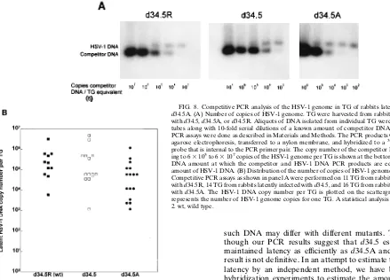
HSV-1 DNA in TG of latently infected rabbits.The number of HSV-1 genomic copies per TG in rabbits latently infected with d34.5, d34.5A, or d34.5R was estimated by competitive PCR assays as described in Materials and Methods. The PCR primer pair produces a 192-nucleotide PCR product from HSV-1 genomic DNA corresponding to a portion of the HSV-1 glycoprotein B (gB) gene. The competitor DNA is identical to this gB region except for a 35-nucleotide deletion internal to the primers that results in a PCR product of 157 nucleotides. The genomic and competitor PCR products were separated, visualized, and quantitated by Southern analysis with a cloned internal probe that is outside the 35-nucleotide deletion. Representative results of such competitor PCR as-says are shown in Fig. 8A. In all three panels, as the intensity of the competitor PCR product decreases from left to right in conjunction with the decreasing amount of input competitor DNA, the intensity of the HSV-1 DNA PCR product increases. For these particular TG, the competitor and HSV-1 DNA band intensities appear equivalent at approximately 63 104 competitor copy equivalents per TG. Thus, these three samples all appear to contain approximately 63104genomic copies of HSV-1 DNA per TG.
[image:7.612.91.265.71.314.2]Identical competitive PCR assays were performed on 11 TG from rabbits latently infected with d34.5R, 14 TG from rabbits
FIG. 5. Virulence of d34.5A in rabbits. Rabbits were infected ocularly with 2
3105
PFU of each virus per eye. Survival was determined 21 days postinfection. P values are in comparison to d34.5A.
FIG. 6. Replication of d34.5A in rabbit eyes. Rabbits were infected with 23 105
PFU of d34.5, d34.5R, or d34.5A per eye as described in Materials and Methods. Tears were collected at the times indicated, and the amount of HSV-1 present was determined by titration with a standard plaque assay as described in Materials and Methods. Each time point represents the average virus titer from six eyes per group. The titers for d34.5R and d34.5A were not different on any day (P.0.05; Student’s t test). On all days the d34.5 virus titers were lower than the d34.5R and d34.5A virus titers (P,0.05; Student’s t test).
on November 9, 2019 by guest
http://jvi.asm.org/
[image:7.612.326.539.385.662.2]latently infected with d34.5, and 16 TG from rabbits latently infected with d34.5A. The estimated number of HSV-1 DNA copies for each of the TG is shown in a scattergram (Fig. 8B). Each symbol represents the estimated number of copies for one TG. The scattergram therefore shows the distribution and spread of the results. The ranges and distributions for all three viruses appear similar. The average number of genomic copies was approximately 53105per TG for d34.5R, 7.13105per
TG for d34.5, and 1.5 3 105 per TG for d34.5A (Table 2). Compared with d34.5R, both d34.5 and d34.5A appeared to have similar numbers of HSV-1 genomes per TG (P50.68 and
P 50.07, respectively; Student’s t test). Thus, if the relative amount of HSV-1 DNA as determined by PCR analysis of latently infected TGs reflects the rate of establishment of la-tency, it would appear that all three viruses established latency at similar rates.
DISCUSSION
Recently we reported an ICP34.5 deletion mutant of HSV-1 strain McKrae (d34.5) that was severely restricted in its ability to spontaneously reactivate in the rabbit eye model of HSV-1 latency (18). Because of the colinearity of ICP34.5 with a portion of LAT, d34.5 also contains a deletion in the 8.3-kb LAT from kb 6.2 to 7.1. Since we had previously shown that LAT is required for efficient spontaneous reactivation (16), it was of interest to determine if the reduced spontaneous reac-tivation of d34.5 was due to the kb 6.2 to 7.1 deletion in LAT or to deletion of ICP34.5. Therefore, in the current study, we reintroduced back into the d34.5 mutant a functional copy of the ICP34.5 gene at a novel location. This mutant, designated
d34.5A, contains the original 917-nucleotide deletion in LAT
(in both viral long repeats) and a single copy of the ICP34.5 gene in the ULof the virus between genes UL37 and UL38.
d34.5A thus has been repaired, or rescued, for one of the two
copies of ICP34.5 while retaining the original kb 6.2 to 7.1 LAT deletion.
In two independent experiments, the spontaneous reactiva-tion rate for d34.5A (9 to 16.1%) was the same as the sponta-neous reactivation rate for wild-type rescued virus (7.7 to 19.6%). This demonstrated that the kb 6.2 to 7.1 region of the 8.3-kb LAT was not required for efficient spontaneous reacti-vation.
The original ICP34.5 deletion mutant, d34.5, had additional defects. Virulence in rabbits was dramatically decreased, as was replication in rabbit eyes and CV-1 cells. All of these defects were due to deletion of ICP34.5, since our LAT mu-tant, containing a complete deletion of the LAT promoter and constructed from the same McKrae parental virus, did not have any of these defects (16). The results reported here indi-cate that spontaneous reactivation and replication in rabbit eyes and CV-1 cells was completely restored by the single copy of the ICP34.5 gene in d34.5A. In contrast, virulence in rabbits was not completely restored in d34.5A. This suggests either that virulence in rabbits requires two copies of the ICP34.5 gene or that the ICP34.5 gene must be in its normal location in the long repeat.
It is of particular interest that in d34.5A the spontaneous reactivation phenotype was completely restored to wild-type levels while virulence remained very low. These results appear to at least partially separate spontaneous reactivation from virulence. They therefore strongly suggest that, in contrast to what has often been assumed, the ability of a particular HSV strain to spontaneously reactivate is not directly related to the virulence of the virus.
We previously reported that the results of a noncompetitive PCR assay appeared to show a trend toward less viral DNA in the TG of rabbits latently infected with d34.5 versus d34.5R. However, the differences were not statistically different. In the current study, our competitive PCR assays also showed a lack of statistical significance. Thus, it appeared that rabbits latently infected with d34.5A had levels of viral DNA in their TG comparable to those in rabbits latently infected with d34.5 or
[image:8.612.83.271.72.584.2]d34.5R. Thus, the ICP34.5 gene did not appear to be required
FIG. 7. Replication of d34.5A in CV-1 cells in culture. Subconfluent mono-layers of CV-1 cells in 60-mm-diameter tissue culture plates were infected with 10 or 0.01 PFU of d34.5, d34.5R, or d34.5A per cell. At the times indicated, the monolayers were harvested and the amount of virus was determined by plaque assay.
on November 9, 2019 by guest
http://jvi.asm.org/
for HSV-1 to efficiently establish latency. Since d34.5R and
d34.5A reactivated spontaneously at statistically higher levels
than d34.5, it is therefore likely that ICP34.5 is either (i) in-volved in reactivation from latency or (ii) required for the virus to replicate efficiently and be detected in tear films following reactivation in the neuron.
The discussion in the preceding paragraph contains an im-plicit assumption for which there is currently no proof, namely, that the total amount of HSV-1 viral DNA present in the TG as determined by PCR assays is a direct and accurate measure of the relative amount of HSV-1 latency. However, PCR assays cannot distinguish between biologically active and biologically inactive HSV-1 DNA. Thus, some of the HSV-1 DNA de-tected by PCR may be degraded viral DNA that does not represent latent DNA capable of reactivation. The amount of
such DNA may differ with different mutants. Therefore, al-though our PCR results suggest that d34.5 established and maintained latency as efficiently as d34.5A and d34.5R, this result is not definitive. In an attempt to estimate the amount of latency by an independent method, we have begun in situ hybridization experiments to estimate the amount of latency on the basis of the percentage of neurons containing the 2-kb LAT RNA. Preliminary results indicate that rabbits latently infected with d34.5 may contain fewer LAT-positive neurons than other latently infected rabbits. Thus, in contrast to the interpretation that would normally be given to our PCR results (i.e., no differences in establishment of latency), the interpre-tation that would normally be given to our preliminary in situ hybridization results suggests that d34.5A may establish latency more efficiently than d34.5.
The apparent complexity of the kb 6.2 to 7.1 LAT region in the long repeat has recently been increased by reports suggest-ing that, in addition to the colinear but antisense ICP34.5 gene, this region contains an independent LAT-sense transcript con-taining a potential ORF that has been designated ORF-P (2, 11, 35). ORF-P is deleted from d34.5 because it is almost completely colinear (but antisense) to the ICP34.5 ORF (Fig. 1B and C). Furthermore, the postulated ORF-P and its pos-tulated promoter are, along with the ICP34.5 ORF, completely contained within the LAT region relocated to the UL of
d34.5A.
ORF-P is potentially of great interest, although its function remains unknown. Formally, some or all of the results in this report that have been attributed to ICP34.5 may instead be due to ORF-P. It should be stressed, however, that the mapping results reported here are independent of whether the rescued phenotypes in d34.5A were due to expression of ICP34.5 or ORF-P. In either case, our results clearly demonstrate that deletion of the kb 6.2 to 7.1 region from the 8.3-kb LAT primary transcript did not significantly affect spontaneous re-activation in the rabbit.
[image:9.612.63.502.73.389.2]In conclusion, genetic mapping of the functional portion(s) of the 8.3-kb LAT gene has been complicated by the colinear-ity with portions of LAT of two important viral genes, ICP0
FIG. 8. Competitive PCR analysis of the HSV-1 genome in TG of rabbits latently infected with d34.5A. (A) Number of copies of HSV-1 genome. TG were harvested from rabbits latently infected with d34.5, d34.5A, or d34.5R. Aliquots of DNA isolated from individual TG were placed into PCR tubes along with 10-fold serial dilutions of a known amount of competitor DNA, and competitive PCR assays were done as described in Materials and Methods. The PCR products were separated by agarose electrophoresis, transferred to a nylon membrane, and hybridized to a32P-labeled cloned
probe that is internal to the PCR primer pair. The copy number of the competitor DNA correspond-ing to 63103to 63107copies of the HSV-1 genome per TG is shown at the bottom. The competitor
DNA amount at which the competitor and HSV-1 DNA PCR products are equal indicates the amount of HSV-1 DNA. (B) Distribution of the number of copies of HSV-1 genomes in different TG. Competitive PCR assays as shown in panel A were performed on 11 TG from rabbits latently infected with d34.5R, 14 TG from rabbits latently infected with d34.5, and 16 TG from rabbits latently infected with d34.5A. The HSV-1 DNA copy number per TG is plotted on the scattergram. Each symbol represents the number of HSV-1 genome copies for one TG. A statistical analysis is shown in Table 2. wt, wild type.
TABLE 2. Estimated number of HSV-1 genomes in TG of latently infected rabbitsa
Virus Avg no. of genomic copies/TG6SD (n)b Pc
d34.5R (wt)d 5310562.83105(11)
d34.5 7.1310563.83105(14) 0.68
d34.5A 1.5310567.53104(16) 0.07
a
Total DNA was isolated from individual TG of rabbits latently infected with either d34.5R, d34.5, or d34.5A. The number of HSV-1 genomic copies per latently infected TG was estimated by competitive PCR assays as described in Materials and Methods.
b
SD, standard deviation. n, number of TG.
c
Student’s t test; double sided. A P value of.0.05 indicates that compared with the number of HSV-1 genomic copies in the d34.5R group, the number of HSV-1 genomic copies in the indicated group is not significantly different.
d
wt, wild type.
on November 9, 2019 by guest
http://jvi.asm.org/
[image:9.612.58.298.605.653.2]and ICP34.5. We have reported here an approach that allows functional genetic mapping of the LAT region that is colinear with ICP34.5. Insertion of a complete functional copy of the ICP34.5 gene in a novel location allowed us to determine that the corresponding (kb 6.2 to 7.1) to LAT region was not in-volved in the function of LAT responsible for enhancing spon-taneous reactivation. A similar approach with the ICP0 gene should allow us to construct a series of deletions in regions of LAT that are normally colinear with ICP0. Combined with similar mapping of LAT regions that do not overlap ICP0, this should allow us to map the functional LAT region(s) and determine the coding region(s) of a potential LAT protein(s) and should ultimately lead to an understanding of the molec-ular mechanism by which LAT enhances spontaneous reacti-vation.
ACKNOWLEDGMENTS
This work was supported by Public Health Service grants EY07566 and EY10243 (S.L.W.), AI32121 (R.L.T.), and NS25789 (N.M.S.); the Discovery Fund for Eye Research (S.L.W., H.G., and A.B.N.); and the Skirball Molecular Ophthalmology Program (S.L.W., H.G., and A.B.N.).
We thank Anita Avery for excellent technical support.
REFERENCES
1. Block, T. M., J. G. Spivack, I. Steiner, S. Deshmane, M. T. McIntosh, R. P. Lirette, and N. W. Fraser. 1990. A herpes simplex virus type 1 latency-associated transcript mutant reactivates with normal kinetics from latent infection. J. Virol. 64:3417–3426.
2. Bohenzky, R. A., M. Lagunoff, B. Roizman, E. K. Wagner, and S. Silverstein. 1995. Two overlapping transcription units which extend across the L-S junc-tion of herpes simplex virus type 1. J. Virol. 69:2889–2897.
3. Chou, J., E. R. Kern, R. J. Whitley, and B. Roizman. 1990. Mapping of herpes simplex virus-neurovirulence to gamma 134.5, a gene nonessential for growth in culture. Science 250:1262–1265.
4. Croen, K. D., J. M. Ostrove, L. J. Dragovic, J. E. Smialek, and S. E. Straus. 1987. Latent herpes simplex virus in human trigeminal ganglia: detection of an immediate early gene ‘‘antisense’’ transcript by in situ hybridization. N. Engl. J. Med. 317:1427–1432.
5. Crowl, R., C. Seamens, P. Lomedico, and S. McAndrew. 1985. Versatile expression vectors for high-level synthesis of cloned gene products in E. coli. Gene 38:31–38.
6. Diviacco, S., P. Norio, L. Zentilin, S. Menzo, M. Clementi, G. Bjamonti, S. Riva, A. Falaschi, and M. Giacca.1992. A novel procedure for quantitative polymerase chain reaction by coamplification of competitive templates. Gene 122:313–320.
7. Gordon, Y. J., B. Johnson, E. Romanowski, and T. Araullo-Cruz. 1988. RNA complimentary to herpes simplex virus type 1 ICP0 gene demonstrated in neurons of human trigeminal ganglia. J. Virol. 62:1832–1835.
8. Hill, J. M., F. Sederati, R. T. Javier, E. K. Wagner, and J. G. Stevens. 1990. Herpes simplex virus latent phase transcription facilitates in vivo reactiva-tion. Virology 174:117–125.
9. Ho, D. Y., and E. S. Mocarski. 1989. Herpes simplex virus latent RNA (LAT) is not required for latent infection in the mouse. Proc. Natl. Acad. Sci. USA 86:7596–7600.
10. Javier, R. T., J. G. Stevens, V. B. Dissette, and E. K. Wagner. 1988. A herpes simplex virus transcript abundant in latently infected neurons is dispensable for establishment of the latent state. Virology 166:254–257.
11. Lagunoff, M., and B. Roizman. 1994. Expression of a herpes simplex virus 1 open reading frame antisense to theg134.5 gene and transcribed by an RNA 39
coterminal with the unspliced latency-associated transcript. J. Virol. 68:6021–6028. 12. Leib, D. A., C. L. Bogard, M. Kosz-Vnenchak, K. A. Hicks, D. M. Coen, D. M. Knipe, and P. A. Schaffer.1989. A deletion mutant of the latency-associated transcript of herpes simplex virus type 1 reactivates from the latent state with reduced frequency. J. Virol. 63:2893–2900.
13. Leib, D. A., K. C. Nadeau, K. C. Rundle, S. A. Rundle, and P. A. Schaffer. 1991. Promoter of the latency-associated transcripts of herpes simplex virus type-1 contains a functional cAMP-response element: role of the latency associated transcripts and cAMP in reactivation of viral latency. Proc. Natl. Acad. Sci. USA 88:48–52.
14. Nesburn, A. B. (ed.). 1983. Report of the corneal disease panel: vision re-search: a national plan 1983–1987, vol. II, part III. The C. V. Mosby Co., St. Louis.
15. Nesburn, A. B., H. Ghiasi, and S. L. Wechsler. 1990. Ocular safety and efficacy of an HSV-1 gD vaccine during primary and latent infection. Invest. Ophthalmol. Visual Sci. 31:77–82.
16. Perng, G. C., E. C. Dunkel, P. A. Geary, S. M. Slanina, H. Ghiasi, R. Kaiwar, A. B. Nesburn, and S. L. Wechsler.1994. The latency-associated transcript gene of herpes simplex virus type 1 (HSV-1) is required for efficient in vivo spontaneous reactivation of HSV-1 from latency. J. Virol. 68:8045–8055. 17. Perng, G. C., H. Ghiasi, R. Kaiwar, A. B. Nesburn, and S. L. Wechsler. 1994.
An improved method for cloning portions of the repeat regions of herpes simplex virus type 1. J. Virol. Methods 46:111–116.
18. Perng, G. C., R. L. Thompson, N. M. Sawtell, W. E. Taylor, S. M. Slanina, H. Ghiasi, R. Kaiwar, A. B. Nesburn, and S. L. Wechsler.1995. An avirulent ICP34.5 deletion mutant of herpes simplex virus type 1 is capable of in vivo spontaneous reactivation. J. Virol. 69:3033–3041.
19. Puissant, C., and L. Houdebine. 1990. An improvement of the single-step method of RNA isolation by acid guanidinium thiocyanate-phenol-chloro-form extraction. BioTechniques 8:148–149.
20. Ramakrishnan, R., D. J. Fink, G. Jiang, P. Desai, J. C. Glorioso, and M. Levine.1994. Competitive quantitative PCR analysis of herpes simplex virus type 1 DNA and latency-associated transcript RNA in latently infected cells of the rat brain. J. Virol. 68:1864–1873.
21. Rock, D. L., A. B. Nesburn, H. Ghiasi, J. Ong, T. L. Lewis, J. R. Lokensgard, and S. L. Wechsler.1987. Detection of latency-related viral RNAs in tri-geminal ganglia of rabbits latently infected with herpes simplex virus type 1. J. Virol. 61:3820–3826.
22. Sacks, W. R., and P. A. Schaffer. 1987. Deletion mutants in the gene encod-ing the herpes simplex virus type 1 immediate early protein ICP0 exhibit impaired growth in cell culture. J. Virol. 61:829–839.
23. Sarisky, R. T., and P. C. Weber. 1994. Role of anisomorphic DNA confor-mations in the negative regulation of a herpes simplex virus type 1 promoter. Virology 204:569–579.
24. Sawtell, N. M., and R. L. Thompson. 1992. Herpes simplex virus type 1 latency-associated transcription unit promotes anatomical site-dependent establishment and reactivation from latency. J. Virol. 66:2157–2169. 25. Spivack, J. G., and N. W. Fraser. 1988. Expression of HSV-1
latency-asso-ciated transcripts in the trigeminal ganglia of mice during acute infection and reactivation of latent infection. J. Virol. 62:1479–1485.
26. Steiner, I., J. G. Spivack, R. P. Lirette, S. M. Brown, A. R. MacLean, J. H. Subak-Sharpe, and N. W. Fraser.1989. Herpes simplex virus type 1 latency associated transcripts are evidently not essential for latent infection. EMBO J. 8:505–511.
27. Steiner, I., J. G. Spivack, D. R. O’Boyle II, E. Lavi, and N. W. Fraser. 1988. Latent herpes simplex virus type 1 transcription in human trigeminal ganglia. J. Virol. 62:3493–3496.
28. Stevens, J. G., E. K. Wagner, G. B. Devi-Rao, M. L. Cook, and L. T. Feldman. 1987. RNA complementary to a herpesvirus alpha gene mRNA is prominent in latently infected neurons. Science 235:1056–1059.
29. Trousdale, M. D., I. Steiner, J. G. Spivack, S. L. Deshmane, S. M. Brown, A. R. MacLean, J. H. Subak-Sharpe, and N. W. Fraser.1991. In vivo and in vitro reactivation impairment of a herpes simplex virus type 1 latency-asso-ciated transcript variant in a rabbit eye model. J. Virol. 65:6989–6993. 30. Wagner, E. K., G. Devi-Rao, L. T. Feldman, A. T. Dobson, Y. Zhang, W. M.
Flanagan, and J. G. Stevens.1988. Physical characterization of the herpes simplex virus latency-associated transcript in neurons. J. Virol. 62:1194–1202.
31. Wagner, E. K., W. M. Flanagan, G. Devi-Rao, Y. Zhang, J. M. Hill, K. P. Anderson, and J. G. Stevens.1988. The herpes simplex virus latency-associ-ated transcript is spliced during the latent phase of infection. J. Virol. 62:4577–4585.
32. Wechsler, S. L., A. B. Nesburn, R. J. Watson, S. Slanina, and H. Ghiasi. 1988. Fine mapping of the major latency-related RNA of herpes simplex virus type 1 in humans. J. Gen. Virol. 69:3101–3106.
33. Wechsler, S. L., A. B. Nesburn, R. Watson, S. M. Slanina, and H. Ghiasi. 1988. Fine mapping of the latency-related gene of herpes simplex virus type 1: alternative splicing produces distinct latency-related RNAs containing open reading frames. J. Virol. 62:4051–4058.
34. Wechsler, S. L., A. B. Nesburn, J. C. Zwaagstra, and H. Ghiasi. 1989. Sequence of the latency related gene of herpes simplex virus type 1. Virology 168:168–172.
35. Yeh, L., and P. A. Schaffer. 1993. A novel class of transcripts expressed with late kinetics in the absence of ICP4 spans the junction between the long and short segments of the herpes simplex virus type 1 genome. J. Virol. 67:7373– 7382.
36. Zwaagstra, J. C., H. Ghiasi, S. M. Slanina, A. B. Nesburn, S. C. Wheatley, K. Lillycrop, J. Wood, D. S. Latchman, K. Patel, and S. L. Wechsler.1990. Activity of herpes simplex virus type 1 latency-associated transcript (LAT) promoter in neuron-derived cells: evidence for neuron specificity and for a large LAT transcript. J. Virol. 64:5019–5028.