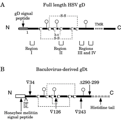
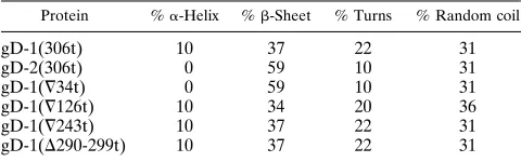
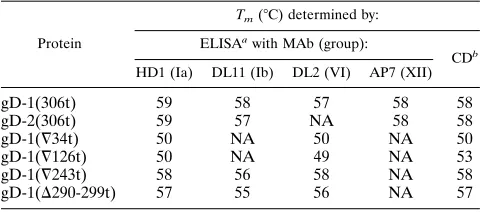
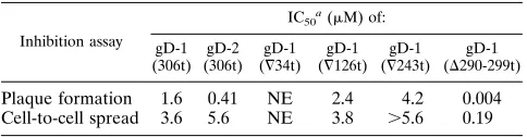
0022-538X/96/$04.0010
Copyrightq1996, American Society for Microbiology
Structure-Function Analysis of Soluble Forms of Herpes
Simplex Virus Glycoprotein D
ANTHONY V. NICOLA,1,2* SHARON H. WILLIS,1,2NIRINJINI N. NAIDOO,1,2†
ROSELYN J. EISENBERG,2,3
ANDGARY H. COHEN1,2
Department of Microbiology1and Center for Oral Health Research,2School of Dental Medicine, and
Department of Pathobiology, School of Veterinary Medicine,3University of
Pennsylvania, Philadelphia, Pennsylvania 19104
Received 1 January 1996/Accepted 11 March 1996
Glycoprotein D (gD) of herpes simplex virus (HSV) is essential for virus entry. Truncated forms of gD lacking the transmembrane and cytoplasmic tail regions have been shown to bind to cells and block plaque formation. Using complementation analysis and a panel of gD mutants, we previously identified four regions of gD (regions I to IV) which are important for virus entry. Here, we used baculovirus vectors to overexpress truncated forms of wild-type gD from HSV type 1 (HSV-1) [gD-1(306t)] and HSV-2 [gD-2(306t)] and four mutants, gD-1(=34t), gD-1(=126t), gD-1(=243t), and gD-1(D290-299t), each having a mutation in one of the four functional regions. We used an enzyme-linked immunosorbent assay and circular dichroism to analyze the structure of these proteins, and we used functional assays to study the role of gD in binding, penetration, and cell-to-cell spread. gD-1 and gD-2 are similar in antigenic structure and thermal stability but vary in secondary structure. Mutant proteins with insertions in region I or II were most altered in structure and stability, while mutants with insertions in region III or IV were less altered. gD-1(306t) and gD-2(306t) inhibited both plaque formation and cell-to-cell transmission of HSV-1. In spite of obvious structural differences, all of the mutant proteins bound to cells, confirming that binding is not the only function of gD. The region I mutant did not inhibit HSV plaque formation or cell-to-cell spread, suggesting that this region is necessary for the function of gD in these processes. Surprisingly, the other three mutant proteins functioned in all of the in vitro assays, indicating that the ability of gD to bind to cells and inhibit infection does not correlate with its ability to initiate infection as measured by the complementation assay. The region IV mutant, gD-1(D290-299t), had an unex-pected enhanced inhibitory effect on HSV infection. Taken together, the results argue against a single func-tional domain in gD. It is likely that different gD structural elements are involved in successive steps of infection.
Herpes simplex virus (HSV) encodes at least 11 glycopro-teins (50). The initial attachment of HSV with cell surface heparan sulfate is mediated by glycoprotein C (gC) and/or gB (12, 22, 23, 61). This is followed by interaction of gD with cellular receptors (27, 28, 32, 36). Then gD and gB, gH, and gL act alone or in combination to trigger pH-independent fusion of the viral envelope and the host cell plasma membrane (50). gD is essential for entry into mammalian cells (33) and has been implicated in cell fusion (50), superinfection restriction (5, 29), and neuroinvasiveness (26). At the amino acid level, gD from HSV type 1 (HSV-1) (gD-1) is 85% identical to its homolog in HSV-2, gD-2 (31, 56, 57). The two proteins are functionally interchangeable (40, 44) and give rise to type-common and type-specific monoclonal antibodies (MAbs) (39). Immunization of animals with purified gD-1 or gD-2 stimulates the production of neutralizing antibodies and a cross-protective immune response to lethal virus challenge (11). Human subunit vaccines containing gD are currently in phase III clinical trials (4, 30).
Antigenic, biochemical, and mutational analyses have led to
a current model of gD structure (8, 17). gD structure is critical for HSV infection and for its efficacy as a vaccine (4, 17). The three disulfide bonds in the extracellular portion of gD are necessary for stability of the structure of the protein as well as the function of the virus (34). The three N-linked oligosaccha-rides (9) are dispensable for gD function in vitro and in vivo (48, 49, 53) but critical for maintenance of antigenic structure (47, 48). However, X-ray crystallography is necessary to solve the true three-dimensional structure of gD (60).
We previously used linker insertion mutagenesis to define functional regions of gD (8). Our working hypothesis is as follows: (i) if a variant is both structurally and functionally intact, then the site altered by that particular mutation is not involved in gD function; (ii) if a variant has lost its ability to function but shows global changes in conformation, processing, or transport, the functional importance of the site cannot be assessed; and (iii) if the mutation does minimal structural damage yet function is abolished, then the site is considered important for gD function. The properties of the insertion variants indicated that four separate regions of gD are impor-tant for function. These were defined as region I (residues 27 through 43), region II (residues 126 through 161), region III (residues 225 through 246), and region IV (residues 277 through 310) (Fig. 1). These results confirmed and extended studies that demonstrated the functional importance of resi-dues 234 to 244 (contained in region III) (19, 40). It was hypothesized that the four functional regions may form a single functional domain.
* Corresponding author. Mailing address: Department of Microbi-ology, School of Dental Medicine, University of Pennsylvania, 4010 Locust St., Philadelphia, PA 19104-6002. Phone: (215) 898-6558. Fax: (215) 898-8385. Electronic mail address: [email protected] .upenn.edu.
† Present address: Department of Biochemistry, University of Wit-watersand, Johannesburg, South Africa.
3815
on November 9, 2019 by guest
http://jvi.asm.org/
Previous studies showed that a mammalian truncated form of gD (gDt) blocked HSV infection in vitro and in vivo (27, 35). Moreover, gDt bound to cell receptors, including the mannose-6-phosphate receptor (3, 27). The baculovirus expression sys-tem has proven useful for analysis of soluble HSV glycopro-teins (46, 54, 59). To further study the gD structure-function relationship, we used baculovirus vectors to overexpress wild-type gD-1(306t) (46), wild-wild-type gD-2(306t), and four gD-1 mu-tants: gD-1(¹34t), gD-1(¹126t), gD-1(¹243t), and gD-1(D 290-299t), each having a mutation in one of the previously characterized functional regions (8, 19, 40). Each protein was truncated prior to the hydrophobic transmembrane region at amino acid 306. We examined the antigenic and biochemical properties of these proteins. To address why these gD mutants do not function in infection and to examine which regions are involved in various aspects of gD function, we tested the two wild-type and four ‘‘nonfunctional’’ forms of baculovirus-de-rived gDt in binding (27, 54), plaque formation (27, 54), and cell-to-cell spread assays.
Our studies show that despite changes in structure and sta-bility, all mutant proteins bound to cells, suggesting that bind-ing is not the only function of gD. The region I mutant failed to inhibit HSV plaque formation and cell-to-cell spread. Un-expectedly, the region II, III, and IV mutants inhibited these processes. Thus, the ability of gD to bind to cells and inhibit infection does not correlate with its ability to initiate infection, suggesting that gD has more than a single functional domain.
MATERIALS AND METHODS
Cells and viruses.African green monkey kidney (Vero) cells were grown in Dulbecco’s minimal essential medium (DMEM) supplemented with 5% fetal calf serum at 378C. Baby hamster kidney (BHK) cells were grown in Eagle’s minimal
essential medium supplemented with 5% fetal calf serum.Spodoptera frugiperda
Sf9 cells (GIBCO BRL) used for producing recombinant baculoviruses and recombinant glycoproteins were propagated in Sf900II medium (GIBCO BRL). HSV-1 strain NS (20) was grown and titered on Vero cells.
Construction of recombinant baculoviruses. (i) Baculovirus expressing wild-type gD-2(306t).Plasmid pWW65 (40) was used as a PCR template to generate
DNA fragments containing the gD-2 gene as previously described (46). The 59
primer was GAAAGATCTAAATACGCCTTAGCAGACC, and the 39primer
was TGCAGCGCGGCGTGGTGGTAGTAGTAGTAAATCTTAAGGC. This strategy was designed to produce gD-2 (from HSV-2 strain 333) truncated at amino acid 306 prior to the hydrophobic transmembrane region and to add four histidine residues to the two histidines already present at residues 305 and 306 of
gD-2. The transfer vector pVTBac (55) was digested withBamHI andEcoRI,
and the gD-2 PCR product was digested withBglII andEcoRI. The fragment was
ligated into pVTBac for 15 h at 158C with T4 DNA ligase (New England
Biolabs). The ligated plasmid was used to transformEscherichia coliXL-1 Blue
competent cells (Stratagene). Plasmid pAN243 was isolated from ampicillin-resistant colonies after screening by restriction enzyme analysis. pAN243 was
recombined into baculovirus (Autographa californicanuclear polyhedrosis virus)
with Baculogold DNA (Pharmingen). Plaques were picked and amplified. Cul-ture supernatants were screened for gD by sodium dodecyl sulfate-polyacryl-amide gel electrophoresis (SDS-PAGE) and Western immunoblotting. The re-sultant recombinant virus is designated bac-gD-2(306t). The protein product is designated 2(306t). The cloning of bac-1(306t), which expresses gD-1(306t) (from HSV-1, Patton strain), was described elsewhere (46).
(ii) Baculoviruses expressing mutant forms of gD-1(306t).DNA fragments were generated by PCR using plasmids D1-N29, D1-H98, D1-H24, and pHC240 (8) as templates and the primers used to construct bac-gD-1(306t) (46). Each PCR product encoded a gD-1 variant bearing an insertion in functional regions I to IV, respectively, which would be truncated at amino acid 306 before the transmembrane region and would contain a six-histidine C-terminal tail. The PCR products were each ligated into the transfer vector pVTBac, as described above, to produce plasmids pAN254, pAN253, pAN251, and pAN258, respec-tively. Each of these was recombined into baculovirus as described above and resulted in viruses designated 1(¹34t), 1(¹126t), bac-1(¹243t), and bac-1(D290-299t). The protein products are designated gD-1(¹34t), gD-1(¹126t), gD-1(¹243t), and gD-1(D290-299t). gD-1(¹34t) has G replacing V at residue 34 and amino acids KIFL inserted after G; gD-1(¹126t) has G replacing A at residue 126 and amino acids KIFP inserted after G; gD-1(¹243t) has amino acids GRSS inserted after residue 243; and gD-1(D290-299t) has amino acids 290 to 299 deleted, R replacing I at residue 290, and amino acids KIFL inserted after R.
Production and purification of gDt.Detailed protocols exist elsewhere for purification of gDt (46, 59). In brief, Sf9 cells were grown in 1-liter suspension cultures and infected with recombinant baculovirus at a multiplicity of infection of 4. At 96 h postinfection, cells were pelleted by centrifugation and the super-natant fluid was passed over a DL6 MAb-Sepharose column. The protein was eluted with freshly prepared 0.1 M ethanolamine (pH 11 [Sigma]), then neutral-ized with 2.5 M Tris-HCl, concentrated with a PM10 membrane (Amicon), and finally dialyzed against phosphate-buffered saline (PBS).
Polyclonal and monoclonal antibodies.Rabbit anti-gD serum R7 (25) was used for Western blotting. Rabbit anti-gB (R69) and anti-gC (R46) sera (18) were used in immunoperoxidase assays (54). Anti-gD MAb DL6 (group II), which recognizes a continuous epitope from residues 272 to 279 (16, 25), was used for immunoaffinity purification and for analysis of antigenic activity, ther-mal stability, and binding. Anti-gD MAbs HD1 (group Ia) (38, 43), DL11 (group Ib) (10, 38), ABD (group III) (45), DL2 (group VI) (10), and AP7 (group XII) (8, 37) recognize discontinuous epitopes and were used for analysis of antigenic structure and thermal stability.
SDS-PAGE analysis.Purified glycoproteins were separated by SDS-PAGE under denaturing conditions in 10% polyacrylamide gels. Following SDS-PAGE, proteins were either stained with Coomassie blue or transferred to nitrocellulose and reacted with MAb DL6.
Antigenic analysis by enzyme-linked immunosorbent assay (ELISA). Ninety-six-well microtiter plates (Corning) were coated with proteins at a concentration
of 8mg/ml in PBS. Nonspecific binding was blocked by addition of blocking
buffer (PBS plus 1% bovine serum albumin [BSA] and 1% chicken ovalbumin). Twofold serial dilutions (in blocking buffer) of ascitic fluids of anti-gD MAb DL11, HD1, DL2, ABD, DL6, or AP7 were added and incubated for 1 h. Plates were washed thrice with PBS, and then protein A-horseradish peroxidase (Boehringer Mannheim) was added and incubated for 1 h. Plates were washed thrice with PBS and then rinsed with 20 mM citrate buffer (pH 4.5). Finally, substrate 2,29-azino-di(3-ethylbenzthiozoline-6-sulfonic acid (ABTS; Moss, Inc.)
was added, and theA405was read with a microtiter plate reader (Dynatech).
[image:2.612.59.296.68.303.2]Secondary-structure analysis.Circular dichroism (CD) spectra were recorded on an AVIV 62DS spectropolarimeter. Spectra were routinely recorded from 260 to 180 nm every 0.5 nm with a time constant of 2 s. Cells with path lengths of 1 FIG. 1. Schematic representations of full-length and truncated HSV gD. (A)
Full-length gD expressed on HSV virion and infected cell surfaces. gD-1 is 369 amino acids (57). gD-2 is 368 amino acids (58). Each has a signal sequence that is cleaved from the mature protein (15). gD has three N-linked sugars (balloons) (9), three disulfide bonds (S-S) (34), and a hydrophobic transmembrane region (TMR). Mutations in gD associated with function are clustered in four regions (I to IV) (8). (B) Schematic representations of baculovirus-derived gD-1(306t),
gD-2(306t), gD-1(¹34t), gD-1(¹126t), gD-1(¹243t), and gD-1(D290-299t). These
gD constructs were cloned into baculovirus as described in the text. These ectodomain forms of gD are truncated at residue 306 and lack the TMR and cytoplasmic tail regions yet retain native conformation (46; this report). The honeybee melittin signal peptide is used in place of the wild-type gD signal, and the proteins have two extra amino acids at the N terminus: DP for gD-1(306t) and variants and DL for gD-2(306t). A histidine tag is at the C terminus of each. The locations of the four insertion mutations (one representing each functional region) are indicated.
on November 9, 2019 by guest
http://jvi.asm.org/
or 0.02 mm were used where appropriate. Protein concentrations were typically
0.3 mM in 25 mM KPibuffer (pH 7.2). A blank spectrum of buffer alone was
subtracted from the sample spectra. All data are expressed in terms of mean
residue ellipticity (u), using a mean residue weight of 116. The data were
nor-malized with the Softsec software package (Softwood Co.). Secondary structure analysis was carried out with the SELCON program (51) supplied in the Softsec package.
Thermal denaturation analysis. (i) CD.The temperature at which half the
protein molecules are denatured (Tm) was determined at 210 nm. This is the
wavelength at which there was the largest difference in signal when spectra
recorded at 25 and 958C were compared. TheTmfor each protein was
deter-mined by raising the temperature of the thermostatted cuvette in 28C steps from 25 to 858C with a 5-min equilibration time.
(ii) ELISA.Eighty micrograms of each protein per ml in PBS was brought to 378C and then to various temperatures from 37 to 1008C for 5 min with a DNA thermal cycler (Perkin-Elmer Cetus). The samples were placed on ice, diluted to
8mg/ml in PBS, and used to coat 96-well plates. The ELISA was then performed
as described above with MAb ascitic fluid dilutions ranging from 1:400 to 1:10,000.
Cell binding assay.Binding of purified glycoproteins to the surface of unin-fected cells was assayed essentially as described previously (54). Vero cell mono-layers in 96-well plates were fixed with 1% paraformaldehyde in PBS for 30 min. To block nonspecific binding sites, cells were incubated with blocking buffer for 30 min. Proteins were serially diluted in dilution buffer (PBS plus 0.5% BSA and 0.5% chicken ovalbumin) and added to fixed cells for 1 h. Cells were washed thrice with washing buffer (PBS plus 0.1% Tween 20). Then, MAb DL6 ascitic fluid in dilution buffer was added for 30 min. Cells were washed three times, and then protein A-horseradish peroxidase and ABTS were added as described above.
Plaque inhibition assay.The effect of purified forms of gDt on HSV plaque production was assayed essentially as described previously (27, 54). Briefly, Vero cell monolayers in 48-well plates were treated with one of the purified
glycopro-teins diluted in 5% DMEM for 1.5 h at 48C. HSV was added (50 PFU per well)
and incubated for 1.5 h at 48C. The cells were overlaid with 5% DMEM
con-taining the competing proteins at the appropriate concentrations. After 24 h at
378C, the medium was removed. The cells were fixed with methanol-acetone
solution (2:1 ratio) for 20 min at2208C and air dried. Virus titers were
deter-mined by an immunoperoxidase assay (54) using anti-gC or anti-gB rabbit poly-clonal antisera.
Inhibition of cell-to-cell spread assay.Fifty PFU of HSV per well was added
to Vero cells in 48-well plates for 1.5 h at 48C. After 3 h at 378C, cells were
overlaid with 5% DMEM containing the competing proteins at the appropriate concentrations. At 24 h postinfection the plates were treated as described above, and spread from single infected cells was scored. When at least three adjacent cells were stained, this group of cells was counted as positive for cell-to-cell spread. Cases in which only single cells were stained were not included in the count.
RESULTS
Production of gDt proteins. Schematic representations of constructs used in this study are shown in Fig. 1. Each protein has the honeybee melittin signal peptide in place of the wild-type gD signal for efficient translocation into the endoplasmic reticulum lumen (55). Each protein is truncated before the transmembrane region at amino acid 306 and has a six-histi-dine tail at the C terminus. gD-1(306t), gD-2(306t), gD-1(¹34t),
gD-1(¹126t), gD-1(¹243t), and gD-1(D290-299t) were purified from supernatants of baculovirus-infected cells by immunoaf-finity chromatography (46, 59). Purified proteins migrated as a single band on SDS-PAGE as detected by protein staining or Western blot (Fig. 2). gD-1(306t) migrated at an approximate molecular size of 45 kDa. gD-1(¹34t), gD-1(¹126t), and gD-1(¹243t) migrated more slowly because of a 4-amino-acid in-sertion in each. gD-1(D290-299t), a deletion-insertion mutant, migrated faster than the wild type. The yield of purified pro-teins ranged from 15 to 20 mg/liter of infected cell supernatant, except for gD-1(¹126t), which yielded 2 mg/liter.
Antigenic analysis of gDt.We previously used electrophore-sis on nondenaturing “native” gels followed by Western blot-ting (10) to show that gD-1(306t) reacted with gD conforma-tion-dependent MAbs and therefore retained its wild-type structure (46). Western and dot blots have also been used to assess the effect of mutation on gD antigenic structure (8, 34, 40, 41, 47, 48). In these studies, assessment of MAb binding was described qualitatively as positive, negative, or weak. As a more quantitative approach, we used an ELISA to examine the antigenic conformation of gD-1(306t) and to compare it with gD-2(306t) and the gD-1 mutant proteins. Reactivity of each protein with six MAbs that recognize different epitopes across gD was quantitated relative to that of gD-1(306t) (Table 1). Each protein reacted equally well with DL6, which binds to a linear epitope (16, 25). gD-1 and gD-2 have common structural features, as is evidenced by reactivity with many type-common MAbs (39), yet they have sequence differences. Moreover, type-specific MAbs have been isolated, indicating that gD-1 and gD-2 are not identical. By quantitative ELISA, gD-1(306t) and gD-2(306t) reacted equally well with HD1 and ABD. How-ever, relative to gD-1(306t), gD-2(306t) had 132 and 139% reactivity with type-common MAbs DL11 and AP7, respec-tively. Thus, the antigenic structures of gD-1 and gD-2 are measurably different.
To quantitate the effect of mutations on gD structure, the four mutant proteins were subjected to ELISA. The antigenic structure of each form of baculovirus-derived gD mirrored that of its full-length, mammalian counterpart as determined by native Western and dot blot (8). Each variant failed to react with AP7 yet retained wild-type levels of reactivity with ABD and reacted with HD1 and DL2; therefore, the variants were antigenically divergent but not globally altered in structure. By quantitative ELISA, the region I and II mutants were more divergent from the wild type than were the region III and IV mutants.
[image:3.612.61.297.70.196.2]CD analysis of gDt.The antigenic analysis summarized in Table 1 is a measure of primary, secondary, and tertiary struc-tures. CD examines protein secondary structure by measuring the difference in absorbance of right and left circularly polar-ized light (7). The CD signal in the far-UV region (180 to 260
[image:3.612.317.556.618.710.2]FIG. 2. SDS-PAGE analysis of gD-1 mutants. Purified proteins were elec-trophoresed under denaturing conditions on 10% polyacrylamide gels and either Coomassie blue stained (A) or Western blotted and probed with rabbit poly-clonal antiserum R7 (B). Protein names are indicated at the top.
TABLE 1. Analysis of gDt by ELISA
Protein
Bindingaof MAb:
DL6 HD1 DL11 ABD DL2 AP7
gD-1(306t) 1.00 1.00 1.00 1.00 1.00 1.00
gD-2(306t) 1.00 1.03 1.32 1.00 0.00 1.39
gD-1(¹34t) 1.00 0.17 0.06 1.00 0.55 0.00 gD-1(¹126t) 1.00 0.43 0.26 1.00 0.59 0.00 gD-1(¹243t) 1.00 1.00 0.63 1.00 0.53 0.00 gD-1(D290-299t) 1.00 1.27 1.07 1.00 0.67 0.00
a
Values shown represent the 50% saturation point for MAb binding relative to gD-1(306t) reactivity, which is normalized to 1.00.
on November 9, 2019 by guest
http://jvi.asm.org/
nm) is used to probe the secondary structure of proteins. To estimate the quantity of secondary structural elements in gD-1(306t) and to compare it with that in gD-2(306t), the CD spectra (Fig. 3) were analyzed (51). gD-1(306t) had ab-sheet content of 34% and an a-helix content of 10% (Table 2). gD-2(306t) had ab-sheet content of 59% and, surprisingly, no detectable a-helix. Thus, the ectodomains of gD-1 and gD-2 are primarilyb-sheet yet differ in overall secondary structure. The effect of insertions in functional regions on secondary structure was also assessed. The CD spectroscopic analysis is shown in Fig. 3B and C. gD-1(¹34t) was 59%b-sheet and had no detectablea-helical content. gD-1(¹126t) diverged slightly from gD-1(306t) inb-sheet and turn content (Table 2). Thus, insertions in region I or II had measurable effects on secondary structure. In contrast, gD-1(¹243t) and gD-1(D290-299t) had the same secondary structure content as gD-1(306t), suggest-ing that these mutations in region III or IV had no detectable effect on secondary structure. These results agree in broad terms with the MAb binding data, suggesting that mutations affected elements of both the secondary and tertiary structures of gD.
Thermal denaturation of gDt.As another measure of struc-ture, we determined the thermal denaturation profile of each form of gDt using both an ELISA and CD.Tm, the
[image:4.612.96.516.71.220.2]tempera-ture at which each protein is 50% reactive with the indicated MAb, is a good indicator of protein stability. The ELISA data for two MAbs, HD1 and DL6, are shown in Fig. 4. Each protein when heated to 378C reacted with HD1 (Fig. 4A). However, when each form of gDt was heated to higher tem-peratures, there was a decline in HD1 reactivity (Fig. 4A). DL6 was used as a control because it recognizes a linear epitope (16, 25). As expected, heating each protein had no effect on DL6 binding (Fig. 4B). Thermal denaturation results with HD1 and three other conformation-dependent MAbs are summarized in Table 3. gD-1(306t) and gD-2(306t) both had Tm values of
588C.
We were interested to determine if theTmof the mutant
proteins would correlate with their antigenic and secondary-structure profiles. gD-1(¹34t) and gD-1(¹126t), which showed the greatest antigenic changes, hadTmvalues of approximately
508C and therefore were more thermally labile than wild-type gD (Table 3). gD-1(¹243t) and gD-1(D290-299t), which were antigenically more similar to gD-1(306t), hadTmvalues of 57
and 568C, respectively (Table 3), and therefore were also sim-ilar in stability to the wild type. Thus, the antigenic,
secondary-structure, and thermal denaturation profiles of the mutant proteins agreed in broad terms, and the last two approaches provide additional ways to examine the effect of mutation on gD structure. Tm values determined by CD (Table 3) were
similar to those obtained by ELISA, indicating that heating had similar effects on both secondary and tertiary structures of each protein.
Binding of gDt to cell surface.Johnson et al. (27) reported that gDt produced in Chinese hamster ovary (CHO) cells bound to cells with a specificity ranging from 30 to 80%. gD-1(306t) and gD-2(306t) produced from baculovirus-infected insect cells bound to Vero cells in a dose-dependent manner with similar specificity (data not shown). When gD-1(306t) was heated to 658C for 5 min, it lost its native conformation (Fig. 4A). However, heat-denatured gD-1(306t) bound to cells as well as the wild type did, suggesting that conformation is not critical for binding. Surprisingly, all four mutant proteins ex-hibited an enhanced ability to bind to the cell surface pared with that of gD-1(306t). Furthermore, each mutant com-peted with gD-1(306t) for binding by 50 to 60% (data not shown). Thus, the failure of these gD variants to complement the infectivity of a gD null virus (8) does not correlate with binding and could be due to their inability to function at a postbinding step. These results support the notion that gD has more than one function in virus infection.
Effect of gDt on HSV plaque formation.Truncated forms of wild-type gDt were previously shown to inhibit plaque produc-tion by HSV (27, 54). Here, gD-1(306t) and gD-2(306t) inhib-ited plaque formation on Vero cells in a dose-dependent man-ner (Fig. 5A). The protein concentration that inhibited HSV-1 plaque production by 50% (IC50) was 1.6mM for gD-1(306t)
FIG. 3. CD spectroscopy of gDt. CD spectra were recorded for the indicated proteins on an AVIV 62DS spectropolarimeter from 260 to 180 nm every 0.5 nm with
a time constant of 2 s and a path length of 1 or 0.02 mm as appropriate. Protein concentrations were typically 0.3 mM in 25 mM KPi, pH 7.2. Blank spectra of buffer
[image:4.612.315.555.626.701.2]alone were subtracted from the sample spectra. The spectra shown are the average of multiple scans recorded at 258C. MRW, mean residue weight.
TABLE 2. Secondary-structure content of gDta
Protein %a-Helix %b-Sheet % Turns % Random coil
gD-1(306t) 10 37 22 31
gD-2(306t) 0 59 10 31
gD-1(¹34t) 0 59 10 31
gD-1(¹126t) 10 34 20 36
gD-1(¹243t) 10 37 22 31
gD-1(D290-299t) 10 37 22 31
a
CD data were analyzed with the program SELCON (51). All data are ex-pressed in terms of mean residue ellipticity (u), using a mean residue weight of 116.
on November 9, 2019 by guest
http://jvi.asm.org/
and 0.41mM for gD-2(306t) (Fig. 5A and Table 4). Equivalent concentrations of BSA caused no inhibition (Fig. 5A). Heat-denatured gD-1(306t) failed to inhibit even when present at 2.8
mM (Fig. 5A). This indicates that conformation is critical for the inhibitory effect of gDt.
Since full-length gD bearing a mutation in any one of four regions fails to function in HSV entry (8), the four truncated mutants were tested for their ability to inhibit HSV plaque formation. If these two assays were measuring the same aspect of gD function, we anticipated that none of the mutants would block infection. As expected, the region I mutant, gD-1(¹34t), did not inhibit plaque production; however, the region II and III mutants showed only a slightly reduced ability to block plaque formation compared with that of the wild type (Fig. 5B). The IC50s of gD-1(¹126t) and gD-1(¹243t) were 2.4 and
4.2mM, respectively. Surprisingly, the region IV mutant, gD-1 (D290-299t), blocked plaque formation 400-fold better than did gD-1(306t). The IC50of gD-1(D290-299t) was 4 nM.
Sim-ilar results were obtained with each form of gDt with BHK cells, although in each case two- to threefold more gDt was required for inhibition (data not shown). Thus, Vero cells were more sensitive to the inhibitory effect of gDt. A strain sensi-tivity to inhibition was also observed. Tenfold less gD-1(306t) was required to inhibit HSV-1 strains KOS and HFEM (data not shown) than to inhibit strain NS (Fig. 5). Since soluble forms of region II, III, and IV variants still inhibit HSV infec-tion of cultured cells, the complementainfec-tion and inhibiinfec-tion of plaque formation assays appear to be measuring different as-pects of gD function. Also, our data suggest that gD has more than one functional domain.
Effect of gDt on cell-to-cell spread of HSV. Since gDt is present in the plaque assay during the entire period of plaque formation (27, 54; this report), it is not clear whether its in-hibitory effect is exerted at the level of initial virus penetration or postinfection at the level of cell-to-cell spread or both. To measure the effect of gDt on HSV spread, the assay was mod-ified. Here, infection was carried out for 3 h at 378C, and then gDt was added. Thus, any reduction in plaque development was due to inhibition by gDt of virus spread from a single infected cell. gD-1(306t) and gD-2(306t) both inhibited cell-to-cell spread of HSV-1 with IC50s of 3.6 and 5.6mM,
respec-tively (Fig. 6A; Table 4). Heat-denatured gD-1(306t) failed to inhibit cell spread, stressing the importance of gD conforma-tion in this process (Fig. 6A). gD-1(¹34t) also did not inhibit HSV spread (Fig. 6B). In contrast, gD-1(¹126t) and gD-1
(¹243t) both blocked this process, although the region III mutant inhibited it to a lesser extent than did the wild type (Fig. 6B). The IC50s for gD-1(¹126t) and gD-1(¹243t) were 3.8
and.5.6mM, respectively (Table 4). gD-1(D290-299t) had an IC50 of 0.19mM, indicating that it blocked cell-to-cell
trans-mission of HSV 20-fold better than did gD-1(306t) (Fig. 6B; Table 4).
DISCUSSION
Immunological, biochemical, and genetic approaches have been used to study the structure-function relationship of gD (17). gD-1 and gD-2 have high structural and functional ho-mology (8, 31, 40, 44, 56). Both proteins have identical disul-fide bond patterns (34), and analogous mutations in gD-1 or gD-2 have similar effects on antigenicity and function of both proteins (8). Extensive mapping of common and type-specific epitopes has been used to predict which regions are on the exterior of the protein and which are in proximity to each other (17). Antigenic studies have also proven useful for un-derstanding function since epitopes recognized by neutralizing MAbs overlap functional regions (8, 39, 40). Recently, we used linker insertion mutagenesis to define four functional regions in gD. We hypothesized that they might fold together to form a single functional domain (8).
[image:5.612.126.484.68.220.2]In this study, we used baculovirus recombinants to produce truncated forms of HSV gD to further investigate the structure
FIG. 4. Effect of temperature on antigenic conformation of gDt. Proteins were heated for 5 min at temperatures ranging from 37 to 1008C, chilled on ice, used to coat 96-well plates, and subjected to ELISA with MAb HD1, which recognizes a discontinuous epitope (A), or MAb DL6, which recognizes a continuous epitope (B).
One hundred percent corresponds to the reactivity (A405) of protein heated to 378C. Each point is the average of duplicates. Several experiments yielded similar results.
TABLE 3. Effect of temperature on structure of gDt
Protein
Tm(8C) determined by:
ELISAawith MAb (group):
CDb
HD1 (Ia) DL11 (Ib) DL2 (VI) AP7 (XII)
gD-1(306t) 59 58 57 58 58
gD-2(306t) 59 57 NA 58 58
gD-1(¹34t) 50 NA 50 NA 50
gD-1(¹126t) 50 NA 49 NA 53
gD-1(¹243t) 58 56 58 NA 58
gD-1(D290-299t) 57 55 56 NA 57
a
See legend to Fig. 4 for details. NA, not applicable.
bThe temperature of a thermostatted cuvette was raised from 25 to 858C in
58C steps with a 3-min equilibration time.Tmvalues were determined at 210 nm,
the wavelength at which the greatest difference was observed by comparing gDt spectra at 25 and 958C.
on November 9, 2019 by guest
http://jvi.asm.org/
[image:5.612.315.555.581.687.2]and function of gD. First, we examined the antigenic and biochemical properties of gD using an ELISA and CD. We compared the structure of wild-type forms of gD from both HSV serotypes and then assessed the effect of mutation on gD-1 using four linker insertion mutants. Second, we used these proteins in functional assays to better understand why these mutants fail to mediate viral entry.
Structural analysis of gD.Our studies confirm that gD-1 and gD-2 are structurally similar but that differences can be dem-onstrated. First, using a quantitative ELISA we found differ-ences in the reactivity of gD-1 and gD-2 with type-common MAbs, suggesting that shared epitopes are presented some-what differently by the two proteins. Second, we found that both proteins are primarily b-sheet in secondary structure. However, gD-1 had more a-helical content than gD-2, and gD-2 had moreb-sheet content than gD-1. Despite the differ-ences in antigenic and secondary structure, both proteins have the sameTmof 588C, indicating that both are fairly thermally
stable.
In the case of the four gD mutants we found that insertions had various effects on structure. In terms of antigenicity, sec-ondary structure, and thermal stability, gD-1(¹34t) and gD-1 (¹126t) were divergent from the wild type whereas gD-1 (¹243t), and gD-1(D290-299t) were more similar. Since full-length versions of these mutants failed to mediate virus entry in the complementation assay (8), it was possible that this was due to a general instability of gD. Although gD-1(¹34t) and gD-1(¹126t) had reducedTmvalues, all of the mutant proteins
retained their structure at the physiologic temperature of 378C, so thermal lability does not explain the failure of any of the four mutants to complement infectivity of a gD null virus. Although gD-1(¹34t) and gD-1(¹126t) are conformationally divergent from the wild type, the latter was capable of inhib-iting infection; thus, the altered structure of these mutants is not the sole explanation for their inability to function.
Functional analysis of gD. Functional studies have pre-sented gD in three different contexts, each implicating gD in inhibition of infectivity. (i) UV-inactivated wild-type but not gD-null virus (28, 32), (ii) soluble gD added to cells (21, 27, 54; this report), and (iii) gD expressed in cells (5, 29) all block HSV entry. Is the ability of gD to inhibit infection related to its role in initiation of infection as in the complementation assay? We addressed this by using soluble forms of gD mutants that failed to mediate entry. We predicted that each mutant would
fail in at least one functional assay and this would help deter-mine why these forms of gD were nonfunctional in entry.
Binding to cells.gD-1(306t) and gD-2(306t) bound to Vero cells with 30 to 40% specificity. Binding of gCt (52), but not gDt (27, 52), was blocked by heparin competition and hepara-tinase treatment of cells, suggesting that the fraction of gDt that bound specifically interacts with a nonheparan sulfate receptor. Heat-denatured gDt and each of the mutant forms of gDt also bound to cells. At face value, this suggests that native conformation is not critical for gD binding and that comple-mented viruses containing these mutants fail to function at a postbinding step in entry. Alternatively, the enhanced binding ability of the four mutants may be due to a site that is more exposed in structurally altered gD and may account for the inability of these complemented viruses to complete the entry process. However, conclusions about gD function using this assay need to be drawn carefully because of high levels of nonspecific binding and difficulty in achieving saturation with gDt (27, 42, 52). We speculate that the binding of gDt in isolation is not analogous to the binding of gD in the context of other viral glycoproteins; indeed, gDt may bind to cell surface molecules not involved in HSV infection.
[image:6.612.122.492.72.219.2]Inhibition of plaque production. To date, gD is unique among HSV glycoproteins in its inhibitory activity, since gBt and gCt fail to block plaque production (27, 54). Here, we show that baculovirus-derived forms of gDt from both HSV serotypes inhibited plaque formation on Vero cells. Since gD-1(¹34t) failed to inhibit infection and cell spread of HSV and its full-length counterpart failed to complement infectivity (8), it is tempting to speculate that this mutation disrupts a com-mon domain necessary for viral entry and inhibition. However, more region I mutants need to be examined before a firm
FIG. 5. Effect of gDt on HSV plaque formation. gDt was added to Vero cell monolayers in 48-well plates for 90 min at 48C. HSV-1 (50 PFU) was added for 90 min at 48C. Cells were overlaid with medium containing appropriate concentrations of gD and shifted to 378C for 24 h. Cells were fixed, and plaques were visualized
by immunoperoxidase staining. Each point represents the average from quadruplicate wells. 1mM gDt536mg/ml. The experiment was repeated several times. Data
shown are from one representative experiment.
TABLE 4. Inhibition of HSV infection by gDt
Inhibition assay
IC50a(mM) of:
gD-1 (306t)
gD-2 (306t)
gD-1
(¹34t) (¹126t)gD-1 (¹243t)gD-1 (D290-299t)gD-1
Plaque formation 1.6 0.41 NE 2.4 4.2 0.004 Cell-to-cell spread 3.6 5.6 NE 3.8 .5.6 0.19
a
Concentration necessary for 50% inhibition of 50 PFU of HSV-1 (NS) as measured by plaque formation on Vero cells. NE, no effect.
on November 9, 2019 by guest
http://jvi.asm.org/
[image:6.612.315.557.647.710.2]conclusion can be drawn. In contrast, soluble forms of three HSV gD mutants that do not function in entry (8), namely, gD-1(¹126t), gD-1(¹243t), and gD-1(D290-299t), were capa-ble of inhibiting virus infection.
This suggests that gD entry domains do not correlate per-fectly with domains responsible for inhibition and that the complementation and inhibition assays are measuring different aspects of gD function. Interestingly, there was not a strict correlation between gD regions involved in superinfection in-terference and those involved in infectivity (1, 6, 8, 13), so it will be of interest to determine if this restriction phenomenon which involves gD expressed on the cell surface is analogous to inhibition by gDt added to cells. HSV entry is a multistep process (12), with gD thought to play a role in binding, fusion, and penetration events (50). Results presented here are con-sistent with the idea that gD is a multifunctional protein with more than a single domain responsible for function, which contrasts with what we previously hypothesized. We speculate that a mutation in gD may disable one but not all functions of virion gD.
Inhibition of cell-to-cell spread.Glycoproteins essential for penetration, gB, gD, gH, and gL, are also required for cell-to-cell spread (36). However, these two processes are not identi-cal since gE, gI, and mannose-6-phosphate residues on gD are involved primarily in spread but not penetration of HSV (2, 14). Johnson et al. (27) demonstrated that gDt inhibited virus penetration as measured by immediate-early gene expression, but it has not been determined if gDt inhibits spread.
Here, to look specifically at spread, gDt was added after virus penetration but prior to spread. 1(306t) and gD-2(306t) directly inhibited cell-to-cell transmission of HSV-1, indicating that gDt inhibits plaque production, at least in part, at the level of virus spread. Since wild-type and mutant forms of gDt had the same inhibitory phenotype in both assays [e.g., gD-1(D290-299t) was the best inhibitor of plaque production and spread, and gD-1(¹34t) was not effective], this supports the notion that gD has a similar role in HSV entry and cell-to-cell spread (24, 36, 37). Also, heat-denatured gDt failed to block plaque production and spread, stressing the importance of gD conformation in both of these processes.
To study the effect of gDt specifically on virus entry, we plan to use HSV mutants that can be detected within several hours of entry. Efforts are under way to determine the mechanism of virus inhibition by gDt. We are also investigating the unex-pected and pronounced inhibitory effect of gD-1(D290-299t) as
well as the effect of gDt on other herpesviruses, HSV-2, and different strains of HSV-1.
ACKNOWLEDGMENTS
This investigation was supported by Public Health Service grants AI-18289 from the National Institute of Allergy and Infectious Dis-eases (NIAID) and NS-30606 from the National Institute of Neuro-logical Diseases and Stroke. A.V.N. is a predoctoral trainee supported by training grant AI-07325 from NIAID.
We thank L. Pereira for supplying MAb HD1, C. Desgranges for ABD, and A. Minson for AP7. We also thank A. Albright for assis-tance with ELISAs.
REFERENCES
1. Brandimarti, R., T. Huang, B. Roizman, and G. Campadelli-Fiume.1995. Mapping of herpes simplex virus 1 genes with mutations which overcome
host restrictions to infection. Proc. Natl. Acad. Sci. USA91:5406–5410.
2. Brunetti, C. R., R. L. Burke, B. Hoflack, T. Ludwig, K. S. Dingwell, and D. C. Johnson.1995. Role of mannose-6-phosphate receptors in herpes simplex
virus entry into cells and cell-to-cell transmission. J. Virol.69:3517–3528.
3. Brunetti, C. R., R. L. Burke, S. Kornfeld, W. Gregory, F. R. Masiarz, K. S. Dingwell, and D. C. Johnson.1994. Herpes simplex virus glycoprotein D acquires mannose 6-phosphate residues and binds to mannose 6-phosphate
receptors. J. Biol. Chem.269:17067–17074.
4. Burke, R. L.1993. HSV vaccine development, p. 367–380.InB. Roizman and C. Lopez (ed.), The herpesviruses, vol. 5. Raven Press, New York. 5. Campadelli-Fiume, G., M. Arsenakis, F. Farabegoli, and B. Roizman.1988.
Entry of herpes simplex virus 1 in BJ cells that constitutively express viral glycoprotein D is by endocytosis and results in degradation of the virus. J.
Virol.62:159–167.
6. Campadelli-Fiume, G., S. Qi, E. Avitabile, L. Foa`-Tomasi, R. Brandimarti, and B. Roizman.1990. Glycoprotein D of herpes simplex virus encodes a domain which precludes penetration of cells expressing the glycoprotein by
superinfecting herpes simplex virus. J. Virol.64:6070–6079.
7. Cantor, C. R., and P. R. Schimmel.1980. Biophysical chemistry, p. 411–433. W. H. Freeman & Co., New York.
8. Chiang, H.-Y., G. H. Cohen, and R. J. Eisenberg.1994. Identification of functional regions of herpes simplex virus glycoprotein gD by using
linker-insertion mutagenesis. J. Virol.68:2529–2543.
9. Cohen, G., D. Long, J. Matthews, M. May, and R. Eisenberg.1983. Glyco-peptides of the type-common glycoprotein gD of herpes simplex virus types
1 and 2. J. Virol.46:679–689.
10. Cohen, G. H., V. J. Isola, J. Kuhns, P. W. Berman, and R. J. Eisenberg.1986. Localization of discontinuous epitopes of herpes simplex virus glycoprotein D: use of a nondenaturing (‘‘native’’ gel) system of polyacrylamide gel
electrophoresis coupled with Western blotting. J. Virol.60:157–166.
11. Cohen, G. H., M. I. Muggeridge, D. Long, D. A. Sodora, and R. J. Eisenberg.
1992. Structural and functional studies of herpes simplex virus glycoprotein
D, p. 217–228.InJ. E. Ciardi (ed.), Genetically engineered vaccines. Plenum
Press, New York.
12. Cooper, N. R.1994. Early events in human herpesvirus infection of cells, p.
365–388.InE. Wimmer (ed.), Cellular receptors for animal viruses. Cold
[image:7.612.125.491.69.219.2]Spring Harbor Laboratory Press, Cold Spring Harbor, N.Y.
FIG. 6. Effect of gDt on HSV cell-to-cell spread. HSV-1 (50 PFU) was added to Vero cell monolayers in 48-well plates for 90 min at 48C. Plates were shifted to 378C for 3 h, and then cells were overlaid with medium containing appropriate concentrations of gDt. At 24 h postinfection cells were fixed, and spread from singly infected cells was visualized by immunoperoxidase staining. Each point represents the average from quadruplicate wells.
on November 9, 2019 by guest
http://jvi.asm.org/
13.Dean, H. J., S. S. Terhune, M. Shieh, N. Susmarski, and P. G. Spear.1994. Single amino acid substitutions in gD of herpes simplex virus 1 confer resistance to gD-mediated interference and cause cell-type-dependent
alter-ations in infectivity. Virology199:67–80.
14.Dingwell, K. S., C. R. Brunetti, R. L. Hendricks, O. Tang, M. Tang, A. J. Rainbow, and D. C. Johnson.1994. Herpes simplex virus glycoproteins E and I facilitate cell-to-cell spread of virus in vivo and across junctions of cultured
cells. J. Virol.68:834–845.
15.Eisenberg, R. J., D. Long, R. Hogue-Angeletti, and G. H. Cohen. 1984. Amino-terminal sequence of glycoprotein D of herpes simplex virus types 1
and 2. J. Virol.49:265–268.
16.Eisenberg, R. J., D. Long, M. Ponce de Leon, J. T. Matthews, P. G. Spear, M. G. Gibson, L. A. Lasky, P. Berman, E. Golub, and G. H. Cohen.1985. Localization of epitopes of herpes simplex virus type 1 glycoprotein D. J.
Virol.53:634–644.
17.Eisenberg, R. J., D. Long, D. L. Sodora, H.-Y. Chiang, W. C. Wilcox, W. R. Abrams, M. I. Muggeridge, and G. H. Cohen.1994. Structure and function
of glycoprotein D of herpes simplex virus, p. 43–65.InY. Becker and G.
Darai (ed.), Frontiers in virology, vol. 3. Springer Verlag, Heidelberg, Ger-many.
18.Eisenberg, R. J., M. Ponce de Leon, H. M. Friedman, L. F. Fries, M. M. Frank, J. C. Hastings, and G. H. Cohen.1987. Complement component C3b binds directly to purified glycoprotein C of herpes simplex virus types 1 and
2. Microb. Pathog.3:423–435.
19.Feenstra, V., M. Hodaie, and D. C. Johnson. 1990. Deletions in herpes simplex virus glycoprotein D define nonessential and essential domains. J.
Virol.64:2096–2102.
20.Friedman, H. M., E. J. Macarak, R. R. MacGregor, J. Wolfe, and N. A. Kefalides.1981. Virus infection of endothelial cells. J. Infect. Dis.143:266– 273.
21.Fuller, A. O., and W.-C. Lee.1992. Herpes simplex virus type 1 entry through a cascade of virus-cell interactions requires different roles of gD and gH in
penetration. J. Virol.66:5002–5012.
22.Herold, B. C., R. J. Visalli, N. Sumarski, C. Brandt, and P. G. Spear.1994. Glycoprotein C-independent binding of herpes simplex virus to cells requires
cell surface heparan sulfate and glycoprotein B. J. Gen. Virol.75:1211–1222.
23.Herold, B. C., D. WuDunn, N. Soltys, and P. G. Spear.1991. Glycoprotein C of herpes simplex virus type 1 plays a principal role in the adsorption of virus
to cells and in infectivity. J. Virol.65:1090–1098.
24.Highlander, S. L., S. L. Sutherland, P. J. Gage, D. C. Johnson, M. Levine, and J. C. Glorioso.1987. Neutralizing monoclonal antibodies specific for
herpes simplex virus glycoprotein D inhibit virus penetration. J. Virol.61:
3356–3364.
25.Isola, V. J., R. J. Eisenberg, G. R. Siebert, C. J. Heilman, W. C. Wilcox, and G. H. Cohen.1989. Fine mapping of antigenic site II of herpes simplex virus
glycoprotein D. J. Virol.63:2325–2334.
26.Izumi, K. M., and J. G. Stevens.1990. Molecular and biological character-ization of a herpes simplex virus type 1 (HSV-1) neuroinvasiveness gene. J.
Exp. Med.172:487–496.
27.Johnson, D. C., R. L. Burke, and T. Gregory.1990. Soluble forms of herpes simplex virus glycoprotein D bind to a limited number of cell surface
recep-tors and inhibit virus entry into cells. J. Virol.64:2569–2576.
28.Johnson, D. C., and M. W. Ligas. 1988. Herpes simplex viruses lacking glycoprotein D are unable to inhibit virus penetration: quantitative evidence
for virus-specific cell surface receptors. J. Virol.62:4605–4612.
29.Johnson, R. M., and P. G. Spear.1989. Herpes simplex virus glycoprotein D
mediates interference with herpes simplex virus infection. J. Virol.63:819–
827.
30.Langenberg, A. G. M., R. L. Burke, S. F. Adair, R. Sekulovich, M. Tigges, C. L. Dekker, and L. Corey.1995. A recombinant glycoprotein vaccine for
herpes simplex virus type 2: safety and efficacy. Ann. Intern. Med.122:889–
898.
31.Lasky, L. A., and D. J. Dowbenko.1984. DNA sequence analysis of the type-common glycoprotein D genes of herpes simplex virus types 1 and 2.
DNA3:23–29.
32.Lee, W.-C., and A. O. Fuller.1993. Herpes simplex virus type 1 and pseu-dorabies virus bind to a common saturable receptor on Vero cells that is not
heparan sulfate. J. Virol.67:5088–5097.
33.Ligas, M. W., and D. C. Johnson.1988. A herpes simplex virus mutant in
which glycoprotein D sequences are replaced byb-galactosidase sequences
binds to but is unable to penetrate into cells. J. Virol.62:1486–1494.
34.Long, D., W. C. Wilcox, W. R. Abrams, G. H. Cohen, and R. J. Eisenberg.
1992. Disulfide bond structure of glycoprotein D of herpes simplex virus
types 1 and 2. J. Virol.66:6668–6685.
35.Martin, L. B., P. C. Montgomery, and T. C. Holland.1992. Soluble glyco-protein D blocks herpes simplex virus type 1 infection of rat eyes. J. Virol.
66:5183–5189.
36.Mettenleiter, T. C.1994. Initiation and spread ofa-herpesvirus infections.
Trends Microbiol.2:2–4.
37.Minson, A. C., T. C. Hodgman, P. Digard, D. C. Hancock, S. E. Bell, and E. A. Buckmaster.1986. An analysis of the biological properties of monoclonal antibodies against glycoprotein D of herpes simplex virus and
identification of amino acid substitutions that confer resistance to
neutral-ization. J. Gen. Virol.67:1001–1013.
38. Muggeridge, M. I., V. J. Isola, R. A. Byrn, T. J. Tucker, A. C. Minson, J. C. Glorioso, G. H. Cohen, and R. J. Eisenberg.1988. Antigenic analysis of a major neutralization site of herpes simplex virus glycoprotein D, using
de-letion mutants and monoclonal antibody-resistant mutants. J. Virol.62:
3274–3280.
39. Muggeridge, M. I., S. R. Roberts, V. J. Isola, G. H. Cohen, and R. J. Eisenberg.1990.
Herpes simplex virus, p. 459–481.InM. H. V. Van Regenmortel and A. R. Neurath
(ed.), Immunochemistry of viruses, vol. II. The basis for serodiagnosis and vaccines. Elsevier Biochemical Press, Amsterdam.
40. Muggeridge, M. I., W. C. Wilcox, G. H. Cohen, and R. J. Eisenberg.1990. Identification of a site on herpes simplex virus type 1 gD that is essential for
infectivity. J. Virol.64:3617–3626.
41. Muggeridge, M. I., T.-T. Wu, D. C. Johnson, J. C. Glorioso, R. J. Eisenberg, and G. H. Cohen.1990. Antigenic and functional analysis of a neutralization
site of HSV-1 glycoprotein D. Virology174:375–387.
42. Nicola, A. V.1995. Unpublished data.
43. Pereira, L., T. Klassen, and J. R. Baringer.1980. Type-common and type-specific monoclonal antibody to herpes simplex virus type 1. Infect. Immun.
29:724–732.
44. Ruyechan, W. T., L. S. Morse, D. M. Knipe, and B. Roizman.1979. Molec-ular genetics of herpes simplex virus. II. Mapping of the major viral glyco-proteins and of the genetic loci specifying the social behavior of infected
cells. J. Virol.29:677–697.
45. Seigneurin, J. M., C. Desgranges, D. Seigneurin, J. Paire, J. C. Renversez, B. Jacquemont, and C. Micouin.1983. Herpes simplex virus glycoprotein D: human monoclonal antibody produced by bone marrow cell line. Science
221:173–175.
46. Sisk, W. P., J. D. Bradley, R. J. Leipold, A. M. Stoltzfus, M. Ponce de Leon, M. Hilf, C. Peng, G. H. Cohen, and R. J. Eisenberg.1994. High-level ex-pression and purification of secreted forms of herpes simplex virus type 1 glycoprotein gD synthesized by baculovirus-infected insect cells. J. Virol.
68:766–775.
47. Sodora, D. L., G. H. Cohen, and R. J. Eisenberg.1989. Influence of asparagine-linked oligosaccharides on antigenicity, processing, and cell surface expression of
herpes simplex virus type 1 glycoprotein D. J. Virol.63:5184–5193.
48. Sodora, D. L., G. H. Cohen, M. I. Muggeridge, and R. J. Eisenberg.1991. Absence of asparagine-linked oligosaccharides from glycoprotein D of her-pes simplex virus type 1 results in a structurally altered but biologically active
protein. J. Virol.65:4424–4431.
49. Sodora, D. L., R. J. Eisenberg, and G. H. Cohen.1991. Characterization of a recombinant herpes simplex virus which expresses a glycoprotein D lacking
asparagine-linked oligosaccharides. J. Virol.65:4432–4441.
50. Spear, P. G.1993. Membrane fusion induced by herpes simplex virus, p.
201–232.InJ. Bentz (ed.), Viral fusion mechanisms. CRC Press, Inc., Boca
Raton, Fla.
51. Sreerama, N., and R. W. Woody.1993. A self-consistent method for the analysis of protein secondary structure from circular dichroism. Anal.
Bio-chem.209:32–44.
52. Tal-Singer, R.1995. Ph.D. thesis. University of Pennsylvania, Philadelphia. 53. Tal-Singer, R., R. J. Eisenberg, T. Valyi-Nagy, N. W. Fraser, and G. H. Cohen.1994. N-linked oligosaccharides on herpes simplex virus glycoprotein gD are not essential for establishment of viral latency or reactivation in the
mouse eye model. Virology202:1050–1053.
54. Tal-Singer, R., C. Peng, M. Ponce de Leon, W. R. Abrams, B. W. Banfield, F. Tufaro, G. H. Cohen, and R. J. Eisenberg.1995. The interaction of herpes simplex virus glycoprotein gC with mammalian cell surface molecules. J.
Virol.69:4471–4483.
55. Tessier, D. C., D. Y. Thomas, H. E. Khouri, F. Laliberte, and T. Vernet.1991. Enhanced secretion from insect cells of a foreign protein fused to the
hon-eybee melittin signal peptide. Gene98:177–183.
56. Watson, R. J.1983. DNA sequence of the herpes simplex virus type 2
glycoprotein D gene. Gene26:307–312.
57. Watson, R. J., J. H. Weis, J. S. Salstrom, and L. W. Enquist.1982. Herpes simplex virus type-1 glycoprotein D gene: nucleotide sequence and
expres-sion inEscherichia coli. Science218:381–384.
58. Watson, R. J., J. H. Weis, J. S. Salstrom, and L. W. Enquist.1984. Bacterial synthesis of herpes simplex virus types 1 and 2 glycoprotein D antigens. J.
Invest. Dermatol.83(Suppl.):102s–111s.
59. Willis, S. H., C. Peng, M. Ponce de Leon, A. V. Nicola, A. H. Rux, G. H. Cohen, and R. J. Eisenberg.Expression and large-scale purification of trun-cated and secreted forms of herpes simplex virus glycoproteins from
bacu-lovirus-infected insect cells.InS. M. Brown and A. R. MacLean (ed.),
Methods in molecular biology, in press.
60. Willis, S. H., J. J. Rux, R. M. Burnett, M. Ponce de Leon, G. H. Cohen, and R. J. Eisenberg.1994. Purification and crystallization of truncated HSV gD
produced in a baculovirus expression system, abstr. 13.In19th International
Herpesvirus Workshop, Vancouver, British Columbia.
61. WuDunn, D., and P. G. Spear.1989. Initial interaction of herpes simplex
virus with cells is binding to heparan sulfate. J. Virol.63:52–58.