Copyright © 1997, American Society for Microbiology
Insertions within Epsilon Affect Synthesis of Minus-Strand DNA
before the Template Switch for Duck Hepatitis B Virus
HAIYAN JIANGANDDANIEL D. LOEB*
McArdle Laboratory for Cancer Research, University of Wisconsin Medical School, Madison, Wisconsin 53706
Received 10 December 1996/Accepted 7 April 1997
Duck hepatitis B virus (DHBV) is a DNA virus that replicates via reverse transcription of a pregenomic RNA (pgRNA). Synthesis of the first strand of DNA (minus-strand DNA) for DHBV can be divided into two steps: (i) synthesis of the first four nucleotides of minus-strand DNA, which is primed by the viral polymerase (P) protein and copied from the sequence 5*-UUAC-3*within the phylogenetically conserved bulge in the encap-sidation signal («) near the 5*end of pgRNA; and (ii) a template switch of the four-nucleotide minus-strand DNA from«to an acceptor site near the 3*end of pgRNA and synthesis of a complete minus-strand DNA. To understand why only four nucleotides of minus-strand DNA were synthesized before the template switch, we introduced small insertions immediately 5* to the UUAC sequence in « and determined whether these «
variants were competent for protein priming and whether minus strands longer than four nucleotides were synthesized. Then we determined, in cell culture, whether the longer minus-strand DNAs were competent to undergo a template switch. Also, we analyzed the structure of the«variants, in solution. We found that the«
variants were functional for protein priming and RNA encapsidation and that the insertions were copied into minus-strand DNA. However, two mutant viruses that contained two different three-nucleotide insertions failed to synthesize minus-strand DNA efficiently from the acceptor site, even though seven nucleotides of the donor and acceptor sites were identical. These results suggest that the length and/or sequence of the minus-strand DNA copied from«can be important for an efficient template switch. The RNA structural analysis of the«
variants indicated alteration in the position and size of the bulge. Overall, these results are consistent with the notion that the template within«is limited to four nucleotides because the remaining two nucleotides located within the bulge are inaccessible for polymerization.
Hepadnaviruses are hepatotropic viruses that cause acute and chronic hepatitis and are closely associated with the de-velopment of hepatocellular carcinoma in mammals (5). This family of enveloped DNA viruses replicate their genomes via reverse transcription of a pregenomic RNA (pgRNA) inter-mediate within cytoplasmic viral nucleocapsids (reviewed in references 10, 19, and 29).
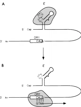
A distinguishing feature of DNA replication of hepadnavi-ruses is that the packaging of pgRNA and the initiation of first-strand DNA (minus-strand DNA) synthesis share a com-mon cis-acting sequence, a stem-loop structure (ε) that resides near the 59end of the pgRNA (9, 11, 12, 14, 22, 23, 26, 35, 37). Interaction of the viral polymerase (P) protein with εis re-quired for both encapsidation of pgRNA and initiation of strand DNA synthesis (24, 38). The synthesis of minus-strand DNA is a complex reaction which includes at least two steps (Fig. 1). In the first step, P protein acts as both the primer and the polymerase for the synthesis of a 4-nucleotide (nt) minus-strand DNA by copying the sequence within a phyloge-netically conserved bulge withinε(4, 33, 35–37, 39, 40). In the second step, the 4-nt minus-strand DNA switches templates from ε to an acceptor site which overlaps a 12-nt sequence termed DR1 near the 39 end of pgRNA, leading to the syn-thesis of mature minus-strand DNA (18, 22, 35, 37).
The importance of synthesizing a minus-strand DNA with proper sequence and length before switching templates is at least twofold. First, since only 4 nt withinεare identical to the sequence at the acceptor site, confining the template to these
sequences ensures complementarity between the minus-strand DNA and the acceptor site. This complementarity facilitates the efficiency of the template switch (18, 22, 34, 37). Second, cessation of DNA synthesis on εis necessary to prevent po-tential RNase H degradation of the 59end of the RNA tem-plate before it serves as a temtem-plate for the synthesis of the 39 end of minus-strand DNA. However, the mechanism which determines the precise boundary of the template withinεfor DNA synthesis before the template switch is not clear. The boundary is not determined by the primary sequence of the template. A variety of mutant sequences have served as tem-plates for minus-strand DNA prior to the template switch (18, 22, 34, 35, 37). Although some of those mutant sequences resulted in aberrant copying of the template region during synthesis of minus-strand DNA before the template switch, they rarely altered the 59boundary of the template (21, 22).
It is not straightforward to study the synthesis of the minus-strand DNA prior to the template switch in cell culture. Be-cause the 4-nt minus-strand DNA synthesized prior to the template switch has yet to be identified in cells supporting duck hepatitis B virus (DHBV) replication, it is not known whether this minus-strand DNA exists as a transient intermediate or a stable molecule during normal viral DNA replication. Infor-mation on synthesis of the minus-strand DNA before the tem-plate switch in cell culture has to be deduced from the analysis of the mature minus-strand DNA, an approach that is limited because only minus-strand DNAs that are capable of carrying out a productive template switch can be examined. We there-fore used an in vitro DNA synthesis system for DHBV (36) to study the synthesis of the minus-strand DNA before the tem-plate switch. This in vitro system has been informative in elu-cidating mechanisms regarding minus-strand DNA synthesis (24, 37, 38, 39). In this work, we studied the consequence of * Corresponding author. Mailing address: McArdle Laboratory for
Cancer Research, University of Wisconsin Medical School, 1400 Uni-versity Ave., Madison, WI 53706. Phone: (608) 262-1260. Fax: (608) 262-2824. E-mail: [email protected].
5345
on November 9, 2019 by guest
http://jvi.asm.org/
inserting nucleotides 59to the template region withinεon the synthesis of minus-strand DNA before the template switch in vitro and on the subsequent template switch process in cell culture. Our findings are consistent with a model in which the occlusion of the two 59-most nucleotides of the bulge by steric hindrance, from either RNA-RNA or RNA-protein interac-tions, defines the 59boundary of the template withinε. Also, these results are consistent with the notion that the proper length and/or sequence of the pretemplate switch minus-strand DNA can be important for an efficient template switch.
MATERIALS AND METHODS
DNA constructs.The European strain DHBV3 was used in all experiments. The nucleotide positions of DHBV3 are numbered from the EcoRI site as specified by Sprengel et al. (31). In this numbering system, transcription of the pgRNA initiates at nt 2529.
p364DRI is a plasmid in which wild-type DHBV sequences from nt 2530 to 3021 were inserted downstream of the T7 RNA polymerase promoter such that the RNA transcripts synthesized from this DNA will initiate at a position 1 nt upstream of the DHBV sequence. pG and pGAG are derivatives of p364DRI containing insertions of the sequence G and GAG between nt 2572 and 2573, respectively. Plasmids pAGC, pGAA, and pGCC contain DHBV sequences from nt 2527 to 3021, including insertions of trinucleotide sequence AGC, GAA, and GCC between nt 2572 and 2573, respectively, under the control of the T3 RNA polymerase promoter. The RNA transcripts synthesized from pAGC, pGAA, and pGCC contain 13 nt of non-DHBV sequences at their 59ends. Plasmids were sequenced to demonstrate the presence of the desired mutation and the absence of fortuitous mutations. Plasmids were linearized at the NsiI site (nt 2845) for the transcription of the RNAs that containε.
Plasmid pHJ55 contains the DHBV sequence from nt 1 to 2526 under the control of the SP6 RNA polymerase promoter in the vector pSP65. In addition, pHJ55 contains the 33-nt coding sequence for the 12CA5 epitope of the influ-enza virus hemagglutinin HA1 protein between DHBV nt 960 and 961, which is within the spacer region of the P gene (33). To synthesize mRNA for translation of P protein, pHJ55 was linearized at the FspI site which is within pSP65.
Plasmid pD1.5G contains 1.5 copies of wild-type DHBV genome (1.5-mer) and supports transcription of the authentic pgRNA upon transfection into LMH cells. Plasmids pG-ε, pGAA-ε, and pGCC-εare 1.5-mers, containing the respec-tive insertions inεon the pgRNA. Plasmid pGCC-DR1 (named 238 in reference 17) is a 1.5-mer in which the AA at nt 2532 to 2533 near the 39end of the pgRNA was replaced with CC. Plasmid pGCC-εDR1 is a 1.5-mer containing both the insertion of GCC inεand the base substitution of CC near the 39end of the pgRNA.
In vitro RNA transcription.RNAs used for in vitro translation, in vitro DNA synthesis reaction, and in vitro RNA structural analysis were synthesized by using a MegaScript Kit (Ambion) or a T7 RiboMax kit (Promega) as instructed by the manufacturer.
In vitro DNA synthesis reaction.The assay for the initiation of minus-strand DNA synthesis (or protein priming) was performed as described by Wang and Seeger (36). One picomole of pHJ55/FspI RNA was used for translation of P protein in a nuclease-treated rabbit reticulocyte lysate (Promega) in a final volume of 50ml by incubation of the mixture at 30°C for 60 min. The protein-priming reaction was then performed by mixing 10ml of the translation reaction mixture with an equal volume of a solution containing 100 mM Tris-HCl (pH 7.5), 30 mM NaCl, 20 mM MgCl2, 2.4 mM dGTP (including 10 mCi of
[a-32P]dGTP), and 4 to 5 pmol of anεRNA template. This mixture was
incu-bated at 30°C for 30 min, and then the reaction was stopped by the addition of 4 volumes of 23sodium dodecyl sulfate (SDS) loading buffer (28), which was then heated at 95°C for 5 min. One-fifth of the volume of each sample was electrophoresed through an SDS–10% polyacrylamide gel. The gel was fixed with isopropanol-acetic acid-water (25:10:60), dried, and exposed to a phosphor screen, which was analyzed on a PhosphorImager 445 SI (Molecular Dynamics). In reactions in which [a-32P]dGTP was replaced with [a-32P]dCTP at the same
concentration, additional dATP, dGTP, and dTTP were also provided at a final concentration of 14mM.
For the synthesis of longer minus-strand DNA which was analyzed by primer extension, the final concentration of deoxynucleoside triphosphates (dNTPs) was adjusted to 100mM, and the DNA synthesis reaction mixture was incubated at 30°C for 60 min.
Immunoprecipitation and primer extension analyses of minus-strand DNA synthesized in vitro.Following the synthesis of DNA in vitro, minus strands covalently linked to the P protein were immunoprecipitated from the rabbit reticulocyte lysate as described previously (2) and subsequently deproteinized by treatment with proteinase K. After DNA synthesis, 5ml of 20% SDS was added to 20ml of the reaction mixture. The mixture was heated at 95°C for 5 min to denature and solubilize proteins, followed by addition of an equal volume of H2O to dilute the SDS concentration to 2%. Next, 250ml of TNET buffer (2.5%
Triton X-100, 190 mM NaCl, 6 mM EDTA, 50 mM Tris-HCl [pH 7.4]) contain-ing 10 mg of preswollen and prewashed protein A-Sepharose CL-4B beads (Pharmacia) and 1ml of monoclonal antibody CA5 (Berkeley Antibody Com-pany), which recognizes the influenza virus hemagglutinin HA1 epitope, were added to the mixture. The mixture was mixed at 4°C for 60 min, pelleted by a 20-s microcentrifugation, and washed three times with 500ml of the washing buffer (0.1% Triton X-100, 0.02% SDS, 150 mM NaCl, 5 mM EDTA, 50 mM Tris-HCl [pH 7.4]). The pellet was suspended in 100ml of TE containing 1 mg of pro-teinase K per ml and 1% SDS and incubated at 37°C for 90 min with occasional mixing. Deproteinized minus-strand DNA was further purified by phenol extrac-tion and ethanol precipitaextrac-tion. The posiextrac-tion of the 59end of minus-strand DNA was determined by a primer extension analysis using an end-labeled oligonucle-otide corresponding to nt 2530 to 2549, using reaction conditions described by Loeb and Tian (18).
Enzymatic probing of RNA structures in vitro.The method for analysis of RNA structure was derived from several published procedures (13–15, 30). Four picomoles ofεRNA transcribed in vitro was placed in 20ml of renaturation buffer (70 mM HEPES [pH 7.4], 10 mM MgCl2, 270 mM KCl) and incubated at
65°C for 5 min, followed by incubation at room temperature for 10 min to allow formation of the secondary structure. The optimal amount of RNase and the time of digestion for eachεRNA were determined emperically. Each RNase digestion reaction mixture contained 5ml of renaturedεRNA, 100ml of RNase digestion buffer (10 mM Tris-HCl [pH 7.5], 10 mM MgCl2, 100 mM KCl), 10mg
of yeast tRNA, and various amounts of RNase T2(GIBCO-BRL) (0, 0.6, 6, and
60 U/ml) or RNase A (0, 0.45, 4.5, and 45 ng/ml). The reaction mixture was incubated on ice for 60 min. Alternatively, each reaction mixture contained either 60 U of RNase T2or 45 ng of RNase A per ml, and was incubated at room
temperature for various lengths of time (0, 5, 10, and 20 min). The cleaved RNAs were then purified by phenol extraction and ethanol precipitation. The positions of the RNase cleavages on the RNA were determined by a primer extension analysis with an end-labeled primer complementary to DHBV nt 2680 to 2700. Briefly, 5ml of solution containing 50 mM HEPES-KOH (pH 7.6), 100 mM KCl, 0.6 pmol of the primer, and the RNA from 1/10 of the cleavage reaction was heated at 90°C for 2 min and transferred to ice and then supplemented to the final volume of 8ml containing additional 50 mM Tris (pH 8.9), 8.25 mM MgCl2,
[image:2.612.92.264.67.292.2]8.25 mM dithiothreitol, 338 mM dNTPs, 15 U of avian myeloblastosis virus reverse transcriptase (Seikagaku American Inc.), and 8 U of RNasin (Promega). The primer extension reaction was then carried out at 45°C for 45 min and was stopped by addition of 4ml of a formamide sequencing loading dye (28). For generation of the sequence ladder, 1 pmol of undigested RNA and 1.2 pmol of
FIG. 1. Initiation of minus-strand DNA synthesis and subsequent template switch for DHBV. The pgRNA, indicated by a line, is the template for the synthesis of minus-strand DNA. The stem-loop structure (ε) on the pgRNA contains the template for the initiation and the synthesis of minus-strand DNA prior to the template switch. The UUAC sequence overlapping DR1 (represent-ed by a rectangle) near the 39end of pgRNA is the acceptor site of the template switch. (A) P protein (shaded area) binds toεand is the primer and polymerase for the synthesis of the first 4 nt of minus-strand DNA by copying the template 59-UUAC-39, which is within the bulge ofε. (B) The 4-nt minus-strand DNA attached to P protein switches templates fromεto the acceptor site and base pairs with the UUAC sequence for elongation of minus-strand DNA synthesis (arrow).
on November 9, 2019 by guest
http://jvi.asm.org/
the same end-labeled primer were annealed in a final volume of 16ml. Four microliters of the annealing mixture was added to four separate tubes containing 0.2ml of 2.5 mM ddATP, ddCTP, ddGTP, or ddTTP. The sequencing reaction mixture was then incubated at 45°C for 45 min. Samples were heated at 95°C for 5 min before loading onto 7.2 M urea–6% acrylamide gel. Analysis of autora-diographic images was performed with a Molecular Dynamics PhosphorImager 445 SI.
Transfection.Transfection of the chicken hepatoma cell line LMH was carried out as described previously (18). Routinely, 4 to 5mg of plasmid DNA was used in each transfection of a 60-mm-diameter plate which was approximately 50% confluent with LMH cells.
Encapsidation assay.Three days posttransfection, intracellular viral nucleo-capsids were harvested as described previously (7). Usually, viral nucleonucleo-capsids from 1/100 of the cell lysate were electrophoresed through a 0.8% agarose–13
Tris-acetate-EDTA gel. Each sample was run in duplicate for detection of nu-cleocapsids and encapsidated nucleic acids. For immunostaining, nunu-cleocapsids were transferred onto a nitrocellulose membrane by capillary action with TNE buffer (10 mM Tris [pH 7.5 to 8.0], 1 mM EDTA, 150 mM NaCl) overnight. The membrane was immunostained with a rabbit anti-DHBc (DHBV core antigen) antibody (a gift from Jesse Summers), followed by incubation with a donkey anti-rabbit immunoglobulin G antibody conjugated with35S (Amersham). For
detection of encapsidated nucleic acids, nucleocapsids were transferred to a nylon membrane (Hybond; Amersham). The nucleic acids were released from the capsids by wetting the membrane in denaturing buffer (0.2 N NaOH, 1.5 M NaCl) for 20 to 30 s, followed by neutralization in buffer (0.2 M Tris-HCl, 1.5 M NaCl) for 5 min. The nucleic acids were UV cross-linked to the membrane. The membrane was then hybridized with a32P-labeled, plus-strand-specific RNA
probe that would detect plus-strand sequences from nt 2845 to 3021 and 1 to 902. Quantification of autoradiographic images of both nucleocapsids and encapsi-dated nucleic acids was performed with a Molecular Dynamics PhosphorImager 445 SI.
Southern blotting, primer extension, and sequence analyses of viral DNAs. Viral DNAs were extracted from the cytoplasmic lysate that was analyzed for encapsidation as described by Staprans et al. (32). Usually 1/30 to 1/10 of purified viral DNAs was subjected to Southern blot analysis with a32P-labeled,
minus-strand-specific, genome-length RNA probe.
For detecting the 59end of minus-strand DNA elongated from DR1, the primer extension analysis was performed on viral DNA, with an end-labeled primer corresponding to nt 2425 to 2447, as described by Loeb and Tian (18).
For determining the 59-terminal sequences of minus-strand DNA, viral DNAs were amplified by a PCR procedure, followed by cloning and sequence analysis, as described in Loeb et al. (17).
RESULTS
The initial aim of this study was to determine why only 4 nt ofεwere copied into minus-strand DNA before the template switch. We proposed two possibilities: (i) only 4 nt of minus-strand DNA could be synthesized irrespective of the number of nucleotides within the bulge ofε, or (ii) the length of minus-strand DNA was determined by the number of nucleotides in the bulge ofε. To distinguish between these two possibilities, we introduced either single nucleotide or trinucleotide inser-tions between posiinser-tions 2572 and 2573 (Table 1) in the phylo-genetically conserved bulge and asked three questions: (i) How do insertions within the bulge affect protein priming and RNA encapsidation? (ii) Can minus-strand DNAs longer than 4 nt undergo a template switch? (iii) How do the insertions affect the structure ofεin solution?
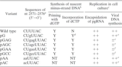
Trinucleotide insertions 5*of the UUAC in«extended the 5* boundary of the template in vitro. First we determined whether theεvariants would support the initiation of minus-strand DNA synthesis at the wild-type position. To address this point, we used two complementary experimental approaches, both of which involve DNA synthesis in vitro. The first ap-proach used an in vitro protein-priming assay in whichεRNA synthesized in vitro was added to P protein translated in a rabbit reticulocyte lysate (36, 37). DNA synthesis was initiated by adding only [a-32P]dGTP, the first nucleotide of minus-strand DNA. The ability of theεvariants to act as a template for protein priming was determined by detecting the covalent linkage of the radiolabel to the P protein in an SDS-polyacryl-amide gel electrophoresis (PAGE) analysis. The four different εvariants that each had 3 nt inserted (GAG, AGC, GAA, and
GCC) immediately 59 to UUAC supported protein priming (Fig. 2A, lanes 3 to 6) at a level comparable to that of the wild-typeε(Fig. 2A, lane 1). However, theεvariant with the 1-nt (G) insertion supported protein priming at a level less than that of the wild type (Fig. 2A, lane 2). In these experi-ments, the incorporation of [a-32P]dGTP into the P protein was dependent on the presence of both the εRNA template and P protein (Fig. 2A, lanes 7 and 8). These results suggested that protein priming was occurring at the normal position for theεvariants.
[image:3.612.316.556.81.214.2]To corroborate the finding that theεvariants supported the initiation of DNA synthesis at the wild-type position, we de-termined the site of initiation of minus-strand DNA synthesis by primer extension. During the DNA synthesis reaction in vitro, a fraction of minus-strand DNA that initiates at the normal site subsequently elongates through εto yield minus strands several hundred nucleotides in length (34, 36). By de-termining the 59terminus of each of these minus-strand DNAs, the position of initiation of protein priming can be inferred (34, 36). Primer extension analysis of minus-strand DNAs synthe-sized in vitro from wild-typeεyielded a major product of 47 nt, indicating a 59 end of minus-strand DNA at the cytosine at position 2576 and several minor products at positions 2581 through 2583 (Fig. 2B, lane 1), which have been seen previ-ously (34, 36). The primer extension product of minus-strand DNAs synthesized from pG RNA was 1 nt longer than that of the wild-type, owing to the 1-nt insertion inε, indicating a 59 end of minus-strand DNA at the wild-type position of initiation (position 2576) (Fig. 2B, lane 2). However, a more abundant 59 end was detected at the A residue at position 2581, located 39 of the bulge on the upper stem ofεon pG RNA (see Fig. 5B for nucleotide sequences and coordinates within ε). This site did not appear to be an initiation site, because pGεRNA did not direct incorporation of [a-32P]dTTP (or [a-32P]dCTP or [a-32P]dATP) as the first nucleotide of minus-strand DNA to a detectable level (data not shown). Therefore, this 59end may result from an aberrant template switch. Minus-strand DNAs synthesized by using εRNAs of pGAG, pAGC, pGAA, and pGCC yielded products 3 nt longer than that of the wild type, indicating their 59ends were at the wild-type position (Fig. 2B, lanes 3 to 6). For pGCC, additional 59ends of minus-strand
TABLE 1. Mutational analyses ofε
Variant nt 2571–2576Sequences ofa (59339)
Synthesis of nascent
minus-strand DNAb Replication in cellculturec
Priming with dGTP
Incorporation
of dCTP Encapsidationof pgRNA Viral DNA synthesis
Wild type CUUUAC Y N 11 11
pG CUgUUAC Y Yd 11 11e
pGAG CUgagUUAC Y Y 11 6e
pAGC CUagcUUAC Y Y 11 6e
pGAA CUgaaUUAC Y Y 11 6f
pGCC CUgccUUAC Y Y 11 6f
pAA aaUUAC NT NT 11 11e
pAC acUUAC NT NT 11 11e
aSequence of insertion or base substitution is shown in lowercase. bY, positive for incorporation; N, negative for incorporation; NT, not tested. c11, 50 to 100% of wild-type level;1, 10 to 50% of wild-type level;6, below 10% of wild-type level.
dIncorporation of dCTP at the fifth position of minus-strand DNA was de-termined by sequencing of the 59terminus of viral minus-strand DNA.
eThe mutant virus contained the indicated mutation withinεand wild-type acceptor site on the pgRNA.
fThe mutant virus contained identical 7-nt sequences at the template region withinεand the acceptor site on the pgRNA.
on November 9, 2019 by guest
http://jvi.asm.org/
DNA were detected at the sequence CUA (positions 2579 through 2581) and at the A residue at position 2583 (Fig. 2B, lane 6). These minus-strand DNAs with 59ends at nucleotides other than C residue likely represent the elongated DNA after
an aberrant template switch, because neither [a-32P]dTTP, [a-32P]dCTP, nor [a-32P]dATP supported the protein-priming reaction (data not shown). In this analysis, the detection of minus-strand DNA dependent on the presence ofεRNA (Fig. 2B, lane 7). The results of the protein-priming and primer extension assays indicated that the εvariants were functional for protein priming at the normal position although additional 59ends of minus-strand DNA could be detected for some of the RNAs.
Next, we determined whether the inserted sequences inε were copied into minus-strand DNA. Because the wild-type template, UUAC, does not contain a guanosine and the ε variants had at least one guanosine inserted at various posi-tions from the site of initiation (i.e., 5th position for pG and pGAG; 6th position for pAGC; and 7th position for pGAA, pGCC, and pGAG), incorporation of dCTP into minus-strand DNA in the protein-priming reaction would indicate that the 59 end of the template had been extended.εRNA transcribed in vitro was mixed with P protein translated in a rabbit reticulo-cyte lysate. To permit the synthesis of minus-strand DNA, 1.2 mM [a-32P]dCTP and 14 mM dATP, dGTP, and dTTP were added. Minus-strand DNA covalently linked to P protein was then detected in an SDS-PAGE analysis.εRNAs of pGAG, pAGC, pGAA, and pGCC all supported incorporation of [a-32P]dCTP into minus-strand DNA (Fig. 2C, lanes 3 to 6), whereas incorporation of [a-32P]dCTP could not be detected for either the wild-typeε(Fig. 2C, lane 1) or theεRNA of pG (Fig. 2C, lane 2). We interpret this result to mean that the 3-nt insertions 59 of the UUAC sequence in ε extended the 59 boundary of the template to at least the seventh nucleotide from the site of minus-strand DNA initiation. However, the 1-nt G insertion at the same site did not affect the 59boundary of the template, at least in vitro (see below).
A single nucleotide insertion 5*of UUAC in«on the pgRNA could be copied into the fifth position of minus-strand DNA synthesized in cell culture. Next we examined the ε variant with the single G insertion in an assay that involved DNA replication in cell culture. We reasoned that if the inserted guanosine was copied into minus-strand DNA and the result-ant minus-strand DNA was competent undergo a template switch and elongation from DR1, we would detect a cytosine, instead of a wild-type thymidine, at the fifth position of minus-strand DNA. We therefore constructed pG-ε, a plasmid com-petent to express a pgRNA with a G residue inserted between nt 2572 and 2573 withinεwhen transfected into LMH cells.
[image:4.612.101.257.71.524.2]First we determined whether pG-εvirus was competent for RNA encapsidation and viral DNA synthesis in cells. Three days posttransfection, cytoplasmic viral nucleocapsids were harvested and subjected to a gel analysis of capsids to deter-mine the extent of RNA encapsidation (7). Capsids synthe-sized by the wild-type and pG-εviruses were readily detected by immunostaining with an anti-DHBc antibody (Fig. 3A). Plus-strand nucleic acid was detected within the capsids for both the wild-type and pG-εviruses, indicating that RNA en-capsidation was readily occurring (Fig. 3B). Upon normaliza-tion of the levels of nucleocapsids (Fig. 3A, lanes 1 and 2), the amount of encapsidated plus-strand nucleic acid for the pG-ε virus (Fig. 3B, lane 2) was about 50% of the level of the wild-type virus (Fig. 3B, lane 1). Next, viral DNAs isolated from these nucleocapsids were analyzed by Southern blotting. Upon normalization of the level of RNA encapsidated, the level of total viral DNA synthesis for pG-εvirus was approxi-mately 80% of that of the wild-type virus (Fig. 3C; compare lane 2 with lane 1). The 59 terminus of minus-strand DNA synthesized in pG-εvirus was at the wild-type position as de-termined by primer extension (data not shown). In summary, FIG. 2. In vitro synthesis of minus-strand DNA forεinsertion mutants (see
Table 1 for indicated mutations). Following translation of P protein in a rabbit reticulocyte lysate,εRNA was added to initiate synthesis of minus-strand DNA. Reactions in lanes 1 to 6 were provided with RNAs from wild-type (WT)ε, pG, pGAG, pAGC, pGAA, and pGCC, respectively.εRNA and P protein were absent in the reactions in lanes 7 and 8, respectively. (A) The DNA synthesis reaction mixture was incubated in the presence of [a-32P]dGTP (1.2mM) at 30°C for 30 min. The initiation product of P protein that forms a complex with dGMP was analyzed by SDS-PAGE (10% gel). The positions and sizes of molecular weight markers are indicated on the right. (B) The DNA synthesis reaction mixture was incubated in the presence of dNTPs (100mM) at 30°C for 60 min. Minus-strand DNA products were purified by immunoprecipitation and protein-ase K treatment. The 59ends of minus-strand DNAs were determined by primer extension analysis. The wild-type site of initiation is indicated by the arrow, and the additional 59ends are marked by asterisks. (C) Assay for incorporation of dCMP into minus-strand DNA. [a-32P]dCTP (1.2mM) and dATP, dGTP, and dTTP (14mM each) were provided in the DNA synthesis reaction at 30°C for 30 min. The complex of minus-strand DNA and P protein was analyzed by SDS-PAGE.
on November 9, 2019 by guest
http://jvi.asm.org/
these results showed that pG-εvirus underwent DNA replica-tion at a level similar to that of the wild-type virus in cell culture.
Next, we determined whether the G inserted withinεin the pG-εvirus was used as a template for the synthesis of the fifth nucleotide of minus-strand DNA by determining the sequence of the fifth position of individual minus-strand DNA molecules from the pG-ε virus. Because the 59-terminal sequences of minus-strand DNA serve as the template for the plus-strand DNA during synthesis of the relaxed circular form of the viral DNA genome (17), we could deduce the 59-terminal sequences of minus-strand DNA by cloning and sequencing the comple-mentary plus-strand DNA. We used a PCR protocol to am-plify, clone, and sequence individual plus strands of DNA of the pG-εvirus. Of 11 viral DNA clones that were sequenced, we found four examples of a C residue at the fifth position in minus-strand DNA. The other seven examples contained the wild-type sequence (T) at the fifth position of minus-strand DNA (Fig. 3D). The sequence of minus-strand DNA from the pG-ε virus demonstrated that the G residue inserted 59 of UUAC inεcould be used as a template for synthesis of minus-strand DNA prior to the template switch. In addition, the level of DNA replication for the pG-εvirus was comparable to the level for the wild-type virus in LMH cells, indicating that the template switch of mutant 5-nt minus-strand DNA fromεto the acceptor site was efficient and that the elongation of minus-strand DNA synthesis from the acceptor site occurred readily despite a 39-terminal mismatch between the minus-strand DNA and the RNA template. However, the reason for the
discordance between results from this cell culture experiment and the in vitro DNA synthesis reaction (see above) is pres-ently not clear.
Overall, the results indicated that nucleotides inserted im-mediately 59 of the UUAC in the phylogenetically conserved bulge were used as a template for the synthesis of a minus-strand DNA longer than 4 nt before the template switch. These results consistent with the notion that the two 59-most nucle-otides of the bulge (59-CU-39), which are not used as a tem-plate by the wild-type virus, were physically occluded from the polymerase. To test whether these nucleotides were part of a sequence specific RNA-RNA or RNA-protein interaction that prevented their use as templates, we changed the CU to AA and AC and determined the abilities of theεvariants to sup-port minus-strand DNA synthesis in cell culture. Both variant viruses were found to synthesize minus-strand DNA as well as wild-type virus (Table 1). In addition, sequence analysis of minus-strand DNA from mutant virus showed that the base substitution of AC did not extend the 59 boundary of the template within ε(data not shown). In summary, the results suggested that the primary sequence CU was not required for limiting synthesis of minus-strand DNA to 4 nt before the template switch.
Mutant viruses with trinucleotide insertion 5* of UUAC were inhibited for efficient synthesis of minus-strand DNA in cell culture.We had shown that insertions of 3 nt immediately 59of the UUAC inεextended the 59boundary of the template during the synthesis of minus-strand DNA in vitro. Next, we determined whether these longer minus-strand DNAs, which are predicted to be 7 nt, could switch templates to DR1 and elongate in cell culture.
Sequence complementarity between the acceptor site and the minus-strand DNA plays a role in the template switch and elongation of minus-strand DNA from DR1 (18, 22, 34, 37). To eliminate the influence of mismatches between the minus-strand DNA and the acceptor site during the template switch, we analyzed mutant viruses with identical sequences at the template and acceptor sites (Fig. 4A). Plasmids pGAA-εand pGCC-εexpress pgRNAs with the 3-nt insertions, GAA and GCC, respectively, between nt 2572 and 2573 inε. The pgRNA expressed from plasmid pGAA-εhas 7 nt of identity between the donor and acceptor templates. Plasmid pGCC-DR1 ex-presses a pgRNA containing AA-to-CC substitution at posi-tions 2532 and 2533 near the 39end of pgRNA, resulting in sequence 59-GccUUAC-39at the acceptor site. This virus sup-ports minus-strand DNA synthesis in cell culture (17). Plasmid pGCC-εDR1 expresses a pgRNA that harbors both the 3-nt insertion of GCC withinεand the base substitutions of CC at the acceptor site, thus creating a 7-nt identity between the donor and acceptor sites on the template. All plasmids support transcription of an otherwise wild-type pgRNA upon transfec-tion into LMH cells.
First, we determined whether the insertions in ε affected RNA encapsidation. Three days posttransfection, cytoplasmic viral nucleocapsids were harvested and analyzed by gel elec-trophoresis. Upon normalizing the level of nucleocapsids de-tected by immunostaining (Fig. 4B, lanes 1 to 5), the amount of encapsidated plus-strand nucleic acid for the mutant viruses (Fig. 4C, lanes 2 to 5) was found to be at a level between 50 and 100% of the level of the wild-type virus (Fig. 4C, lane 1). These results indicated that the pgRNAs with the 3-nt insertion withinεwere competent for encapsidation.
[image:5.612.61.296.68.272.2]Next, we determined whether the template switch had oc-curred by detecting the presence of minus-strand DNA elon-gated from the acceptor site at DR1. Viral DNAs isolated from cytoplasmic capsids containing equal amounts of encapsidated FIG. 3. Synthesis of minus-strand DNA for mutant virus pG-εin LMH cells.
Three days posttransfection in LMH cells, cytoplasmic viral nucleocapsids were harvested. (A and B) Gel analysis of capsids to measure the level of encapsidated plus-strand viral nucleic acid per nucleocapsid. (A) Viral nucleocapsids detected by immunostaining with anti-DHBc antibody. (B) Encapsidated viral nucleic acids detected by hybridization with a plus-strand-specific RNA probe. (C) Southern blot analysis of viral DNA isolated from cytoplasmic nucleocapsids with a minus-strand-specific probe. RC, relaxed circular form of viral DNA; DL, duplex linear form; SS, single-stranded form. Lane 1, wild-type (WT) virus; lane 2, mutant virus pG-ε. (D) 59-terminal sequences of viral minus-strand DNA from mutant virus pG-ε. Top line represents the pgRNA transcribed from pG-ε, showing sequences at the template region within εand at the acceptor site overlapping DR1. The inserted guanosine withinεis shown in lowercase. 59 -terminal sequences of minus-strand DNAs obtained by sequence analysis of cloned viral DNAs are shown below the pgRNA. Of a total of 11 clones that were sequenced, the number of clones harboring the mutant or wild-type sequence is
indicated on the left.
on November 9, 2019 by guest
http://jvi.asm.org/
RNA, as determined by the gel assay of capsids (see above), were subjected to primer extension analysis. The 59 end of minus-strand DNA synthesized by the virus pGCC-DR1 was detected at the wild-type position within DR1 at a level similar to that for the wild type (Fig. 4D; compare lane 5 with lane 1). The level of minus-strand DNA elongated from DR1 in virus pGCC-εranged from 1 to 3% of the wild-type level (Fig. 4D, lane 3). More importantly, the virus pGCC-εDR1, in which a 7-nt identity between the template inεand the acceptor site was restored, did not support the synthesis of elongated minus-strand DNA from DR1 at a level any better than virus pGCC-ε (Fig. 4D, lane 4). The level of minus-strand DNA with its 59 end at DR1 for the pGAA-εvirus ranged from 5 to 20% of the level of the wild-type virus (Fig. 4D, lane 2). Corroborating these primer extension results, a reduced level of DNA was detected for the pGAA-εand pGCC-εDR1 mutant viruses in a Southern blot analysis (data not shown). In addition, the Southern blot results were consistent with the notion that the
minus-strand DNAs of the mutant viruses had not undergone template switches to other locations on the template (data not shown). In summary, the two mutant viruses (pGAA-ε and pGCC-εDR1) that were predicted to synthesize a 7-nt minus-strand DNA fromε(on the basis of the in vitro analysis) and had a 7-nt identity between the donor and acceptor sites had a reduced level of minus-strand DNA elongated from the accep-tor site.
Similarly, the mutant viruses pGAG-εand pAGC-ε, which contained the insertion of GAG or AGC withinεand a wild-type acceptor site, supported normal levels of RNA encapsi-dation but elongated minus-strand DNA from DR1 poorly (Table 1).
Structural analysis indicated that insertions 5*of UUAC in
«did not result in a larger bulge.Next we determined whether the nucleotides inserted in the bulge region ofεresulted in a larger single-stranded bulge. To determine this, we performed structural analyses by enzymatically probing the variant ε RNAs. The εRNAs used in the structural analysis were the same as those used in the DNA synthesis reactions performed in vitro.εRNAs were incubated at room temperature to allow the formation of secondary structure and then subjected to digestion with RNase T2, which cleaves single-stranded regions of RNA independent of the sequence. The positions of the RNase T2cleavages were then determined by primer extension analysis of the digestedεRNAs.
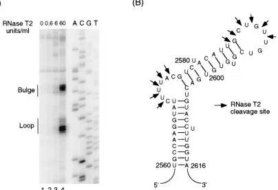
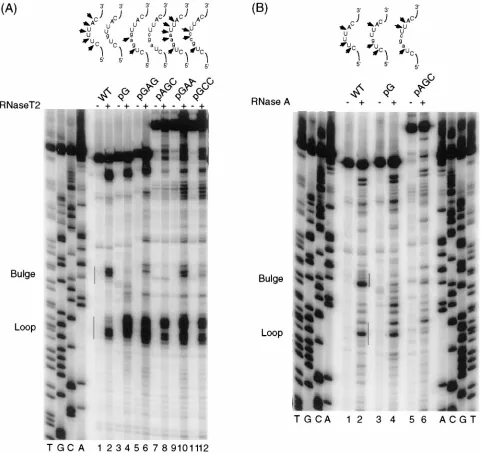
For the wild-typeεRNA, two prominent regions of cleavage were detected after RNase T2digestion (Fig. 5A). One region, from nt 2571 through 2575 (CUUUA), represents the bulge. The second region, from nt 2585 through 2590 (UGCUGU), represents the loop (Fig. 5A, lane 4). The positions of the cleavages within both the bulge and loop regions determined did not coincide exactly with those predicted by the phyloge-netic analysis (12) (Fig. 5B; also see Discussion). Next, we analyzed the mutant ε RNAs with RNase T2. The cleavage positions for the mutant εRNAs were different from those determined for the wild-typeε(Fig. 5A; Fig. 6A, lane 2). For pGAG RNA, only the U at position 2572 and the A within the inserted sequence were cleaved by RNase T2(Fig. 6A, lane 6). For the pGAA RNA, both the AA within the insertion and the nucleotides surrounding the insertion at positions 2572, 2573, and 2574 were cleaved (Fig. 6A, lane 10). For the pGCC RNA, cleavages were detected at the U at position 2572 and at the CC within the inserted sequence (Fig. 6A, lane 12). However, for both pG and pAGC RNAs, no prominent cleavages by RNase T2 were detected from nt 2571 through 2575, which correspond to the bulge region in the wild-type ε (Fig. 6A; compare lanes 4 and 8 to lane 2). Additional cleavages were detected for all of the ε variants that were not seen in the wild-type RNA, suggesting that the inserted sequences had induced changes at distal sites in the structure. Several posi-tions within the upper stem became sensitive to RNase T2 cleavages, such as the U at position 2578 in the lower part of the upper stem (Fig. 6A; compare lanes 4, 6, 10, and 12 to lane 2) and the uppermost base pair in the upper stem adjacent to the loop, suggesting an enlarged loop region (Fig. 6A; compare lanes 6 and 10 to lane 2).
To determine whether the absence of cleavage within the template region of the pG and pAGC RNAs was peculiar to the RNase T2 digestion and not reflective of the single-stranded nature of the RNA, the pG and pAGC RNAs were also subjected to digestion by RNase A, a single-strand-specific RNase that preferentially cleaves 39to U and C residues. For the wild-typeε, RNase A cleavages were detected 39to the CU at positions 2571 to 2572 and the U at position 2574 (Fig. 6B, lane 2). The wild-type cleavage sites were also detected on the FIG. 4. Synthesis of minus-strand DNA for mutant viruses with 3-nt
inser-tions withinεin LMH cells. (A) Schematic representation of the pgRNA of wild-type (WT) and mutant viruses. Nucleotide sequence surrounding the donor and the acceptor sites is shown. Sequence of base substitutions or insertions are in lowercase. (B and C) Gel analysis of capsids to measure the level of encap-sidated plus-strand viral nucleic acid per nucleocapsid. (B) Nucleocapsids de-tected by immunostaining with anti-DHBc antibody. (C) Encapsidated nucleic acid detected by hybridization with plus-strand-specific probe. (D) Primer ex-tension analysis to determine the 59ends of minus-strand DNAs. Viral DNAs isolated from cytoplasmic capsids containing equivalent amounts of encapsidated plus-strand nucleic acids, as determined in panel C, were used in each primer extension reaction. The arrow indicates the position of 59end of minus-strand DNA.
on November 9, 2019 by guest
http://jvi.asm.org/
pG RNA, indicating a single-stranded bulge region (Fig. 6B; compare lane 4 to lane 2). However, for pAGC RNA, only nucleotides surrounding the insertion, such as the U at position 2572 and the UU at positions 2573 and 2574, were cleaved by RNase A (Fig. 6B, lane 6).
In summary, nucleotides inserted between positions 2572 and 2573, which were used as a template for the synthesis of minus-strand DNA, did not predictably increase the size of the single-stranded bulge. Instead, the insertions altered the size and position of the bulge and partially disrupted the upper stem, thus enlarging the loop on protein-free RNA in solution.
DISCUSSION
For DHBV, we have found that insertions within the bulge region of ε, 59 of the UUAC sequence, can be copied into DNA during the initiation of minus-strand DNA synthesis. Although these inserted nucleotides were used as a template for DNA synthesis, these insertions did not appear to corre-spondingly increase the size of the single-stranded bulge on the εRNA. We have also shown that the two 59-most nucleotides of the bulge could be changed without decreasing the level of minus-strand DNA synthesized in cell culture. In addition, we have found that viruses with insertions within the template region ofε, which were competent for RNA encapsidation and the priming of minus-strand DNA synthesis, were unable to synthesize normal levels of minus-strand DNA elongated from DR1.
A model for defining the 5*boundary of the RNA template in
« for minus-strand DNA synthesis prior to the template switch. Our finding that 1 or 3 nt inserted 59 to the UUAC
within DHBV εwere copied into minus-strand DNA is con-sistent with a model in which the 59 boundary of the RNA template within ε is set because the sequence beyond the UUAC is occluded from the active site of the polymerase, as suggested by Rieger and Nassal (27). Such an occlusion could be the result of an RNA-RNA or RNA-protein interaction. This putative interaction does not require the specific se-quences of the 2 nt beyond the UUAC in the bulge, because when the sequence of the two 59-most nucleotides of the bulge was changed, the wild-type 59boundary of the RNA template inεwas retained, resulting in wild-type 59-terminal sequences of minus-strand DNA in the mutant virus (data not shown).
An example of physical occlusion playing a role in defining the boundary of an RNA template for DNA synthesis can be found with telomerase, a ribonucleoprotein that contains an RNA template and a reverse transcriptase, which is responsi-ble for the synthesis and maintenance of the telomeres at chromosome ends (for a review, see reference 6). The scheme of telomere synthesis by telomerase involves synthesis of a short DNA product followed by a template switch. The RNA template region for ciliate telomerase is within a highly or-dered RNA structure. A conserved 6-nt motif, located 2 nt upstream of the template region, sets the 59 boundary of the RNA template. This motif is thought to engage in either RNA-protein or RNA-RNA interaction that physically prevents the polymerization of DNA beyond the 59boundary of the tem-plate (3, 16).
[image:7.612.114.518.73.349.2]Alternative conformation of the stem-loop structure does not prevent productive interactions with P protein.The pres-ence of the bulge region, but not its specific primary sequpres-ence, FIG. 5. Structural analysis of wild-typeεRNA by enzymatic probing with RNase T2. (A) One picomole of renatured RNA was incubated with RNase T2on ice for 30 min. Positions of cleavages were determined by primer extension analysis with an end-labeled primer complementary to the 39end of the RNA. Lanes 1 to 4, reactions with increasing concentrations of RNase T2(0, 0.6, 6, and 60 U/ml). The sequence ladder was generated with the same primer and undigested wild-typeε RNA as the template. The bulge and the loop regions are marked on the left. (B) Diagram of the stem-loop structure ofεpredicted by phylogenetic studies. Arrows indicate cleavage sites by RNase T2mapped in panel A.
on November 9, 2019 by guest
http://jvi.asm.org/
is necessary for binding of P protein toε(24). However, the correlation between the size of the bulge and the function ofε has not been examined. Our determination of the mutant RNA structures by enzymatic probing indicates that insertions within the phylogenetically conserved bulge region might alter the size and the position of the bulge withinεand alter the struc-ture of the upper stem and the loop regions. Nonetheless, these ε variants with altered structures were competent to interact with P protein for both RNA encapsidation and initi-ation of minus-strand DNA synthesis. These results suggest
[image:8.612.67.548.69.522.2]that a bulge region of exactly 6 nt is not an absolute prereq-uisite for the initial recognition ofεby the P protein. However, localized conformational changes of RNA structure induced by protein binding have been reported for human immunodefi-ciency virus TAR RNA with Tat protein (1), human immuno-deficiency virus Rev-responsive element RNA with Rev pro-tein (25), and adenovirus VA RNA with propro-tein kinase DAI (8). It is possible that upon binding of the P protein, the mutant εRNAs undergo a conformational change such that the tem-plate region becomes single stranded for DNA synthesis. To FIG. 6. Enzymatic probing analyses ofεvariant RNAs. One picomole of renatured RNA was subjected to RNase digestion. Positions of cleavages were determined by primer extension analysis with an end-labeled primer which anneals to a position near the 39end of the RNA. (A) RNase T2digestion. Cleavage reaction mixtures were incubated on ice for 30 min. Lanes 1 and 2, wild-type (WT)ε; lanes 3 and 4, pG; lanes 5 and 6, pGAG; lanes 7 and 8, pAGC; lanes 9 and 10, pGAA; lanes 11 and 12, pGCC. Lanes 1, 3, 5, 7, 9, and 11, control reactions with no RNase T2; lanes 2, 4, 6, 8, 10, and 12, reactions with 60 U of RNase T2per ml. (B) RNase A digestion. Reaction mixtures were incubated at room temperature for 20 min. Lanes 1 and 2, WTε; lanes 3 and 4, pG; lanes 5 and 6, pAGC. Lanes 1, 3, and 5, control reactions with no RNase A; lanes 2, 4, and 6, reactions with 45 ng of RNase A per ml. Sequence ladders were generated with the same primer, using undigested pGAG RNA as the template. The bulge and the loop regions in the wild-typeεare indicated by bars. For eachεRNA, the cleavage sites of RNase T2or RNase A detected in the region spanning from nt 2571 through 2576 are indicated by arrows in a diagram above the gel. WT bulge sequences are shown in uppercase; inserted sequences in theεvariants are shown in lowercase.
on November 9, 2019 by guest
http://jvi.asm.org/
address this possibility, determination of theεstructure bound by the P protein would be informative.
The synthesis of a longer minus-strand DNA before the template switch results in decreased synthesis of mature mi-nus-strand DNA. Although the mutant viruses pGAA-ε and pGCC-εDR1 were competent for packaging of pgRNA in LMH cells, they failed to efficiently synthesize minus-strand DNA after the template switch by two criteria: the low level of minus-strand DNA elongated from DR1 as detected by the primer extension analysis, and the low level of viral DNA detected in a Southern blot analysis (data not shown). Because these mutants were competent for the initiation of minus-strand DNA synthesis, we conclude that these mutant viruses are defective for a process after the initiation of minus-strand DNA synthesis and before or during the elongation of DNA from DR1.
A deficiency in carrying out the template switch would ac-count for the reduction in the level of minus-strand DNA elongated from DR1 for pGAA-ε and pGCC-εDR1 viruses. The longer minus-strand DNA copied fromεfor the mutant viruses, which would be equal to or longer than 7 nt, may less readily dissociate from its template within ε than does the wild-type 4-nt minus-strand DNA because of a greater number of base pairs. This reduced propensity to dissociate from its template would mean a less efficient template switch for the mutant viruses. A greater reduction in the level of minus-strand DNA for the pGCC-εDR1 virus than for the pGAA-ε virus is consistent with this notion, considering that the minus-strand DNA copied fromεfor the pGCC-εDR1 virus would have a higher GC content, and therefore more hydrogen bonds with the RNA withinε, than the equivalent minus-strand DNA of pGAA-εvirus. Alternatively, if the minus-strand DNA cop-ied fromεfor the pGAA-εand the pGCC-εDR1 viruses was longer than 7 nt, then this greater than 7-nt minus-strand DNA would contain mismatches at its 39terminus with the acceptor site, leading to a reduction in the amount of minus-strand DNA elongated from the acceptor site. However, we can rule out the possibility that minus-strand DNA initiated withinεfor the mutant viruses has efficiently extended fromεto the 59end of the pgRNA, which could be either a cause for or a conse-quence of an inefficient template switch. Minus-strand DNA elongated fromεto the 59end of the pgRNA for the pGAA-ε and the pGCC-εDR1 viruses would yield a DNA molecule 50 nt in length, which was not detected at a significant level in a primer extension analysis (data not shown). In addition, the apparent molecular weights of the complex of minus-strand DNA and P protein synthesized in vitro for theεvariants and the wild type were very similar as determined in an SDS-PAGE analysis (Fig. 2 and data not shown), suggesting that minus-strand DNA copied from theεvariants is not much longer than 7 nt, if it is longer at all.
The template switch during the synthesis of minus-strand DNA is a highly specific process, and the sequence comple-mentarity of 4 nt between the nascent minus-strand DNA and the acceptor site is not sufficient to account for this specificity. In theory, a longer nascent minus-strand DNA could increase the specificity and thus facilitate the template switch, but DHBV does not use this as part of the mechanism. Our results indicate that before the template switch, it is possible to syn-thesize a minus-strand DNA that is longer than 4 nt. However, these longer minus-strand DNAs appear to be defective for the template switch, arguing that synthesizing a longer minus-strand DNA prior to the template switch is not a suitable means to ensure specificity of the template switch. The speci-ficity of the template switch remains an enigma that awaits further study.
ACKNOWLEDGMENTS
We are grateful to Paul Ahlquist, Jeff Ross, and Bill Sugden for many helpful suggestions and advice on this work and the manuscript. We thank Louis Mansky, Mike Havert, and Karlyn Mueller-Hill for critical reading of the manuscript and Ilse Riegel for editorial assis-tance.
This work was supported by NIH grants GM50263 and CA07175. D.D.L. is the recipient of American Cancer Society Junior Faculty Research Award JFRA-651.
REFERENCES
1. Aboul, E. F., J. Karn, and G. Varani. 1995. The structure of the human immunodeficiency virus type-1 TAR RNA reveals principles of RNA recog-nition by Tat protein. J. Mol. Biol. 253:313–332.
2. Anderson, D. J., and G. Blobel. 1979. Immunoprecipitation of proteins from cell-free translations. Methods Enzymol. 96:111–120.
3. Autexier, C., and C. W. Greider. 1995. Boundary elements of the
Tetrahy-mena telomerase RNA template and alignment domains. Genes Dev.
9:2227–2239.
4. Bartenschlager, R., and H. Schaller. 1988. The amino terminal domain of the hepadnaviral P gene encodes the terminal protein (genome-linked pro-tein) believed to prime reverse transcription. EMBO J. 7:4185–4192. 5. Beasley, R. P. 1988. Hepatitis B virus, the major etiology of hepatocellular
carcinoma. Cancer 61:1942–1956.
6. Blackburn, E. H. 1993. Telomerase, p. 557–576. In R. F. Gesteland, and J. F. Atkins (ed.), The RNA world. Cold Spring Harbor Laboratory Press, Cold Spring Harbor, N.Y.
7. Calvert, J., and J. Summers. 1994. Two regions of an avian hepadnavirus RNA pregenome are required in cis for encapsidation. J. Virol. 68:2084– 2090.
8. Clarke, P. A., and M. B. Mathews. 1995. Interactions between the double-stranded RNA binding motif and RNA: definition of the binding site for the interferon-induced protein kinase DAI (PKR) on adenovirus VA RNA. RNA 1:7–20.
9. Fallows, D. A., and S. P. Goff. 1995. Mutations in theεsequences of human hepatitis B virus affect both RNA encapsidation and reverse transcription. J. Virol. 69:3067–3073.
10. Ganem, D., and H. E. Varmus. 1987. The molecular biology of the hepatitis B viruses. Annu. Rev. Biochem. 56:651–693.
11. Hirsch, R. C., D. D. Loeb, J. R. Pollack, and D. Ganem. 1991. cis-acting sequences required for encapsidation of duck hepatitis B virus pregenomic RNA. J. Virol. 65:3309–3316.
12. Junker-Niepmann, M., R. Bartenschlager, and H. Schaller. 1990. A short
cis-acting sequence is required for hepatitis B virus pregenome
encapsida-tion and sufficient for packaging of foreign RNA. EMBO J. 9:3389–3396. 13. Knapp, G. 1989. Enzymatic approaches to probing of RNA secondary and
tertiary structure. Methods Enzymol. 180:192–212.
14. Knaus, T., and M. Nassal. 1993. The encapsidation signal on the hepatitis B virus RNA pregenome forms a stem-loop structure that is critical for its function. Nucleic Acids Res. 21:3967–3975.
15. Kroi, A., and P. Carbon. 1989. A guide for probing native small nuclear RNA and ribonucleoprotein structures. Methods Enzymol. 180:212–227. 16. Lingner, J., L. L. Hendrick, and T. R. Cech. 1994. Telomerase RNAs of
different ciliates have a common secondary structure and a permuted tem-plate. Genes Dev. 8:1984–1998.
17. Loeb, D. D., K. J. Gulya, and R. Tian. 1997. Sequence identity of the terminal redundancies on the minus-strand DNA template are necessary but not sufficient for the template switch during hepadnaviral plus-strand DNA synthesis. J. Virol. 71:152–160.
18. Loeb, D. D., and R. Tian. 1995. Transfer of the minus strand of DNA during hepadnavirus replication is not invariable but prefers a specific location. J. Virol. 69:6886–6891.
19. Loeb, D. D., and D. Ganem. 1992. The reverse transcription pathway of the hepatitis B viruses, p. 329–355. In A. M. Skalka and S. P. Goff (ed.), Reverse transcriptase. Cold Spring Harbor Laboratory Press, Cold Spring Harbor, N.Y.
20. Loeb, D. D., R. C. Hirsch, and D. Ganem. 1991. Sequence-independent RNA cleavages generate the primers for plus strand DNA synthesis in hepatitis B viruses: implications for other reverse transcribing elements. EMBO J. 10: 3533–3540.
21. Loeb, D. D., et al. Unpublished results.
22. Nassal, M., and A. Rieger. 1996. A bulged region of the hepatitis B virus RNA encapsidation signal contains the replication origin for discontinuous first-strand DNA synthesis. J. Virol. 70:2764–2773.
23. Pollack, J. R., and D. Ganem. 1993. An RNA stem-loop structure directs hepatitis B virus genomic RNA encapsidation. J. Virol. 67:3254–3263. 24. Pollack, J. R., and D. Ganem. 1994. Site-specific RNA binding by a hepatitis
B virus reverse transcriptase initiates two distinct reactions: RNA packaging and DNA synthesis. J. Virol. 68:5579–5587.
25. Renwick, S. B., A. D. Critchley, C. J. Adams, S. M. Kelly, N. C. Price, and
on November 9, 2019 by guest
http://jvi.asm.org/
P. G. Stockley.1995. Probing the details of the HIV-1 Rev-Rev-responsive element interaction: effects of modified nucleotides on protein affinity and conformational changes during complex formation. Biochem. J. 308(Pt.2): 447–453.
26. Rieger, A., and M. Nassal. 1996. Specific hepatitis B virus minus-strand DNA synthesis requires only the 59encapsidation signal and the 39-proximal direct repeat DR1. J. Virol. 70:585–589.
27. Rieger, A., and M. Nassal. 1995. Distinct requirements for primary sequence in the 59- and 39-part of a bulge in the hepatitis B virus RNA encapsidation signal revealed by a combined in vivo selection/in vitro amplification system. Nucleic Acids Res. 23:3909–3915.
28. Sambrook, J., E. F. Fritsch, and T. Maniatis. 1989. Molecular cloning: a laboratory manual, 2nd ed. Cold Spring Harbor Laboratory Press, Cold Spring Harbor, N.Y.
29. Seeger, C., J. Summers, and W. S. Mason. 1991. Viral DNA synthesis. Curr. Top. Microbiol. Immunol. 168:41–60.
30. Skinner, M. A., V. R. Racaniello, G. Dunn, J. Cooper, P. D. Minor, and J. W. Almond.1989. New model for the secondary structure of the 59non-coding RNA of poliovirus is supported by biochemical and genetic data that also show that RNA secondary structure is important in neurovirulence. J. Mol. Biol. 207:379–392.
31. Sprengel, R., C. Kuhn, H. Will, and H. Schaller. 1985. Comparative se-quence analysis of duck and human hepatitis B virus genomes. J. Med. Virol. 15:323–333.
32. Staprans, S., D. D. Loeb, and D. Ganem. 1991. Mutations affecting hepad-navirus plus-strand DNA synthesis dissociate primer cleavage from translo-cation and reveal the origin of linear viral DNA. J. Virol. 65:1255–1262. 33. Tavis, J. E., and D. Ganem. 1993. Expression of functional hepatitis B virus
polymerase in yeast reveals it to be the sole viral protein required for correct initiation of reverse transcription. Proc. Natl. Acad. Sci. USA 90:4107–4011. 34. Tavis, J. E., and D. Ganem. 1995. RNA sequences controlling the initiation and transfer of duck hepatitis B virus minus-strand DNA. J. Virol. 69:4283– 4291.
35. Tavis, J. E., S. Perri, and D. Ganem. 1994. Hepadnavirus reverse transcrip-tion initiates within the stem-loop of the RNA packaging signal and employs a novel strand transfer. J. Virol. 68:3536–3543.
36. Wang, G. H., and C. Seeger. 1992. The reverse transcriptase of hepatitis B virus acts as a protein primer for viral DNA synthesis. Cell 71:663–670. 37. Wang, G. H., and C. Seeger. 1993. Novel mechanism for reverse transcription
in hepatitis B viruses. J. Virol. 67:6507–6512.
38. Wang, G. H., F. Zoulim, E. H. Leber, J. Kitson, and C. Seeger. 1994. Role of RNA in enzymatic activity of the reverse transcriptase of hepatitis B viruses. J. Virol. 68:8437–8442.
39. Weber, M., V. Bronsema, H. Bartos, A. Bosserhoff, R. Bartenschlager, and H. Schaller.1994. Hepadnavirus P protein utilizes a tyrosine residue in the TP domain to prime reverse transcription. J. Virol. 68:2994–2999. 40. Zoulim, F., and C. Seeger. 1994. Reverse transcription in hepatitis B viruses
is primed by a tyrosine residue of the polymerase. J. Virol. 68:6–13.