JOURNALOFVIROLOGY,Dec. 1994, p.8045-8055 0022-538X/94/$04.00+0
Copyright © 1994,AmericanSociety forMicrobiology
The
Latency-Associated
Transcript Gene of
Herpes
Simplex
Virus
Type
1
(HSV-1) Is Required for Efficient
In
Vivo
Spontaneous
Reactivation
of
HSV-1 from
Latency
GUEY-CHUEN PERNG,' EDMUND C. DUNKEL,2 PATRICIA A. GEARY,2SUSANM. SLANINA,1 HOMAYONGHIASI,1'3 RAVI KAIWAR,' ANTHONY B.NESBURN,13ANDSTEVENL. WECHSLER 3*
OphthalmologyResearchLaboratories, Cedars-Sinai Medical Center,LosAngeles, California 900481;Molecular Virology, Schepens Eye ResearchInstitute, Boston, Massachusetts021142;and Department of
Ophthalmology,
University ofCalifornia LosAngeles School of Medicine, LosAngeles, Califomia 900243 Received 23 June 1994/Accepted 23 August1994
During herpes simplex virus type 1 (HSV-1) neuronal latency, the only viral RNA detected is from the
latency-associated transcript (LAT)gene.We have madeaLAT deletionmutantof McKrae,anHSV-1 strain
withaveryhighinvivospontaneousreactivationrate.Thismutant(dLAT2903) lackstheLATpromoterand thefirst 1.6 kb ofthe 5'endof LAT.dLAT2903wascomparedwith its parental virusandwitharescuedvirus
containing arestored LATgene (dLAT2903R). Replicationof the LATmutantin tissue culture, rabbiteyes,
and rabbit trigeminal ganglia was similar to that of the rescued and parental viruses. On the basis of semiquantitative PCR analysis ofthe amount ofHSV-1 DNA in trigeminal ganglia, the LAT mutant was
unimpairedinitsabilitytoestablishlatency.Incontrast, spontaneousreactivationofdLAT2903in the rabbit ocularmodel ofHSV-1latency and reactivationwasdecreasedtoapproximately33%of normal. This decrease
was highly significant (P < 0.0001) and demonstrates that in an HSV-1 strain with a high spontaneous reactivationrate,deletion ofLATcandramaticallydecrease invivospontaneousreactivation. We alsoreport
herethatdeletion ofLAT appeared toeliminate ratherthanjustreduce invivoinduced reactivation.
Herpes simplex virus (HSV) establishes a lifelong latent
infection inhost sensory neurons. A hallmark ofHSV latent infection is the propensity of the latent virus toreactivate at various times and producerecurrentdisease.Recurrent HSV-1 corneal infection, whichcanleadtoblindness due toscarring
ofthecornea, is themostcommon cause ofinfectious
blind-nessinthedevelopedworld(18).
Although the mechanism(s) by which HSV establishes and reactivates from latency is an area of intense study, much
remains unknown. The latency-associated transcript (LAT) RNAsaretheonly HSV RNAsthathavebeen detectedduring
neuronallatency (24, 30). LAThastherefore been implicated in latency. LAT produces a family of LATRNAs including
onesof2, 1.3to1.5,and8.3 kb(4, 8, 24, 27, 29, 30, 33-37, 40). The LATs overlap, in an antisense direction, two important viralgenes, ICPOandICP34.5.
To look fora phenotype for LAT, investigators in several laboratorieshave made andanalyzed avarietyof HSV-1 LAT mutants in mice and rabbits (1, 10-12, 14, 15, 25, 28, 32). HSV-1 reactivates spontaneously in the rabbit but notin the
mouse. The parental strains used to construct the published LATmutantsdonothavehighspontaneousreactivationrates (evenin therabbit). Thus,mostofthese studies have examined the effect of LATonexplantand/orinduced reactivationonly. Since the phenotypes for explant, induced, and spontaneous reactivation arenotnecessarilydue tothesamegeneorsetsof
genes, none of the studies employing explant or induced
reactivationhave directlyaddressed the possiblerole ofLAT inspontaneousinvivo reactivation.Onlytwostudies havetried toassessspontaneousin vivo reactivation ofaLAT mutant. In
one study (10) a LATmutant did not significantly decrease
*Correspondingauthor.Mailingaddress:OphthalmologyResearch
Laboratories, Cedars-Sinai Medical Center, Davis Bldg. Rm. 5072,
8700Beverly Blvd.,LosAngeles,CA 90048.
spontaneous reactivation. In a second study (2), in which
spontaneousreactivationwasreportedasbeingsignificant, the
authors nonetheless indicated that "The low frequency of spontaneous reactivation demonstrated by all viruses used makes meaningful investigation of the differences between experimentsimpossible. At thisjuncture,wewouldstress the highly significant differences in induced reactivation between LAT+ and LAT-viruses." Thus,the involvement of LAT in spontaneous in vivo reactivation remains to be definitively confirmed.
We report here the construction ofa mutantwith a LAT deletionmutation in McKrae. This HSV-1 strain hasa much
higherinvivospontaneous reactivationratethan theparental strain of any previously reported LAT mutant. This LAT mutant(dLAT2903)wascomparedwith itsparentalvirusand with a rescued virus in which the LAT deletion has been
repaired (dLAT2903R). We found that withtheexception of reactivation, dLAT2903wasnormalfor allbiological
parame-ters examined. In contrast, spontaneous reactivation of dLAT2903 in the rabbit ocular model of HSV-1 latencyand reactivation was decreased to approximately 33% ofnormal. This demonstrates an important role for LAT in in vivo
spontaneousreactivation.
Previous studies have concluded that LATmutantsdecrease but do not eliminate induced reactivation. However, these reports did nottake into account thebackground of
sponta-neous reactivation. We report here that attempts to induce reactivation of dLAT2903 did not increase the reactivation rate above thebackgroundofspontaneousreactivation.Thus,
weshow here for the first time that(i)inanHSV-1 strainwith
ahighspontaneousreactivation rate,deletion ofLAT signifi-cantlydecreased spontaneousreactivation,and(ii)deletionof LAT not only decreased but appeared to completely block induced reactivation. These highly significant differences in spontaneous (P< 0.0001) and induced(P< 0.0001)
reactiva-8045
Vol. 68,No. 12
on November 9, 2019 by guest
http://jvi.asm.org/
tionwereprobablyduetodirecteffectsofLATonreactivation,
sincedLAT2903appearedtoestablishlatencyatanormalrate.
MATERIALS AND METHODS
Virus and cells. All mutants were derived from HSV-1
McKrae. The parental McKrae virus and all mutants were
triple plaquepurifiedandpassaged onlyonce ortwicepriorto
use. Rabbit tearfilmswereculturedon primary rabbit kidney
cellmonolayerstolookforthepresenceofreactivated HSV-1. Rabbitskin(RS)cells(fromRickThompson)wereused forall
other experiments, unless otherwise indicated. All cells were
growninEagle'sminimalessential mediumsupplementedwith 10% fetal calfserum.
Construction ofthe LAT deletion mutant dLAT2903. The previouslyclonedBamHI Bfragment of HSV-1 McKrae (22)
was digested with EcoRV and HpaI to remove a 1.8-kb
fragmentbeginningupstreamof the LATpromoterTATAbox and extending 705 nucleotides into the 2-kb LAT (see the bottom ofFig. 1). Pacllinker (8 bp) (New England Biolabs, Beverly, Mass.)wasligatedtothedigested site. ThePacllinker containingplasmidwas digested withPaclrestriction enzyme,
purified byagarosegelelectrophoresis, electroeluted fromthe gel,religated, andusedtotransformEscherichiacoli lambdaR RICI857 (5, 22). Plasmid DNAwas screened and purified by
standardprocedures (16).The LATdeletionmutantwasmade
and isolated bypreviously published procedures (25). Briefly, the purified plasmid DNAwith the 1.8-kb LAT deletion was
cotransfectedwith purified infectious McKrae DNA into RS cells by the calcium phosphate transfection procedure. LAT deletion mutants were generated by homologous recombina-tion.Virusesfromthecotransfectionwereplated,and isolated
plaqueswerepicked and screened for the deletion by restric-tion digestion and Southern analysis. Selected plaques were
triple plaque purified andreanalyzed by restriction digestion and Southern analysis to ensure that both copies of LAT contained thedeletion. Afinal plaquewaspurified and desig-nated dLAT2903. Finally, by using the same methods, the
dLAT2903Rrescuedvirus, inwhich the deletionwasrestored, was generated by cotransfection and homologous
recombina-tionofinfectiousdLAT2903 virus DNA withthe original intact BamHI-B-containingplasmid. As above,plaqueswereisolated
fromthecotransfection mixand screenedby restriction diges-tion and Southern analysis. One plaque was triple plaque
purified andreanalyzed by restriction digestion and Southern analysistoensurethatbothcopies ofLAThad beenrestored. Rabbits.NewZealandWhite(NZW)female rabbits,8to10 weeksold(Irish Farms),wereused for allexperiments.Rabbits
were treated in accordance with Association for Researchin Vision and Ophthalmology, American Association for Labo-ratory Animal Care, andNationalInstitutes ofHealth guide-lines.
Rabbitmodelofocular HSV-1 infection,latency,and
spon-taneousreactivation.Rabbitswerebilaterallyinfected without
scarification oranesthesia by placing2 x 105 PFU of HSV-1
pereye into the conjunctival cul-de-sac, closingthe eye, and
rubbingthelidgently against theeyefor 30s(24).At this dose
ofvirus,virtuallyall ofthesurvivingrabbitsharborabilateral
latentHSVinfection inbothtrigeminalganglia, resultingina
high group rate of spontaneous reactivation with HSV-1
McKrae. Latency is assumedto have been established by 28 days postinfection. Acute ocular infection of all eyes was
confirmed by HSV-1 positive tear film cultures collected on
days3 and 4postinfection.
Eyedisease.Thepresenceofeyediseasewasdetermined by
examining the rabbit eyes on days 1, 3, 5, 7, 10, and 14
postinfection for herpes simplex dendritic and
geographical
ulceration, iritis, andconjunctivitis byslit lamp
biomicroscopy
with 1% fluorescein (20, 21). Each eye disease parameter was scoredon ascale of 0to4
(0,
nodisease;
1, 25% involvement of the cornea; 2,50%;
3,75%;
4,100%).
Ocular shedding. To test for
spontaneously
reactivatedvirus, beginning on day 30 postinfection, tear film specimens
were collected daily from each eye as previously described (19), using anylontipped swab. The swab was then
placed
in 0.5 ml of tissue culture medium andsqueezed,
and the inoculated medium was used to infect primary rabbit kidneycell monolayers. These cell monolayers were observed in a maskedfashionby phase-light microscopy for upto30daysfor HSV-1 cytopathic effects. All positive monolayers were blind passaged onto fresh cells to confirm the presence of virus. DNA waspurified from selected positive culturesderived from rabbits latently infected with the LAT deletion mutant dLAT2903 and analyzed by restriction enzyme digestion and
Southern blots to confirm that the reactivated virus was identical to the input virus. This was found to be the case inall instances.
Induced reactivation. Induction was done either by ionto-phoresis of 6-hydroxydopamine to the
cornea
followed by topical application of epinephrine as we previously described(19) or by intrastromal injection of sterile saline as previously described (9). Induced ocular shedding was monitored as above.
Virus replication during acute infection. Tear films were collected as described above on various days postinfection. In experiment 1, a standard plaque assay was done with 10-fold serial dilutionsof the inoculated medium. Inexperiment 2, the amount of virus was estimated by inoculating cells with10-fold dilutions of the inoculated medium and scoring the results as virus positive or negative. The amount of virus in trigeminal ganglia (TGs) was similarly estimated beginning with individ-ual disrupted TGs in 1 ml of medium.
Purification of DNA from TGs of latently infected rabbits. Individual TGs were removed from euthanized rabbits and stored at -80°C until use. To remove debris, individual TGs were transferred to sterile Eppendorf tubes containing 1.0 ml of 100% ethanol at room temperature. Each tube was vortexed for 1 min, the TG was pelleted in a microcentrifuge for 2
min
and resuspended in 100% ethanol, and the procedure was repeated twice. The final pelleted TG was vacuum dried and suspended in 500
RI
of Tris-EDTA (TE) containing 0.1% sodium dodecyl sulfate and 100,ug
of proteinaseK per ml. The mixture was incubated at55°C
for 16 h. The treated mixture was extracted once with water-saturated phenol and then with chloroform-isoamyl alcohol (24:1). The final supernatant was precipitated with 2.5 volume of 95% ethanol and vacuum dried.Semiquantitation of latent HSV-1 DNA. Latent HSV-1 DNA was semiquantitated by PCR by the methods of Coen and coworkers (3, 13) with minor modifications. The DNA extracted from each TG was resuspended in 100
pI
of double-distilled water. Half (50pI)
of the DNA was used for ampli-fication of HSV-1 DNA, and the other half was used for amplification of rabbit actin DNA. We split the sample and ran the primer sets in parallel, because our experience is that running the primer sets together often produces anomalies. The primer set used for virus DNA amplification was from the thymidine kinase (TK) gene of HSV-1 as previously reported (31). The size of the PCR product is 110 bp. The internal probe used to detect this product is from the same study (31). The actin primer set used for amplification of cellular actin and theon November 9, 2019 by guest
http://jvi.asm.org/
SPONTANEOUS REACI7VATION REQUIRES LAT 8047
0Tomu 0
TRL UL IL IRS TR
Genomic 118kb 120kb 122kb 124kb 126kb 128kb 130kb
rucodde
postion
ICPO ICP34.5 ICP4
2KB LAT --- 8.3LAT
120,298-120,446 18,640 118773
EcoRV TATA I 119kb
LATprofKter
Hpal 120kb
2 kb LAT
LATdeIbeiondLAT29 03 ---1.8kbdele EcoRV
118,640
Hpa
120,448
FIG. 1. Schematicrepresentation of theLATdeletionmutantdLAT2903.The LATgeneislocated in the longrepeatregions (terminal long
repeatand internal long repeat) of the HSV-1genome,and the virus is thushomozygousfor the LATgene.Forsimplicity, only thecopyof the
LATgenein the internalrepeatregion is shown schematically.AnidenticalcopyofLATis alsopresentin the terminal longrepeat.The prototypic
viral genomicstructurewithmapunits is shown along thetop.Theportion of theinternalrepeatregion containing LAT is blownuptoshow the
relative locationsof the LAT, ICPO, gamma-34.5, and ICP4genes.The regioncontainingthe 5' end of LAT is further blownuptorevealadditional
details, includingthe LATpromoterregionand TATA box and the locations of the EcoRV andHpaIrestriction sites that define the ends of the deletionindLAT2903. Thedashed lineatthe bottomofthe figure indicatesthe location of the deletion. Nucleotide numbersarebasedonthe completesequenceof strain17syn+ (17,23).
internal probeusedtodetect the 124-bp PCR productwereas
previously reported (2).
Thirty cycles of amplification were done with Taq
poly-merase (Boehringer Mannheim Biochemicals, Indianapolis,
Ind.) and the supplied buffer. Cycling reactions were
per-formed with athermalcycler (Appligene, Pleasanton, Calif.). Cycleswereasfollows: (i)denaturation at94°Cfor 1min; (ii) annealingat5°Cbelow themeltingtemperatureof the primers for 1.5 min; and(iii) extension at74°Cfor 2.5 min.The final cyclewasterminated witha 10-minextension periodat74°C. Theamplifiedproductswerefractionatedona1.8%Sea-Kem
agarose gel running in Tris-acetate-EDTA buffer. Alternate
lanes contained the TK PCR product and the actin PCR productfrom thesameTG.
The specificity of the amplified DNA products was
con-firmed by Southern blot hybridization of the PCR products with an internal probe. The Southern blots were exposed overnight to preflashed Kodak X-rayfilm at -70°Cwith an
intensifying screen.The filmswere developed, andthe DNA bands were scanned with a laser densitometry scanner. The relative amountof HSV-1 DNAwas expressedas the ratioof theintensityof the TK PCR bandtotheintensityof the actin PCRband. To maintaingood separation ofthe TKand actin PCRproducts (i.e.,to avoidoverexposed bands from infring-ing on lighter bands), every other lane was left empty. A twofold dilution series ofaknown amount ofviral DNAwas
mixed with uninfected TGs,DNAwas isolated andsubjected to PCR under the same conditions, and the results were
plottedon alog-log plot (see Fig. 7A).This ensuredthat under
the conditions used the finalPCR amplification productwas
directly related to the input amount of viral DNA in the experimental samples.
Statisticalanalyses.Statisticalanalyseswereperformedwith Instat,apersonalcomputersoftwareprogram.Foranalyses by eitherthe Studentttest,theMann-Whitneyranksumtest,the chi-squaretest,orthe Fisherexacttest, resultswereconsidered statisticallysignificantwhenP< 0.05. Foranalysisof correla-tions (the Spearman nonparametric correlation test), results
wereconsideredto showacorrelation when the absoluter> 0.5 and P< 0.05.
RESULTS
Structure of the LATdeletionmutantdLAT2903.To
exam-ine the importance of LAT in spontaneous reactivation, we
constructed a LAT deletion mutant ofHSV-1 McKrae. Mc-Kraewasusedastheparentalvirus becauseit has thehighest spontaneous reactivation rate of any commonlyused HSV-1 strain. Inparticular, McKraehasamuchhigher spontaneous
reactivationratethananyoftheHSV-1strainspreviouslyused forconstruction ofLAT mutants.
Onedifficultyinconstructing aLATdeletionmutant is that
LAT completely overlaps the ICPO and gamma-34.5 genes
(Fig. 1, center). Deletion of any of the LAT region that
overlaps either of these genes would make it difficult to
definitively assignto LATanyphenotypic alterationsseen in
LAT
1I
II
I1k
WddtypeLATgene
121-kb
120,882
122kb
VOL.68, 1994
on November 9, 2019 by guest
http://jvi.asm.org/
[image:3.612.123.506.72.343.2]sucha mutant.Therefore, ourstrategywas to removethe LAT promoter and as much of the 5' LAT region as possible
without infringingonanynon-LAT genes.
dLAT2903wasconstructedbystandardhomologous recom-binationasdescribed in Materials and Methods. It containsa 1.8-kb deletion from nucleotides 118640 (an EcoRV site) to 120466 (aHpaI site) (Fig. 1, bottom). This deletion extends from 138 nucleotides upstream of the LAT promoter TATA boxto approximately 705 nucleotides into the 2-kb LAT and encompasses approximately 0.2 kb of the promoter and the first 1.6 kb of the primary transcript. Southern analysis of restriction enzyme digestions was used to verify that dLAT 2903 contained thisdeletion inbothcopies of LAT(datanot shown).Arescuedvirus,dLAT2903R,inwhich bothcopiesof the deleted LAT region are fully restored was made by
homologous recombination ofdLAT2903with anappropriate
cloned restriction fragment from McKrae as described in Materials and Methods.
As constructed, dLAT2903 is incapable of LAT
transcrip-tion. It isgenerally accepted that the abundant 2-kb LATand
possible less-abundant LATs are all derived from a single primary LAT transcript of approximately 8.3 kb (39). This 8.3-kb LAT is transcribed under the control of the
well-mapped and well-characterized LAT promoter (6, 15, 26, 37-40), which is deleted from dLAT2903. It is well docu-mented that deletion of this LAT promoter producesviruses that make no detectable LAT-related RNAs
(12).
Nonethe-less,thepossibilityofacrypticpromoterjust priortothestart of the 2-kb LAT that isresponsiblefortranscriptionofsomeof the 2-kb LAT has been raised (7). Since the deletion in
dLAT2903 has removed the authentic LAT promoter, the
hypothetical crypticpromoter,andover700nucleotidesof the startof the 2-kbLAT, thismutantis unable to expresseither theprimary8.3-kbLAT orthe 2-kb LAT. We have confirmed thisbyNorthern
(RNA)
blotanalysisof theRNAproducedin RScellmonolayers infected withdLAT2903orMcKrae(data notshown).Replication ofdLAT2903 in tissue culture. Monolayersof RS cellswere infected with dLAT2903, dLAT2903R, or Mc-Kraeat amultiplicityof infection of 0.01or10 PFU percell. At various times, monolayers were harvested by freeze-thawing
and virus titers were determined by plaque assay (Fig. 2). Replicationof the mutant, that of the rescued mutant, and that of theparentalwild typewere similaratbothinput
multiplic-itiesatall times examined. Infact,at amultiplicityof infection of0.01 the growth kinetics of dLAT2903 and McKrae were
indistinguishable, whileat amultiplicityofinfection of 10 the
apparentlyminorbutinsignificantdifferences favored theLAT deletionmutant.Thesimilaritiesingrowth rates ofdLAT2903, dLAT2903R,and McKraeindicated that deletion of LAT did not affect viral replication in RS cell tissue culture.
ReplicationofdLAT2903 invivo.Toexamine the ability of dLAT2903toreplicate in rabbit eyes relative to McKrae, tear filmswerecollected ondays 3, 5, and 7 postinfection from each of three rabbits (six eyes) infected with 2 x 10⁵ PFU of McKrae per eye, 2 x 10⁵ PFU ofdLAT2903 per eye, or (in casedLAT2903replicates poorly) 2 x 10⁶PFU ofdLAT2903
per eye.Theamountof virus in each tear film was determined
by standard plaque assays (Fig. 3A). On each of the 3 days, therewasnostatistically significant difference in the amount of detected virus (P> 0.05; Mann-Whitney nonparametrictest). Thus, deletion of LAT did notadversely affect the ability of HSV-1 toreplicate in rabbit eyes. In fact, the tendency was for
dLAT2903toreplicatebetter than McKrae, although, as stated
above,this difference was not significant. Although the amount ofvirus detected in eyes infected at the higher input dose of
A. MOI=0.01
1( 14 E~
aMcKrae -o-dLAT2903
dLAT2903R
Hours post infection
B. MOI=10
109r
14
1(
1
4-14 _mMcKrae
-0-dLAT2903 [-dLAT2903R
0 12 24 36 48 60
[image:4.612.374.495.73.428.2]Hourspost infection
FIG. 2. ReplicationofdLAT2903 in tissue culture.Semiconfluent monolayersof RS cellswereinfected withMcKrae, theLATdeletion
mutantdLAT2903, orthe rescuedmutantdLAT2903R. Followinga
1-hadsorption, the monolayerswerewashed with medium and refed with fresh medium containing 10% fetal calf serum. At the times indicated afterinfection,eachmonolayerwasfreeze-thawedtwice, and the amount of total virus per milliliter was determined by plaque assays.(A)Initial infectionwas0.01 PFU percell;(B)initial infection
was 10PFUpercell.MOI, multiplicityof infection.
dLAT2903
(2
X 106PFU per eyecomparedwith 2 X 10⁵PFU pereye)
appeared slightly larger, this differencewas alsonotstatistically
different.To comparedLAT2903 withdLAT2903Rforthe abilityto replicate in rabbit eyes and TGs, nine rabbits (18 eyes per
virus) were ocularly infected with each virus as described in Materials andMethods. Ondays 3, 5,and7, three rabbits per groupweresacrificed and TGswereharvested foranalysis of infectious virusasdescribedin Materialsand Methods. On the samedays,tears werecollected from all eyespriortosacrifice
(nine
rabbits per virusonday 3,sixrabbits pervirus onday 5, andthree rabbits per virusonday 7).Theamountof viruswas estimatedby
analyzing 10-fold dilutions for the ability to produce cytopathiceffects(Fig. 3B).Asin the above compar-ison of dLAT2903 and McKrae, on each day the average amountof virus detected per eyewasnotdifferent for theLAT deletion mutantandthe rescued virus (P> 0.3 foreachday;Student t
test).
The average amount of infectious virus de-tected in TGs(Fig. 3C)wasalsonotdifferentonthesedays foron November 9, 2019 by guest
http://jvi.asm.org/
SPONTANEOUS REACTIVATION REQUIRES LAT 8049
A.
Replication in eyes, Exp't IB.
Replication in eye, Exp't 2C.
Replication in TGs, Exp't 20
I. 0.
Dayspostinfection
2 4 6 8 "' 2 4 6
Dayspost infection Dayspostinfection
FIG. 3. Replication of dLAT2903 in rabbit eyes and TGs. (A) Rabbits were infected with 2 x 10⁵PFU ofMcKrae per eye, 2x 10⁵PFU of theLATdeletionmutantdLAT2903per eye, or 2x 10⁶ PFUofdLAT2903per eye, asdescribed in Materials and Methods. At 2 h postinfection (day 0)and ondays3, 5,and7postinfection,tearfilmswerecollected as described in Materials and Methods, and the amount of infectious virus per eye wasdetermined byplaque assays. The average of titers from six eyes for each virus at each time point are shown. (B) Nine rabbits per group wereinfected withdLAT2903ordLAT2903R (2x 10⁵ PFU per eye), tear films were collected as above, and 10-fold serial dilutions were analyzed fortheabilitytoproduce cytopathiceffect. The average results are shown for 18 eyes per virus on day 3, 12 eyes per virus on day 5, and 6 eyes pervirusonday7.(C) Six rabbitsper groupfrom panelB weresacrificed on days 3, 5, and 7. TGs were disrupted, and 10-fold serial dilutions were analyzedasabove. The averageresultsforsixTGs per virus per timepoint are shown.
dLAT2903 and dLAT2903R(sixganglia per group per day; P > 0.1foreach day).Thus, deletion of LAT did not appear to alter acute HSV-1 replication in the rabbit eye or the rabbit TGs. Therefore,LATdid not appear to play a significant role in replication of HSV-1 in the eye, spread of HSV-1 to the TGs,orreplication of HSV-1 in the TGs.
Eye disease induced bythe LAT deletion mutant. Infection of rabbit eyes with HSV-1 McKrae produces ocular disease similartothatseenin humans.Conjunctivitis, dendritic ulcer-ation, geographical ulceration, and clouding were measured during acute infection of 18 eyes infected with the LAT
deletionmutantand 18 eyesinfected with theparentalMcKrae virus. Thesemeasurements weredonedaily byslit-lamp biomi-croscopyasdescribed in Materials and Methods. The average
peak eye disease in each group is shown inFig. 4. No
statis-tically significant differences were seen between
dLAT2903
and McKrae infection for any of the eye disease parameters
(P> 0.05 for all parameters; Studentttest). Thus, deletion of LAThad nosignificant effectoneyedisease. This suggests that LATdoesnotplayanimportantrole in eyedisease in the rabbit. Neurovirulence ofdLAT2903. Neurovirulence and sponta-neous and induced reactivation of dLAT2903 in bilaterally ocularlyinfected rabbitswereanalyzedin vivo intwo
indepen-dentexperiments. Inexperiment 1,rabbitswere infected with
dLAT2903 or McKrae. There was no statistically
significant
difference in the survivalratesbetween McKrae
(4
of9,44%)
anddLAT2903
(11
of18,61%;
P=0.47,Fisherexacttest,two sided) (Table 1).In experiment 2, rabbits were infected with dLAT2903 or dLAT2903R. The survival ratesfor the LAT deletion mutant and the LAT rescued viruseswere identical
(12
of20,
60%;
P = 1.25) (Table 1). Thisrate wasalso similarto thatseenin
experiment1for McKrae
(P
=1.0). Thus,
deletionof LATdid notappeartoaltersurvival,suggestingthatLATdoesnotplay
acritical role inneurovirulence of HSV-1 in the rabbit.
[image:5.612.67.566.76.288.2]Spontaneous reactivation oftheLATdeletionmutant. Rab-bit eyes were infected with dLAT2903, McKrae, or dLAT 2903R.Beginning30dayspostinfection (at which time latency had already been established), all eyes were swabbed once a
FIG. 4. Eye disease in rabbits infected with dLAT2903. Rabbits were bilaterally ocularly infected with McKrae or dLAT2903 as
described in Materials andMethods. Diseasewasscoredonascale of
0to4(nodiseaseto100% comealinvolvement)ondays 3, 5, 7, 10, 14,
and 21 as described in Materials and Methods. The average peak
scoresfor 18eyesper groupfor each diseaseparameterareshown. 0)
0)
U-a.
3.5
3
2.5
a1
0
a 1.5
:I
0.5
0
*McKrae
dLAT2903
.l
,~~~
Conjunctivitis Dendrites Geographic Clouding
ulcers
VOL. 68, 1994
on November 9, 2019 by guest
http://jvi.asm.org/
[image:5.612.336.551.449.666.2]TABLE 1. Effect of a LAT deletion on survival andspontaneous and inducedreactivation'
No.of spontaneous No. ofspontaneous No.ofspontaneous No.of eyesinduced
Expt
No.ofsurvivors/reactivation-
reactivation-positive reactivation toreactivate/totaltotalno.(%) eyes/total no. (%) episodes/eye no. (%)
Expt 1
dLAT2903 11/18 (61) 21/484(4) 9/22(41) 0.5(n=22) 0/22(0)
McKrae 4/9 (44) 27/176(15) 7/8(88) 1.7(n=8) 3/8(38)
P(dLAT2903vsMcKrae) 0.47b <0.0001b 0.04b 0.02c O.O1b
Expt 2
dLAT2903 12/20 (60) 24/816(3) 9/24(38) 0.7(n=24) 1/24(4)
dLAT2903R 12/20 (60) 62/816(8) 18/24(75) 1.1(n=24) 10/24(42)
P(dLAT2903vsdLAT2903R) 1.25 <0.0001b 0.02b 0.02c 0.004b
aRabbitswerebilaterallyocularly infected with dLAT2903(LAT-),McKrae(LAT+),ordLAT2903R(LAT+)virusesasdescribedin MaterialsandMethods. The infectiousdose of all viruses was 2x 10⁵PFUpereye except forone-half ofthedLAT2903 infectedrabbits inexperiment1,whichwereinfectedwith2x 106PFU
pereye. Nosignificantdifferences in results were seen between thesedoses,and the resultswerepooledforconvenience.Survivalwasassessed 21dayspostinfection.
Spontaneousreactivation was assessedbeginningonday28postinfection(22 days,experiment 1;34days, experiment2)asdescribed inMaterials and Methods. Reactivationswereinducedbyiontophoresisorbyintracornealinjectionasdescribedin Materials and Methods.
bFisherexacttest, two sided. The groups are consideredsignificantlydifferent if P<0.05.
cMann-Whitneyrank sum test, two sided. The groups are consideredsignificantlydifferent if P<0.05.
day to collect tear films for analysis of reactivated virus as described in Materials and Methods. Experiment 1compared the LATdeletionmutantdLAT2903 with theparentalMcKrae virus. Experiment 2 compared dLAT2903 with the rescued dLAT2903R virus.
The cumulative number of virus-positive tear film cultures
during 22 days (experiment 1) or 34 days (experiment 2) is shown in Fig. 5. Because of the different number of rabbits (and eyes) in the two experiments and between the LAT-positive and LAT-negative viruses in experiment 1, the data were standardized to represent cumulative positive cultures
per eye. The cumulative spontaneous reactivation rate in rabbits latently infected with dLAT2903 appeared greatly reduced comparedwiththat inMcKrae-infected rabbits(Fig. 5A) ordLAT2903R-infected rabbits (Fig.5B).
A statistical analysisofpositive(spontaneously reactivated) culturesversusnegative culturesis shown inTable1,column 3. In experiment 1, 15% (27 of 176) of the McKrae cultures contained spontaneously reactivated virus. In contrast, only 4% (21 of 484, 3.75-fold fewer) of the tear films from eyes infected with the LAT deletion mutant were positive for reactivated virus. This difference was highly significant (P <
FIG. 5. Cumulativespontaneous reactivation inrabbits infected with the LAT deletionmutant,dLAT2903.Rabbitswerebilaterallyocularly
infectedwith 2x10⁵ PFU of either McKrae,dLAT2903,ordLAT2903Rper eye asdescribed in Materials andMethods. Tearfilmswerecollected
dailyfor 22days(A)or34days (B) beginningonday 30 postinfection (day 1). Thetearfilmswereplatedonindicator cells and observed forup
to30daysforthepresenceof spontaneously reactivated virusasdescribed in Materials and Methods. The resultsareplottedasthe cumulative number ofvirus-positive cultures divided by the total number of cultures dividedbythenumber ofeyes.This gives the cumulative number of the
averagepositivetearfilmculturesper eye oneachday.Atthe endof each experiment, thedLAT2903-infectedeyesaveragedapproximatelyone
positive culture each. TheMcKrae (A)-infectedeyesaveragedoverthree positive cultureseach, and thedLAT2903R(B)-infectedeyesaveraged almost threepositive cultureseach.Thetotal number of cultures and positive cultures is shown in Table1,column3.(A) Experiment 1. Solid
squaresrepresentthe results from16eyeslatently infected with McKrae;opensquaresrepresentthe results from 22eyeslatentlyinfected with
dLAT2903. (B) Experiment2.Solidsquares,24McKraeeyes; open squares,24dLAT2903Reyes.
AA
SpontaneousReactivation
4
-t3.5
--e 3
-:
2.5S
2!
2
0
a.
.5
E
0 5 10 15 20 25
Dayof tearfilmcollection
B SpontaneousReactivation
3-
942.5-0'
e!
0
0.
CL~ ~ ~ ~ ~ ~ d
E
2 0.5 0~
0 N
0 10 20 30 40
Day oftearfilmcollection
on November 9, 2019 by guest
http://jvi.asm.org/
[image:6.612.56.555.87.211.2]SPONTANEOUS REACTIVATION REQUIRES LAT 8051
45%
LAT+ 40%
35%
um
0
a)
- 25%
0>' 20%
0
15%
0.
10% LAT0
Spontaneous Induced
reactivation reactivation
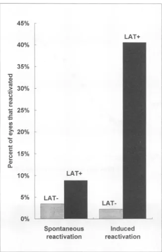
FIG. 6. Summary ofspontaneousand induced reactivation of the LAT deletionmutantdLAT2903.The results ofexperiments1 and 2
werepooledfor the LAT- virus(dLAT2903) and the LAT+viruses
(McKrae plus dLAT2903R). The combined results for spontaneous
reactivation (Table 1, column 3) and induced reactivation (Table 1, column 6) are shown. Combined spontaneous reactivation:
LAT-virus,45 of1,300 cultures, 3.5%; LAT+ viruses,89of 992cultures,9%
(P<0.0001,Fisherexacttest).Combinedinduced reactivation:
LAT-virus, 1of 46eyes,2%; LAT+ viruses,13 of 32eyes,41%(P<0.0001).
0.0001). Similar resultswere obtained in experiment2. dIAT 2903 had a spontaneous reactivation rate ofonly 3% (24 of 816), almost threefold less than with dLAT2903R (8%, 62 of 816). As inexperiment 1,this differencewashighly significant
(P<0.0001) (combinedresults showngraphicallyinFig. 6,left side). Thus,deletion ofLATproducedapronounceddecrease in spontaneous reactivation. This suggests that LAT is an
importantfactor inspontaneous reactivation in the rabbiteye
model.
Because the above analyses do not take into account the
number ofeyesin each of thegroups, the datawere analyzed
byadditional methods. The fraction ofvirus-positive cultures
for eacheye ineachgroup (i.e.,the fraction of time each eye
was virus positive) was calculated, and these fractions were
analyzed by the Student t test. In both experiments, the
decreased spontaneous reactivation rate ofthe LAT deletion mutant compared with the parental or rescued virus was
statistically significant (McKraeversusdLAT2903,P = 0.021;
dLAT2903Rversus dLAT2903,P = 0.015).
The number of eyes in each group that had at least one
spontaneous reactivation is shown in Table 1, column 4. In
experiment 1, 88% (7 of 8) of the McKrae infected eyes reactivated spontaneously while only 41% (9 of 22) of the
dLAT2903-infectedeyesreactivatedspontaneously (P=0.04). Inexperiment 2,75% (18 of24)of the eyesinfectedwith the rescuedLAT mutant dLAT2903R reactivated spontaneously, comparedwithonly38%(9of24)of theeyesinfectedwith the LAT deletion mutant. Again, this difference was statistically
significant (P= 0.02).
5%
LAT-Another method of
examining
theeffect of theLATdeletiononspontaneousreactivation isto
analyze
the number of times that spontaneousreactivation is detected in each eye,regard-less of the
length
of time that virus is present. This isequivalent
to the number of
episodes
in which reactivated virus is detected in the tears, with consecutivedays
ofpositive
culturesbeing
treatedas asingle
event.Thus,
aneyethat sheds virus for asingle day
and an eye that sheds virus for five consecutivedays
would both constitute oneepisode
of spontaneousreac-tivation. The average number of spontaneousreactivations per eyeineach group calculated in thismanneris shown in Table
1,
column 5. Inexperiment 1,
the number of spontaneous reactivations in the dLAT2903-infected eyesaveraged
0.5compared
with 1.7 for McKrae-infected eyes(P
=0.02).
Inexperiment 2,
dLAT2903-infectedeyesaveraged
0.7 spontane-ous reactivationcompared
with 1.1episodes
per eye fordLAT2903R-infectedeyes
(P
=0.02).
All of the aboveanaly-ses
strongly
supportthenotion that LATplays
asignificant
andimportant
role in spontaneous reactivation in the rabbit eye model.Induced invivo reactivation.
Following
theperiod
oftear film collection foranalysis
of spontaneousreactivations,
in vivo reactivations were inducedby
iontophoresis
of6-hydroxydo-pamine
followedby topical
epinephrine
(experiment
1)
orinjection
of sterile water between thelayers
of the cornea(experiment
2) (see
MaterialsandMethods).
Invivo inducedreactivationwasscored
positive
only
iftearfilmsonday
3 or4(or
both) following
inductionwerepositive
for virus(Table
1,
column
6). Approximately
40% of the eyes infected with theparental
McKrae virus(3
of8)
or the rescued LAT deletion mutant(10
of24)
wereinducedtoreactivate.Incontrast,noneof the
dLAT2903-infected
eyeswere inducedto reactivate inexperiment
1(0
of22;
P =0.01)
andonly
one of thedLAT2903-infected
eyesappeared
tobeinduced toreactivate inexperiment
2(1
of24,
4%;
P =0.004) (combined
resultsshown
graphically
inFig. 6,
right
side).
Inexperiment 2,
tear films were collected for two additionaldays.
No additionalvirus-positive
eyesweredetectedonday
5or6postinduction
indLAT2903-infected
rabbits.Thus,
the lack of induced reacti-vation ondays
3 and 4postinduction
was notdue to aslight
delay
in the induced reactivation ofd[AT2903.
The
single dLAT2903
virus-positive
eyewasviruspositive
onboth
days
3 and 4postinduction. Thus,
of the 48 culturesassayed
onthesetwodays
inexperiment
2and the44culturesassayed
onthesetwodays
inexperiment 1,
approximately
2%(2
of92)
wereviruspositive. Since,
as determinedabove,
the spontaneousreactivationratefordLAT2903
was3to4%,
the 2% reactivation rate detectedfollowing
induction was not above thebackground
spontaneousreactivationrateexpected
for this mutant virus
during
thisperiod.
Thus,
thesingle
reactivation event seen withdLAT2903
may have been the result of spontaneous rather than induced reactivation. These results suggest that theLAT deletion mutantresponded
verypoorly
(if
atall)
tostimulicapable
ofinducing
reactivationofwild-type
(and
rescued)
virus.Rate ofestablishment of
latency.
Deletion of LATmight
result in a reducedrate ofestablishing
latency,
and thiscould accountfor the apparentreducedreactivationratesseenwithdLAT2903.Toaddress
this,
atthetermination ofexperiment
2 the rabbitsweresacrificed and both TGswereremoved. TotalDNAwasisolated from each
TG,
and the relativeamountsofHSV-1-specific
DNA weredetermined for each TGby
semi-quantitative
PCRanalysis
asdescribed inMaterials andMeth-ods. Twosetsof PCR
primers
wereused. Onesetwasspecific
fora
region
of the HSV-1TKgene. Theothersetwasspecific
for a
portion
ofcellular actin. This was used as an internalVOL. 68, 1994
on November 9, 2019 by guest
http://jvi.asm.org/
[image:7.612.102.265.76.329.2]controltostandardize the recovery of DNA from the TGs and to standardize any differences in efficiency of DNA transfer
during Southern blotting. The relative intensity of each PCR
product bandfollowing Southern analysis was determined by
laserscanningasdescribed in Materials and Methods, and the
ratio of each HSV-1specificband and thecorresponding actin bandwascalculated. This number represents the standardized relative amount of HSV-1 DNA.
Toensurethat thePCR was linear over the range of HSV-1 DNA present in the samples and also to produce a standard curvefrom which the amount of HSV-1 DNA in the latent TGs couldbeextrapolated,acontrol experimentwasruninparallel. Twofold serialdilutions containing known amounts of HSV-1
genomicDNA inthe range ofapproximately
103
to106copies were added to test tubes each containing a control TG from uninfected rabbits. The total DNA was then isolated andsubjected to PCRby the same procedures used for the DNA from thelatently infected rabbits. The autoradiographic
inten-sities of theHSV-1 PCRproducts of the serially diluted DNA were plotted on a log-log scale versus the known amount of
input HSV-1 DNA (Fig. 7A). The result was a relatively
straight line, indicating thatin this range of HSV-1 DNA our PCR assaywas quantitative.
An autoradiogram ofatypical set of experimental PCRs is shown inFig.7B.Visually, the intensity of viral DNA (D) from the dLAT2903R rescued virus (R) and the dLAT2903 LAT deletion virus (L) appeared similar, relative to the intensity of theircorrespondingactin(A)DNA.Following laser densitom-etryscanning of this and similar autoradiograms, the amount ofHSV-1 DNArelativetoactin from 24individual TGs from the dLAT2903R-infected rabbits was calculated to be 1.82 ± 0.45
(Table
2,column2).Therelativeamountof HSV-1 DNA inthe24individual TGs from the dLAT2903 mutant was 2.28 ± 0.69. Although the LAT deletion mutant-infected TGsappeared to contain an average of 27% more latent HSV-1 DNAthan the rescuedvirus, this difference was not statistically
significant (P
= 0.62). These results strongly suggest that the dLAT2903 LAT deletionmutantwas notdefective in its ability toestablishlatency.Thus, following ocular infection, LAT does not appear to play an important role in the establishment oflatencyin rabbitTGs. These results therefore also indicate that the decreased spontaneous and induced reactivation rates seen with the LAT deletion mutant dLAT2903 were not due to a decreased ability of this virus to establish latency. Thus, the notion that LATplays adirect role in the in vivo reactivation process in the rabbit is strongly supported.
Asjudgedby extrapolation to the standard plot in Fig. 7A, theamountofexperimentalDNAPCRproducts corresponded to averages ofapproximately 8 x 10⁵ and 1 x 10⁶ copies of viral DNA per TG in the rabbits latently infected with
dLAT2903Rand dLAT2903, respectively (Table 2, column 3). Intherabbitslatently infected with dLAT2903R, the number of HSV-1genomic copies varied from approximately 4.0x 10⁴ to 4.4 x
106
per TG, with one DNA-negative TG (<1,000copies). In the rabbits latently infected with dLAT2903, the number of HSV-1 genomic copies varied from approximately 5.2 x 10⁴ to 7.4 x 10⁶, with two DNA-negative TGs. This approximately 100-fold range of values is similar to that reportedfor the number of copies of HSV-1 DNA in TGs of
latentlyinfected mice (13).
Lack of correlation between the relative amount of latent HSV-1 DNA in each TG and the observed spontaneous and induced reactivation rates for the corresponding eye. The
analysesdescribed above made it possible to look for correla-tions between the amount of HSV-1 DNA in each TG (a measureoflatency) and the amount of spontaneous or induced
A
z
5 E
.a
Q
%--S E
=U,n
B
1O03 1104 105 106 107
HSV-1 DNA molecules per1J10thofa TG
B-1
-A
-A"*E*edb'7
D A D A D A D A D A
R R L L L
FIG. 7. Southern blot analysis of the PCR products from total DNAisolatedfromlatently infectedTGs. (A) As a control standard, twofoldserial dilutionsofHSV-1genomicDNA representing approx-imately 106 to 103 molecules were each mixed with a TG from an uninfected rabbit and total DNAwas isolated and subjected to the identical PCR protocols used for panel B. The autoradiographic intensity of an internalTKprobe, usedto determinethe amount of HSV-1 TK PCR product, was plotted against the copy number ofinput genomic DNA on a log-log graph. The computer program used to analyze the intensity of the autoradiographic bands reports small numbers for dark areas and large numbers for light areas. Thus, increasingPCRproduct goes from the bottom to the top of they axis. (B) At the conclusion of experiment 2, TGs were harvested from sacrificedrabbits.As described in Materials and Methods, total DNA wasindividually isolated from each TG and aliquots were subjected to PCR withprimer pairs specificfor either the HSV-1 TK gene or the gene for cellular actin. The PCR products were then separated by agarose electrophoresis, blotted, hybridized to an internal probe for theTKproductand the actin product, and exposed to X-ray film for autoradiography. Adjacent lanes contain the TK and actin PCR productsfromthesame TGs. The results from these and similar blots (24dLAT2903 and 24 dLAT2903R DNA samples) were quantitated, and the ratioof the intensity of hybridization to the TK and actin productswasdetermined.By using this normalized intensity of hybrid-ization of the TKPCRproduct, the amount of DNA in each TG was alsoestimated byextrapolation to theplotin panel A. The results are shown in Table 2.
reactivation in the corresponding eye. Although searches for correlations on a group basis have been previously attempted (25), to ourknowledge this was the first attempt to look for correlations between individual sets of TGs and eyes. A Spearman nonparametric correlation test was done with the data for the TGs and eyes from rabbits latently infected with dLAT2903RordLAT2903.An absolute r > 0.5 andP < 0.05 would indicate a correlation. No correlation was found be-tween the relative amount of latent HSV-1 DNA and sponta-neous or induced reactivations in either group (Table 2, columns 4 and 5) (absolute r<0.3, P > 0.2). Thus, at the limits of thecurrent levels ofsensitivity, we were unable to detect any
on November 9, 2019 by guest
http://jvi.asm.org/
[image:8.612.333.533.69.330.2]SPONTANEOUS REACTIVATION REQUIRES LAT 8053
TABLE 2. Relative and semiquantitative amounts of latent HSV-1 DNA in rabbit TGs and analysis of correlations betweenHSV-1 DNA and reactivation in thecorresponding
eye"
Correlation:relative amt of Correlation:relative amt of Virus Relative amt of HSV-1 Range (mean) of HSV-1 genome HSV-1 DNA with HSV-1 DNA withinduced
DNA inTGsbc copiesin latently infectedTGsd
spontaneousreactivation reactivation
dLAT2903 2.28 +0.69 (n =24) 5.2x
104-7.4
x106(1X 10⁶) r= -0.05, P=0.81e
r= -0.09, P=0.69e dLAT2903R 1.82+0.45 (n =24) 4.0x 10⁴-4.4x10⁶ (8 x 10⁵) r=-0.23, P=0.28 r = -0.09, P = 0.76a The relative amount of HSV-1 DNA in individual TGs from rabbits latently infected withdLAT2903ordLAT2903Rwasdeterminedas described in Materialsand
Methods.
bMean ±standarderror.
cP(dLAT2903vsdLAT2903R)= 0.62 (Student t test, two sided).
dThe estimated amount of DNA was quantitated for each of 24 latently infectedTGspervirusby extrapolation of thenormalizedintensity of the PCRproducts
tothe graphshowninFig. 7A.
eSpearman nonparametriccorrelation test. An absolute r > 0.5 combined with a P < 0.05 would indicate a correlation. These results show no correlation.
correlation between the level of ganglionic latency and reacti-vation.
DISCUSSION
Allprevious HSV-1 LAT mutants have been constructed in virus strains that do not have high in vivo spontaneous reactivation rates. Studies to examine the role of LAT in reactivation from latency have therefore almost exclusively used in vitro explant reactivation models or in vivo induced reactivation models. Thus, there is little direct information regarding the potential role of LAT in in vivo spontaneous reactivation. To overcome this, we have constructed a LAT deletion mutant in HSV-1 McKrae and studied the effect of this mutationonin vivo spontaneousreactivation in the rabbit ocularmodel of HSV-1 latency and reactivation. McKrae was
chosen as the parental virus because it has the highest spon-taneousreactivationrateof any of the commonly used HSV-1
laboratory strains.
Since LAT overlaps at least two important HSV-1 genes,
ICPOandICP34.5, adeletionof the entire LAT gene cannot be usedtoexamine therole ofLATin spontaneousreactivation.
We therefore constructed dLAT2903to contain alarge LAT promoter deletion and the largest possible deletion of the 5' end ofLAT, whileensuringthatnoother known HSV-1 genes weredisturbed. Thiswasdone fortwo reasons.First,to ensure that no LAT transcription could occur from the LAT pro-moter, we deleted the entire region containing the LAT promoterrather thanjust deleting or altering asmall region assumed to be crucial, such as the TATA box. Second, to ensurethatnotranscriptionof the 2-kb LATcouldoccurfrom a potential crypticpromoterjust upstream of the 2-kb LAT,
the deletion also covers this potential cryptic 2-kb LAT promoter and the first third of the 2-kb LAT itself. Thus, dLAT2903isincapableoftranscribingany LATfrom the bona
fide LAT promoter or from the potential
cryptic
promoter. This was confirmed by Northern blots. Other, smaller LAT promoter deletion mutants have previously been shown toproduce no detectable LAT RNA in tissueculture or
during
neuronallatency
(12,
14,28).
dLAT2903 was compared with the
parental
McKrae virus andwiththerescuedvirus, dLAT2903R. Exceptfor differences inreactivation,the LAT deletionmutant wasindistinguishable
from the parental and rescued viruses. The LAT deletion mutant
replicated
in tissue culturewith thesamekinetics and tothesamefinal titeraswild-type
virus,
regardless
of the initialmultiplicityofinfection.
Likewise,
replication
ofdLAT2903in rabbit eyes and rabbit TGswasthe same asthat ofwild-type
virus. Therate atwhichdLAT2903established
latency
wasalsoindistinguishablefrom that of the
wild-type
viruses.Incontrast,dLAT2903 was significantly restricted in its ability to reactivate spontaneously.
Inthe experiments reported here, the spontaneous
reactiva-tion rate for McKrae was 15%, compared with 8% for the rescued LAT mutant dLAT2903R. We have found that such variation in spontaneous reactivation rates from experiment to
experimentis common,evenwith the same virus stock. These variations are thought to be due to variations in variables such asthe age and health of the groups of rabbits used at different times and the fact that New ZealandWhite rabbits are not an inbred strain. Thus, the spontaneous reactivation rates of 8% fordLAT2903R and15%for McKrae are both consistent with ourprevious McKrae studies. In fact, substitution of the results for dIAT2903R in place of McKrae in experiment 1 still indicated a statistically significant decrease in the spontaneous reactivation rate of dLAT2903 (P < 0.05, Fisher exact test). Thus, the higher spontaneous reactivation rate seen in exper-iment 1 with McKrae did not alter the conclusion that in
experiment 1 (aswell as in experiment 2) the LAT deletion mutanthad a reduced spontaneous reactivation rate.
Therateofin vivo spontaneous reactivation for dLAT2903
averaged approximately one-third that of the parental or rescued virus(spontaneous reactivation rates combined from bothexperiments: dLAT2903,3%; McKraeplusdLAT2903R,
9%). Thus,deletion of LAT significantlyreduced but did not eliminate spontaneous reactivation. This suggests that there may be two categories of spontaneous reactivations, LAT-dependentspontaneousreactivations(whichrepresent
approx-imately two-thirds of the spontaneous reactivations in this
system) and LAT-independent spontaneous reactivations
(which representapproximatelyone-third of the spontaneous reactivations in thissystem).
In contrastto the one-third residual spontaneous reactiva-tion seenwithdLAT2903,ourresults suggest thatinducedin vivo reactivation in dLAT2903 may have been eliminated
rather than reduced. Only 2% of eyes(1 of
46)
from rabbitslatentlyinfected withdLAT2903 showed reactivation
following
in vivoinduction,comparedwith41% of eyes
(13
of32)
from rabbits latently infected with dLAT2903R and McKrae. In-duced reactivationwas measuredondays
3 and4postinduc-tion,and thesingle
dLAT2903-reactivating
eyewaspositive
on bothdays.Since all other eyeswerenegativeonbothdays,
the "induced"sheddingrate was2of 92(2%).
Thisrateislikely
to havebeen duetothe spontaneous reactivation ofdLAT2903(3
to
4%).
Thus, deletion of the LAT promoter may have rendered dLAT2903refractorytoinduced in vivoreactivation.On the basis of the above
discussion,
we propose thefollowingpossibility:
(i)
in therabbit ocular model of HSV-1 latency andreactivation,LATisrequired
for in vivoinduction of reactivation;(ii)
approximately
two-thirds of spontaneous VOL. 68, 1994on November 9, 2019 by guest
http://jvi.asm.org/
reactivationsareLATdependent andmaytherefore represent in vivo induced reactivationsinwhich theinducingfactor(s) is not elucidated; and (iii) approximately one-third of spontane-ousreactivationsareindependent of LAT andrepresentalow level ofbackgroundspontaneous reactivations.
Regardless of whether the above speculation is correct or whetherLATsimply increasestheprobabilityofaspontaneous reactivation event by threefold, LAT appears to be a major factor inboth in vivoinduced reactivations and classic spon-taneousreactivations.Assumingthat themechanismsof spon-taneous and induced reactivations in humans are accurately reflectedin therabbitmodel, ourresultsstrongly suggestthat LAT isacrucial factor inclinically importantHSV-1 reactiva-tions.
ACKNOWLEDGMENTS
This work wassupportedbyPublic Health Service grants EY07566 andEY10243,theDiscovery Fund for Eye Research, and the Skirball MolecularOphthalmologyProgram.
REFERENCES
1. Block, T. M., J. G. Spivack,I. Steiner, S. Deshmane, M. T. McIntosh,R. P. Lirette, and N. W. Fraser. 1990. A herpes simplex virus type 1latency-associated transcript mutant reactivates with normal kineticsfromlatentinfection. J. Virol. 64:3417-3426. 2. Bloom, D. C., G. B.Devi-Rao,J. M. Hill, J. G. Stevens, and E.K.
Wagner. 1994. Molecular analysis of herpes simplex virus type 1 duringepinephrine-induced reactivation of latently infected rab-bits in vivo. J. Virol. 68:1283-1292.
3. Coen, D. M. 1992. Quantitation of rare DNAs by PCR, p. 15.3.1-15.3.8. InF.M.Ausubel, R. Brent, R. E. Kingston, et al. (ed.), Current protocols in molecular biology, suppl. 17. Wiley-Interscience,NewYork.
4. Croen, K. D., J. M. Ostrove, L. J. Dragovic, J. E. Smialek, and S. E.Straus. 1987.Latentherpessimplex virus in human trigem-inal ganglia: detection of an immediate early gene "antisense" transcriptby in situhybridization.N. Engl. J. Med. 317:1427-1432. 5. Crowl, R., C. Seamens, P. Lomedico, and S. McAndrew. 1985. Versatileexpressionvectorsfor high-level synthesis of cloned gene productsin E.coli. Gene38:31-38.
6. Dobson, A. T., F. Sederati, G.Devi-Rao,W. M. Flanagan, M. J. Farrell, J. G. Stevens, E. K. Wagner, and L. T. Feldman. 1989. Identification of the latency-associated transcript promoter by expression ofrabbitbeta-globin mRNA in mouse sensory nerve ganglia latently infectedwitharecombinant herpes simplex virus. J.Virol.63:3844-3851.
7. Goins, W. F., L. R. Sternberg,K. D. Croen, P.R. Krause, R. L. Hendricks,D. J.Fink, S. E. Straus, M.Levin,and J. C. Glorioso. 1994. A novel latency-active promoter is contained within the herpes simplex virus type 1UL flanking repeats. J. Virol. 68:2239-2252.
8. Gordon, Y. J., B.Johnson,E. Romanowski, and T. Araullo-Cruz. 1988. RNAcomplimentary toherpes simplex virus type 1 ICPO
gene demonstrated in neurons ofhuman trigeminal ganglia. J. Virol. 62:1832-1835.
9. Gordon, Y. J., E. Romanowski, and T. Araullo-Cruz. 1990. A fast, simplereactivationmethod forthestudy of HSV-1 latency in the rabbitocular model. Invest. Ophthalmol. Vis. Sci. 31:921-924. 10. Hill, J. M., F. Sedarati, R. T. Javier, E. K. Wagner, and J. G.
Stevens. 1990. Herpes simplex virus latent phase transcription facilitates in vivoreactivation. Virology 174:117-125.
11. Ho, D. Y., and E. S.Mocarski. 1989. Herpes simplex virus latent RNA(LAT) is not required for latent infection in the mouse. Proc. Natl. Acad. Sci. USA86:7596-7600.
12. Javier, R. T., J. G.Stevens,V. B.Dissette, and E.K.Wagner. 1988. A herpes simplexvirus transcript abundant in latently infected neurons is dispensable for establishment of the latent state. Virology166:254-257.
13. Katz, J. P., E. T. Bodin, and D. M. Coen. 1990. Quantitative polymerasechainreactionanalysis of herpes simplex virus DNA in
ganglia of mice infected withreplication-incompetent mutants.J. Virol. 64:4288-4295.
14. Leib, D. A., C. L. Bogard, M. Kosz-Vnenchak, K. A.Hicks, D. M. Coen, D. M. Knipe, and P. A. Schaffer. 1989. Adeletion mutantof the latency associated transcript ofherpes simplex virus type 1 reactivates from the latent state with reduced frequency. J. Virol. 63:2893-2900.
15. Leib, D. A.,K. C. Nadeau, K. C. Rundle, S. A. Rundle, and P. A. Schaffer. 1991. Promoter of the latency-associated transcripts of herpes simplex virustype-1 contains a functional cAMP-response element: role of the latency associated transcripts and cAMP in reactivation of viral latency. Proc.Natl.Acad. Sci. USA 88:48-52. 16. Maniatis, T., E. F. Fritsch, and J.
Sambrook.
1982. Molecular cloning: a laboratory manual. Cold Spring Harbor Laboratory, Cold Spring Harbor, N.Y.17. McGeoch, D. J., M. B.Dalrymple,A. J.Davison, A. Dolan, M. C. Frame, D. McNab, L. J. Perry, J. E. Scott, and P. Taylor. 1988. The complete DNA sequence of the long unique region in the genome of herpes simplex virus type 1. J. Gen. Virol. 69:1531-1574. 18. Nesburn, A. B. (ed.). 1983. Report of the corneal disease panel:
vision research: anational plan 1983-1987, vol. II, part III. The C. V. Mosby Co., St. Louis.
19. Nesburn, A. B., H. Ghiasi, and S. L. Wechsler. 1990. Ocular safety and efficacy of an HSV-1 gD vaccine during primary and latent infection. Invest. Ophthalmol. Vis. Sci. 31:77-82.
20. Nesburn, A. B., C. Robinson, and R. Dickinson. 1974. Adenine arabinoside effect on experimental idoxuridine-resistant herpes simplex infection. Invest. Ophthalmol. 4:302-304.
21. Nesburn,A. B., and M. D. Trousdale. 1981. Treatment of acute and chronic ocular herpes infection with acyclovir, p. 309-314. In R. Sundmacher (ed.), Herpetische Augenerkrankungen. J. F. Berg-mann Verlag, Munich.
22. Perng, G. C., H. Ghiasi, R. Kaiwar, A. B. Nesburn, and S. L. Wechsler. 1994. An improved method for cloning portions of the repeat regions of herpes simplex virus type 1. J. Virol. Methods 46: 111-116.
23. Perry, L. J., and D. J. McGeoch. 1988. The DNA sequences of the long repeat region and adjoining parts of the long unique region in the genome of herpes simplex virus type 1. J. Gen. Virol. 69:2831-2846.
24. Rock, D. L., A. B. Nesburn, H. Ghiasi, J. Ong, T. L. Lewis, J. R. Lokensgard, and S. L. Wechsler. 1987. Detection of latency-related viral RNAs in trigeminal ganglia of rabbits latently in-fected with herpes simplex virus type 1. J. Virol. 61:3820-3826. 25. Sawtell, N. M., andR.L. Thompson. 1992. Herpes simplex virus
type 1 latency-associated transcription unit promotes anatomical site-dependent establishment and reactivation from latency. J. Virol. 66:2157-2169.
26. Shimozuma, M., W. L. Drew, R. C. Miner, W. L. Epstein, and K. Fukuyama. 1992. Direct inactivation of herpes simplex virus type-2 by rat epidermal protein. Antiviral Res. 18:179-189.
27. Spivack, J. G., and N. W. Fraser. 1988. Expression of HSV-1 latency-associated transcripts in the trigeminal ganglia of mice during acute infection and reactivation of latent infection. J. Virol. 62:1479-1485.
28. Steiner, I., J. G. Spivack, R. P. Lirette, S. M. Brown, A. R. MacLean, J. H. Subak-Sharpe, and N. W. Fraser. 1989. Herpes simplex virus type 1 latency associated transcripts are evidently not essential for latent infection. EMBO J. 8:505-511.
29. Steiner, I., J. G. Spivack, D. R. O'Boyle II, E. Lavi, and N. W. Fraser. 1988. Latent herpes simplex virus type 1 transcription in human trigeminal ganglia. J. Virol. 62:3493-3496.
30. Stevens, J. G., E.K.Wagner, G. B. Devi-Rao, M. L. Cook, and L. T. Feldman. 1987. RNA complementary to a herpesvirus alpha gene mRNA is prominant in latently infected neurons. Science 235: 1056-1059.
31. Takasu, T., Y. Furuta,K.C. Sato, S. Fukuda, Y. Inuyama, andK. Nagashima. 1992. Detection of latent herpes simplex virus DNA and RNA in human geniculate ganglia by the polymerase chain reaction. Acta Otolaryngol. 112:1004-1011.
32. Trousdale, M. D., I. Steiner, J. G. Spivack, S. L. Deshmane, S. M. Brown, A. R. MacLean, J. H. Subak-Sharpe, and N. W. Fraser. 1991. In vivo and in vitro reactivation impairment of a herpes
on November 9, 2019 by guest
http://jvi.asm.org/
SPONTANEOUS REACIIVATION REQUIRES LAT 8055
simplex virus type 1 latency-associated transcript variant in a
rabbiteyemodel. J. Virol. 65:6989-6993.
33. Wagner, E. K., G. Devi-Rao, L. T. Feldman, A. T. Dobson, Y. Zhang, W. M. Flanagan, and J. G. Stevens. 1988. Physical characterization of the herpes simplex virus latency-associated transcript inneurons. J. Virol.62:1194-1202.
34. Wagner,E.K.,W.M.Flanagan, G. Devi-Rao, Y. Zhang,J. M. Hill,
K.P.Anderson, andJ. G. Stevens. 1988. The herpes simplexvirus
latencyassociated transcript is spliced during the latent phase of infection.J. Virol. 62:4577-4585.
35. Wechsler, S. L., A. B. Nesburn, R. J. Watson,S. Slanina,and H.
Ghiasi. 1988. Fine mapping of the major latencyrelated-RNA of herpessimplex virustype 1 inhumans.J. Gen. Virol.69:3101-3106. 36. Wechsler, S. L., A. B. Nesburn,R.J. Watson, S. M. Slanina,and
H. Ghiasi. 1988. Fine mapping of the latency related gene of herpessimplex virustype1: alternative splicing produces distinct latency-related RNAs containing open reading frames. J. Virol.
62:4051-4058.
37. Wechsler, S. L.,A. B. Nesburn,J. C. Zwaagstra, andH.Ghiasi. 1989. Sequence of the latencyrelatedgeneof herpes simplex virus
type1.Virology 168:168-172.
38. Zwaagstra,J.C., H. Ghiasi,A. B. Nesburn, andS.L.Wechsler. 1989. In vitro promoteractivityassociated with the latency
asso-ciated transcriptgeneof herpessimplex virustype1.J.Gen.Virol.
70:2163-2169.
39. Zwaagstra, J. C., H. Ghiasi, A. B.Nesburn, andS. L. Wechsler. 1991. Identification ofamajorregulatorysequenceinthe latency associated transcript (LAT)promoterofherpes simplex virustype
1(HSV-1). Virology 182:287-297.
40. Zwaagstra, J. C., H. Ghiasi, S. M. Slanina,A. B.Nesburn, S. C. Wheatley, K. Lillycrop, J. Wood, D. S. Latchman,K. Patel, and
S. L. Wechsler. 1990. Activity of herpes simplex virus type 1
latency-associated transcript (LAT) promoter in neuronderived cells: evidence forneuron specificity and for a large LAT
tran-script.J. Virol. 64:5019-5028. VOL. 68, 1994