Study of the effect of amniotic membrane grafting on corneal ulcers.
Full text
Figure


Related documents
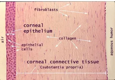
Normal corneal sublayers imaged by in vivo confocal microscopy: (A) surface epithelium with prominent, bright nuclei (arrow); (B) Basal epithelial cells showing only
Part IV LASIK Late Postoperative Complications: Dry Eye Syndrome, Epithelial Ingrowth, Corneal Ectasia, and Other Complications 43 Dry Eye Syndrome: Ocular Surface Syndrome.
We found that Pax6 gene expression is absent in the ocular surface of the Dkk2 mutant (Fig. 4B), whereas the nuclei of corneal epithelial basal cells of wild-type corneas show
Watanabe, et al, Functional bioengineered corneal epithelial sheet grafts from corneal stem cells expanded ex vivo on a temperature-responsive cell culture surface,
Aims: To assess the efficacy of corneal collagen cross-linking (CXL) combined with amniotic membrane graft in the management of severe melting ulcers in the dog and cat using a
The density of basal epithelial cells, keratocytes and endothelial cells, and the status of the subbasal nerve fibers were evaluated using in vivo corneal confocal microscopy..
