AETIOLOGICAL PROFILE AND
CLINICO-ECHOCARDIOGRAPHIC FEATURES OF
CONGESTIVE HEART FAILURE
Dissertation Submitted to
THE TAMIL NADU DR. M.G.R MEDICAL UNIVERSITY
In partial fulfillment of the regulations for the award of the degree of
M.D. BRANCH – I
GENERAL MEDICINE
GOVT. STANLEY MEDICAL COLLEGE & HOSPITAL THE TAMIL NADU DR. M.G.R. MEDICAL UNIVERSITY
CHENNAI, INDIA.
CERTIFICATE
This is to certify that the dissertation titled “AETIOLOGICAL PROFILE AND
CLINICO ECHOCARDIOGRAPHIC FEATURES OF CONGESTIVE HEART
FAILURE” is the bona fide original work
of Dr. K. VASANTH in partial fulfillment of the requirements for M.D.
Branch – I (General Medicine) Examination of the Tamil Nadu Dr. M.G.R Medical
University to be held in MARCH 2009. The period of study was from August 2007 to
August 2008.
PROF.V.RUCKMANI,M.D.,
Professor and Head Dept. of Medicine, Govt. Stanley Medical College and Hospital Chennai-600 001.
DR.J.MOHANASUNDARAM.M.D.,D.N.B.,Ph.D,
DEAN
DECLARATION
I, Dr. K. VASANTH hereby solemnly declare that the
dissertation titled “AETIOLOGICAL PROFILE AND CLINICO
ECHOCARDIOGRAPHIC FEATURES OF CONGESTIVE
HEART FAILURE” was done by me at Govt. Stanley Medical College and Hospital
from August 2007 to August 2008 under the supervision and guidance of my Unit Chief
and Head of Department of Medicine Prof.V.RUCKMANI, M.D.,
This dissertation is submitted to Tamil Nadu DR. M.G.R Medical University,
towards partial fulfillment of requirement for the award of M.D. Degree (Branch - I) in
General Medicine.
Place: Chennai. Date:
(DR. K. VASANTH)
ACKNOWLEDGEMENTS
I sincerely thank the Dean, Govt. Stanley Medical College and Hospital,
Dr.J.MOHANASUNDARAM,M.D.,D.N.B.Ph.D., for permitting me to avail the
facilities of the college &
Hospital for my dissertation work.
I am indebted to Prof.V.RUCKMANI,M.D.,Professor and Head of the
Department of Medicine, Govt. Stanley Medical College and Hospital for this foresight,
guidance, periodical supervision & encouragement to me to do the study.
I whole heartedly express my sincere gratitude and thanks to
Prof.S.NATARAJAN.M.D.,Professor of Medicine, Govt. Stanley Medical College and
Hospital for being a Source of inspiration, excellent guidance, valuable instructions and
help in every stage of study which has made this dissertation work possible.
I am thankful to Prof.S.SHIVAKUMAR,M.D.,and
Prof.T.VENKATAKRISHNAN,M.D., Govt. Stanley Medical College & Hospital for
their encouragement in doing this study.
I am extremely thankful to my Unit Assistant Professors
Dr.MURALIDHARAN, Dr.GOWTHAM, Dr.ARUN, Dr.SUJIT, Dr.SURESH,
Dr.MOHANRAO and Dr.THILAGAVATHY for
their valuable guidance, reference material and encouragement throughout the study.
I am also thankful to my colleagues who shared their knowledge and also the
INDEX
CONTENTS PAGE NO
1. INTRODUCTION 1
2. REVIEW OF LITERATURE 2
3. AIM OF THE STUDY 28
4. METHODOLOGY 29
5. OBSERVATIONS & DATA 33
DATA ANALYSIS 6. DISCUSSION 56
7. CONCLUSION 59
ANNEXURES
A. BIBLIOGRAPHY
B. PROFORMA
INTRODUCTION
Heart failure is the end stage of all diseases of the heart and is a
major cause of morbidity and mortality. Since 1970s the treatment of CHF`has been
transformed, resulting in major benefit to patients. This advance has been the
consequence of better understanding of the pathophysiology, investigations, the
introduction of newer drugs and cardiac transplantation. The traditional treatment of
heart failure with digoxin and diuretics has been replaced by diuretics, ACE inhibitors
and drugs directed against the origins of heart failure such as aspirin and lipid lowering
drugs. Newer objectives are optimization of the quality of life, avoidance of hospital
admissions prevention of progression of damage to the myocardium and prolongation of
life.
So it becomes important to conduct clinical and Para clinical studies to know
about the status, precipitating factors and complications of the disease. Only with a
reliable study, changes in the modality of approach in controlling, diagnosing and
treating the disease can be done. Here, an attempt has been made to study on selected
REVIEW OF LITERATURE
History:
Heart failure, angina and the pulse were known in the ancient
Egyptian and early Greek civilizations. (Dallas 1993, Horine 1941). Hippocrates
described cardiac cachexia, most vividly reports of the benefits of foxglove exists in
Roman literature. (Moore, 1985) Hering used nitrate in 1853 to treat heart failure, the
first use of vasodilator. Bruton later in 1867 described the use of amyl nitrate to treat
angina. ACE inhibitors were shown to be of benefit in terms of mortality in patients with
heart failure for the first time in 1987. The large trail of digoxin, showing no effect on
overall mortality, was reported in 1996.
Definitions of heart failure
1. A pathophysiological state in which an abnormality of cardiac function is
responsible for the failure of the heart to pump blood at a rate commensurate with
the requirements of the metabolizing tissues (Braunwald 1994).
2. Congestive heart failure represents a complex clinical syndrome,
characterized by abnormalities of left ventricular function and
neurohormonal regulation, which are accompanied by effort
intolerance, fluid retention and reduced longevity.(Packer 1988).
3. Symptoms of heart failure, objective evidence of cardiac dysfunction and
response to treatment directed towards the heart failure. (Task force of the
EPIDEMIOLOGY:
Heart failure is a relatively common disorder. It is estimated that 4.6 million
persons in the United States are being treated for heart failure, with 550,000 new cases
diagnosed each year. (Dallas 1999, Massie - 1977)
The prevalence of heart failure increases dramatically with age,
occurring in 1 to 2 percent of persons aged 50 to 59 and up to 10 percent of individuals
older than the age of 75. (Ho et al 1993)
Approximately 80 percent of all heart failure admissions occur in
patients older than 65, as a result, heart failure is the leading discharge diagnosis in
persons 65 years or older in the United States (Rich 1999).
Despite a steady decline in the incidence of coronary artery disease and stroke,
both the incidence and prevalence of heart failure continue to rise.
Between 1985 and 1995 the number of heart failure hospitalizations
increased by 5 percent and 8, 70,000 hospital discharges for heart failure
occurred in 1996. (Haldeman – 1999)
In the United States approximately 45,000 deaths each year are
primarily caused by heart failure and heart failure is listed as a contributing cause in
260,000 deaths (Dallas 1999).
In smaller mid western areas, recent assessment has suggested a
prevalence as high as 6% of population. The same prevalence of 6-7% also has been
The overall prevalence of heart failure is 3-20 per 1000 population
although this exceeds 100 per 1000 in those aged 65 yrs and over. The
annual incidence of heart failure is 1-5 per 1000 and the relative incidence doubles for
each decade of life after the age of 45 yrs. (Davis Hobbs, Lip – 2003). The overall
incidence is likely to increase in the future because of both an aging population and
therapeutic advances in the management of acute myocardial infarction leading to
improved survival in patients with impaired cardiac function. (Cowie et al - 1999).
Three major factors such as age, race and gender influence the
prevalence and outcome in patients with heart failure.
Age:
The most important factor is age. The prevalence of CHF is less than 1% in
patients < 50 yrs of age regardless of gender. At older than 50 years of age however, the
prevalence increases to approximately 5% for patients between 50 and 70 years of age,
nearly 10% for all patients over 70 yrs of age and perhaps as high as 15% for patients
over the age of 80.
The impact of age alone leads to a significant continued increase in the prevalence
of heart failure as recent statistics have suggested that there will be near doubling of
Race:
The second major influencing factor is race. There has been a higher prevalence of
heart failure in blacks compares to whites.
More importantly black patients develop heart failure at a younger age than white
patients. (Bourassa et al 1993).
Gender:
The third major factor influencing the heart failure is gender. The
national heart and nutrition education survey (NHANES) from 1988-1994
estimated that there is an increased prevalence of congestive heart failure in men aged
70 years and younger. In contrast, women aged older than 70 years have a higher
prevalence. This difference may be in part owing to the increased average life
expectancy in women (Anderson 2001).
The highest prevalence of CHF is in black men followed by black women.
Data derived from the National centre of Health Statistics report
that total life expectancy for the United States Population is 76.7 years, Life expectancy
is 73.8 years for men and 79.5 years for Women (2000 estimate).
While the overall incidence of congestive heart failure is probably equal between
the two genders there are several unique features that may influence the prevalence of
congestive heart failure in women, including a higher average ejection fraction for an
equivalent amount of symptoms than men at all ages. This may reflect a higher
1998)
The Framingham data shows an age adjusted annual incidence of heart failure of
0.14% in women and 0.23% in men. Survival in women is
generally better than in men.
AETIOLOGY:
The relative importance of aetiological factors in heart failure is
dependent on the nature of the population being studied, as coronary artery disease and
hypertension are common causes of heart failure in western countries, whereas valvular
heart diseases and nutritional cardiac diseases are more common in the developing
world. (Zannad - 1999)
The common causes of heart failure include:
1. coronary artery disease, Ischemia
2. hypertension
3. cardiomyopathies
4. valvular heart disease and congenital heart diseases
5. arrhythmias
6. alcohol and drugs
- alcohol
- cardiac depressant drugs (beta blockers and calcium
7. high output failure
- anemia, thyrotoxicosis, beriberi, etc
8. pericardial diseases
9. primary right heart failure
-Pulmonary hypertension eg. pulmonary embolism, corpulmonale
-tricuspid incompetence
Coronary artery disease and its risk factors:
Coronary artery disease is the commonest cause of heart failure. In the studies of
left ventricular dysfunction (SOL VD) Coronary artery disease accounted for about 75%
of the cases of chronic heart failure.
(Khadra Saleem, Rand 1998)
Coronary artery disease and hypertension (either alone or in the combination)
were implicated as the cause in over 70% of cases of heart failure in the fragmentation
study. (Ho – 1993)
Coronary risk factors, such as smoking and diabetes are also
risk markers for the development of heart failure. Smoking is an independent and strong
risk factor for the development of heart failure in men, although the findings in women
are less consistent.
In Framingham heart study, diabetes and left ventricular
hypertrophy were the most significant risk markers for the development of heart failure.
cholesterol concentration are also independent risk factors for heart failure. Clearly these
risk factors may increase the risk of heart failure through their effects on coronary artery
disease, although diabetes alone may induce important structural and functional changes
in the myocardium, which further increase the risk of heart failure.
Hypertension:
Hypertension has been associated with an increased risk of heart
failure in several epidemiological studies. In the Framingham heart study,
hypertension was reported as the cause of heart failure either alone or in
association with other factors, in over 70% of cases on the basis of non
invasive assessment. (Ho - 1993). However hypertension is probably a more common
cause of heart failure in selected patient groups, including females and black
populations. Hypertension predisposes to the development of heart failure via a number
of pathological mechanisms, including left ventricular hypertrophy.
Left ventricular hypertrophy is associated with left ventricular systolic and
diastolic dysfunction and an increased risk of myocardial infarction and it predisposes to
both atrial and ventricular arrhythmias. Electrocardiographic left ventricular hypertrophy
is strongly correlated with the development of heart failure, as it associated with a 14
fold increase in the risk of heart failure in those aged 65 years or under.
Valvular heart disease:
Rheumatic heart disease may have declined in certain parts of the
other developing nations. In the Framingham heart study, rheumatic heart disease
accounted for heart failure in 2% of men and 3% of women,
although the overall incidence of valvular heart disease has been steadily
decreasing in the Framingham cohort over the past 30 years (HO - 1993).
MR and AS are the most common causes of heart failure, secondary to valvular
disease. MR and AR lead to volume overload, in contrast with AS which leads to
pressure overload. The progression of heart failure in patients with valvular heart disease
is dependent on the nature and extent of the valvular disease. In aortic stenosis heart
failure develops at a relatively late stage and without, valve replacement, it is associated
with a poor prognosis. In contrast, patients with chronic mitral or aortic regurgitation
generally decline in a slower and more progressive manner. (Teerlink, et al 1991).
Cardiomyopathies:
Cardiomyopathies are defined as the disease of the heart muscle that are not
secondary to coronary heart disease, hypertension or others. As primary disease of heart
muscle, cardiomyopathies are less common causes of heart failure, but awareness of
their existence is necessary to make a diagnosis. Cardiomyopathies are separated into
four functional categories dilated, hypertrophic, restrictive and obliterative. These
groups can include rare specific heart muscle diseases such as hemochromatosis in
which cardiac involvement occurs as part of a systemic disorder. Dilated
cardiomyopathy is a more common cause of heart failure than hypertrophic and
developing countries. (Oakley 1997).
Arrhythmias:
Cardiac arrhythmias are more common in patients with heart failure and
associated structural heart diseases, including hypertensive patients with left ventricular
hypertrophy.
In the Hillingdon heart failure study 30% of patients presented for the first time
with heart failure had atrial fibrillation and over 60% of patients admitted urgently with
atrial fibrillation to a Glasgow hospital had echocardiographic evidence of impaired left
ventricular function (Stevenson, 1995).
Alcohol and drugs:
Alcohol has a direct toxic effect on the heart which may lead to acute heart failure
or heart failure as a result of arrhythmias, commonly atrial fibrillation. Excessive
chronic alcohol consumption also leads to dilated cardiomyopathy.
Alcohol is the identifiable cause of heart failure in 2-3% of cases.
Chemotherapeutic agents (doxorubicin) and antiviral drugs (zidovudine)
have been implicated in heart failure, through direct toxic effects on the
myocardium. (Maki et al - 1998)
Endocrine causes:
High output heart failure is most often seen in patients with anaemia and
thyrotoxicosis. Myxedema may present with heart failure as a result of myocardial
Corpulmonale:
Corpulmonale is defined as enlargement of the right ventricle
secondary to abnormalities of the lungs, thorax, pulmonary ventilation or
circulation. It sometimes leads to right ventricular failure, with an elevation of
trasnsmural right ventricular end diastolic pressure. (Rich et al 2005).
Pericardial Diseases:
Pericardial diseases like tuberculosis, CRP and malignant
involvement may also cause congestive heart failure.
Nutritional Causes:
Vitamin deficiency like wet beriberi, anaemia and deficiency of
hematopoetic factors leading to heart failure, continue to produce a problem especially
in developing countries like India.
PATHOPHYSIOLOGY:
Heart failure is the multisystem disorder which is characterized by the
abnormalities of cardiac, skeletal muscle and renal function with stimulation of the
sympathetic nervous system and a complex pattern of neurohormonal changes.
Myocardial Systolic Dysfunction:
The primary abnormality in non valvular HF is an impairment in the
left ventricular (LV) function leading to fall in cardiac output. This fall in
cardiac output leads to activation of several neurohormonal compensatory
mechanisms aimed at improving the mechanical environment of heart.
rate, increases myocardial contractility and peripheral
vasoconstriction. Activation of Renin-Angiotensin-Aldosterone system
(RAAS) results in vasoconstriction and increase in blood volume with salt and water
retention. Concentration of vasopressin and natriuretic peptides increase. Furthermore
there may be progressive cardiac dilatation or alterations in cardiac structure or both
(Borgeon, Burnett, 1997).
Renin – Angiotensin – Aldesterone system (RAAS)
Simulation of RAAS leads to increased concentration of Renin,
Angiotensin II (AT II) and Aldosterone. AT II is a potent vasoconstrictor of renal and
systemic circulation where it stimulates release of Nor Adrenaline from sympathetic
nerve terminals, which inhibits vagal tone and promotes release of Aldosterone. This
leads to sodium and water retention, in addition, AT – II has an important effects on
cardiac myocytes and may contribute to endothelial dysfunction. (Francis et al, 1990)
Sympathetic Nervous System:
Sympathetic nervous system is activated in HF via low and high
pressure baroreceptors as an early compensatory mechanism which provides inotropic
support and maintain cardiac output. In long term, the ability of the myocardium to
respond to chronic high concentration of catecholamines is activated by down regulation
of ß-receptors. This may be associated with baroreceptor dysfunction and further
Natriuretic Peptides:
There are three natriuretic peptides of similar structure and these exert a wide
range of effects on heart, kidneys and cardio vascular system.
Atrial natriuretic peptide (ANP) is released from atria in response to stretch,
leading to natriuresis and vasodilatation. In humans, Brain
natriuretic peptide (BNP) is also released from heart and its actions are similar to those
of ANP. C- type natriuretic peptide is limited to vascular
endothelium and CNS and has only limited effects on natriuresis and
vasodilatation. (Van Chang et al, 2001 and Moe et al, 1993)
Vasopressin:
Vasopressin concentration is also increased in severe chronic HF. High
concentration of the hormone are particularly common in patients receiving diuretic
treatment and this may contribute to development of hyponatremia (Francis et al, 1990
and Goldsmith, 1986).
Endothelin:
Secreted by vascular endothelial cells, is a potent vasoconstrictor on renal
vasculature. Its concentration is also correlated with indices of
severity such as pulmonary capillary wedge pressure (PCWP) and need for
Patterns of Neurohormonal activation and prognosis
Asymptomatic left ventricular dysfunction:
Plasma noradrenaline concentration increases early in development of left
ventricular dysfunction and plasma renin activity usually increases in patients receiving
diuretic treatment. Nor adrenaline concentration in patients with asymptomatic LV
dysfunction is a strong and independent predictor of development of symptomatic HF
and long term morality.
In severe untreated chronic HF, concentrations of renin, AT – II,
Aldosterone, nor adrenaline and ANP are all increased. Plasma levels correlate with both
the severity of HF and the long term prognosis. Patients with chronic HF and increased
plasma nor adrenaline concentration do also have a worse prognosis (Esler, 1997).
Diastolic dysfunction:
Diastolic dysfunction refers to clinical syndrome of HF with
preserved Left Ventricle Ejection Fraction (EF – 0.4 or more) in the absence of major
valvular disease. In diastolic HF, LV cavity is stiff due to increased LV mass. It relaxes
slowly in early diastole and offers greater resistance to filling in late diastole so that
diastolic pressure is increased. The low cardiac output manifests as fatigue while the
increase in end diastolic pressure is transmitted backwards through valve less pulmonary
veins to pulmonary capillaries resulting in exertional dyspnea (Vasa Levy, 2000).
Mechanisms contributing to abnormal LV diastolic properties
myocardial changes with or without associated hypertrophy (Kitzman et al, 2002). The
prognosis of diastolic HF is generally better than that of systolic Heart failure.
Diastolic Heart failure is common in clinical practice. The
Diagnosis of diastolic HF may be considered in patients with HF who have normal LV
EF (0.4 or more) (Ibrahim, 2003).
Myocardial Dysfunction due to Remodeling, Hibernation and Stunning:
After extensive myocardial infarction, cardiac contractility is
frequently impaired and neurohormonal activation leads to regional
eccentric and concentric hypertrophy of Non-infarcted segment with
expansion of infarct zone. This is known as Ventricular Remodeling.
Particular risk factors for this development of progressive ventricular
dilatation after an MI include large infarcts, anterior infarctions, occlusion of artery
related to infarction and hypertension.
Myocardial dysfunction may also occur in response to stunning
which describes delayed recovery of myocardial function despite restoration of coronary
blood flow in the absence of irreversible damage. This is in contrast to hibernating
myocardium which describes persistent myocardial dysfunction due to reduced
perfusion although cardiac myocytes remain viable and myocardial contraction may
TYPES OF HEART FAILURE
Forward Failure:
It is defined as the inability of the heart to maintain effective stroke
volume to meet the metabolic demands of the body.
Backward Failure:
Small transient inequality between the two ventricles resulting in
acute pulmonary edema.
Right sided heart failure:
Due to stagnation of blood in the right side of the heart and
right heart failure signs develop.
Left sided heart failure:
Due to poor LV contractile function in aortic stenosis or massive MI where the
signs of left sided heart failure develops.
Acute heart failure Vs chronic heart failure:
The clinical manifestations of HF depends mainly on the rate at
which the syndrome develops and specifically whether sufficient time has
elapsed for compensatory mechanisms to become operative and for fluid to accumulate
Low output Vs high output failure:
Heart failure in low cardiac output at rest or in milder cases during exertion
characterizes most forms of cardiovascular diseases.
High output heart failure occurs in variety of high output states like
thyrotoxicosis, Paget’s disease, AV fistulas anaemia and beriberi. (Stevenson 1989).
Systolic Vs diastolic HF:
Implicit in the physiological definition of heart failure (inability to pump an
adequate volume of blood and or to do so only from an abnormally elevated filling
pressure) is that heart failure can be caused by an abnormality in systolic function
leading to a defect in expulsion of blood leading to systolic heart failure or by an
abnormality in diastolic function leading to a defect in ventricular filling (diastolic
failure). This may be due to slowed or incomplete ventricular relaxation, which may be
transient as occurs in ischaemia, or sustained, as occurs in concentric myocardial
hypertrophy or restrictive cardiomyopathy secondary to infiltrative conditions like
amyloidosis.
The principal clinical manifestations of systolic failure result from
an inadequate cardiac output and secondary salt and water retention – forward heart
failure (Gaasch 1994).
Whereas the major consequence of diastolic HF is related to the
elevation of ventricular filling pressure upstream to the ventricular cavity,
1995)
Clinical features and complications:
Patients with HF present with a variety of symptoms, most of
which are nonspecific. The common symptoms of congestive HF include
fatigue, dyspnoea, pedal edema and exercise intolerance or symptoms that relate to the
underlying cause.
Symptoms and signs in heart failure:
Symptoms
Dyspnoea
Orthopnoea
Palpitation
Chest pain
Reduced exercise tolerance, lethargy, fatigue
Nocturnal cough
Wheeze
Ankle swelling
Signs
Cachexia and muscular wasting
Tachycardia
Pulsus alternans
Increased JVP
Displaced apex beat
RV heave
Crepitations or wheeze
3rd heart sound
Oedema
Hepatomegaly, ascites
Symptoms:
Dyspnoea:
Exertional breathlessness is the frequent presenting symptom in heart failure,
although it is a common symptom in patients with pulmonary disease. Dyspnoea is
NYHA Classification of dyspnea:
Class I – no limitation of physical activity. ordinary physical activity does not cause
undue symptoms.
Class II – slight limitation of physical activity. ordinary physical activity causes undue
fatigue, dyspnea or palpitation.
Class III – moderate limitation of physical activity. less than ordinary activity causes
symptoms.
Class IV – Severe restriction of physical activity. unable to perform to any activity
without symptoms.
Orthopnea:
Orthopnea is a more specific symptom. PND results from increased
LV filling pressure and therefore has a greater sensitivity and predictive
value (Maning 1995).
Fatigue and Lethargy
Fatigue and Lethargy in CHF are due to impaired muscle blood flow and poor
tissue perfusion.
Oedema
Swelling of ankles and feet is another common presenting feature.
Heart failure may manifest as oedema, right hypochondrial pain (liver
congestion) and loss of appetite (due to bowel congestion). An increase in
weight loss are important markers of disease severity. (Milne 1985)
Physical Signs
Physical examination has serious limitations as many patients
particularly those with less severe heart failure, have few abnormal signs. In addition
some physical signs are difficult to interpret, and if present, may occasionally be related
to diseasesother than HF.
Oedema and tachycardia, for example are too intensive to have any useful
predictive value and although pulmonary crepitations may have a high diagnostic
specificity. Increased JVP has a high specificity in
diagnosing HF in patients who are known to have cardiac disease.
Displaced apex beat in patients with myocardial infraction and 3rd
heart sound have a relatively high specificity.
Framingham Criteria for Diagnosis of Congestive Heart Failure:
Major Criteria
1) Paroxysmal nocturnal dyspnea
2) Neck vein distension
4) Cardiomegaly
5) Acute pulmonary edema
6) S3 gallop
7) Increased Venous Pressure (>16cm H2O)
8) Positive Hepatojugular reflux
Minor Criteria:
1) Extremity edema
2) Night Cough
3) Dyspnea on exertion
4) Hepatomegaly
5) Pleural effusion
6) Vital capacity reduced by one-third from normal
7) Tachycardia (≥ 120 bpm)
Major or Minor:
Weight loss ≥ 4.5 kg over 5 days of treatment
To establish a clinical diagnosis of congestive heart failure by these
AIM and OBJECTIVES
1) To study about the etiological profile of congestive heart failure.
2) To study about the clinical features of congestive heart failure.
MATERIALS and METHODS
Place : Department of Medicine
Design : Observational Study
Period : August 2007 to August 2008
Sample Size : 100 Patients
Collaborating : Cardiology
Departments
Inclusion Criteria
1) All patients above 12 years of age with clinical evidence of congestive heart
failure.
2) Patients of Both sexes.
Exclusion Criteria
1) Patients less than 12 years of age
2) Hemodynamically unstable patients
3) Pregnant women
METHODS:
1. Thorough history.
2. Complete physical examination.
3. Chest X-ray.
4. ECG.
5. Complete Blood Count
6. Renal Function Tests.
7. Echo cardiography
Definitions used for the study
1) Dyspnea:
Subjective awareness of the sensation of breathing
2) Orthopnea:
Dyspnea on assuming recumbency.
3) Angina:
Retrosternal chest discomfort due to myocardial ischemia.
4) Palpitation:
Diabetes:
American diabetes association criteria for the diagnosis of
diabetes mellitus.
1. Symptoms of diabetes plus blood glucose concentration ≥ 200 mg/dl or
2. Fasting plasma glucose ≥ 126 mg/dl or
3. Two hour plasma glucose ≥ 200 mg/dl during an oral glucose
tolerance test.
Hypertension:
Clinical blood pressure of > 140/90 mm Hg confirmed on two
separate occasions. (JNC > criteria)
Ischemic Heart Disease:
IHD and CAD is typically defined as a > 50% stenosis of any
epicardial coronary artery.
Manifestations of CAD include stable angina, acute coronary
Syndrome (ACS), congestive heart failure, sudden cardiac death and silent ischemia.
cardiography
Hypokinesia
Akinesia
Dyskinesia
Rheumatic Heart Disease:
Evidence of valvular involvement in the form of thickening, fibrosis or
calcification.
Ejection Fraction:
Depressed ejection fraction < 40%
Normal ejection fraction > 50%
Dilated Cardiomyopathy:
Demonstration of global hypokinesia of left ventricle without regional wall
motion abnormalities.
Total Number of Patients – 100
Male - 67
Female - 33
Mean Age of Patients = 50 yrs
TABLE - 1
AGE(Years) MALE FEMALE TOTAL (n=100)
12 - 20 4 4 8
21 - 30 5 2 7
31 - 40 5 4 9
41 - 50 12 9 21
51 - 60 21 9 30
61 - 70 15 3 18
AGE & SEX DISTRIBUTION
4 5 5
12 21 15 6 4 2 4 9 9 3 1 8 7 9 21 30 18 7 0 5 10 15 20 25 30 35
12 20 21-30 31-40 41-50 51-60 61-70 >70
TABLE - 2
PRESENTING FEATURES
Dyspnea class Male Female Total(n=100)
Class I 0 0 0
Class II 2 0 2
Class III 28 11 39
Class IV 37 22 59
Dyspnea is the most common presenting symptom. 100% of patients had
PRESENTING FEATURES
0 2
28
37
0 0
11
22
0 2
39
59
0 10 20 30 40 50 60 70
Class I Class II Class III Class IV
Dyspnea Class
To
tal(
n=1
00)
TABLE - 3
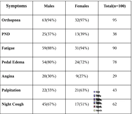
SYMPTOM COMPLEX
Symptoms
Males Females Total(n=100)Orthopnea 63(94%) 32(97%) 95
PND 25(37%) 13(39%) 38
Fatigue 59(88%) 31(94%) 90
Pedal Edema 54(80%) 24(72%) 78
Angina 20(30%) 9(27%) 29
Palpitation 22(33%) 21(63%) 43
Night Cough 45(67%) 17(51%) 62
0 20 40 60 80 100
1st Qtr 2nd Qtr 3rd Qtr 4th Qtr
East West North 0 20 40 60 80 100
1st Qtr 2nd Qtr 3rd Qtr 4th Qtr
East West North 0 20 40 60 80 100
1st Qtr 2nd Qtr 3rd Qtr 4th Qtr
East West North 0 20 40 60 80 100
1st Qtr 2nd Qtr 3rd Qtr 4th Qtr
East West North
Next to dyspnea, Orthopnea is the most common symptom,
followed by fatiguability and pedal edema. Significant percentage of females had
SYMPTOM COMPLEX
95 38 90 78 29 43 62 63(94%) 25(37%) 59(88%) 54(80%) 20(30%) 22(33%) 45(67%) 32(97%) 13(39%) 31(94%) 24(72%) 9(27%) 21(63%) 17(51%) 0 10 20 30 40 50 60 70 80 90 100 Orthopnea PND Fatigue
Pedal Ede
ma Angi na
Palpitatio n
TABLE - 4
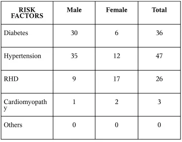
MAJOR RISK FACTORS
RISK
FACTORS Male Female Total
Diabetes 30 6 36
Hypertension 35 12 47
RHD 9 17 26
Cardiomyopath
y 1 2 3
Others 0 0 0
Already existing structural heart disease is the major risk factor
for CHF. 75% of patients had a history of heart disease in the past. Ischemic heart
MAJOR RISK FACTORS
30 35 40 9 1 0 6 12 6 17 2 0 36 47 46 26 3 0 0 5 10 15 20 25 30 35 40 45 50Diabetes Hypertension IHD RHD Cardiomyopathy Others
RISK FACTORS
TABLE - 5
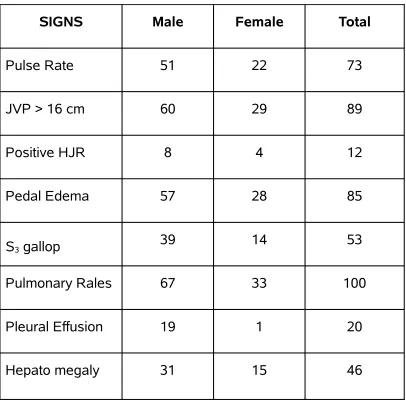
SIGNS
SIGNS Male Female Total
Pulse Rate 51 22 73
JVP > 16 cm 60 29 89
Positive HJR 8 4 12
Pedal Edema 57 28 85
S3 gallop 39 14 53
Pulmonary Rales 67 33 100
Pleural Effusion 19 1 20
Hepato megaly 31 15 46
The most common sign in CHF is Basal pulmonary
SIGNS
51 60 8 57 39 67 19 31 22 29 4 28 14 33 1 15 73 89 12 85 53 100 20 46 0 20 40 60 80 100 120Pulse R
ate
JVP > 1
6 cm
Positive
HJR
Pedal E
dema S3 gallo
p
Pulmona
ry Rales
Pleural E
TABLE - 6
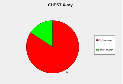
CHEST X - ray
Sign Total
Cardio megaly 91
Pleural effusion 17
Cardiomegaly is a sensitive sign of CHF occurring in
91% of patients.
CHEST X-ray
91 17
Cardio megaly
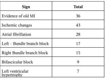
TABLE - 7
ECG
Sign Total
Evidence of old MI 36
Ischemic changes 43
Atrial fibrillation 28
Left – Bundle branch block 17
Right Bundle branch block 15
Bifascicular block 9
Left ventricular
hypertrophy 7
Electrocardiography evidence of IHD is found in 43% of patients. Atrial
fibrillation is the only arrhythmia which had been documented apart from sinus
ECG
36
43 28
17 15
9 7
TABLE -7
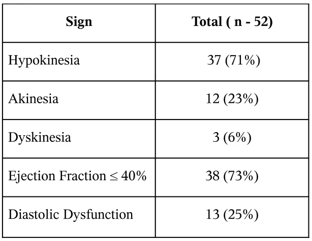
ECHOCARDIOGRAPHIC FINDINGS
Ischemic Heart Disease
Sign Total ( n - 52)
Hypokinesia 37 (71%)
Akinesia 12 (23%)
Dyskinesia 3 (6%)
Ejection Fraction ≤ 40% 38 (73%)
Diastolic Dysfunction 13 (25%)
Depressed Ejection fraction is found in 73% of patients of
ISCHEMIC HEART DISEASE
13(25%) 38(73%) 3(6%) 12(23%) 37(71%) 0 5 10 15 20 25 30 35 40hypokinesia akinesia dyskinesia Ejection Fraction ≤ 40% Diastolic dysfunction
Sign
To
tal
(n
- 5
2)
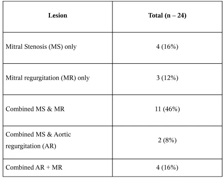
hypokinesia akinesia dyskinesiaTABLE - 8
Rheumatic Heart Disease
Lesion Total (n – 24)
Mitral Stenosis (MS) only 4 (16%)
Mitral regurgitation (MR) only 3 (12%)
Combined MS & MR 11 (46%)
Combined MS & Aortic
regurgitation (AR) 2 (8%)
Combined AR + MR 4 (16%)
Among the Rheumatic Heart diseases, combined MS & MR accounted for a large
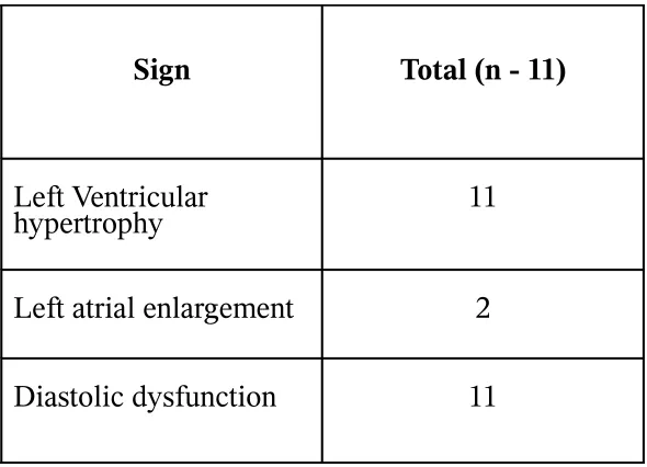
TABLE – 9
Hypertensive Heart Disease
Sign Total (n - 11)
Left Ventricular
hypertrophy 11
Left atrial enlargement 2
Hypertensive Heart Disease
11
2 11
Left ventricular hypertrophy
left atrial enlargement
[image:51.612.157.457.449.653.2]Diastolic dysfunction
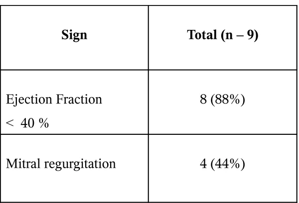
TABLE -10
Dilated Cardiomyopathy
Sign Total (n – 9)
Ejection Fraction < 40 %
8 (88%)
Dilated Cardiomyopathy
8(88%) 4(44%)
Ejection fraction
[image:52.612.105.512.54.342.2]Mitral regurgitation
TABLE -11
ETIOLOGICAL PROFILE
Diagnosis Male (n - 67) Female (n - 33) Total (n - 100)
Ischemic Heart
disease 41 (61%) 11 (33%) 52
Hypertensive Heart
Rheumatic Heart
disease 9 (13%) 15 (45%) 24
Dilated
Cardiomyopathy 6 (9%) 3 (9%) 9
Anemia 3 (4%) 1 (3%) 4
Ischemic Heart disease accounts for a majority (52%) of cases of CHF.
Rheumatic heart disease is the most common cause of Heart failure in females followed
ETIOLOGICAL PROFILE
52 11 24 9 3(4%) 6(9%) 9(13%) 8(12%) 41(61%) 1(3%) 3(9%) 15(45%) 3(9%) 11(33%) 4 0 10 20 30 40 50 60Ischemic Heart disease Hypertensive Heart disease Rheumatic Heart disease Dilated Cardiomyopathy Anemia
DIAGNOSIS
TO TA L(n = 1 00)Male (n - 67)
Female (n - 33)
TABLE-12
Age and Heart Failure
Diagnosis Mean Age (yrs) Total
Ischemic Heart disease 59.2 52
Rheumatic Heart
disease 32.5 24
Hypertensive Heart
disease 52.1 11
Dilated
Cardiomyopathy 45.1 9
Anemia 32.7 4
The mean Age of patients with CHF due to IHD was 59.2. The Mean age was low
Age and Heart Failure
59.2 32.5 52.1 45.1 32.7 52 24 11 9 4 0 10 20 30 40 50 60 70 Ischemic Heart disease Rheumatic Heart disease Hypertensive Heart disease Dilated Cardiomyopathy Anemia Diagnosis ToDISCUSSION
The total number of patients analyzed were 100, out of which 67%
were male patients. The Mean Age of patients found in the study was 50
years. Patients between the age group of 51 – 60 years accounted for a large proportion
of cases.
The most common presenting complaint is dyspnea. Majority (59%) of patients
with dyspnea were in class IV failure. This proportion is much higher compared to Euro
heart failure survey where only 34% of patients had class IV failure.
Orthopnea is the second most common symptom constituting 95% of patients
followed by Fatigue (90%) and Pedal Edema (78%).
RISK FACTORS:
Ischemic Heart disease is the most common structural heart disease
predisposing to the development of chronic congestive Heart failure
followed by hypertension and Diabetes. This finding correlate well with
of heart failure in this study as opposed to the western data.
SIGNS:
Basal pulmonary rales is the most consistent sign associated with CHF followed by
elevation of JVP (89%) and Pedal Edema (85%).
Chest skiagram revealed enlarged cardiac shadow in a majority of patients. As such
chest X-ray can be considered as a sensitive test for the
initial diagnosis of CHF. Electro cardiographic evidence of Ischemic Heart disease is
found in 43% of patients.
AETIOLOGY:
The most common cause of CHF in this study was Ischemic heart disease (52%)
followed by Rheumatic heart disease (24%) and hypertensive heart disease. This study
shows a higher prevalence of Rheumatic heart disease as a cause of CHF compared to
western data. Combined valvular lesions are associated with increased risk of congestive
heart failure.
ECHO CARDIOGRAPHY:
patients had depressed ejection fraction (≤ 40%). Among patients with IHD 73% of
patients had depressed ejection fraction. Diastolic dysfunction is present in 24 % of
patients.
AGE AND CHF:
The Mean Age of patients with CHF due to IHD was 59.2 years whereas RHD leads
to CHF at an earlier age (32.5 years). The presence of diastolic dysfunction in
Hypertensive Heart disease leads to CHF at an earlier age (52.1 years) compared to
CONCLUSION
1. In this study 67% of patients were males and 33% of patients
were females. The mean age of patients was 50 years.
2. The highest number of patients with CHF was in the age group
of 51 – 60 years.
3. Ischemic heart disease was the most common risk factor for the
development of chronic CHF.
4. Dyspnea was the most common presenting symptom and
majority of them (59%) were in class IV failure.
5. Basal pulmonary rales was the most common clinical sign of
CHF followed by elevated JVP and pedal edema.
6. Chest X-ray showed evidence of cardio megaly in majority
(91%) of patients.
7. Atrial fibrillation was the only arrhythmia documented apart
from sinus tachycardia.
8. The most common cause of congestive Heart failure is Ischemic
heart disease (52%) followed by rheumatic Heart disease (24%)
9. Among the patients with non-valvular heart disease, 61% had
depressed ejection fraction (≤ 40%) while 24 % had Diastolic
dysfunction
10. Rheumatic heart disease accounted for a majority of cases of
CHF, relatively at an earlier age (Mean 32.5 years) compared
to IHD (Mean 59.2 years).
BIBLIOGRAPHY
1. Anderson RN: United States Life tables, 1998: National Vital
Statistics Reports. In Hyaltsville, (Eds) National centre for healthy
Statistics, 2001: vol 48, no, 18.
2. American diabetics Association: Clinical practice recommendation
2002. Diabetes Care 2004, 27: 51.
3. Bourassa MG, Gurae O, Bangdiwala S,et al. Natural History and
patterns of current practice in heart failure. J. Am. Coll Cardiol,
1993,22: 14 – 19.
4. Braunwald, E. Report of the task force on Research in heart failure,
Bethesa M.D., National Heart, Lung and Blood Institute, 1994.
5. Cowie MR, Wood DA, Coats AJS, Thompson SG, Poole Wilson PA,Suresh V, et
al. Incidence and aetiology of heart failure: a population- based study. Eur heart J
1999; 20: 421 – 8.
6. The CONSENSUS Trail study group, Effect or enalapiril on morality in severe
congestive heart failure: results of the cooperative north Scandinavian enalapiril
survival study (CONSENSUS). N Engl. J Med. 1987: 1429-35.
7. Chobanian Av, Bakris GL, Black HR, et al. Seventh report of the joint National
committee on the prevention, Detection, Evaluation and treatment of High blood
pressure. Hypertension 2003: 42: 1206 – 1252.
Braunwald (ed), Philadelphia Sounders.
9. Davis R.C. Hobbs, FDR Lip. GH History and Epidemiology. ABC of Heart
failure: BMU: 2003: 1-4.
10.Dallus, Tx American heart association 2000, Heart and stroke
statistical update, 2000: vol 3, 56-68.
11.Dallas Volte S, A short historical survey of heart failure. J. Chin
Invest 1993: 71: S167 – S176.
12.Esler M, Kaye D, Lambert G, et, al. Adrenergic nervous system in
heart failure, Am J Cardiol 1997, 80: 71 – 141.
13.Francis GS, Benedict C, John stone De, et al. Comparison of Neuro endocrine
activation in patients with left ventricular dysfunction with and without
congestive heart failure. A sub study of the studies of LV dysfunction. (SOLVD)
Circulation 1990, 82: 1724 – 1729.
14.Feildman MD, Alderman JD, Aroesty JM et al. Depression of systolic and
diastolic myocardial reserve during artial tachycardia in patients with dilated
cardiomyopathy. J Chin Invest 1998, 82: 1661.
15.Gheorghiade M, Bonow RO. Chronic heart failure in the United States : A
manifestation of coronary artery disease circulation 1998, 97 : 282-289.
16.Grantham JA, Borgeon DD, Burnet et al. Pathophysiological and potential
therapeutic roles in CCF. Am J Physiol 1997, 92: 12, 1077-1083.
response to water loading in CHF. Am J Cardiol 1986, 58: 295.
18.Gaash WH. Diagnosis and treatment of heart failure based on left ventricular
function Am J of cardiology 1999,65:23
19.Ghali JK Kadakin S. Cooper R, Farlinz precipitating factors leading to
decomposition of heart failure: Trails among urban blacks. Arch Intern Med.
1988, 148: 2013.
5) Goldman L, Hasimto B, Cook EF, Loscalzo A. Comparitive reproducibility and
validity of symptoms for assessing cardiovascular functional class: Advantages of
a new specific activity scale. Circulation 1981, 64: 1227.
6) Horine Ef. An epitome of ancient pulse core: Bull Hist, Med, 1941: 10: 209-249.
7) Ho KKL, Pinksy JL, Kannel WB, et al, The epidemiology of heart failure : The
framingham study. J Am Col Cardiol 1993, 22: 6A – 13A.
8) Haldeman GA, Croft JB, Giles WH. Rashidee A. Hospitalisation of patients with
heart failure: National Hospital discharge survey. 1985 10 1995. Am Heart J.1999,
137: 352.
9) Ibrahim BS. The frequency of systolic versus diastolic heart failure in an Egyptian
cohort. Eur J Heart fail 2003, 5: 41-5.
10) International Agency for Research on Cancer: Tobacco smoke and
Involuntary smoking, lARC Monographs on the Evaluation of Carcinogenic Risks
to Humans. Lyon, France, 2003, Vol 83.
analysis from the study of Left Ventricular dysfunction (SOLVD) Trial Jam Coll
Cardiol, 1988, 31: 419 – 425.
12) Kitzman DW, Little WC, Brubaker PH, Anderson RD, Hindley WG,
Manberger Ct et al, Pathophysiological characterization of Isolated diastolic heart
failure in comparison to systolic heart failure. Ia MA 2002: 288: 2144 – 50.
13) Lip GYH, Sarwar S, Ahmed J, Lee S, Kapoor V, Child D, et al, A survey of
heart failure in general practice the West Birmingham heart failure project. Eur J
Gen Prac. 1997: 3 : 85-9.
14) Mancini DM, Wilson JR, Bolinger L, et al. Invivo magnetic resonance
spectroscopy measurement of deoxymyoglobin during exercise in patients with
heart failure. Demonstration of abnormal muscle metabolism despite adequate
oxygenation Circulation, 1994, 90: 500.
15)Mannin HL, Schwartzateiu RM. Mechanisms of disease: Pathophysiology of
dyspnoea: N Eng J Med, 1995 333: 1547.
16) Milne EN, Pistolesi M, Miniati M, Giuntiwi C: The radiologic distinction
between cardiogenic and non cardiogenic pulmonary oedema. ASR Am J
Roentgenol 1985, 144: 879.
17) Massie BM, Shah NB: Evolving trends in the epidemiologic factors of heart
failure: Rationale for preventive strategies and comprehensive disease
Management, Am Heart J 1997, 133: 703.
exercise on Adrenergic activity and heart rate validity in patients with a history of
alcohol induced AF. Am J Cardiol, 1998, 82: 317.
19) Moe GW, Grima EA, Wong NL, et al. Dual natriuretic peptide system in
experimental heart failure, J Am Coll Cadiol 1993, 22: 891.
20) Moore DA, William withering and Digitalis BMJ, 1985: 290 – 324.
21) Oakley C, Biology, Diagnosis, investigations and management of
cardiomyopathies. BMJ 1992: 315: 1520 – 4.
22)Parker M, Survival in patients with chronic heart failure and its potential
modification by drug therapy. In Cohn JN (ed Drug treatment of heart failure, 2nd
ed. Seeannus NJ, ATC international, 1988, p273.
23) RKG MW, Nease RF. Cost effective analysis in clinical practice: A case of
heart failure, Arch internal Med 1999, 159: 1690.
24)Richs. Pulmonary hypertension and Corpulmonale. In Braunwalds Heart diseases
7th edition. Zippe D, et al, Saunders publications philadephia, USA, 2005, 1113-
1115.
25) Rahimtoola Sh. The hibernating myocardium. Am Heart J 1989, 117:
2111-21.
26) Senni M, Triboulloy CM, Rodeheffer RJ, et al. Congestive heart failure in
the community. Arch Inten Med, 1999, 157: 29-34.
27)Stevenson WG. Mechanism and management of arrhythmias in heart failure.
28) Schoffer J, Tews A, Langes K et al. Relationship between myocardial nor
epinephrine content and left ventricular function and endomyocardial biopsy
study. Eur Heart J 1987, 8 : 748.
29) Stevenson LW, Perloff SK. The limited reliability of physical signs for
estimating hemodynamics in chronic heart failure JAMA 1989, 261: 884.
30) The task force on Heart failure of the European Society of Cardiology,
Guidelines for the diagnosis of heart failure. Eur Hear J 1995: 6: 741 – 51.
31) JN. Goldhaber SZ, P, Pfeffer in A, An overview of contemporary etiologies
of CH. Am Heart J 1991; 121: 1852.
32) Tsutamoto T, Hisanaga T, Fukai D, et al. Prognostic value of plasma
intercellular, adhesion molecule and endothelin l, concentration in patients with
chronic congestive heart failure. Am J Cardiol, 1995 76: 803 – 808.
33)Van Cheng BS, Kazanagra K, Garcia A, et al. A rapid bedside test for B type
natriuretic peptide treatment outcomes in patients admitted with decompensated
heart failure. J Am Coll. Cardiol, 2001, 37 : 386 – 391.
34) Vasan RS, Levy D. Defining diastolic heart failure: a call for standardized
diagnostic criteria. Circulation 2000, 101: 2118 – 21.
35)Vasan RS, Benjamin EJ, Levy D, Prevalence, clinical features and prognosis of.
diastolic heart failure: an epidemiologic perspective. J Am Coll Cardiol, 1995,
26 : 1565
peptide produced by vascular endothelial cells, Nature 1988, 332: 411.
37) Yusuf S, Sleight, Pogne J et al. Effects of an angiotensin converting enzyme
inhibitor, ramipril on cardio vascular events in high risk patients: the outcome
prevention evaluation study. N Engl J Med, 2000, 342:145.
38) Zuccala G, Galtel C, Manas Grarina E, et al. Left Ventricular dysfunction. A
clue to cognitive impairment in older patients with heart failure. J Neurol
PROFORMA
HEART FAILURE
Name : I.P.NO :
Age : Sex :
Occupation : Income :
Residence :
Present Illness :
1.Breathlessness Yes / No
Class I II III IV
2.Orthopnea Yes / No
3.PND Yes / No
4.Night Cough Yes / No
5.Chest Pain Yes / No
6.Palpitation Yes / No
7.Syncope Yes / No
8.Pedal Edema Yes / No
9.Oliguria Yes / No
Past History :
1.Diabetes Yes / No
2.Hypertension Yes / No
3.IHD Yes / No
4.Cardiomyopathy Yes / No
5.Congenital Heart
Disease Yes / No
6.RHD Yes / No
7.Thyroid Disorders Yes / No
8.Anemia Yes / No
9.CKD Yes / No
10.Others Yes / No
Personal History
1.Smoking Yes / No
2.Alcoholism Yes / No
3.Drug Abuse Yes / No
4.STI Yes / No
Family History
1.Diabetes Yes / No
2.Hypertension Yes / No
Examination :
1.Consciousness Normal / Altered
2.Pallor Yes / No
3.Dyspnea Yes / No
3.Cyanosis Yes / No
4.Clubbing Yes / No
5.Extremity Edema Yes / No
6.JVP Raised / Not Raised
7.Blood Pressure : Temperature : Weight : 8.Pulse :
9.Respiratory Rate :
Cardio-Vascular System :
1.Heart Sounds
2.Murmur
3.S 3
Respiratory System:
1.Rales Yes / No
2.Pleural Effusion Yes / No
Abdomen:
CNS :
Investigations :
1.Hemogram:
Hb
Tc
Dc
ESR
2. Blood:
Sugar
Urea
Creatinine
Na K Cl Hco3
3.Chest X-Ray:
4.ECG :
5.Echocardiography:
Final Diagnosis :
ABBREVIATIONS
ACE – Angiotensin Converting Enzyme
AS – Aortis Stenosis
AR – Aortic Regurgitation
BP –Blood Pressure
CHF – Congestive Heart Failure
CM – Cardiomegaly
DC – differential count
CXR – Chest X-Ray
DM – Diabetes Mellitus
DCMP – Dilated Cardiomyopathy
ESR – erythrocyte sedimentation rate
EF – Ejection Fraction
Hb – Hemoglobin
HT – Hypertension
HJR – Hepatojugular Reflex
HHD – Hypertensive Heart Disease
IHD – Ischemic Heart Disease
JVP – Jugular Venous Pressure
LV – Left Ventricle
MS – Mitral Stenosis
MR – Mitral Regurgitation
PF – Pleural Effusion
RHD – Rheumatic Heart Disease
RV – Right Ventricle
TC – total white blood cell count
MASTER CHART
1 – Yes