Evaluation and comparison of PCR with serological
methods for the early diagnosis of spotted fever
group of rickettsial infections.
A Dissertation submitted to the Tamil Nadu, Dr.M.G.R. Medical
University, Chennai for the M.D. Degree in Microbiology.
CERTIFICATION
This is to certify that the dissertation entitled, “Evaluation and comparison of PCR with serological methods for the early diagnosis of spotted fever group of rickettsial infections” is the bonafide work of Dr.T.Sohanlal, toward the M.D. Branch-IV (Microbiology) Degree examination of the Tamil Nadu Dr.M.G.R. Medical University, to be conducted in March 2009.
Dr.John Antony Jude Prakash, MD Dr.Mary S Mathews, MD
Guide Professor & Head
Associate Professor of Microbiology Dept. of Microbiology
Dept. of Microbiology Christian Medical College
Christian Medical College Vellore – 632 004
Vellore – 632 004 India
CONTENTS
Content
Page
I.
Introduction
1
II.
Aims and Objectives
4
III.
Review of Literature
5
IV.
Materials and Methods
29
V.
Results
40
VI.
Discussion
57
VII.
Summary and Conclusion
65
VIII.
Bibliography
67
Introduction
Rickettsial infections are known causes of illness and death worldwide. Evidence is accumulating that these diseases especially scrub typhus and spotted fever are re-emerging in India (15, 36-38, 61, 68)
As these diseases present as acute undifferentiated febrile illness, a high index of suspicion is essential in diagnosing this condition. This is further compounded by non-availability of specific tests to diagnose rickettsial infections in most centers. The less sensitive but inexpensive, Weil–Felix test, still remains the main stay of laboratory diagnosis/confirmation of these infections in India. Many clinicians still rely on dramatic response to doxycycline therapy for confirming rickettsial disease in doubtful cases (15, 37)
In case of acute infections a significant antibody titer is observed at the end of first week, concomitant with the detection of IgM antibodies, whereas IgG antibodies appear at the end of second week (6, 26). The immunofluorescence assay for detecting rickettsial antibodies adapted to a method format, the micro-immunofluorescence test (MIF) is the investigation of choice. This test which allows the simultaneous detection of IgG and IgM antibodies is available only in reference laboratories (49).
ELISA is demonstrated to be as sensitive and specific as MIF for the diagnosis of spotted fever (6). Moreover, the ELISA has been found to be more sensitive than the MIF for the detection of low levels of antibodies present in the early stages of infection/disease (7, 27). In addition, the ELISA can be automated and is less prone to intra-observer and inter-observer variation.
Direct detection of rickettsia by inoculation of guinea pigs, eggs or cell culture is hazardous and is to be attempted only in a reference laboratory (27). At present, the direct diagnostic test that is useful during the acute illness is immunohistolochemical examination of cutaneous biopsy of a rash (52). Availability of a highly sensitive and versatile technique for detection of rickettsial DNA like, the PCR has provided a means of diagnosis even before seroconversion. The sensitivity of the PCR assay depends on the specimen and the technique used.
fever group of rickettsia. They are the genes encoding the 16S rRNA, the 17kDa protein, citrate synthase, OmpA and OmpB (4, 35, 63, 72).
Aim of the study:
To determine the utility of serological and molecular techniques in the diagnosis of spotted fever group of rickettsial infections.
Objectives of the study:
A. To standardize a polymerase chain reaction (PCR) assay for the detection of spotted fever group rickettsia.
B. To evaluate the utility of PCR and IgM ELISA in confirming the clinical diagnosis of spotted fever.
C. Comparison of whole blood and skin biopsy of the rash (specimen) for detection of spotted fever group (SFG) rickettsia by PCR.
D. Confirmation of PCR amplification by sequencing of three randomly selected PCR products of the two gene targets used.
Review of literature
History
:
In 1899, Edward E. Maxey was the first to describe the clinical condition named Rocky Mountain spotted fever (RMSF), the prototypical tick-borne rickettsiosis. In 1906, Howard T. Ricketts reported the role of the wood tick in the transmission of the causative agent, subsequently named Rickettsia rickettsii. Conor and Bruch in 1909 differentiated Mediterranean spotted fever (MSF) from viral exanthems. In 1919, S. Burt Wolbach proved that ticks were the vectors that maintained Rickettsia rickettsii in nature. He also described the histopathology of the lesions of RMSF. For almost a century, R. rickettsii was the only tick-borne rickettsia conclusively associated with human disease in the Western world. During the 20th century, other SFG rickettsiae have been detected in North American ticks. They include Rickettsia parkeri in 1939, Rickettsia montanensis (previously named R. montana) in 1963, and Rickettsia rhipicephali in 1978 (46). However, these rickettsiae are generally considered nonpathogenic to humans (53, 57).
campaign that targeted the elimination of ticks. The observation of small bacillus that could not be cultivated by Ricketts revealed that bacterial diseases could be transmitted from pests to people via vectors. These findings were published in the in 1909 and the Rickettsia are named in honor of Howard Ricketts for his pioneering work. In 1915, Weil and Felix devised a tube agglutination test named after them(46) which is still used in resource poor settings, especially in India for the diagnosis of rickettsial infection.
In India, the clinically entity termed as Indian tick typhus (ITT) was described to be prevalent since the beginning of the 20th century but has not yet been isolated from cases (46).
Agents:
All over the world rickettsiae are emerging or re-emerging as major pathogens. The spotted
fevers group (SFG) composes a large group of tick, mite and flea-borne zoonotic infections
that are caused by closely related rickettsiae.
Taxonomic status:
The order Rickettsiales includes the families Rickettsiaceae and Anaplasmataceae.
The most numerous and important genus is Rickettsia. Analysis of the 16S rRNA
(
rrs
)
gene sequences has suggested that Coxiellaburnetii, the agent of Q-Fever andBartonella quintana, which causes trench fever are phylogenetically unrelated to the alpha1 subgroup of proteobacteria which includes the genus Rickettsii (1, 75).
Rickettsiae were traditionally divided into three groups using phenotypic criteria. They are the typhus group, the spotted fever group, and the scrub typhus group. In 1995, Tamura et al proposed reclassification of Rickettsia tsutsugamushi the causative agent of scrub typhus in to a new genus Orientia tsutsugamushi using 16S rRNA (rrs) gene sequence analysis (69). According to Stothard and Fuerst, Rickettsia bellii and Rickettsia canadensis phylogenetically predate the typhus –spotted fever group split and need to be included in to a separate group named the ancestral group. At present,
Transitional group (flea borne spotted fever) (ref)
Rickettsia felis
Gillespie et al (2007) have opined that Rickettsia felis should be included in a new group called the transitional group (TRG) based on detailed characterization of the conjugative plasmid (pRF) genes. (11)
Epidemiology of Spotted fever:
Only a few tick-borne rickettsioses are present on more than one continent, most are confined to a particular area, limited by the presence of their vectors (57). Between 1984 and 2004, nine species or subspecies of tick-borne spotted fever rickettsiae were designated as emerging pathogens throughout the world. They are R. japonica (Japan), “R. conorii caspia” (Astrakhan and Kosovo) R. africae (sub-Saharan Africa and West Indies), R. honei (Flinders Island, Tasmania, Australia, Thailand), R. slovaca (Europe), “R. sibirica mongolitimonae” (China and Africa), R. heilongjiangensis (China), R. aeschlimannii (Africa and Europe) and R. parkeri (USA) (44).
Rocky Mountain spotted fever (RMSF):
been reported for this illness in the pre-antibiotic era (46). The tick vectors involved in transmission are from the Dermacentor and Amblyomma genera. The infection is endemic in the rural areas of the Atlantic seaboard and mid-western USA; here most cases occur from April to August (71). In addition infection has been reported from Central and South America also (46). Inoculation eschars are rarely seen in Rocky Mountain spotted fever, and a generalized exanthema, typically purpuric, maybe absent in some 10% of the cases. Some patients with Rocky Mountain spotted fever may develop meningitis. The presence of purpuric rash in such patients will lead to difficulties in diagnosis as it cannot be distinguished from meningococcemia . About 500 cases of Rocky mountain spotted fever (RMSF) are reported yearly in the USA, predominantly in children below 16 years of age cases with peak incidence from April to August.
In sub-Saharan Africa, seven spotted fever group pathogenic rickettsias are known to occur. They are Rickettsia conorii conorii, R. conorii caspia, R. africae, R. aeschlimannii; R. sibirica mongolitimonae; R. felis and R. massiliae. In addition to the occurrence of the ubiquitous murine typhus, epidemic typhus affects tens to hundreds of thousands of persons who live in Sub-Saharan with civil war, famine and poor conditions (40).
African tick bite fever (ATBF):
of sub-Saharan Africa and the French West Indies (22). The vectors involved in transmission are ticks of the Amblyomma genus, especially Amblyomma hebraeum and Amblyomma variegatum. The illness is usually acquired in rural areas where wild game or domestic cattle are present. The tick vectors are active throughout the year are aggressive and actively attack humans. An exposed individual thus gets multiple tick bites and many members in a given group, maybe attacked at the same time. In addition, a prospective cohort study of 940 short-term travellers to rural sub-equatorial Africa, demonstrated that African tick bite fever occurred at an incidence ranging from 4.0–5.3% (18).
Mediterranean spotted fever (MSF):
Mediterranean spotted fever, also known as ‘boutonneuse’ fever, is caused by Rickettsia conorii is endemic around the Mediterranean basin, the Middle East, India, and in parts of sub-Saharan Africa. It is transmitted by the dog tick, Rhipicephalus sanguineus. In an endemic area like southern France, the incidence is as high as 50 cases / 100,000 inhabitants, with most cases occurring from May to September (58). MSF usually occurs in urban and suburban areas. A single inoculation eschar (in 70% of the cases) and a generalized maculopapular rash (in >95% of the cases) are the important clinical features in addition to fever (59).
disease is endemic in India, where it was described as early as the beginning of the 20th century (17). The disease resembles Mediterranean spotted fever except that the rash in Indian tick typhus is purpuric, and an inoculation eschar is extremely uncommon (16). Seven cases among labourers working in tea estates in Kerala have been diagnosed by MIF (68).
Astrakhan fever:
Since 1983, some 1000 cases of Astrakhan fever, caused by R. conorii subsp. caspia have been reported in the Caspian basin (70). The name is derived from a region of Russia located by the Caspian Sea called as Astrakhan where cases have occurred since the 1970s. Rash is seen in most patients whereas as tache noire (eschar) is seen in about a quarter of the cases. The dog tick Rhipicephalus pumilio is the principal vector (70). Astrakhan fever rickettsia has also been detected in R. sanguineus ticks collected from dogs and military personnel in Kosovo The clinical features are similar to that of Mediterranean spotted fever but an inoculation eschar maybe seen in only 20% of the cases (9).
Queensland tick typhus:
Queensland tick typhus caused by Rickettsia australis occurs in rural eastern Australia. It is transmitted by ticks of the Ixodes genus and 60 cases have been reported since 1946 (64).
Fatalities are rare (17).
North Asian tick typhus (Siberian tick typhus):
Rickettsia sibirica causes North Asian tick typhus (Siberian tick typhus). This disease is endemic in large parts of rural northern Asia, including the former USSR, China and Mongolia with sero-positivity rates exceeding 60% in some endemic areas. Most cases occur between May and September (31).
Rickettsial pox:
Rickettsial pox caused by Rickettsia akari is the only known mite-borne spotted fever group rickettsiosis. It is transmitted by the bite of bloodsucking infected mites (Allodermanyssus sanguineus) and characterized by a primary eschar, fever and a
papulo-vesicular rash. Culture confirmed cases have been reported from the USA,
Ukraine and Croatia and in 1997 from South Africa (21).
Japanese spotted fever (JSF):
tick surveys suggests that Haemophysalis flava and Haemophysalis hystericis as vectors of JSF. Two other serotypes or species of spotted fever group rickettsiae have been isolated from ticks in Japan; one is closely related to R. helvetica and the other whose genotype is unknown, is closely related to a Slovakian genotype (33). In a study undertaken in the Western Province of Sri Lanka, 31 patients with possible rickettsioses were identified. Scrub typhus was diagnosed in 19 and spotted fever in eight (50).
Though Rickettsia aeschlimannii was first isolated from ticks in 1997 only two
human cases have been reported world-wide till 2004 (17). R. sibirica subsp.
mongolotimonae and R. heilongjiangensis cause disease in China, Mongolia, Siberia,
Armenia and Pakistan (76).
Flea-borne spotted fever: Flea-borne spotted fever is caused by R. felis and is
transmitted by the cat flea, Ctenophalides felis. Human infection has been described
since 1994 from USA, Mexico, Brazil, Germany, Spain, Tunisia, Thailand, South
Korea and Laos (48).
Data from India:
species examined, R. conori was found in Boophilus microplus and Rhipicephalus haemaphysalis. Antibodies against the R. conori antigen were detected in the sera of Rattus blanfordi, R.r. rufescens and Suncus murinus by complement fixation test (41). More recently, Kamarasu et al (2007) have opined that scrub typhus and rickettsial illness are distributed in Tamil Nadu based on a serological study conducted over two years (2004 & 2005). In 2004, 115 of 306 samples and in 89 of 964 samples tested demonstrated positive titers for scrub typhus by the Weil-Felix test. Positive titers for other rickettsial illness were observed in 44 samples (19). There is ample evidence from our centre that the number of cases of scrub typhus and spotted fever tends to increase during the cooler months (Prakash JAJ, unpublished).
Pathogenesis:
Till date 20 rickettsial genomes have been fully sequenced. Totally 17 rickettsial surface cell antigen (sca) genes numbered 1 to 17 have been described (39). The rOmpA and rOmpB(sca5) are high molecular weight proteins encoded by surface cell antigen gene (sca) and help in the attachment of bacteria to the host cells (30). The sca4 gene is known as gene D, it encodes for a phospholipase D. Phosholipase D has been postulated to be responsible for the cytotoxic effect on host cells infected by rickettsia (60) and for survival of rickettsiae in arthropods. Phosholipase A2 is responsible for mediating host cell entry and allows the spotted fever rickettsia to escape phagocytosis by lysis of the phagosomal membrane(74). Immune responses are induced by rOmpA, rOmpB and sca4 (geneD) (60).
The preliminary event in rickettsial infection is entry of organisms into the skin,
through the bite of the arthropod vector. Rickettsiae have a predilection for
endothelial cells and hence infect all organs having a vasculature (74). They attach to
host cells with the help of adhesions OmpA and OmpB (46). Once attached, they
induce the host cell to phagocytose by receptor mediated endocytosis (74).
Phosholipase A2 enables the SFG rickettsiae to escape from the host cell phagosome
allows the cell to cell spread of bacteria leading to a disseminated infection without
host cell lysis (12). Infection of the endothelial cells leads to increased permeability
of the microvasculature and is due to the disruption of the adherens junctions between
these cells (74).
Pathology:
Histopathologic examination reveals features of septic vasculitis. In the early lesions the infiltrates consist of a perivascular lymphohistiocytic infiltrate along with extravasated red blood cells and oedema of the dermis. This progresses later to leukocytoclastic vasculitis. Basal cell vacuolization, lymphocytic exocytosis fibrin thrombi and capillary wall necrosis is also seen infrequently (25).
Clinical features:
In tick borne rickettsioses symptoms appear 6 to 10 days after the tick bite. Characteristically an inoculation eschar is seen at the site of the tick bite in addition to fever, headache, muscle pain rash and local lymphadenopathy (2). The rash in spotted fever is macular and blanching, becomes erythematous, maculopapular with petechial hemorrhages in fully evolved cases (25). Purpura fulminans is seen in severe cases. The rash typically begins on the extremities and then spreads centripetally and often involves the palms and soles .
As soon as infection is suspected samples are taken for confirmation of diagnosis and specific therapy is to be instituted without delay for best results. The drug of choice is doxycycline even for young children (25). Chloramphenicol is a useful alternative especially in those allergic to doxycycline. Other drugs used are josamycin for pregnant women (3 g per day for 7 days). Chloramphenicol also can be used for pregnant women except near term.
Laboratory diagnosis of spotted fever:
Isolation of spotted fever group (SFG) rickettsiae:
The ultimate method for diagnosis and also discovery of a new species is isolation of the organism in culture. As all rickettsiae including spotted fever group (SFG) rickettsia are obligate intracellular parasites cultivation will be successful only when attempted in media containing cells or by inoculation into susceptible animals (27). The ability to cultivate rickettsia provides a constant source of antigens which can be used for serological assays. Culture needs to be undertaken in Biosafety level III containment facilities and requires extensively trained personnel. In addition, the best results are obtained when the sample is inoculated immediately on receipt in the laboratory and when treatment has been delayed till sample is collected.
Inoculation of animals and embryonated eggs: In the past guinea pigs and mice have been used for isolation and also for eliminating mycoplasma from contaminated cell cultures . Yolk sac inoculation of embryonated chicken eggs has also been used previously for isolation of rickettsia. These methods have been replaced now by the more efficient and comparatively less cumbersome cell culture methods including the shell vial culture techniques (34).
Cell culture: Various adherent cell lines have been used successfully to isolate rickettsia in biosafety containment level III facilities. The cell lines commonly used are Vero (African green monkey kidney), L929 (mouse fibroblast), HEL (human embryonic lung fibroblasts), XTC2 (Xenopus laevis, tick cell line) and MRC5 (human lung fibroblast) (27, 51)
As the sensitivity is low and also time taken for detection is longer, conventional cell culture has been replaced by shell vial culture in the reference centers undertaking rickettsial isolation by culture.
Shell vial culture technique: This technique which was originally described for
cytomegalovirus isolation has been adapted by Marrero and Raoult (1989) for
isolation of R. conorii using HEL fibroblasts (34). In 1991, Kelly et al, demonstrated
that isolation of rickettsiae is better and occurs earlier when Vero or L929 shell vial
should be centrifuged after the material has been inoculated. The reason for this being that
centrifugation enhances rickettsial attachment and cell penetration (2, 23). In spite of the increased
sensitivity of this technique, about a third of rickettsiae, have been shown to be nonviable on
subsequent passage (46). Confirmation is by microscopic examination after Acridine orange or
Gimenez or immunofluorescence staining or PCR (2).
The time taken for growth is usually 48 -72 hours) (27). La Scola and Raoult have reported that in 34 (43%) of the 79 samples from untreated patients, R conorii was isolated by shell vial culture technique. They also reported that the sensitivity of isolation increases to 50% when CECs are used (13).
Immunohistochemistry: Spotted fever group rickettsia can be detected in tissues by using immunohistochemcal methods. Immunofluorescence technique and immunoperoxidase methods have been used to detect rickettsia in tissue specimens both fresh or formalin treated and paraffin embedded. Though the specificity is 100%, sensitivity ranges from 53 % and 75% (55, 73) are superior to stains such as Giemsa or Giminez . False-negative results have been attributed to therapy with tetracycline or chloramphenicol more than 24 hours prior to sampling and inability to uobtain a tissue sample from the focus of vasculitis (73). Immunohistochemical assays for antigen detection provide better resolution of cells around the detected rickettsiae and can be performed on archived specimens (42)
The most frequently used methods for diagnosis are serological assays. The major problem encountered in serological diagnosis is the occurrence of cross-reactions. Cross reactivity is due to the similarity of antigens of pathogens within the same genus and sometimes from different genera (54).
Weil-Felix test:
Weil-Felix test is still the only assay available in many diagnostic laboratories in India. This assay has been replaced by more sensitive assays such as MIF (micro-immunofluorescence assay) and ELISA for detection of IgM and IgG antibodies. The Weil-Felix test uses Proteus antigens as these have antigens which share epitopes with rickettsial antigens with the exception of R. akari (54) The Proteus vulgaris OX-2 antigen reacts with sera from persons infected with SFG rickettsiae. The P. vulgaris OX-19 antigen reacts with sera from individuals suffering form typhus and spotted fever (RMSF) . As this assay detects predominantly IgM antibodies it is negative in Brill-Zinsser disease, were an IgG response is observed. The advantages of this test is that a single sample can be processed, can be performed as a tube agglutination or a micro-plate agglutination and antigens can be easily produced in-house (minimal risk) especially in resource poor situations .
The complement fixation (CF) test has been used in the past for the detection of antibodies specific for rickettsiae. Though purified rickettsial antigens which are species specific have been used, cross-reacting antibodies among the SFG and typhus groups have been observed (65). In addition to requiring culture facilities and expertise for production of these antigens, results depend on how the antigen was produced and how much was used.
Micro-immunofluorescence assay:
Micro-immunofluorescence assay uses the micro-method format to detect rickettsial antibodies. This test is an adaptation of the immunoflourescence assay and is considered the serologic reference test for diagnosis of rickettsioses (49). The significant titer for detection of IgG and IgM antibodies to Rickettsia conorii (MSF) is ≥ 128 & ≥64. For infections caused by other spotted fever rickettsia, the cutoff titers are ≥ 64 for IgG and/or ≥32 for IgM antibodies (2).
The diagnostic titer will vary from region to region and is dependent on the endemicity of the infection in the population being studied. For RMSF, a sensitivity ranging from 84.6% to 100% and a specificity of 99.8 to 100% has been reported (2). A sensitivity of 46% to 100% has been documented for MSF by MIF (2). MIF assays using the whole selection of antigens are available only in reference centers.
ELISA:
co-workers in 1983 (6). In addition, ELISA detects the low levels of antibodies seen during late convalescence. Keysary and Strenger have concluded that lipo-polysacharide (LPS) ELISA is able to differentiate between MSF and murine typhus in patients whose sera show cross reactions by MIF (24). ELISA needs minimal equipment and expertise. Hence it can serve as a useful diagnostic tool in centers which lack the infrastructure necessary to provide a MIF assay or culture or PCR for diagnosis of rickettsial infections.
We use a commercially available IgM ELISA for spotted fever which uses Rickettsia rickettsii as the antigen. The manufacture (PanBio Ltd, Brisbane, Australia) states that cross reactions can occur with the typhus group rickettsiae. A sensitivity and specificity of 98.2% and 93.6% has been demonstrated by using the MIF as the reference test.
Other serological tests:
Western blot (WB) assay after cross-absorption (CA) has been used in reference centers to help to determine the rickettsial agent responsible for illness by antibody evaluation (27).A study done by Raoult et al (2001) demonstrated that 81 of the 414 subjects evaluated were positive by MIF with CA and WB (56). A specificity and positive predictive value of 100% was seen with both techniques (56).
According to Parola et al (2005) a rickettsia is said to be the agent responsible for infection if the IgG or IgM antibody titer against this rickettsia is at least two or more serial dilutions greater than IgG or IgM antibody titers against other rickettsia. When the above criteria is not satisfied, Western blot assays with or without cross-absorption needs to be performed (47).
It is also important to remind clinicians that collection of acute and convalescent-phase serum specimens, separated by several weeks, is necessary to confirm disease.
Polymerase chain reaction
citrate synthase gene (gltA), ompA, ompB and geneD (encodes for phosholipase D) and these have been used successfully by many researchers for detection and characterization of rickettsia (46) .
Fournier et al have devised a nested PCR known as suicide PCR in which single use primers which were never used in the laboratory previously were used. Skin biopsy specimens from 103 individuals diagnosed to have rickettsioses were studied. Culture, standard PCR and suicide PCR was performed on all. Of these 32 (31.0%) were culture positive, 47 (45.6%) were positive by standard PCR. Skin biopsy specimens of 70 (68.0%) of the 103 patients were positive by “suicide PCR”(10).
Yeon-Joo Choi et-al evaluated a nested PCR detect rickettsial DNA using gltA and rOmpB gene primers in 200 serum samples from febrile individuals in South Korea. Rickettsial DNA was detected in 24 individuals. Rickettsia conorii, R. japonica, and R. felis and R. typhi were the rickettsia identified by sequence analysis of the amplicons (3).
A nested PCR to detect a 267 bp amplicon of the rOmpB gene of SFG rickettsiae was designed and evaluated by the same group using primers derived from the R.conorii strain Seven. As low as seven copies per assay, were detected by this nested PCR. SFG rickettsial DNA was found in 71 of the 100 MIF positive serum samples tested (5).
by amplifying a 214-bp DNA fragment of the R conorii 17-kDa protein genes (29). Restriction fragment length polymorphism (RFLP) was used to differentiate between these two groups of rickettsiae. The assay was shown to have a detection limit of 10 copies per assay. They were able to detect the requisite target in all the five serum samples and six of the seven tissue samples from the MSF cases that succumbed to their illness (29).
Tzianabos et al (1989) suggested that it is possible to detect spotted fever group rickettsial DNA from blood clots by amplifying a 246 bp fragment of 17-kDa antigen gene by PCR. Of the nine culture positive RMSF patients, only seven were positive by PCR. The standard PCR described by Tzianabos et al can detect 100 fg of rickettsial DNA which is equivalent to detection of 60 rickettsiae (72).
Massung etal (2001), designed a nested protocol for amplifying the same gene (17kDa) for detection of DNA from the typhus group rickettsiae. Using this protocol typhus group rickettsial DNA was amplified from the CSF of a patient with meningitis from New Mexico. A combination of the above mentioned two PCR protocols has been recommended by Parola et al (2005) for detection of SFG rickettsial DNA from blood, serum, skin biopsy and tick specimens (46). Moreover, they have opined that for routine identification of rickettsia, detection of the OmpA and gltA genes by PCR will suffice (46).
Stenos et al (2005) designed a qPCR assay that detected a 74bp fragment of the citrate synthase gene (gltA) of rickettsia belonging to the spotted fever and typhus group. The assay was found to detect one target copy number per reaction. Therefore, the authors feel that this assay would be very useful for the diagnosis of rickettsial illnesses, when rickettsial numbers are very low (67).
Eremeeva et al (2003) described a SYBR Green assay which detects a 154bp fragment of the rOmpA gene of spotted fever rickettsia. The detection limit of this assay was found to be 5 copies per reaction. The qPCR was found to be as sensitive as the plaque assay and was useful in the quantitation of rickettsia from infected tissues (8).
Paris et al (2008) designed a multiplex qPCR for detection of scrub typhus group, typhus group and spotted fever group of rickettsiae using the 47kDa, gltA and OmpB genes as targets. The amplicon sizes were 74 bp for gltA, 118 bp for 47 kDa and 252 bp for ompB genes. Detection limits were 24 copies/µl, 5 copies/ µl & 1 copy/µl respectively. Melt-curve analysis was used to detect group specific amplicons. The assay was subjected to 54 samples, of which all cell-culture and 75% of characterized clinical buffy coat samples were correctly identified (43).
Materials and methods
Subjects:
Children above the age of 6 months and adults with fever (≥5 and ≤15 days) and rash were enrolled into the study, if found eligible as per the inclusion and exclusion criteria enumerated below. These subjects were included after evaluation by a dermatologist and after ruling out drug reactions and viral exanthems, by history and examination (A clinical proforma was also filled up as given in Appendix II at the time of sample collection. Informed consent was obtained from all individuals recruited: Appendix I).
Inclusion criteria: 1. Age ≥ 6 months
2. Fever ≥5 and ≤15 days
3. Rash: non confluent maculopapular rash with or without purpura/purpura fulminans.
Exclusion criteria: 1. Age ≤ 6 months
Samples:
Skin biopsy from the rash, whole blood and serum utilized for the study. A 3×3mm sized piece of skin was taken by punch biopsy as per standard protocol after administration of local anesthesia to the site. Whole blood (5ml) was collected in EDTA tube (Vacuette, Greiner Bio-one GmbH, Kremsmunster, Austria) and clotted blood in a serum collection tube (BD Vacutainer, BD, Franklin Lakes, NJ, USA) for serology.
Study period: November 2006 to April 2008
Tests done on the samples collected from patients recruited into the study Serum: Spotted fever IgM ELISA, scrub typhus IgM ELISA and Weil-Felix test Whole blood: Nested PCR for detection of citrate synthase gene (gltA) and 17kDa gene of rickettsia.
Skin biopsy: Nested PCR for detection of citrate synthase gene (gltA) and 17kDa gene of rickettsia.
All the above mentioned tests were evaluated for the diagnosis of spotted fever using the case definition given below.
Case definition of spotted fever used in this study:
to scrub typhus by ELISA.
Statistical analysis:
Sample size: In this study the clinical inclusion criteria used is specific for spotted fever. About 30% laboratory confirmation has been described in literature, in a less clinically defined group. As the present study used more rigid clinical criteria, the reasonable expectation of prevalence was considered as 40-50%. Therefore, sample size was calculated with an expected prevalence of 50%, desired precision of 15% and 95% confidence interval. Using these criteria the minimal number of samples to be tested was determined as 43. (Epi Info 6 version 6.04b to c).
Test sensitivity, specificity, negative predictive value, and positive predictive value were calculated by using a 2 X 2 table using the case definition described above. The Kappa co-efficient (for assessing the agreement of serological and molecular tests) was calculated using (Epi Info 6 version 6.04b to c)
Serology
the Rickettsia rickettsii antigen.
The Weil-Felix test was done with in-house antigens prepared and interpreted as per the protocol followed currently in the department. A titer of ≥80 with OX 19 and / or OX 2 was considered positive for rickettsial infection and OX K ≥80 was taken as scrub typhus positive.
PCR:
Whole blood and skin biopsy specimens (3mm X 3mm, punch biopsy) of each of these patients stored at -70° C before DNA extraction.
DNA extraction from whole blood:
DNA was extracted from frozen whole blood using Flexi Gene DNA kit (Qiagen GmbH, Hilden, Germany) as per procedure described by the manufacturer. Prior to extraction the whole blood sample was subjected to a pre-purification procedure using a lysis buffer (containing 20mM Tris, 10 mM EDTA and pH 8). The Flexigene protocol subsequently followed for extraction is as described below:
1. Buffer FG 1 (25ml/sample) was transferred into a 50 ml centrifuge tube. Whole blood (5-6 ml) was added and mixed by inverting the tube five times. 2. The mixture was centrifuged for 5 min at 2000g.
3. The supernatant was discarded and the tube was inverted on a clean piece of absorbent paper for 2 min, taking care that the pellet remained in the tube. 4. Buffer FG2/QIAGEN Protease (prepared earlier) was added and the closed
5. Once homogenization was complete, the tube was inverted three times and placed in a heating block and incubated at 65º C for 10 min.
6. Isopropanol (5 ml) was added and mixed thoroughly by inversion until the DNA precipitate became visible as threads or a clump.
7. The mixture was centrifuged for 3 min at 2000xg.
8. The supernatant was discarded and the tube inverted on a clean piece of absorbent paper for 2 min, taking care that the pellet remained in the tube. 9. Five ml of 70% ethanol was now added and the mixture vortexed for 5
seconds and centrifuged for 3 min at 2000xg
10. The supernatant was discarded and the tube was inverted on a clean piece of absorbent paper for 5 min, taking care that the pellet remained in the tube 11. The DNA pellet obtained was air dried until all the liquid evaporated).
12. One ml of Buffer FG3 was added to the dried pellet and the same was vortexed for 5 seconds at low speed, the DNA was completely dissolved by incubating for one hour at 65º C in a heating block.
The extracted DNA was stored as 10 µl aliquots at -70° C pending testing by PCR. DNA Extraction from skin biopsy:
DNA was extracted using QIAamp Blood DNA Mini Kit (Qiagen GmbH, Hilden, Germany) for DNA purification from tissue as per manufacturer’s instruction as follows.
2. To this mixture, 20 µl Proteinase K was added and mixed by vortexing followed by incubation at 56ºC until the tissue was completely lysed.
3. The sample was pulse vortexed for 15 seconds after adding 200 µl of Buffer AL, followed by incubation at 70ºC for 10 min.
4. Absolute alcohol (200 µl) was added to the sample and mixing done by pulse vortexing for 15 seconds.
5. The resulting mixture from step 4 was carefully applied to the to the QIAamp spin column. Centrifugation was done at 6000xg (8000rpm) for 60 seconds and the tube containing the filtrate was discarded.
6. The spin column was placed into a new clean two ml collection tube and 500 µl buffer AW1 was added. The column was centrifuged at 6000xg (8000rpm) for 60 seconds, the tube containing the filtrate was discarded. The spin column was placed in a new, clean 2 ml collection tube.
7. Five hundred micro-liters (500 µl) of buffer AW2 was added into the spin column and centrifuged at 20000xg (14000rpm) for 3 minutes. The tube containing the filtrate was discarded.
iii) PCR amplification:
Citrate synthase gene (specific for all rickettsiae) (4, 63) and the 17kDa antigen (specific for spotted fever rickettsiae) (35, 72) are amplified by nested protocol. The thermal cycler used was the ‘Veriti’ 96 well Fast Thermal Cycler (Applied Biosystems, Foster City, CA, USA). Primers were synthesized by Sigma-Genosys, Bangalore, India. Genomic DNA from Rickettsia parkeri, kindly provided by Professor JS Dumler, Division of Medical Microbiology, Department of Pathology, Johns Hopkins Medical Institutions, Baltimore, MD, USA was used as the positive control.
Standardization of the PCR assays used:
Standardization was done using annealing temperature starting from 45°C to 56°C° and MgCl2 concentrations starting from 2 mM to 12 mM with an increasing interval of 2 mMol /reaction. Subsequently the optimal temperature and duration for extension was determined. The optimal conditions obtained for each are given below for both the primary & secondary reactions for the two targets detected.
a) Detection of Citrate Synthase Gene (4) Primary reaction (first round)
primers already described (4, 63).The sequence of the primers used is as given below: RpCS. 877p (forward): 5’ - GGG GAC CTG CTC ACG GCG G – 3’
RpCS. 1258n (reverse): 5’ - ATT GCA AAA AGT ACA GTG AAC C –3’ PCR Mix:
A 8 µl portion of extracted DNA was amplified in a 100 µl reaction mixture containing 10 pmol of each primer, 200 µM each of dATP, dCTP, dGTP, dTTP, 1.25 Units of Taq DNA polymerase (Applied Biosystems, Foster City, CA, USA), 5 mM MgCl2 and 10 x PCR buffer without MgCl2 and PCR grade water (MilliQ, Millipore Corporation, Bedford, MA, USA) to make up the volume to 100 µl.
Amplification parameters:
PCR amplification included an initial denaturation of 95°C for 2 minutes followed by 35 cycles of denaturation at 95°C for 30 seconds, annealing at 50°C for 20 seconds, extension at 68°C for 25 seconds and concluded with a final extension at 72°C for 3 minutes.
Secondary reaction
A 338 fragment of the citrate synthase gene was amplified using the primers whose sequences are given below (4).
RpCS.896p (forward): 5’ - GGC TAA TGA AGC AGT GAT AA– 3’ RpCS. 1,233n (reverse): 5’ - GCG ACG GTA TAC CCA TAG C– 3’ PCR mix:
Units of Taq DNA polymerase (Applied Biosystems, Foster City, CA, USA), 3 mM MgCl2 and 10 x PCR buffer without MgCl2 and PCR grade water (MilliQ, Millipore Corporation, Bedford, MA, USA) to make up the volume to 50 µl.
Amplification parameters:
PCR amplification included an initial denaturation of 95°C for 2 minutes followed by 35 cycles of denaturation at 95°C for 30 seconds, annealing at 52°C for 30 seconds, extension at 68°C for 30 seconds and concluded with a final extension at 72°C for 3 minutes.
b) Detection of 17kDa antigen encoding gene: (35, 72). A 246 bp fragment of the coding region of the 17-kDa antigen gene amplified by using nested protocol Primary reaction
The primers R17122 and R17500 sequences described (35) are as given below R17122 (forward): 5’ - -CAGAGTGCTATGAACAAACAAGG– 3’
R17500 (reverse): 5’ - CTTGCCATTGCCCATCAGGTTG– 3’ PCR Mix:
A 8 µl portion of extracted DNA will be amplified in a 100 µl reaction mixture containing 10 pmol of each primer, 200 µmol each of dATP, dCTP, dGTP, dTTP, 1.25 Units of Taq DNA polymerase (Applied Biosystems, Foster City, CA, USA), 5 µM MgCl2 and 10 x PCR buffer without MgCl2 and PCR grade water (MilliQ, Millipore Corporation, Bedford, MA, USA) to make up the volume to 100 µl.
Amplification consisted of initial denaturation at 94°C for 3minutes, followed by of 40 cycles comprising, denaturation at 94 for 30 seconds, annealing at 52°C for 30 seconds, extension at 67°C for 30 seconds and final extension at 72°C for 3 minutes. Secondary reaction
The primers Tz15 and Tz16 described by Tzianabos et al in 1989 (72) were used and the sequences are as given below
Tz 15 (forward) 5’-TTC TCA ATT CGG TAA GGG C-3’ Tz 16 (reverse) 5’-ATA TTG ACC AGT GCT ATT TC-3’ PCR Mix:
A 1µl portion of amplicon is taken as template and amplified in a 50 µl reaction
mixture containing 10 pmol of each primer, 200 µM each of dATP, dCTP dGTP, dTTP, 1.25 Units of Taq DNA polymerase (Amplitaq, Applied Biosystems, Foster City, CA, USA)), 2 µmol MgCl2 and 10 x PCR buffer without MgCl2 and PCR grade water (MilliQ, Millipore Corporation, Bedford, MA, USA) to make up the volume to 50 µl.
Amplification parameters:
Detection of PCR product:
The citrate synthase gene PCR amplified product [10 µl] and the 17kDa protein gene product (10 µl) was electrophoresed in a submarine electrophoresis unit in a 1.5% agarose gel stained with ethidium bromide. The amplified product was visualized using a gel documentation system (Bio-Rad Laboratories, Hercules, CA, USA).
Sequencing
Results
[image:43.612.170.507.414.650.2]From November 2006 to April 2008, 59 individuals with fever and rash were recruited into this study. Of these, 32 were males (54.2%) and 27 were females (45.8%). A diagnosis of spotted fever was made in an individual who had fever of five to fifteen day duration with rash, in whom defervescence of fever occurred within 48 hours of doxycycline therapy with negative serology for scrub typhus. Using this case definition, 42 of the 59 subjects enrolled were diagnosed as spotted fever. Among these 42 cases, 22 (52.4%) were males and 20 (47.6%) were females. Figs 1-3 show the different types of rash encountered in our patients.
Fig. 2. A spotted fever case with purpuric rash. Note the erythematous papule with central purpuric area.
[image:44.612.190.516.436.671.2]Fig. 4. PCR for citrate synthase gene (gltA). Gel picture showing the 338 bp gene product amplified by nested PCR of skin biopsy specimens.
Positive samples: 3, 4, 9 and 17 Positive controls: sample 7 and 18 Negative control: 16
Fig. 5. PCR for 17-kDa gene. Amplified 246 bp partial gene product in gel analysis.
[image:45.612.110.568.110.271.2] [image:45.612.112.571.441.593.2]The age distribution of patients with fever and rash including those who have been diagnosed as spotted fever is as given in fig.6.
Fig. 6. Age wise distribution of patients with fever and rash
AP TAMIL NADU
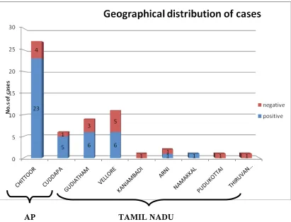
Fig. 7. Geographical location of study subjects and spotted fever cases
The maximum numbers of cases were from Chittoor district in Andhra Pradesh. Of the 27 cases of fever with rash recruited into the study from this area, 23 (85%) were diagnosed as spotted fever as per the case definition used.
Fig. 8. The geographical distribution of the patients depicted in the histogram (fig. 7)
The mean duration of fever among study subjects was 9.8 ± 3.4 days; among those with spotted fever was 10.5 ± 3.4 days and in those who were not diagnosed as spotted fever was 8.2 ± 3.3 days.
synthetase gene (gltA) and 17-kDa gene detected 38 (90.48%) and 23 (54.76%) cases respectively. Among the17 patients who were not spotted fever, ELISA has given two false positives and skin biopsy gltA PCR has given one false positive result. It is interesting to note that the skin biopsy PCR for 17-kDa gene did not show any false positives but failed to detect 15 (39.47%) of the 38 spotted fever cases detected by skin biopsy gltA PCR. Both PCRs failed to detect four (9.52%) spotted fever cases whereas ELISA was unable to detect five (11.90%) of 42 diagnosed as spotted fever according to the case definition.
In addition to the IgM ELISA for scrub typhus and spotted fever, the Weil-Felix test was also performed on the serum samples. The Weil-Felix test was positive in 14 (33.33%) of the spotted fever cases. Though the sensitivity of the Weil-Felix test is low, it did not show any false positive results.
Table 1: Comparison of citrate synthase gene PCR with IgM ELISA in the diagnosis of spotted fever
Clinical Spotted fever
Negative Positive
gltA PCR and ELISA POS 0 34
gltA PCR and ELISA NEG 14 1
gltA PCR POS, ELISA NEG 1 4
gltA PCR NEG, ELISA POS 2 3
Observed agreement between tests: 88%
Kappa coefficient: 0.72 (>0.40 significant) Z value : 5.37 (>1.96 significant) P value : <0.0005
Table 2 shows the performance of the IgM ELISA in the diagnosis of spotted fever. Of the 39 spotted fever IgM ELISA positives, 37 were diagnosed as spotted fever using the case definition. In 34 of these patients, gltA PCR was positive (33 skin biopsy and one whole blood). In three spotted fever cases where serology was positive, skin biopsies and whole blood gltA PCR could not detect rickettsial DNA. The skin biopsy gltA PCR detected four spotted fever cases that were negative by serology for spotted fever by IgM ELISA.
This patient also exhibited IgM antibodies to scrub typhus by ELISA (patient had eschar) and subsequently expired in spite of treatment with doxycycline. The two spotted fever IgM ELISA false positives were negative by gltA PCR and 17-kDa PCR done on skin biopsy and blood and also by the Weil Felix test. However, these two samples were positive for IgM antibodies to scrub typhus by ELISA and showed the expected clinical response to doxycycline.
Table 2: Evaluation of ELISA in the diagnosis of spotted fever
Clinical Spotted fever
Positive Negative TOTAL
ELISA POS 37 2 39
ELISA NEG 5 15 20
TOTAL 42 17 59
(A value of ≥16 PanBio units was considered as positive) Sensitivity of ELISA : 88.10%
Specificity of ELISA : 88.24%
Positive predictive value : 94.87%
Negative predictive value : 75.00 %
Table 3 demonstrates the ability of the citrate synthase gene PCR in the diagnosis of
spotted fever. The aforementioned PCR assay detected 38 (36 skin biopsy + two
whole blood) of the 42 cases of spotted fever. In four of these cases gltA PCR was
negative (both skin biopsy and whole blood) but in three of these cases IgM ELISA
assays (done on both samples), spotted fever IgM ELISA and scrub typhus IgM
ELISA with therapeutic response to doxycycline. In the spotted fever negative group,
the lone false positive result by gltA PCR (skin biopsy positive, whole blood
negative) was also negative by spotted fever IgM ELISA, 17-kDa gene PCR (both
skin biopsy and whole blood). This patient was positive by scrub typhus IgM ELISA
[image:52.612.98.534.385.460.2]and succumbed despite treatment with doxycycline.
Table 3: Evaluation of PCR for detection of rickettsial citrate synthase (gltA)
gene in the diagnosis of spotted fever
Clinical Spotted fever
Positive Negative TOTAL
PCR POS 38 1 39
PCR NEG 4 16 20
42 17 59
Sensitivity of PCR : 90.48%
Specificity of PCR : 94.12%
Positive predictive value : 97.44%
Negative predictive value : 80.00%
gltA PCR positives, one sample was considered as a false positive result. Whole blood PCR for gltA gene picked up 13 cases of which two were skin biopsy PCR negative for gltA. The 17-kDa gene PCR done using DNA from skin biopsy and whole blood was negative in these two patients (Both these two patients were spotted fever IgM ELISA positive). In 11 spotted fever cases, DNA extracted from both skin biopsy and whole blood was positive by gltA PCR
All the 23 skin biopsy samples from cases of spotted fever, positive by 17-kDa gene were also positive by gltA PCR. All the four whole blood 17-kDa gene PCR positives were positive by skin biopsy 17-kDa gene PCR and gltA PCR (skin biopsy and whole blood) and diagnosed as spotted fever as per the case definition used in this study.
The gltA gene gave the best yield among the two gene targets used for PCR detection of rickettsial DNA (i.e. gltA and 17-kDa gene) from both skin biopsy and whole blood. Totally 38 patients diagnosed as spotted fever patients were PCR positive by gltA gene PCR (skin biopsy = 36, whole blood = 2). Of these 38 spotted fever cases, PCR for 17kDa gene picked up 23 (60.52%) positives when DNA from skin biopsy was the source. Whole blood 17-kDa gene PCR could detect only four patients with spotted fever. Table 4enumerates the perfomance of the two PCR assays when done on the two samples used in this study
Table 5: Evaluation of Weil-Felix test in the diagnosis of spotted fever (n = 59)
Clinical Spotted fever
Positive Negative TOTAL
Weil-Felix pos. 14 0 14
Weil-Felix neg. 28 17 45
42 17 59
Significant titer for spotted fever by Weil-Felix test: OX19 and /or OX2 ≥80
Sensitivity : 33.33%
Specificity : 100.0%
Positive predictive value : 100.0%
Negative predictive value : 37.77%
Fig. 9. Diagnosis of spotted fever: Sensitivity of tests used and the duration of fever.
The relation between fever duration at the time of presentation and the ability of PCR and ELISA in diagnosing spotted fever from the 5th to 15th day of illness is enumerated above (fig. 9). On the 5th day of illness, sensitivity of ELISA is 33% and that of PCR is 66%. Eighth day onwards the sensitivity of ELISA is 100% and PCR sensitivity varies from 50% to 100%.
0 20 40 60 80 100 120
Days 5 6 7 8 9 10 12 13 14 15
Spotted Fever
3 3 5 2 2 11 3 1 2 10
Nos. of ELISA positive
1 1
4
2 2 11 3 1 2 10
Nos. of PCR positive
2
3 5 2 2
Table 6. Details of cases showing false positive results by PCR and ELISA
Legend:
* Response- Defervescence of fever within 48 hrs of doxycycline therapy ** Eschar seen on right fore arm
*** Purpura fulminans noted in this patient
Patient A: Skin biopsy gltA PCR false positive as the fever did not respond to doxycycline (scrub typhus IgM ELISA is positive and eschar present on the right fore arm). This patient had a fatal outcome.
Fig. 10. Sequence electrophorogram of citrate synthase (gltA) gene amplified from patient no.18.
The forward sequence of the gltA gene obtained by sequencing is depicted above (fig. 10). This sequence showed a homology of 98% similarity (with 99% coverage) for Rickettsia mongolitimonae (DQ097081), Rickettsia marmionii (AY737684), Rickettsia sibirica (U59734), Rickettsia africae (U59733) and Rickettsia honei (U59726) by BLAST analysis (Megablast) which belong to the spotted fever group (SFG) rickettsia.
for Rickettsia japonica (AB359457), Rickettsia honei strain (AF060704), Rickettsia parkeri (EF689732), Rickettsia conorii str. Malish 7 (AE008675), Rickettsia rickettsii (AY281069), Rickettsia peacockii (AF260571) and Rickettsia sibirica (AF445384) when analyzed by BLAST (Megablast).
Totally three amplicons each of the gltA and 17-kDa gene target were sequenced. The sequenced amplicons differed by three base pairs for gltA and one base pair for the 17-kDa gene target when compared with sequences which were found highly similar (≥ 98%) by BLAST analysis (Megablast).
Discussion:
amenable to antimicrobial treatment; spotted fever is one such clinical condition that is emerging in India. Other rickettsial infections such as typhus fever and scrub typhus also need to be considered in a cases presenting with fever and rash. Vasculitis is the basic pathology in all the aforementioned infections as the agents preferentially infect endothelial cells (74). In many centers in India, the Weil-Felix test, whose sensitivity is low, is the only sero-diagnostic test available . Earlier reports have documented the endemicity of rickettsioses among children and adults in the Himalayan belt, Maharashtra, TamilNadu, Karnataka and Kerala. Previous studies done from this centre have clearly demonstrated the occurrence of spotted fever and scrub typhus as major causes of preventable morbidity and mortality. Moreover occurrence of a novel spotted fever rickettsia has been demonstrated using serological and molecular techniques (61).
illness (2) and for the reasons mentioned above is available only in reference laboratories.
Currently, the most widely used system for primary isolation in reference laboratories is cell culture especially the centrifugation shell vial technique using human embryonic lung (HEL) fibroblasts . Culture has to be kept for at least three weeks and confirmation of culture has to be done by immunoflourescence staining or by PCR. Hence, though it is the most definitive test, it is time consuming and labour intensive. Culture techniques are used in reference laboratories to provide a source of antigens for serological tests and for raising antisera for antigen detection by immunohistochemistry.
Antigen detection using immunohistochemical methods on formalin-fixed, paraffin-embedded tissue specimens obtained at autopsy or cutaneous biopsy samples (particularly eschars) is confirmatory. The major drawback of this test is it suffers from lack of sensitivity though it is 100% specific for diagnosis of spotted fever. Under the best circumstances immunohistochemistry shows a sensitivity of 70% (55, 73). As this test requires the availability of specific antisera (which currently is not available commercially), it is performed only in reference laboratories which have facilities for raising these antisera.
rickettsiae, serological tests are the mainstay of diagnosis of these infections. The insensitive Weil-Felix test, based on the detection of antibodies to various heterophile Proteus antigens that cross-react with rickettsiae, is still used in many developing countries like India for sero-diagnosis of rickettsial infections. The test of choice of for diagnosis of spotted fever is an IFA adapted to a micromethod format. This has been termed the micro-immunofluorescence test (MIF) (49) and allows the detection of IgM and IgG antibodies or both. The sensitivity of this test varies from 26 % to 100% depending on the time of sampling and the agent being studied (2). The full panel of antigens to perform these assays and detect the etiological agent is currently available only at reference laboratories for reasons already cited. Nowadays, rickettsial antibodies can also be detected by the ELISA method, which is considered as good as the IFA for diagnosis of spotted fever.
The major limitation of serology is the occurrence of cross-reactions because of intra-generic antigenic similarity and occasionally across different genera. In addition, acute and convalescent-phase serum specimens, separated by several weeks, are necessary to confirm disease. Cross-absorption (CA) techniques and Western blotting (WB) are used in reference centers to distinguish the rickettsiae responsible for infection by antibody evaluation (54)
least two serial dilutions higher than titers of IgG or IgM antibody against other rickettsial antigens. When differences in titers between several antigens are lower than 2 dilutions, Western blot assays and, if needed, cross-absorption studies are performed” (45).
Rickettsiae, including spotted fever group rickettsiae have been detected by PCR amplification from samples such as blood, skin biopsy samples, and arthropod tissues (2). As none of the PCR assays to date are specific for individual rickettsial species, amplified products are further analyzed by methods like RFLP (62) and or sequencing to identify the species detected. An advantage of sequencing the PCR amplification product is it enables us to obtain a precise identification of a new isolate. Sequencing part of the genes coding for, citrate synthase (gltA), a 17-kDa protein, ompA and ompB has been used to characterize and identify new species of spotted fever group of rickettsia (4, 35, 63, 72).
Various studies done (38, 61, 66, 68) in our centre have reported that spotted fever is prevalent in south India and needs to be considered among the differential diagnosis in patients presenting with fever and rash. This prospective study was undertaken to evaluate the usefulness of molecular and serological methods in the diagnosis of spotted fever.
and exclusion criteria. Using the case definition a diagnosis of spotted fever was made in 42 of the 59 subjects studied. The case definition used was based on duration of fever, presence of rash, response to doxycycline and negative serology for scrub typhus. Almost 60% (25 of 42 cases) of the cases were children aged 6 years and below.
Nested PCR assays to detect the citrate synthase gene (gltA) and the 17-kDa antigen gene targets have been evaluated previously for the diagnosis of spotted fever (4, 35).
These two PCR assays were performed on two specimens, namely whole blood and skin biopsy from each subject recruited into this study. The IgM ELISA for scrub typhus and spotted fever, the Weil Felix test was performed on all the serum samples form patients recruited into the study and interpreted as per the current protocol followed in our laboratory.
The 338 bp amplification product of gltA target (for SFG and TG rickettsiae) was generated by nested PCR in 38 of the 42 spotted fever cases when PCR results obtained for skin biopsy and whole blood DNA were combined. The sensitivity and specificity of gltA PCR is 90.48% and 94.18% respectively when PCR results from
both samples are combined. DNA extracted from whole blood was positive by gltA PCR in 13 cases. In 11 of these cases skin biopsy also gave the desired result. On the two skin biopsy gltA PCR negative samples experiments to rule out inhibitors could
container was checked for the same. For one of these patients, the skin biopsy sent for
histopathological examination was inadequate. In four cases both the skin biopsy and whole blood was positive by 17-kDa PCR. An additional 19 skin biopsies were positive for spotted fever group rickettsial DNA by 17-kDa PCR. In one case diagnosed as spotted fever (as per the case definition) serology and molecular tests for spotted fever were negative. This patient presented on the fifth day of illness and defervescence of fever was observed within 28 hours of initiating treatment with doxycycline. Scrub typhus IgM antibodies could not be detected by ELISA in this individual. According to our case definition this is a case of spotted fever which is false negative by the serological and molecular assays used in the current study. Other rickettsial illness such as typhus fever and Ehrlichiosis where a similar response to specific therapy is seen are also possible diagnosis. Sequencing performed on three skin biopsy amplicons each for the gltA gene and 17-kDa gene followed by BLAST analysis provided confirmatory evidence for specificity of target amplification. The three amplicons sequenced (of each target) showed the same sequence but differed by three base pairs (gltA) and one base pair from the nearest match/best match, when compared with existing/published sequences in the GenBank. This suggests that probably one strain or species (which could be novel) of spotted fever group rickettsiae is circulating in our area. This assumption based on the available data needs to be assessed further by using other targets such as ompA and ompB genes.
sequencing of these amplicons will be able to prove whether the amplified sequence belongs to a SFG and TG rickettsiae. The sequence analysis of the three gltA gene amplicons gave unequivocal evidence that only spotted fever rickettsiae were amplified in at least the sequenced samples. The two blood gltA PCR true positives and the lone false positive need to be sequenced to confirm amplification by nested PCR. Sequence analysis of all amplicons will enable us to direct enquiries for specific detection of novel rickettsial genotypes circulating in the community.
In this prospective study, the IgM ELISA and nested PCR for detection of the gltA
gene have shown comparable sensitivity and specificity in the diagnosis of spotted
fever. Our data suggests that whole blood for detection of rickettsial DNA by nested
PCR is not as suitable as skin biopsy specimens. As we used a pre-purification step
and a standard kit to obtain high quality DNA from whole blood, presence of PCR
inhibitors is ruled out. Fournier et al (2004) noted a similar finding and advocated the
use of suicide PCR to improve sensitivity(10). Tzianabos et al (1989) have opined
that the amount of circulating rickettsiae may be below the threshold of detection. As
the detection limit of the gltA and the 17-kDa gene PCR is seven and sixty rickettsial
copies per reaction respectively, this may be the reason for the decreased sensitivity
observed with whole blood.
Latha et al in 2004 have opined that rainfall seemed to be the most important climatic factor affecting seasonal variation in tick activity (28). As this region receives most of its rainfall during the cooler months, surveys evaluating tick activity and disease incidence need to be undertaken to confirm this.
Furthermore, studies using the micro-immunofluorescence test (MIF) and the highly sensitive and specific quantitative real-time PCR assay (Prakash JAJ, unpublished)
are needed. These studies will help in understanding the vector- host relationship in
this endemic area. A study using the real-time multiplex PCR assay will also be
helpful in arriving at a decision regarding the utility of whole blood in the diagnosis
of spotted fever. Lastly, rapid and specific confirmation of the etiology will help in
Summary and conclusion
A prospective study was under taken to evaluate the efficacy of serological and molecular technique in the diagnosis of spotted fever in 59 clinically suspected cases in South-India.
1. A nested PCR to detect gltA gene and 17-kDa gene of spotted fever group rickettsia was standardized.
2. Using the case definition, 42 cases were diagnosed as spotted fever. ELISA detected 37 cases where as 38 cases were positive by gltA PCR, of these two were detected only in blood. The 17-kDa antigen gene was detected in 23 of the 38 gltA PCR positive cases.
3. The gltA PCR and ELISA showed a sensitivity of 90.48% and 88.1% respectively. A specificity of 94.28%, 88.24% was observed for these two assays.
4. Two false positive results by ELISA and one by skin biopsy gltA PCR were observed.
5. Sequencing of three amplified products for each of the targets confirmed PCR amplification of spotted fever DNA.