1
APPLICABILITY OF THE CUTANEOUS
LUPUS ERYTHEMATOUS DISEASE AREA
AND SEVERITY INDEX (CLASI) IN
PATIENTS WITH SYSTEMIC LUPUS
ERYTHEMATOSUS (SLE)
A
DISSERTATION SUBMITTED IN PARTIAL FULFILLMENT OF
M.D. (DERMATOLOGY, VENEREOLOGY AND LEPROSY)
BRANCH XX - EXAMINATION OF THE DR. M.G.R.MEDICAL
UNIVERSITY, TAMILNADU
2
CERTIFICATE
This is to certify that the dissertation entitled
“
Applicability of the
Cutaneous Lupus Erythematous disease area and severity index (CLASI) in
patients with Systemic Lupus Erythematosus (SLE)
”
is the bonafide original
work done by
Dr. Pankaj Sharad Salphale.
This study was undertaken at
the
Christian Medical College Hospital, Vellore
from the year 2007 under
my direct guidance and supervision, in partial fulfillment of the requirement
for the award of the
M.D. degree in Dermatology, Venereology and
Leprosy of the Tamilnadu Dr.M.G.R. Medical University.
Guide: Head of the department:
Dr. Renu George Dr. Susanne Abraham
Professor Professor & Head
Dept. of Dermatology Dept. of Dermatology Christian Medical College Christian Medical College
3
ACKNOWLEDGEMENTS
I would like to record my sincere gratitude to All the patients who participated in the study with their whole hearted co-operation.
Dr. Renu George for the valuable guidance, encouragement and trust to make this work possible.
Dr. Debashish Danda, my co guide for his valuable suggestions and guidance in this work.
Dr Pushpa Eapen, Dr. Susanne Abraham, Dr Laxmisha Chandrashekhar, Dr Dincy Peter and my colleagues for helping me with the thesis.
Mr Lazarus Raj Premkumar for the photographs, Dr. Prashant Soma for the Telugu translations, Miss Nithya Joseph for the statistical analysis and Mr Sathiyamurthi for technical help.
4
Contents
S. No.
Description
Page. No
1. Introduction
1
2.
Aims and Objectives
3
3.
Review of Literature
5
4.
Patients and Methods
23
5. Results
30
6. Discussion
54
7. Conclusions
61
8. Summary
63
9.
Limitations
65
10.
Bibliography
67
11. Appendices
5
INTRODUCTION
Systemic lupus erythematosus (SLE) is a multisystem, autoimmune connective tissue disorder with a wide range of clinical features. Dermatological manifestations are among the most frequent presenting signs and remain a major source of disease flares throughout the course of the illness.1 Assessment of activity of and damage caused by cutaneous disease is essential from research as well as practice point of view. Most of the indices for systemic activity assessment include cutaneous manifestations as one of the components. In 2005, an exclusive index for the cutaneous disease named CLASI (Cutaneous lupus area and severity index) was formulated and applied to research .2 CLASI assesses the activity of and damage caused by cutaneous lupus erythematosus and has so far been applied to only LE specific lesions.3 Any given LE patient may manifest more than one type of specific skin lesion, but, in most patients one form of LE-specific skin involvement predominates.1
The uniqueness of CLASI lies in its ability to separate damage and activity as such a distinction is essential in any disease that can cause severe persistent organ damage.
6
AIMS & OBJECTIVES
1. To study the applicability of Cutaneous Lupus Erythematosus Disease Area and Severity index (CLASI) in specific lesions of cutaneous lupus erythematosus occurring in SLE patients in our population.
2. To assess the disease activity of patients with Systemic Lupus Erythematosus (SLE) and skin lesions using the Systemic Lupus Erythematosus Disease Activity Index (SLEDAI).
7
REVIEW OF LITERATURE
Cutaneous lupus erythematosus (CLE) is a chronic autoimmune disease with specific and non-specific clinical manifestations. While there are indices like SLEDAI (Systemic lupus erythematosus disease activity index) 4, BILAG (British Isles Lupus Assessment Group) 6 and SLAM (Systemic lupus activity measure) 7 to measure activity of systemic disease in SLE, there was, till recently, no exclusive index to measure cutaneous activity.
CLASI came into existence in 2005 after it was validated in a Philadelphia Hospital, USA.2 This index scores damage and activity of the cutaneous disease separately. The activity measurement attempts to quantify the level of active inflammation in the skin, scalp and oral mucosa. The damage measurement attempts to quantify the “footprint” of destruction left behind by the previous inflammation.8 When used in routine practice for patient follow up it can provide useful information on the response to therapy and for planning new or alteration of existing therapy.
EPIDEMIOLOGY
8
A prevalence study in India carried out in a rural population near Delhi found a point prevalence of 3 per 100, 000.11 Malaviya et al reported that the median age of onset of SLE was 24.5 years and the sex ratio (F:M) was 11:1.12 In a study done from CMC, Vellore that described the clinical profile of 65 patients with LE, the mean ages of onset were 31.3 years in DLE, 33 years in SCLE and 26.8 years in SLE.13 Though the age differences were not significant, the male to female ratios of 1.3:1 and 1:7 in DLE and SLE respectively were statistically significant.
CLINICAL CLASSIFICATION OF CUTANEOUS LUPUS ERYTHEMATOSUS
(CLE)
Skin lesions are common in SLE and were found in up to 90% of a studied cohort.1
The vast majority of patients will have LE-related skin disease sometime during the course of their illness. Discoid lesions, malar rash, oral ulcers and photosensitivity constitute 4 of the 11 criteria for the diagnosis of SLE devised by the American College of Rheumatology (ACR) in 1982 and modified in 1997.14 There are many patients with LE-specific skin disease that do not have, or may never develop SLE.15
9 LE-SPECIFIC SKIN LESIONS
Discoid LE (DLE)
It presents as photo- distributed erythematous papules and / or plaques with adherent scaling extending into patulous follicular orifices. Lesions can be localized or disseminated. Localized DLE is confined to the head and neck. Scalp involvement occurs in 60% of DLE patients, and is the only area involved in approximately 10%.16 Of patients presenting with DLE lesions, 5% to 10% will subsequently develop clear-cut evidence of systemic disease.16 A studydescribing the clinical profile of 65 patients of LE in CMC, Vellore had 32 SLE patients of which 7 patients (21.8%) had DLE lesions.13
Sub acute cutaneous LE (SCLE)
SCLE was first defined by Sontheimer et al. It presents as non-scarring, erythematous, papulosquamous and/or annular skin lesions, occurring in a symmetrical, photo distributed pattern.17, 18 Patients presenting with SCLE have accounted for 5–30% of variously reported LE populations.19 In the CMC study, 5 out of 65 patients (7.7%) had SCLE.13
10 Acute cutaneous LE (ACLE)
ACLE can occur as localized or generalized disease. Localized ACLE is often referred to as the ‘malar rash’ or ‘butterfly rash’ of SLE.21 There is a tendency to spare the nasolabial folds. Generalized ACLE presents as a widespread morbilliform or exanthematous eruption in a photo distribution sparing the knuckles and bullous or toxic epidermal necrolysis- like acute cutaneous LE skin lesions.3 Usually, the generalized form of ACLE is associated with increased disease activity of SLE and is often accompanied by mucosal changes affecting the mouth (hard palate, buccal mucosa, gingiva, and uvula), nose, pharynx, and vagina.
Bullous LE
Bullous LE typically affects young adults in the second, third, or fourth decade of life. The criteria utilized for making the diagnosis include the diagnosis of SLE based on the ACR criteria, vesicles and bullae arising on, but not limited to, sun-exposed skin and routine histopathologic findings compatible with dermatitis herpetiformis.22
Tumid lupus erythematosus
11 Lupus panniculitis
Lupus panniculitis occurs frequently in adults between the ages of 20 and 60 years (female: male ratio 2:1).24 It usually presents as painful (later asymptomatic) or tender, indurated, subcutaneous nodules on the face, proximal extremities and the buttocks. The overlying skin may be normal or ulcerated. The nodules may resolve with deeply indented scars.21
Chilblain lupus
Chilblain or perniotic LE presents as purple red papules, nodules or plaques on the toes, fingers and face and is precipitated by cold, damp climates. It is usually seen in females.25
Mucosal LE
Cardinali C et al reported a 5.1% prevalence of mucous membrane lesions among the 186 SLE patients who were studied for specific skin lesions.27A wide spectrum of oral mucosal lesions is found in cutaneous and systemic forms of lupus erythematosus such as cheilitis, erythematous patches, discoid lesions, “honeycomb patches”, lichen planus like lesions and discrete ulcers.28
LE-NON-SPECIFIC LESIONS
12
have an increased disease activity when compared to those with specific lesions and to those with both kinds of lesions. Cardinali C et al5 found LE non –specific skin lesions in 31% of the 186 studied patients. The most common non-specific manifestation was Raynaud’s phenomenon that was seen in 39.6% followed by nonscarring alopecia seen in 31% patients.27
Cutaneous vascular disease
This includes vasculitis, vasculopathy, periungual telangiectasia, livedo reticularis, thrombophlebitis, Raynaud’s phenomenon, and erythromelalgia.
Vasculitis: About 10-20% of SLE patients have some form of vasculitis.3 Cutaneous vasculitis often correlates with active SLE .29 In the CMC study, purpuric lesions were encountered in 20% of SCLE and in 15.6% of SLE patients .13
Vasculopathy: This may present as atrophie blanche, leg ulcers or Degos disease like
lesions. It is frequently associated with the presence of antiphospholipid antibodies.3 The latter may be associated with livedo reticularis .
Digital lesions are polymorphous, and often considered clinically as vasculitis. These may present as ulceration or gangrene, pitting scars, micro infarcts, urticarial lesions, petechiae or purpura, erythematous non tender lesions and nodules .30
13
vasculitis was found by Wysenbeek AJet al who studied alopecia in a cohort of 74 SLE patients.32 It correlated with exacerbation of disease activity as assessed by disease activity index.
The incidence of alopecia areata was studied in 39 patients by Werth VP et al and was found to be 10%.33 It was concluded that alopecia areata may have an association with LE.
Lupus hair is a form of transient alopecia in chronically active SLE patients. Thin, weakened hairs are found at the periphery of the scalp especially the frontal area. These hairs easily fragment above the surface of the scalp .3, 16
The other non-specific findings include sclerodactyly, rheumatoid nodules, calcinosis cutis, urticaria, LE-nonspecific bullous lesions, lichen planus, erythema multiforme and cutis laxa or anetoderma .3
Antiphospholipid syndrome: It is defined as the occurrence of venous thrombosis, arterial
thrombosis, recurrent pregnancy loss and /or thrombocytopenia in the setting of moderate to high titer lupus anticoagulant or anticardiolipin antibody.1 It may accompany LE or may occur as a primary disease. The cutaneous manifestations include cutaneous necrosis, livedo reticularis, superficial thrombophlebitis, digital gangrene, porcelain white scars, splinter hemorrhages and ulcerations of the legs.1, 34
Calcinosis cutis: Though less common, it has been reported in patients with SLE in the
14 Cutaneous lupus mucinosis
It presents as indurated erythematous papules and plaques typically on the arms and trunk. The histology shows diffuse dermal mucin deposits but lacks the classic vacuolar changes classically seen in LE-specific skin lesions.15
Pigmented LE
Cutaneous lupus erythematosus may present as primary pigmented lesions and the histopathology of this reveals the picture of DLE. In the CMC study, 7 patients were described who presented with asymptomatic hyperpigmented macules. 13 Biopsy of these lesions revealed features of DLE.A retrospective review of cutaneous biopsies in elderly patients presenting clinically with single, hyper pigmented macular lesions showed a histopathologic correlation .35
CUTANEOUS LUPUS ERYTHEMATOSUS IN CHILDREN
The most common clinical features in childhood DLE are discoid plaques and photosensitivity. Cutaneous lesions are usually on the sun-exposed areas .36
Discoid lupus erythematosus is uncommon in childhood. 36-44 The clinical features are similar to those of adults in presentation and chronic course .36 The range of histological and immunofluorescence features in children with DLE is similar to that in adults .45
15 HISTOPATHOLOGY
DLE: The lesions of DLE show hyperkeratosis with keratotic follicular plugging, variable
acanthosis and epidermal atrophy. There is basement membrane thickening and an interface dermatitis involving the follicles and the epidermis accompanied by a moderate to heavy superficial and deep perivascular and periappendageal lymphocytic infiltrate.50,51 Basal epithelial layer destruction and pigmentary incontinence are characteristic. Pronounced dermal mucin deposition is usually present in lesions of DLE .52
SCLE: The lesions show a mild hyperkeratosis and mild to moderate epidermal atrophy.
Liquefaction degeneration is usually seen at dermo-epidermal interface and occasionally in follicles. Colloid bodies, pigment incontinence, dermal edema and dermal mucinous degeneration are occasional. There is suprabasilar exocytosis of lymphocytes showing satellitosis to necrotic keratinocytes. The interface change exhibits a hybrid pattern comprising cell poor vacuolar foci alternating with zones of lichenoid dermatitis. 52 Bangert et al reported that epidermal atrophy was more common in SCLE in comparison to DLE. They concluded that both DLE and SCLE differ quantitatively in the degrees of certain changes.53 The results of this study were confirmed by Mooney E et al.54
ACLE: The histological changes seen in usually show only discrete interface dermatitis
16
In a series of 15 cases of tumid LE that was studied by Alexiades-Armenakas et al23 dermo- epidermal junction involvement was absent in 80% of the cases and was focal in 20% cases. The hallmark finding present in the study was abundance of mucin in the dermis.
Histology of lupus panniculitis shows lobular panniculitis with a dense inflammatory infiltrate of lymphocytes and plasma cells as well as mucin deposits between fat cells.21
Histology of cutaneous lupus mucinosis reveals dermal mucin deposits and moderate to severe mononuclear infiltrate around the blood vessels and hair follicles. The epidermal and junctional changes of SLE can manifest over a period of time .58
DIRECT IMMUNOFLUORESCENCE TEST IN CUTANEOUS LUPUS
ERYTHERMATOSUS
The test is a valuable adjunct in the diagnosis of SLE when clinical features and laboratory investigations are inconclusive .34
17
In a study in Indian patients, the lupus band test was found to be highly sensitive. It was positive for lesional skin of all untreated patients with subacute cutaneous LE and systemic LE but was not found useful on nonlesional skin.13
CUTANEOUS DISEASE ACTIVITY IN SLE
Appropriate categorization of cutaneous lupus is important for limiting adverse outcomes such as scarring as well as in assessing risks for systemic disease .26 A minority of patients with DLE will have associated systemic findings, whereas most patients with ACLE have aggressive visceral involvement. Patients with SCLE rarely have life-threatening systemic involvement. Nonspecific skin lesions frequently are seen in patients with active disease. They are histopathologically indistinct for LE and may be an indicator of disease activity.26
Therapeutic decisions in cutaneous LE should be based on evaluation of disease activity and severity. Severity denotes the gravity of the manifestation while activity implies a continuous phenomenon ranging from no activity to maximal activity.7
The existing outcome measures available for SLE are not sensitive enough to measure the activity of CLE. The cutaneous manifestations of SLE are the least systematically studied aspects of this illness. General scores like Dermatology index of disease severity (DIDS) are too crude in the body surface area assessment in diseases which affect relatively small areas of the skin like CLE.60
18
designed and validated as an instrument to be used in therapeutic trials in patients with CLE.
CUTANEOUS LUPUS ERTHEMATOSUS DISEASE AREA AND SEVERITY
INDEX (CLASI)
This scoring system was developed by investigators at the Veterans Affairs Hospital, Philadelphia and the dermatology departments at the University of Pennsylvania school of Medicine, Philadelphia and Jefferson Medical College, Philadelphia, USA. The design of the CLASI and its characteristics was based on a review of the literature on LE and outcome instruments used in dermatology.2 It was assessed for its content validity by a group of seven American dermato-rheumatologists and the ACR (American College of Rheumatology) Response Criteria Committee on SLE.
19
may have a significant impact on the quality of patient’s life and self-esteem separate damage and activity scores are designed.2
The area of skin involved by CLE often reflects disease activity and extent of the disease .62 The CLASI describes the extent of the disease in terms of the intensity of involvement of anatomical areas. The lesions of CLE are variable in size and may be confluent. Disease improvement may lead to division of larger confluent lesions into greater number of smaller lesions leading to a paradoxically higher score .2 The area involved by CLE is also difficult to calculate as it involves only small areas of the skin in contrast to psoriasis and atopic dermatitis where body surface area assessment is used in the scores PASI 63 and SCORAD 64,65respectively. Area measurements are often hard to reproduce due to inconsistencies in assessment of body surface areas and involved areas between investigators.
The indices that assess skin disease depend primarily on erythema as an indicator of skin activity. Erythema is a prominent and easily recognizable sign even in darkly pigmented skin. It is a direct reflection of the hyperemia that accompanies inflammation. Though erythema may be under-appreciated in black skin, in the experience of Bonilla— Martinez Z et al this was not a problem as they applied CLASI in African -American patients.66 A trained clinician can readily make out erythema except in the darkest skin tones. The absolute value of erythema due to skin tone is not considered relevant as long as the difference in erythema that is attributed to therapy is appreciated.
20
detailed documentation. DLE often affects only the head, but it can be disfiguring and more serious than the widespread SCLE which usually resolves without scarring. The involvement of visible areas causes greater psychological morbidity to patients and needs greater therapeutic attention by the physician than that of hidden areas. The disease specific shift is taken into account by the weight placed on visible areas in CLASI.62
STRUCTURE OF CLASI 2 (Appendix-B)
CLASI is designed as a table with rows denoting anatomic areas and columns include scores for major clinical symptoms. Separate scores for activity and damage are calculated. The activity is assessed by erythema, scale or hypertrophy, mucous membrane involvement, acute hair loss and nonscarring alopecia.
The damage is scored in terms of dyspigmentation, scarring of lesions and/or panniculitis and scarring of the scalp. The dyspigmentation score is doubled if it has remained visible for more than 12 months and is taken to be permanent.
The scores are calculated by simple addition based on the extent of symptoms. The severity of involvement for each symptom is calculated according to the worst affected lesion within that area for each symptom .2 The maximum possible scores for activity and damage are 70 and 56, respectively.8
21
additional subscales to measure the subgroup’s particular characteristics like such as bullae and induration.66
The studies on the applicability of CLASI are limited. Kreuter A et al recently applied the CLASI to a clinical study of 10 patients of SCLE who responded to monotherapy with mycophenolate mofetil. The improvement was assessed by CLASI over a 3 month follow up, the mean scores falling by statistically significant levels. 19
Krathen M S et al evaluated the validity CLASI for use by rheumatologists via reliability testing.8 They studied 14 subjects of which 10 had cutaneous lupus erythematosus, one had a mimicker skin disease only (a cutaneous lesion that may appear clinically similar to CLE) and three had both types of lesions. They were rated with the CLASI by academic-based dermatologists and rheumatologists. Reliability testing confirmed its use by both dermatologists and rheumatologists.
22 DISEASE ACTIVITY INDICES IN SLE
Disease activity is defined as a reversible manifestation of an underlying inflammatory process. It reflects the type and severity of organ involvement at each point in time. Assessment of disease activity is very important as many treatment decisions depend on the accuracy of the physician’s clinical judgment of disease activity.4 In clinical practice, concepts of both activity and severity are used to evaluate patients. Activity implies a continuous phenomenon (an interval scale) ranging from no activity to maximal activity. Severity implies gravity of the manifestation.67
About 60 consensual systems were developed and applied by rheumatologists to quantify systemic disease activity in their patients with SLE. Of these the SLEDAI (Systemic Lupus Erythematosus Disease Activity Index) 4 BILAG (The British Isles Lupus Assessment Group)6 and the SLAM (Systemic Lupus Activity Measure)7 designed to assess systemic activity of LE appeared useful for dermatological needs. Goodfield M et al pointed that these indices are more in favour of systemic manifestations.61 Hence there was need to establish criteria that are more suitable to characterize the disease specific parameters of cutaneous LE.
The indices have been shown to be valid when used by physicians from different countries. The reproducibility, validity and sensitivity to change of these commonly used indices have been confirmed.68
SYSTEMIC LUPUS ERYTHEMATOSUS DISEASE ACTIVITY INDEX (SLEDAI)
(Appendix C)4
23
measures disease activity within the last 10 days. The advantage of SLEDAI is the relatively small number of items and the ease of scoring.
It is designed as a one page weighted form consisting of 24 items. An item is noted when present in a patient. The score is calculated by summing the predetermined weights for the items that are present. Life threatening items are assigned higher weights. The maximum possible score is 105. It includes immunology results in the form of positive scoring for an abnormal level of anti-dsDNA antibodies or complement (C3 or C4). The mucocutaneous manifestations that are scored are a new onset inflammatory rash, mucosal ulcers, alopecia and vasculitis. The latter includes ulceration, gangrene, tender finger nodules, periungual infarction and a biopsy proof of vasculitis.
Systemic disease activity categories have been defined on the basis of SLEDAI scores: no activity (SLEDAI = 0), mild activity (SLEDAI = 1-5), moderate activity (SLEDAI = 6-10), high activity (SLEDAI = 11-19), and very high activity (SLEDAI 20 and above).69 Fitzgerald J D applied SLEDAI in a retrospective chart study and showed that disease activity can be reliably reproduced through such study.70
24
PATIENTS AND METHODS
The study was conducted from 1st May 2007 to 30th August 2008 (16 months). It was a prospective, cross sectional study done in the Department of Dermatology, Venereology and Leprosy. The subjects were those attending the outpatient department and inpatients referred to us from the Medical, Pediatrics and rheumatology units.
Inclusion Criteria:
1. All patients with systemic lupus erythematosus having lupus specific and non-specific lesions.3
Exclusion Criteria:
1. Patients without a cutaneous manifestation of LE.
2. Patients not willing to be included.
3. Neonatal lupus.
Ninety-three patients with a diagnosis of SLE according to the 1997 modification of ARA criteria 14 (Appendix D) were included in the study. Three patients were excluded as they were subsequently diagnosed to have mixed connective tissue disease.
25
The presenting mucocutaneous complaints and their respective durations were noted. The body sites affected by lupus specific and non-specific manifestations were recorded. A history of drug intake (corticosteroids, immunosuppressants and hydroxychloroquine) and the topical applications used by the patient in the three months prior to their presentation was noted. Subjects were informed about the purpose of the study (Appendix F) and informed consent (Appendix G) was obtained. Separate child and adolescent assent forms (Appendices H1 & H2) were used for patients in the age groups of 7- 12 years and 13-17 years respectively. Clinical photographs of lesions were taken after patient consent or parenteral consent in case of children.
Clinical Examination
Each patient was examined by the principal investigator for skin lesions. They were classified as specific or non-specific according to the Gilliam classification (Appendix A) 3 for skin lesions of LE. The diagnosis of CLE was based on clinical features and confirmed by skin biopsy, whenever necessary. The lupus specific lesions of acute, subacute and chronic cutaneous lupus erythematosus were recorded. Cutaneous vascular lesions such as palpable purpura, urticarial vasculitis, Raynaud’s phenomenon as well as other non-specific manifestations were recorded in the proforma.
Laboratory investigations
26
Skin biopsy and Direct immunofluorescence (DIF) test
Biopsy of the lesional skin for histopathology was done whenever necessary. The histological features were classified as specific, non-specific and equivocal. It was reported as specific if it showed characteristic features of LE as reported by Lever.50 If the histological features were highly suggestive, but not characteristic, they were classified as equivocal. The term nonspecific was used when features were not suggestive of LE.
For direct immunofluorescence (DIF) the skin biopsy samples were freshly frozen. Cryostat sections were immunostained by the DIF method using antibodies to IgG, IgA, IgM and C3. It was considered positive if it showed multiple immunoreactants (inclusive of IgG) at the basement membrane zone in a granular or linear pattern.
Scoring of the CLASI (Appendix B)
The activity and the damage scores of specific lesions of CLE were calculated using the physician rating. The maximum possible CLASI activity and damage scores are 70 and 56 respectively.
Thirteen anatomical sites were examined for the most severely affected cutaneous lupus-associated lesion.
Scoring of activity
27
lesions. Mucous membrane involvement was recorded as present or absent. Acute hair loss was defined as occurring within the last 30 days or as reported by the patient. Scalp alopecia was graded on a scale of 0-3 from absent alopecia to alopecia that was focal or patchy in more than one scalp quadrant.
For the purpose of defining a scalp quadrant, the scalp was divided by two intersecting lines-one connecting the highest points of both ear lobes and an imaginary line running through the midline of the scalp dividing it into right and left halves. If there was a lesion within the quadrant, the same was considered as involved.
The total activity score was calculated by summation of the scores of erythema, scale, mucosal involvement and alopecia.
Scoring of damage
Damage was assessed by noting dyspigmentation, scarring/atrophy, and scarring alopecia. These parameters were assessed in similar body locations as in the activity assessment.
Dyspigmentation was noted as present or absent. If the dyspigmentation lasted at least 12 months, the score of the same was doubled. Scarring and/or atrophy were graded from absent to severely atrophic scarring or panniculitis on a 0 to 2 scale. Scalp scarring was graded on a 0 to 6 scale from absent scarring to involvement of the whole skull.
28
The total damage score was calculated by the summation of the score for dyspigmentation, scarring of skin and scarring alopecia.
Systemic lupus erythematosus disease activity index (SLEDAI) (Appendix C)4
SLEDAI was used to assess the systemic disease activity. Apart from the central nervous system manifestations of the disease(seizures, psychosis, cranial nerve disorder, lupus headache and cerebrovascular accident), patients were examined for leg and digital ulcers, gangrene, tender finger nodules, joint swelling or tenderness and questioned about proximal muscle ache or weakness. Biopsy proof of vasculitis was recorded. Urine analysis findings of hematuria, proteinuria, pyuria and casts were noted. Cutaneous manifestations scored for the purpose of SLEDAI were: new onset inflammatory rash, new onset or recurrence of oral or nasal mucosal ulcers and new onset or recurrence of diffuse or patchy hair loss. Laboratory findings noted were total leukocyte counts, platelet counts, total complement and C3, C4 levels and estimation of double stranded DNA antibodies.
Each of the 24 items on the SLEDAI scoring system was recorded according to their presence or absence at the first patient visit or in the previous ten days. These items were given the pre-assigned scores. The individual scores were added to give the total SLEDAI score. Activity categories were defined on the basis of SLEDAI scores from no activity (SLEDAI = 0) to a high activity (SLEDAI 20 and higher).69
29 Statistical analysis
The variables analyzed were the CLASI activity score, CLASI damage score, SLEDAI score, duration of SLE and the duration of the skin lesions. Nonparametric correlation (Spearman correlation coefficient) was done to study the correlation between CLASI activity scores and the SLEDAI scores as well as the correlation of the CLASI damage score with the duration of the disease and the duration of skin lesions. Linear regression analysis was done to quantify the relationship between duration of the disease and of the skin lesions and the CLASI damage score.
30
RESULTS
TABLE 1- AGE AND GENDER OF THE PATIENTS
Age (years)
Males (n=6)
Females (n=87)
Total Number (n=93)
Percentage (%)
5-9 0 1 1 1.07
10-19 1 14 15 16.12 20-29 2 34 36 38.70 30-39 2 17 19 20.43 40-49 1 12 13 13.97 50-59 0 6 6 6.45
>60 0 3 3 3.22
Demographic profile
Ninety- three patients met the inclusion criteria of the study. Three patients were excluded as they were subsequently diagnosed to have mixed connective tissue disease. The majority of patients were from West Bengal (40.9%), the southern states of Tamilnadu, Andhra Pradesh and Kerala (29%) and Jharkhand (10.75%). The remaining patients (19.3%) were mostly from the North Eastern and other Indian states and Bangladesh.
Age and gender (Table 1)
There were 87 adults (>15 years) and 6 children (≤ 15 years). The mean age of the patients was 29.8±12.73 years (range 5-65). There were 87 females and 6 males. The mean age of males was 28.5±10.46 years (range 13-40) and that of females was 29.9
31 Clinical profile
TABLE 2-PRESENTING COMPLAINTS
Presenting complaints
Number of patients (n=93)
Percentage (%)
Mean duration (Months)
Alopecia 67 72% 12.17
Photosensitivity 64 68% 14.3
Oral ulcers 57 61% 9.73
Skin lesions 31 33.3% 10.58
Presenting complaints (Table 2)
The most common presenting complaint was alopecia (67 patients; 72%) followed by photosensitivity (64 patients; 68%) and oral ulcers (57 patients; 61%). Skin lesions were the first manifestation in 31 patients (33.3%).
The most common site for skin lesions was the face (85%) followed by involvement of the trunk (75%) and the upper limbs (70%).
32
TABLE 3-TYPES OF SKIN LESIONS ON GILLIAM’S CRITERIA3
Type of skin lesions in patients
Number of patients (n=93)
Percentage (%)
Specific skin lesions 19 20.43
Non-specific skin lesions 17 18.28
Specific and non-specific skin lesions
56 60.21
Classification of skin lesions based on Gilliam’s criteria (Table 3)
More than half the patients (60.21%) presented with a combination of specific and specific lesions while 19 (20.43%) had only specific and 17 (18.28%) had only non-specific lesions. Specific lesions of LE alone or in combination with non-non-specific lesion of LE were observed in 75 (80.65%) patients.
TABLE 4-TYPES OF SPECIFIC LESIONS
Type of specific lesion Number of patients (n=93) Percentage (%)
Discoid lesions 51 54.83
Malar rash 29 31.19
Generalised acute cutaneous
LE 26 27.96
Papulosquamous
rash(SCLE) 7 7.53
Tumid lesions 4 4.31
[image:32.612.100.513.489.698.2]33 LE-specific skin lesions (Table 4)
[image:33.612.103.474.265.550.2]The most common specific cutaneous lesions were discoid lesions that were seen in 51(54.83%) patients followed by malar rash in 29 patients (31.19%) and generalised acute cutaneous LE in 26 patients (27.9%). Papulosquamous lesions of subacute lupus erythematosus (SCLE) were seen in 7.53% patients.
TABLE 5-TYPES OF NON-SPECIFIC LESIONS
Non-specific lesions Number of patients Percentage
Alopecia±Lupus hair 59 63.44
Mucosal ulcers 39 41.93
Palpable purpura 18 19.36
Targetoid lesions 9 9.68
Livedo reticularis 7 7.53
Raynaud’s phenomenon 4 4.31
Leg ulcers 3 3.23
Digital ulcers 3 3.23
Urticaria 2 2.15
Alopecia areata 1 1.08
Sclerodactyly 1 1.08
Bullous lesions 1 1.08
Finger nodules 1 1.08
Urticarial vasculitis 1 1.08
LE non-specific skin lesions (Table 5)
34
TABLE 6 - HISTOPATHOLOGY AND DIRECT IMMUNOFLUORESCENCE
FEATURES OF SPECIFIC AND NON-SPECIFIC LESIONS
S.No. Clinical HPE DIF
1 Ulcer/purpura Stasis dermatitis/LCV Perivascular IgA and C3 2 GACLE Erythema multiforme
like Negative
3 DLE Specific ---
4 GACLE Equivocal IgM only
5 Tumid lesion Non-specific ---
6 GACLE Non-specific Positivity of BM for IgG,M and C3
7 Purpura LCV ---
8 DLE Equivocal Positivity of BM for IgG, A, M and C3
9 Papulosquam
lesions SCLE Specific ---
10 Leg ulcer Inadequate ---
11 Purpura LCV ---
12 Ulcer Non-specific ---
13 GACLE Non-specific Positivity of BM for IgG, IgA, IgM and C3
14 GACLE Non-specific Positivity of BM for IgG, IgA, IgM and C3
15 Malar rash Equivocal C3 only
16 DLE Non-specific ---
17 DLE Equivocal ---
18 Targetoid Non-specific ---
19 GACLE Equivocal ---
20 Discoid Specific ---
21 Purpura LCV Negative
22 Discoid
Perifollicualr and perivasular inflammation
---
23 Nodules Panniculitis ---
24 Discoid Specific Positivity of BM for IgG, IgA, IgM and C3
25 Hypopigmented
macules Non-specific ----
35 (Contd…)
S.No. Clinical HPE DIF
27 DLE Specific Positivity of BM for IgG,
IgA,IgM and C3
28 Malar rash Specific ---
29 Cutaneous
vasculitis LCV ---
30 GACLE Erythema multiforme like
Fine granular staining for IgG, IgM and C3
31 Purpura LCV ---
32 GACLE ACLE Fine gran discon stainfor
IgG,,IgM , IgA and C3 33 GACLE Erythema multiforme
like
Coarse gran positivity of BM for IgG, IgM and C3 34 Lupus
mucinosis ACLE
Fine gran positivity of BM for IgG, IgA, IgM and C3
35 DLE DLE ---
36 Purpura LCV ---
37 Ecchymosis Non-specific ---
38 GACLE Specific Fishnet fluorescence for IgG 39 Malar rash Non-specific ---
40 Malar/GACLE Non-specific ---
41 Purpura LCV
Thick linear deposits of IgG,M and A in BMZ and IgG on vessel wall
42 Discoid Non-specific IgM only
43 Malar rash Equivocal IgM only
44 Discoid Specific ---
45 Discoid Specific Specific IgA,IgG and C3 46 GACLE Erythema multiforme
like Negative
47 Malar rash Non-specific ---
48 GACLE Specific
49 GACLE ACLE Negative
DLE-Discoid lupus erythematosus, LCV-leucocytoclastic vasculitis, GACLE-Generalise acute cutaneous
LE, ACLE-Acute cutaneous lupus erythematosus, papulosquam-papulosquamous lesions, SCLE-Sub acute
36 Results of histopathology and DIF (Table 6)
Skin biopsy of the representative skin lesion was done in 49 out of 93 (52.68%) patients. Biopsies were done from 35/49 patients with LE specific lesions. Twenty -four out of the 35 (68.5%) patients showed histological features of LE and 11/35 (31.42%) patients showed non- specific features.
37
TABLE 7-PHYSICIAN RATED CUTANEOUS LUPUS ACTIVITY SCORE IN 74
PATIENTS WITH SPECIFIC SKIN LESIONS OF SLE AND THEIR
RESPECTIVE SLEDAI SCORES
S.
N
o
Age / Sex Type
of LE
Specific Lesion Mucosal
In
volvem
ent
Patient repor
te d Al o p ec ia Non Sca ri n g Al o p ec ia (Grade) No. O f si te s score d (Sk in) CL A S A Ac ti v ity store (Max=70 ) S LEDA I Store (Max=105)
1 42/
F Discoid N Y Y(1) 3 7 7
2 21/
F Discoid N Y Y(1) 7 12 29
3 21/
F Discoid N Y Y(3) 10 19 15
4 65/
F Discoid Y Y Y(1) 10 22 19
5 13/ F
Discoid
/GACLE Y Y Y(1) 9 14 17
6 35/ F
PLR/(Tumid
lesions) Y N Y(1) 7 11 20
7 22/ F
Discoid
/GACLE Y Y Y(3) 11 24 20
8 17/ F
Discoid
/GACLE Y Y Y(3) 13 29 28
9 33/
F Discoid/malar N Y N(0) 4 10 17
10 26/ F
Discoid GACLE/papsq
uam
Y Y Y(1) 6 15 15
11 52/
F Discoid N N Y(1) 1 2 4
12 54/ F
Discoid
/papsquam N Y Y(1) 4 8 12
13 18/ F
Malar
rash/GACLE Y Y Y(1) 7 17 24
14 17/ F
Discoid
/GACLE Y Y Y(1) 6 11 19
15 32/
F GACLE N N Y(1) 8 10 15
16 55/
F Discoid N Y Y(1) 4 9 6
17 18/
38 S.No Ag e / Se x Typ e of LE S p ecifi c
Lesion Muc
o sal Involvement Patien t re p o rted Alop ecia No n Sc a ri n g Alop ecia (Grade) N o
. Of sites
sc o red (Skin ) CLA S A
Activity store (Max=70) SLEDAI S
tore
(Ma
x
=105)
18 57/
F Malar N Y Y(1) 2 3 20
19 31/
F Papsquam N Y Y(1) 10 26 8
20 20/
F Discoid N Y Y(3) 3 9 6
21 24/ F
Malar
/GACLE Y Y Y(1) 4 10 19
22 18/ F
Discoid /Malar /papsquam
Y
Y Y(1) 11 28 23
23 34/ F
Malar
/GACLE Y Y Y(1) 6 22 23
24 27/
F Discoid N Y Y(1) 6 14 2
25 30/ F
Malar
/Papsquam Y Y Y(1) 7 20 16
26 25/
F Discoid Y Y Y(1) 7 15 13
27 24/ F
Discoid
/Malar N Y Y(1) 3 7 6
28 22/
F Discoid N N N(0) 6 6 14
29 20/
F Discoid N N N(0) 2 4 2
30 34/ F
Discoid
/Malar Y Y Y(1) 8 20 8
31 16/ F
Discoid
l/Malar N Y Y(1) 8 21 8
32 23/
F Discoid Y Y Y(1) 12 37 10
33 46/ F
Discoid N Y Y(1) 4 11 12
34 16/ F
Malar rash/GACLE
Y N N(0) 11 22 4
35 32/ F
Discoid lesions
Y Y Y(1) 9 27 10
36 23/ F
Discoid lesions
N Y Y(3) 9 27 10
37 12/ F
Malar rash/Discoid lesions
N Y Y(1) 3 8 4
39 S.No Ag e / Se x Typ e of LE S p ecifi c
Lesion Muc
o sal Involvement Patien t re p o rted Alop ecia No n Sc a ri n g Alop ecia (Grade) N o
. Of sites
sc o red (Skin ) CLA S A
Activity store (Max=70) SLEDAI S
tore (Ma x =105) 38 20/ F Malar rash/GACLE
Y Y Y(1) 11 28 12
39 30/ F
GACLE Y Y Y(1) 8 17 30
40 19/ F
Discoid/GAC LE
N Y Y(1) 5 11 12
41 27/ F
Malar/GACLE Y Y Y(1) 9 22 17
42 20/ F
Malar/GACLE N Y Y(1) 9 15 6
43 20/ F
Discoid Y Y Y(1) 5 13 4
44 38/ M
Malar/GACLE Y N N(0) 13 30 22
45 30/ F
Malar/GACLE Y Y Y(1) 10 22 12
46 20/ F
Discoid Y Y Y(1) 1 4 28
47 26/ F
Malar/Discoid Y Y Y(1) 3 10 22
48 61/ F
Discoid/GAC LE
Y Y Y(1) 11 29 20
49 28/ F
Discoid N Y Y(3) 2 5 4
50 46/ F
Papsquam Y Y Y(1) 7 20 10
51 36/ F
Discoid N Y Y(1) 1 4 10
52 12/ F
GACLE Y Y Y(1) 12 15 15
53 44/ F
Discoid lesions
Y N N(0) 9 25 12
54 15/ F
Malar rash N Y Y(1) 2 5 32
55 22/ F
Malar/GACLE N Y Y(1) 6 13 18
56 13/ F
Discoid N Y Y(1) 1 4 6
57 63/ F
Discoid N Y Y(1) 11 28 9
58 47/ F
Malar/Discoid Y Y Y(1) 3 6 16
S.No Ag e / Se x Typ e of LE S p ecifi c
Lesion Muc
o sal Involvement Patien t re p o rted Alop ecia No n Sc a ri n g Alop ecia (Grade) N o
. Of sites
sc o red (Skin ) CLA S A
Activity store (Max=70) SLEDAI S
tore (Ma x =105) 59 21/ F Malar/GACLE /Discoid
Y Y Y(1) 13 36 8
60 20/ M
Malar/discoid N N N(0) 5 10 6
61 40/ F
Discoid Y Y Y(3) 5 12 4
62 40/ F
Papsquam/disc oid
N N Y(1) 9 16 6
63 37/ F
Malar/(Tumid) N Y Y(1) 2 10 6
64 32/ M
Discoid N N N(0) 3 4 17
65 26/ F
Discoid N Y N(0) 10 26 8
66 28/ M
Malar N Y N(0) 1 3 25
67 28/ F
Malar/GACLE Y Y Y(1) 11 26 10
68 42/ F
Discoid Y Y Y(1) 3 10 15
69 31/ F
Discoid Y Y Y(1) 9 23 19
70 22/ F
Malar N N N(0) 1 2 6
71 52/ F
Discoid/GAC LE
Y Y Y(1) 8 19 10
72 23/ F
Malar/GACLE /Discoid
Y Y Y(1) 9 24 20
73 38/ F
Malar/GACLE /Discoid
Y Y Y(1) 13 39 14
74 25/ F
Discoid Y N N(0) 4 8 17
GACLE---Generalised acute cutaneous lupus erythematosus, Discoid-Discoid lesions, Malar-Malar Rash,
Papsquam- Papulosquamous lesions,Y-Yes, N-No
Grades of clinical nonscarring alopecia: Grade 0-Absent, Grade 1-diffuse,non-inflammatory, 2-focal or
patchy in one quadrant, 3-focal or patchy in more than one quadrant
Physician rated CLASI activity score (Table 7)
CLASI activity was scored for the 74/93 (79.56%) patients with specific lesions. Mean CLASI activity score was 15.6±9.28 (range 2-39; maximum score =70)
The average number of sites scored was 6.58 (range 1 to 13). The CLASI activity score was ≥20 in 27 patients of whom 15 (55.5%) patients had 10 or more sites of involvement. There was a trend towards increasing activity scores in patients with higher number of involved sites.
Fifty- one patients (68.9%) had discoid lesions, 41(55.4%) patients had lesions of acute cutaneous LE and 7 (9.45%) patients had papulosquamous lesions of subacute cutaneous LE. Twenty- three (31.08%) patients had more than one type of LE specific skin lesions.
Mucosal involvement was seen in 39 (52.7%) patients. Sixty-one (82.43%) patients had self reported alopecia. Nonscarring alopecia was clinically seen in 62 (83.78%) patients.
Most of the patients had an associated alopecia or oral mucosal involvement. Specific cutaneous lesions without alopecia or oral mucosal involvement were exclusively seen in 5 (6.75%) patients.
erythema and 13.33% had a grade 3 erythema. Erythema was less frequent on the covered areas (legs, feet, and abdomen).
[image:42.612.101.426.294.472.2]Grade I scaling was most commonly observed on the ear and facial lesions (100%) followed by those on the scalp (97.5%). Grade II scaling was observed in 5 (6.75%) patients.
TABLE 8- MORPHOLOGY OF THE LESIONS AND THE CLASI ACTIVITY
SCORES
Types of lesions(n=74) Mean CLASI
activity score
SCLE(n=2) 23 ACLE/DLE/SCLE(n=2) 21.5
ACLE/SCLE(n=1) 20 ACLE/DLE(n=18) 18.22 ACLE(n=20) 15.15 DLE(n=29) 13.48 DLE/SCLE(n=2) 12
Morphology of the skin lesions and CLASI activity scores (Table 8)
There was a relationship between the morphology of the lesions and mean CLASI activity scores. Patients with SCLE, and ACLE occurring with other types of LE specific lesions had higher activity scores than those with only discoid lesions or ACLE.
TABLE 9: CLASI damage score, duration of SLE and the duration of skin lesions Serial Nu mber Ag e/ se x Du ra ti o n o f disease( mo nt h s) Du ra ti o n o f sk in lesions (month s) Nu m b
er o f
si
tes
for damage(ski
n
/scalp) Dys
p igment ati o n Y/ N(s ites ) Scarri ng sk in Y/ N (num ber of si tes) Scarri ng scalp Y/ N (G ra de ) N umb er of si te s
for damage CL
ASI da
m
age
score (MAX
=56)
1 42/F 36 36 12/0 Y(12) Y(1) N(0) 12 13
2 21/F 4 4 5/1 Y(6) N(0) N(0) 6 6
3 21/F 36 5 0/1 Y(1) N(0) Y(6) 1 7
4 65/F 30 30 1/0 Y(1) N(0) N(0) 1 1
5 13/F 6 6 3/0 Y(2) Y(1) N(0) 3 3
6 35/F 8 1.5 2/1 Y(3) N(0) N(0) 3 3
7 22/F 24 3 6/1 Y(7) Y(1) Y(4) 7 11
8 17/F 60 60 7/1 Y(8) Y(3) Y(5) 8 27
9 33/F 24 1 3/0 Y(3) Y(1) N(0) 3 8
10 26/F 4 1 1/0 Y(1) N(0) N(0) 1 1
11 52/F 36 36 8/1 Y(9) N(0) N(0) 9 18
12 54/F 12 8 9/1 Y(10) N(0) N(0) 10 10
13 17/F 6 24 5/0 Y(5) Y(3) N(0) 5 8
14 55/F 48 12 3/0 Y(3) N(0) N(0) 3 3
15 31/F 24 12 3/1 Y(4) N(0) Y(6) 4 14
16 20/F 24 24 2/1 Y(3) N(0) Y(5) 3 11
17 24/F 3 3 2/0 Y(2) N(0) N(0) 2 2
18 18/F 8 10 8/1 Y(9) N(0) N(0) 9 9
19 27/F 36 36 8/1 Y(9) Y(1) Y(5) 9 25
20 30/F 12 12 4/0 Y(4) N(0) N(0) 4 8
21 24/F 6 6 1/0 Y(1) N(0) N(0) 1 1
(Contd…) Ser ial Num b er Ag e/ sex Du ra ti o n o f di se a se( m o nt h s) Du ra ti o n o f sk in lesions (mon ths) Numb
er o f sites
for d a mage(sk in /s calp) Dys p ig ment ation Y/ N (s ites ) Sca rri n g s k in Y/ N (num b
er of sites)
S
carri
ng scal
p
Y/N (Grade) Numb
er of sites fo r d a mage C L AS I d a mage score (MAX=56)
23 22/F 3 0.75 6/0 Y(5) Y(1) N(0) 6 10
24 20/F 24 1 2/0 Y(2) Y(2) N(0) 2 8
25 34/F 60 60 11/1 Y(12) N(0) N(0) 12 24
26 16/F 24 24 2/0 Y(2) N(0) N(0) 2 4
27 23/F 48 48 3/1 Y(4) N(0) Y(5) 4 13
28 46/F 120 36 2/1 Y(3) N(0) Y(4) 3 10
29 16/F 3 0.5 1/0 Y(1) N(0) N(0) 1 1
30 32/F 6 6 7/0 Y(7) N(0) N(0) 7 7
31 23/F 18 18 6/1 Y(7) N(0) Y(6) 7 30
32 20/F 6 6 2/0 Y(2) N(0) N(0) 2 2
33 30/F 10 1 1/0 Y(1) N(0) N(0) 1 1
34 19/F 6 0.5 1/0 Y(1) N(0) N(0) 1 1
35 27/F 36 1 1/0 Y(1) N(0) N(0) 1 1
36 20/F 6 1 9/1 Y(5) Y(7) Y(6) 10 18
37 38/ M
3 3 1/0 Y(1) N(0) N(0) 1 1
38 20/F 6 2 1/0 Y(1) N(0) N(0) 1 1
39 26/F 6 1 4/0 Y(3) Y(1) N(0) 4 4
40 61/F 60 0.5 3/1 Y(4) Y(2) Y(4) 4 14
41 28/F 2 1.5 11/1 Y(12) N(0) Y(4) 12 16
42 46/F 6 6 2/1 Y(3) N(0) Y(6) 3 9
43 36/F 132 3 1/0 Y(1) Y(1) N(0) 1 2
44 44/F 120 9 8/1 Y(6) Y(5) Y(6) 9 28
(Contd…)
Ser
ial Num
b
er
Age/sex Duration of disease(
mo n ths) Duration o f sk in le sion s (mon ths) Number o f sites for d a mage(sk in /s ca lp ) Dys p igmentation Y/N(si tes ) Scarri ng sk in Y/N (number of sites) Scarri ng scal p
Y/N (Grade) Number of sites for d
a mage CLASI d a mage score (MAX=56)
46 13/F 24 24 1/0 Y(1) N(0) N(0) 1 1
47 63/F 24 12 3/1 Y(3) Y(2) N(0) 4 5
48 47/F 36 36 8/1 Y(9) N(0) N(0) 9 18
49 21/F 12 12 1/1 Y(2) N(0) N(0) 2 4
50 40/F 84 84 2/1 Y(3) Y(3) Y(6) 3 13
51 32/M 2 2 2/0 Y(2) N(0) N(0) 2 2
52 26/F 42 42 4/0 Y(4) Y(2) N(0) 4 10
53 28/F 2 1 1/0 Y(1) N(0) N(0) 1 1
54 42/F 6 6 1/0 Y(1) N(0) N(0) 1 1
55 31/F 6 6 8/1 Y(9) N(0) N(0) 9 9
56 52/F 2 2 7/0 Y(7) N(0) N(0) 7 7
57 23/F 8 1 5/0 Y(5) N(0) N(0) 5 5
58 38/F 6 0.5 5/0 Y(5) N(0) N(0) 5 5
59 25/F 2 2 1/1 Y(2) N(0) N(0) 2 2
CLASI damage score (Table 9)
98.3% patients showed dyspigmentation. It was of less than 12 months duration in 37 (63.79%). The mean number of sites scored for dyspigmentation was 3.22 (range 1 to 12). Scarring of the skin was seen in 18(24%) patients. The mean number of sites scored for scarring were 2.27 (range 1 to 7).
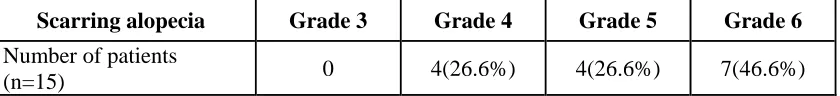
TABLE 10-SCARRING OF THE SCALP
Scarring alopecia Grade 3 Grade 4 Grade 5 Grade 6
Number of patients
(n=15) 0 4(26.6%) 4(26.6%) 7(46.6%)
Scarring scalp: Grade 3-in one scalp quadrant, Grade 4- in two scalp quadrants, 5- in three scalp
quadrants, 6-scarring alopecia affecting the whole skull.
Scarring of the scalp (Table10)
Scarring of the scalp was seen in 15 patients. 7 (46.6%) had scarring alopecia affecting all four quadrants of the scalp.
14 patients (16.66%) were scored for both scarring and non scarring alopecia as the two types of alopecia co-existed in some lesions.
TABLE 11-SLEDAI PARAMETERS AND THE NUMBER OF PATIENTS
SLEDAI VARIABLES NUMBER OF PATIENTS
(n=93)
PERCENTAGE (%)
Seizures 1 1.07
Psychosis 0 0
Organic brain syndrome 4 4.3
Visual disturbances 0 0
Cranial nerve disorder 0 0
Lupus headache 0 0
Cerebrovascular accident 0 0
Vasculitis 13 13.97
Arthritis 18 19.35
Myositis 5 5.37
Urinary casts 22 23.65
Hematuria 25 26.88
Proteinuria 22 23.65
Pyuria 22 23.65
New Rash 55 59.14
Alopecia 74 79.57
Mucosal ulcers 39 41.93
Pleurisy 4 4.3
Pericarditis 2 2.15
Low total complement 37 39.78
Elevated dS DNA 57 61.29
Leukopenia 13 13.97
Thrombocytopenia 10 10.75
Fever 19 20.43
Systemic lupus erythematosus disease activity index (SLEDAI) (Table 11)
in whom CLASI activity score was used was 13.45± 7.43(range 2 to 32). Low (1-5), moderate (6-10), high (11-19) and very high (≥20) activity scores were seen in 9 (12.16%), 23 (31.08%), 26 (35.13%) and 16 (21.62%) patients respectively
The most commonly scored cutaneous parameter in SLEDAI was alopecia that was observed in 79.57% patients. This was followed by an inflammatory type rash (specific as well as non-specific LE related) and mucosal ulcers observed in 55 (59.14%) and 39 (41.93%) patients respectively.
Among the systemic manifestations, arthritis was seen in 18 patients (19.35%) followed by vasculitis (gangrene, tender finger nodules or biopsy proven vasculitis) in 13 patients (13.97%).
Elevation of Anti-ds DNA beyond the reference range of the testing laboratory was the most common laboratory parameter seen in 57 patients (61.29%)
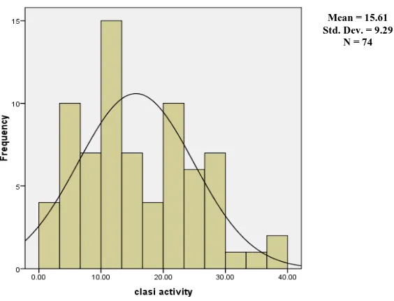
Figure 1 - Distribution of SLEDAI scores
Figure 2 - Distribution of CLASI activity scores
The CLASI and SLEDAI scores with their mean values less than twice the standard distribution have a positively skewed distribution.
Mean = 13.45 Std. Dev. = 7.438
N = 74
Mean = 15.61 Std. Dev. = 9.29
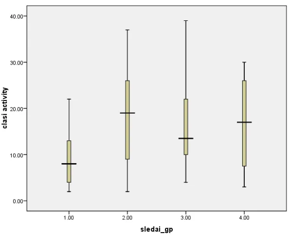
[image:49.612.175.457.416.631.2]Figure 3-Correlation between CLASI activity score and SLEDAI score
The box and whisker diagram (Figure 3) shows the four groups (1=low, 2=moderate, 3=high and 4=very high activity) of the SLEDAI severity score represented as sledai_gp against the CLASI activity score represented as clasi activity. The band at the middle of the box represents median (50th centile). The bottom and top of the box are the 25th and 75th percentile. The ends of the whiskers represent the maximum and minimum values of the data. The distribution of the variables is asymmetric as seen by the unequal length of the whiskers and the off centre positions of the median values. With increasing SLEDAI severity scores there is no corresponding increase in CLASI activity score.
Figure 4- Correlation between CLASI damage score (clasi_damage) and duration of
disease (dur_disease)
Figure 5-Correlation between CLASI damage score (clasi_damage) and duration of
[image:51.612.178.461.448.674.2]The scatter plots (Figures 4,5) display values for CLASI damage score that is the dependent variable and the duration of the disease and the duration of skin lesions. The data is displayed as a collection of points, each having the value of one variable determining the position on the horizontal axis and the value of the other variable determining the position on the vertical axis. The pattern of dots slopes from lower left to upper right, it suggests a positive correlation between the variables being studied.
Correlation done between the duration of disease (dur_disease) and CLASI damage score (Spearman’s correlation coefficient=0.477) and between the duration of skin lesions (dur_slesion) and CLASI damage (Spearman’s correlation coefficient=0.472) was statistically significant (p<0.01)
DISCUSSION
Cutaneous lupus erythematosus disease area and severity index (CLASI) is a relatively new tool formulated in 2005 by American dermatologists at the Veterans Affairs Hospital, Philadelphia, USA.2 The CLASI consists of physician rated activity and damage scores. There have been only a few studies done worldwide on the application of CLASI in patients with specific lesions of LE in patients with and without systemic involvement. Similarly, studies on its applicability in patients of pigmented skin are also limited.2,8,19,66 Our aim was to study the applicability of CLASI in specific lesions of cutaneous lupus in patients with SLE. We also studied the correlation of the CLASI activity score with clinical activity of SLE using the SLEDAI (Systemic lupus erythematosus disease activity index), an index used for assessing systemic disease activity.4
This study was done in the Department of Dermatology, Venereology and Leprosy. There were 93 patients recruited in this study which comprised 87 adults and 6 children. Among the adults, 5 were males and 82 were females. The male to female ratio of 1:14.5 was similar to a study by Yell J A et al31 and was much higher than earlier studies done in this centre and from Europe where it was 1:713 and 1:5.6 70 respectively.
patients (31.19%) and generalised acute cutaneous LE in 26 patients (27.9%). Cardinali C et al 27 in their retrospective study on 58 Italian patients with SLE found that discoid lesions, ACLE lesions and SCLE were seen in 32.75%, 39.65% and 13.79% of cases respectively. In the Hopkins Lupus cohort of 570 patients, the most common specific cutaneous manifestation was malar rash seen in 64% of the patients.1 In a study done on 32 patients of SLE from this centre, discoid lesions were seen in 21.8%, maculopapular rash in 46.8% and malar erythema in 34.3% patients.13
The most prevalent non-specific lesion in our study was alopecia (63.44%) followed by oral ulcers (41.93%) and palpable purpura (19.36%). The prevalence of LE non-specific lesions in a study by Cardinali C et al was 31% and the most common lesion in their patient series was Raynaud’s phenomenon.27 In a review of cutaneous disease among 73 SLE patients by Yell JA et al 31 the prevalence of alopecia was lower (54%) than in our series. Oral ulcers were seen in 41.93% of our patients that was comparable to the frequency in the Hopkins cohort.1
CLASI is reportedly an excellent scoring tool to assess the activity of cutaneous lupus. The most severely affected lesions within each anatomical area of the body is scored. Serial assessment of the activity and damage using CLASI is useful in studying the outcome of therapy as has been shown in a study by Kreuter A et al.19 In this cross sectional study, the physician rated CLASI activity and damage score was done on 75 patients. Previous studies have been done on limited number of patients ranging from 8-14 patients.2, 8, 19, 66
scores in our patients was 15.6 (range 2-39). This score was comparable to that seen in other studies.19,72 The range of CLASI activity score done in a recent pilot study analysing the impact of therapy on the CLE disease severity and the quality of life at time of entry in the study was 8-4972 while Kreuter A et al 19 reported the activity scores ranging from 5-25 in a study done on patients with SCLE.
The parameters that are used to assess the activity of CLE are erythema, scale/hypertrophy, mucous membrane involvement, acute hair loss, or non-scarring alopecia.62 CLASI activity score is largely based on the extent of the erythema. The latter is prominent, easily recognised and can be reliably assessed even in black skin .62 Erythema was an easily recognizable feature in our patients too. 56.6% patients had grade I (pink, faint) and 34.54% patients had grade II (red) erythema. Only 8.86% patients had a grade III erythema (dark red, violaceous, crusted or hemorrhagic). Erythema was best appreciated on the face including the malar area.
Grade I scaling was appreciated in 100% patients whereas hypertrophic scaling was seen only in 5 (6.75%) patients. The latter was seen on lesions over the scalp, nose, arms and legs. Scaling was a common feature of lesions on the ears .
Among the other parameters scored to assess activity, mucosal ulcers were seen in more than 50% patients, sixty-one (82.43%) patients had self reported alopecia and non-scarring alopecia was present in 62 (83.78%) patients.
The factors affecting the CLASI activity score in our patients were studied.
number of lesions.5 A similar observation was made in our study in relation to specific skin lesions of LE. It was found that the activity score increased proportionately with the number of anatomical areas involved. In our study, higher CLASI activity scores were also seen in patients with SCLE and ACLE occurring in combination with other specific lesions as compared to those with only discoid lesions. The five patients who had only specific skin lesions without alopecia or mucosal involvement had lower mean CLASI activity score compared to those who in addition had the latter two parameters.
The CLASI damage score was done on 59 out of the 75 patients. The parameters in the CLASI damage score are: dyspigmentation, cutaneous scarring /atrophy/ panniculitis and scarring of the scalp. The damage score in our patients ranged from 1 to 30. The CLASI damage score was studied for its correlation with the duration of SLE and the duration of the skin lesions. A statistically significant correlation was seen with the duration of SLE and the duration of skin lesions (p<0.01). It was also found that for a unit increase in the duration of the disease and of skin lesions there was a proportionate increase in the damage score.
SLEDAI was developed by Bombardier C et al at the University of Toronto.4 It is a global index containing 24 weighted objective clinical and laboratory variables and measures disease activity within the last 10 days. The maximum possible score is 105. The advantage of SLEDAI is the relative small number of items and the ease of scoring. Systemic disease activity categories have been defined on the basis of SLEDAI scores: no activity (SLEDAI = 0), mild activity (SLEDAI = 1-5), moderate activity (SLEDAI = 6-10), high activity (SLEDAI = 11-19), and very high activity (SLEDAI 20 and above).69
of use. The SLEDAI activity score of our patients varied from 2 to 32. Low (1-5), moderate (6-10), high (11-19) and very high (≥20) activity scores were seen in 9 (12.16%), 23 (31.08%), 26 (35.13%) and 16 (21.62%) patients respectively. 16 (21.62%) patients had a SLEDAI score of greater than 20 that indicated very high disease activity. Majority of patients had high disease activity with the SLEDAI scores between 12 to 19.
Correlation between SLEDAI and CLASI activity scores
CONCLUSIONS
1. This study has shown that CLASI is an effective tool to assess cutaneous activity and damage of specific lesions of LE. The mean CLASI activity score was 15.6 (range 2 to 39) and the mean damage score was 8.24 (range 1 to 30). The scores seen in our patients were comparable to other studies.
2. The mean CLASI activity scores were higher in those who had higher number of anatomical sites affected and those with SCLE, and ACLE occurring in combination with other specific lesions.
3. The CLASI damage scores correlated with the duration of SLE (p<0.01) and also with the duration of skin lesions (p<0.01).
(p=0.16).
SUMMARY
LIMITATIONS
1. All patients included in this study had prior disease modifying treatment. CLASI activity scores may have been higher if patients were not on prior treatment.
BIBLIOGRAPHY
1. Petri M. Dermatologic Lupus: Hopkins Lupus Cohort. Semin Cutan Med Surg 1998;17: 219-27.
2. Joerg A, Lynne T, Jesse B A. The CLASI (Cutaneous Lupus Erythematosus Disease Area and Severity Index): An Outcome Instrument for Cutaneous Lupus Erythematosus. J Invest Dermatol 2005; 125:889–94.
3. Werth V P . Clinical manifestations of cutaneous lupus erythematosus. Autoimmunity Reviews 2005;4: 296–302.
4. Bombardier C, Gladman D, Urowitz M. Derivation of the SLEDAI. A disease activity index for lupus patients. Arthritis Rheum 1992;35: 630 – 40.
5. ZecÏevic RD , Vojvodic D , Ristic B , Pavlovic MD, Stefanovic D and Karadaglic D. Skin lesions-an indicator of disease activity in systemic lupus erythematosus? Lupus 2001; 10: 364-67
7. Liang MH, Socher SA, Larson MG, Schur PH. Reliability and validity of six systems for the clinical assessment of disease activity in systemic lupus erythematosus. Arthritis Rheum 1989;32:1107-18.
8. Krathen MS, Dunham J,Gaines E et al. The Cutaneous Lupus Erythematosus Disease Activity and Severity Index: expansion for rheumatology and dermatology. Arthritis Rheum. 2008;59:338-44.
9. Li C K, Isenberg D A. Systemic lupus Erythematosus. Medicine 2006; 34:445-52.
10. Danchenko N, Satia JA, Anthony MS. Epidemiology of systemic lupus erythematosus: a comparison of worldwide disease burden. Lupus 2006; 15: 308-318.
11. Malaviya AN, Singh RR, Singh YN, Kapoor SK, Kumar A.Prevalence of systemic lupus erythematosus in India. Lupus 1993; 2: 115-18.
12. Malaviya AN, Chandrasekaran AN, Kumar A, Shamar PN.Systemic lupus erythematosus in India. Lupus 1997;6:690-700
13. George R, Mathai R, Kurian S. Cutaneous lupus erythematosus in India: Immunofluorescence profile. Int J Dermatol 1992;31:265-69.
14. Hochberg MC. Updating the American College of Rheumatology revised criteria for the classification of systemic lupus erythematosus [letter]. Arthritis Rheum 1997;40:1725.
15. McCauliffe DP. Cutaneous Lupus Erythematosus. Semin Cutan Med Surg 2001;20:14-26