BLOOD LOSS IN NORMAL LABOUR
Dissertation submitted to
The Tamilnadu Dr. M.G.R. Medical University
in partial fulfillment of the regulations for the award of the degree of
M.D. – Branch II
OBSTETRICS AND GYNAECOLOGY
K.A.P. Viswanathan Government Medical College
Tiruchirappalli
The Tamilnadu Dr. M.G.R. Medical University Chennai
This is certify that the dissertation entitled “ROLE OF TRANEXAMIC
ACID IN REDUCING BLOOD LOSS IN NORMAL LABOUR” is a
bonafide work done by Dr. B. SOWMYA at K.A.P. Viswanathan Government Medical College, Trichy. This dissertation is submitted to Tamilnadu Dr. M.G.R. Medical University in partial fulfillment of University rules and regulations for the award of M.D. degree in Obstetrics and Gynaecology.
Prof. Dr. PREMAVATHY PRABHU ELANGO, M.D., D.G.O.,
Professor & Head of the department Obstertrict and Gynaecology K.A.P.V. Govt Medical College, Trichy.
Prof. Dr. A. KARTHIKEYAN, M.D., Forensic Medicine
Dean
I Dr. B. Sowmya, solemnly declare that the dissertation titled, “ROLE OF TRANEXAMIC ACID IN REDUCING BLOOD LOSS IN NORMAL
LABOUR” is a bonafide work done by me at K.A.P.V. Government Medical
College, Trichy, during 2010 – 2011 under the guidance and supervision of
Prof. Dr. PREMAVATHY PRABHU ELANGO, M.D., D.G.O., Professor
and Head of the department, Obstertrics and Gynaecology. This dissertation is submitted to the Tamilnadu Dr. M.G.R. Medical University, in partial fulfillment of University rules and regulations for the award of M.D. Degree (Branch – II) in Obstetrics and Gynaecology.
Place: Trichy
Date :
My deepest thanks to Prof. Premavathy M.D., D.G.O., Professor and Head of the Department of obstetrics and gynaecology for her expert guidance and support for the completion of the study. She has taken pains to go through my dissertation and made necessary correction as and when needed.
I express my thanks to The Dean, Trichy Medical College and Annal Gandhi Memorial Government Hospital, Trichy for extending his support and granting me permission to undertake this study.
My deep sense of Gratitude to Dr. Kalarani M.D., D.G.O., and Dr. Bama Ramesh M.D., D.G.O., Dr. Mumtaz M.D., D.G.O., Professors
Department of Obstetrics and Gynaecology for their support and valuable suggestions.
I extend my heartfelt thanks to all my Assistant Professors, Department of Obstetrics and Gynaecology for their utmost cooperation throughout this study.
PR PULSE RATE
SBP SYSTOLIC BLOOD PRESSURE
DBP DIASTOLIC BLOOD PRESSURE
RR RESPIRATORY RATE
HT HEIGHT
WT WEIGHT
BMI BODY MASS INDEX
PPH POST PARTUM HAEMORRHAGE
HB HAEMOGLOBIN
POD POST OPERATIVE DAY
NICU NEONATAL INTENSIVE CARE UNIT
LFT LIVER FUNCTION TEST
RFT RENAL FUNCTION TEST
IUD INTRA UTERINE FETAL DEATH
AMTSL ACTIVE MANAGEMENT OF THIRD STAGE OF LABOUR
VWD VON WILLEBRAND’S DISEASE
ITP IDIOPATHIC THROMBOCYTOPENIC PURPURA
TTP THROMBOTIC THROMBOCYTOPENIC PURPURA
DIC DISSEMINATED INTRA VASCULAR COAGULATION
DUB DYSFUNCTIONAL UTERINE BLEEDING
TXA TRANEXAMIC ACID
CVS CARDIO VASCULAR SYSTEM
RS RESPIRATORY SYSTEM
HELLP HEMOLYSIS, ELEVATED LIVER ENZYMES, LOW PLATELETS
CT CLOTTING TIME
BT BLEEDING TIME
CBC COMPLETE BLOOD COUNT
CVP CENTRAL VENOUS PRESSURE
PCV PACKED CELL VOLUME
CONTENTS
S. No. Title Page
No.
1. Introduction 1
2. Aim of the Study 4
3. Review of Literature 5
4. Materials and Methods 40
5. Results and Analysis 45
5. Discussion 62
6. Summary 68
7. Conclusion 70
8. Bibliography
INTRODUCTION
Gods interest in the human race is nowhere better evinced than in obstetrics – Martin H. Fischer.
Child birth is one of the most marvelous and memorable segment in a woman’s life. The art of vaginal birth is a unique experience and calls for a celebration. Although the incidence of caesarean section is increasing throughout the world, the art of vaginal birth has not lost its charm. Birth is a life changing event. The care given to woman during labour has the potential to affect her both physically and emotionally in the short and long term.
Though labour is a physiological process, it is often associated with morbidity and mortality. The most common cause being blood loss. Life threatening obstetric hemorrhage occurs in approximately 1 per 1000 deliveries. The most recent Practice Bulletin from the American College of Obstetrics and Gynaecology places the estimate at 1,40,000 maternal deaths per year or 1 woman every 4 minutes. There are a number of drugs available for the management of PPH. The recent ones being the anti fibrinolytics and recombinant factor VIIa.
Postpartum haemorrhage (PPH) remains a leading cause of maternal mortality, especially in developing countries (Ronsmons 2006). In confidential enquiries into maternal deaths in South Africa (2005 to 2007) (Confidential
enquiries 2006), 383 maternal deaths due to PPH were reported and the
(17.5%) were caused by uterine atony, where uterotonics were required to control the bleeding. Other cases of maternal death from PPH were due to uterine rupture (37 in women with previous caesarean sections and 43 in women without previous caesarean sections), retained placenta (88), inverted uterus (seven), and other genital tract trauma including caesarean section (141). The great majority were thus not due to uterine atony, and attempts to address the problem need to go beyond the use of uterotonic drugs. Because of the difficulty of randomised trials in women presenting with PPH, the use of tranexamic acid for preventing PPH in high-risk women could be regarded as a proxy for assessing its use for treating PPH. In particular, high-risk factors which may not be responsive to uterotonics, such as placenta praevia and lacerations from instrumental delivery, may respond to tranexamic acid. If tranexamic acid is found to be effective in the prevention of PPH in such high-risk women, its use could be extrapolated to the treatment of PPH (as has been the case for most treatments for PPH, such as oxytocin and ergometrine).
Hence antifibrinolytics will be effective in reducing blood loss by interacting with the fibrinolytic mechanism.
AIM OF THE STUDY
1. To evaluate the efficacy of parenteral Tranexamic Acid in reducing blood loss during normal labour.
REVIEW OF LITERATURE
Post partum hemorrhage is the leading cause of maternal mortality. All woman who carry a pregnancy beyond 20 weeks gestation are at risk of PPH and its sequelae. In the developing world, several countries have maternal mortality rates in excess of 1000 women per 100,000 live births and WHO statistics suggest that 25% of maternal deaths are due to PPH, accounting for more than 100,000 maternal deaths per year.
The leading causes of pregnancy related death are hemorrhage, embolism and hypertensive disorders of pregnancy – Berg CJ et al 1996.
Prendiville et al. in Bristol Third Stage Trial found an incidence of
postpartum bleeding of 5.9% in actively managed group and 17.9% in physiologically managed group. They concluded that intervention in third stage of labour reduces the risk of PPH by 30 – 40%.
MrCormick et al. published a systematic review of studies that
assessed the efficacy of intervention in 3rd stage – concluded that intervention reduced the incidence of PPH, decreased the need for blood transfusion and decreased the need for additional uterotonic drugs.
Malcolm Potts 2006 – Darwinian evolution is not about what is nice,
Idara Udofia et al. 2008 – Any intervention aimed at preventing PPH
will reduce maternal mortality by more than one quarter.
Blood loss during normal labour
Normal pregnancy is accompanied by changes in the haemostatic mechanisms, particularly increase in the levels of factor VII, VIII, X and fibrinogen and a pronounced decrease in fibrinolytic activity. Parturition presents a serious challenge to the vascular compartment, but it has been generally held (Taylor, 1966; Donald, 1969) that contraction of the myometrium “the living ligatures” of the uterus - is the mechanism which, mainly controls blood loss at delivery. In the other situations where there is injury to the vascular tree an efficient blood coagulation system plays a vital part in achieving effective haemostasis and preserving the blood volume.
Physiological adaptations in pregnancy
Antepartum adaptations for physiological blood loss at delivery include a 42% increase in plasma volume and a 24% increase in red blood cell volume by the third trimester. Progressively complicated deliveries are accompanied by greater degrees of blood loss.
Vaginal delivery – 500ml Caesarean section – 1000ml Caesarean hysterectomy – 1500ml Emergency hysterectomy – 3500ml
However, these thresholds do not take into account the pre-existing health status and blood loss of as little as 200ml can be life threatening for a woman with severe anemia or cardiac disease (Kongnyuy et al., 2009).
GREENTOP GUIDELINES (RCOG 2007) – defines PPH as
• 1 blood volume loss / 24 hrs
• 150 ml / minute blood loss
• 50% blood volume loss / 3 hrs
Some of the factors leading to increased blood loss in the third stage labour are as follows:
• Mean vaginal blood loss is higher in multiparae than in primiparae
• In primiparae, forceps delivery is associated with greater blood loss than
• Patients with an episiotomy and a laceration lose significantly more
blood than those without such insult. Episiotomies contribute to 154 ml to the average blood loss.
Incidence of PPH
2% to 11% - when blood loss is estimated visually (Brent et al., 1967) 20% - when blood loss is estimated by quantitative methods (Newton et al.,
1961)
Types of PPH
1° PPH / early PPH – which occurs within 24 hrs of delivery
2° PPH / late PPH – which occurs 24 hrs to 6 weeks after delivery Most cases of PPH almost 99% are due to 1° PPH
Pathophysiology
placenta, the uterus initiates a process of contraction and retraction, shortening its fiber and kinking the supplying blood vessels, like physiologic sutures or "living ligatures."
If the uterus fails to contract, or the placenta fails to separate or deliver, then significant hemorrhage may ensue. Uterine atony, or diminished myometrial contractility, accounts for 80% of postpartum hemorrhage. The other major causes include abnormal placental attachment or retained placental tissue, laceration of tissues or blood vessels in the pelvis and genital tract, and maternal coagulopathies. An additional, though uncommon, cause is inversion of the uterus during placental delivery.
Potential causes for increased blood loss during labour:
1. Tone loss 2. Trauma
3. Tissue retention 4. Thrombotic defect
Of these, tone loss is the commonest and thrombotic defect is the most difficult to treat.
1. TONE LOSS : 80%, the following are the predisposing factors
• Multiparity
• Uterine overdistension – multiple pregnancy, hydramnios, macrosomia
• Prolonged labour
• Anemia
• Chorioamnionitis
• Uterine abnormalities or fibroids
• Previous H/O PPH
• Induced labour
• Inadvertent use of oxytocics
• Drugs – halogenated anaesthetics, MgSO4, nifidepine, beta agonists,
diazoxide
• Placenta – accreta, praevia, abruption
2. TRAUMA : 20%
Lacerations and haematomas resulting from birth trauma can cause significant blood loss that can be lessened by haemostasis and timely repair. Factors causing increased blood loss are:
• Obstructed labour
• Big baby
• Pfannensteil incision
• Transverse lie
• Classical caesarean section
• Poor obstetrician’s skill
3. TISSUE RETENSION : 10%
Risk factors
• Advanced maternal age
• High parity
• Previous invasive placenta
• Previous caesarean section
• Placenta praevia
• Previous H/O manual removal of placenta
4. THROMBOTIC DEFECT: 5%
• Coagulopathies – ITP, TTP, VWD, hemophilia
• Acquired – HELLP, abruption, IUD, DIC, septicemia
PPH is a potentially life threatening complication of both vaginal and caesarean delivery. Associated morbidity is related to the direct consequences of blood loss as well as the potential complications of haemostatic and resuscitative interventions.
Consequence of uncontrolled haemorrhage include
• Hypovolemic shock and associated organ failure including renal failure, stroke, myocardial infarction
• Postpartum hypopituitarism (Sheehan syndrome): Acute blood loss and / or hypovolemic shock during and after childbirth can lead to hypoperfusion of the pituitary and subsequent necrosis. Although often asymptomatic, it may present with an inability to breastfeed, fatigue, hypogonadism, amenorrhea, and hypotension.
Consequences of fluid resuscitation
• Fluid overload can lead to extremity edema and pulmonary edema. The latter is less common in young healthy women, but it should be suspected in the setting of large fluid and blood product resuscitation.
• Dilutional coagulopathy occurs when crystalloids and/or serum-poor blood products are given in large volume.
Risks from exposure to blood products
• Allergic or febrile reactions have an incidence of about 1 case per 333
population.
• Anaphylactic reactions occur in 1 in 20,000 to 1 in 47,000 blood
products transfused.
• Transfusion-related acute lung injury (TRALI) occurs in 1 out of every
5,000 transfusions, but more often with high plasma containing products like fresh frozen plasma (FFP) and platelets. It often starts within 1-2 hours of the transfusion, but it can happen anytime up to 6 hours after a transfusion. The symptom complex includes severe bilateral pulmonary edema, severe hypoxemia, tachycardia, cyanosis, hypotension, and fever.
• Acute immune hemolytic reaction, though rare, is the most serious type
• Delayed hemolytic reaction: This type of reaction happens when the
body slowly attacks antigens (other than ABO antigens) on the transfused blood cells. Symptoms occur days to weeks after a transfusion. Affected patients are either asymptomatic or have mild symptoms, which may include jaundice, low-grade fever, and a low hemoglobin or hematocrit
Infection
Hepatitis is the most common disease transmitted by blood transfusions. According to the American Red Cross, about 1 blood transfusion in 205,000 transmits a hepatitis B infection, and 1 blood transfusion in about 2 million transmits hepatitis C.
Other rare but potential infections include HIV (risk of 1 in 2.5 million), Lyme disease, babesiosis, and malaria. Donors are screened for potential exposure so transmission is very rare. Rarely, blood may be contaminated with tiny amounts of skin bacteria during donation. Platelets are the most likely blood product to be affected by contamination from skin flora.
• Metabolic reactions: With large volume and rapid transfusions, patients
are at risk of encountering 3 metabolic reactions: hypothermia, hyperkalemia, and citrate toxicity.
• Hypothermia results from the transfusion of unwarmed crystalloid or
product additive that binds serum calcium and can cause hypocalcemia with large-volume transfusions. Hemolysis occurs with red blood cell storage releasing increasing amounts of intracellular potassium with time. Transfusions of older red blood cells increase the risk of hyperkalemia.
Risks associated with surgical intervention
• Intubation and anesthesia complications: Pregnant women have an
increased risk for aspiration, failed intubation, and death from failed ventilation when compared with nonpregnant patients. Respiratory injury or infection, myocardial infarction, myocardial arrhythmia, stroke, or allergic reactions to anesthetic medications may also rarely occur.
Bleeding: Continued bleeding from the genital tract or a bleeding
complication from the surgery may occur.
Infection: Sepsis, wound infection, or pneumonia is possible.
• Deep venous thrombosis and/or pulmonary embolism: Risk is increased
due to postpartum and postoperative associated hypercoagulability as well as from relative immobility in the operative and postoperative period.
• Hysterectomy
• Asherman syndrome, which is secondary (non-hormone mediated)
amenorrhea due to uterine scarring that develops after infection and/or curettage performed to remove placental fragments
BLOOD LOSS ASSESSMENT
In order to assess the efficiency of the drug, the blood loss assessment has to be standardized.
Clinical methods
1. By subjective characters 2. Visual estimation
1. Estimation by subjective characters
• Shock index
SI = HEART RATE / SYSTOLIC BP
NORMAL = 0.5 – 0.7, with significant hemorrhage, it increases to 0.9 – 1.1
• Rule of 30
1. If systolic BP falls by 30 mmHg
2. Heart rate rises by 30 bpm
3. Respiratory rate rises by 30 breaths per minute
4. Hb or Hct drops by 30%
Patient is said to have lost atleast 30% of blood volume if all the above exists.
• Measurement of CVP
Normal : 8 – 14 cm of water. If CVP is low 0-6 cm of water, it means the blood volume is low in relation to cardiac capacity. If CVP is high > 15 cm of water, it means the blood volume is high in relation to cardiac capacity.
• Classification of haemorrhage
PARAMETER CLASS I CLASS II CLASS III CLASS IV
Blood loss (ml) < 750 750 – 1500 1500 - 2000 > 2000 Blood loss (%) < 15 15 – 30 30 – 40 > 40 Pulse rate/min < 100 > 100 > 120 > 140
Blood pressure N ↓ ↓ ↓
Respiratory rate/min 14 – 20 20 – 30 30 – 40 > 40 Urine output (ml/hr) > 30 20 – 30 5 – 15 Negligible CNS symptoms Normal Anxious Confused Lethargic
2. Visual estimation
This is the most frequently practiced method. The incidence of PPH is underestimated in the visual estimation by 89% - Brant & Duthie et al.
Budny et al. reported a strong positive association between calculated
Dr. P. Bore et al. 2006
• 10 × 10 cm swab = 60 ml
• 30 × 30 cm swab = 140 ml
• 45 × 45 cm swab = 350 ml
• 1 kg soaked swabs = 1000 ml
• 50 cm diameter floor spill = 500 ml
• 75 cm diameter floor spill = 1000 ml
• 100 cm diameter floor spill = 1500 ml
Gravimetric methods
• Patient weighing method
• Swab weighing method
By measuring the weight of the patient or swabs prior to and after delivery. - Patient weighing method: allowance must be made for drain,
dressings, infection, tissue removal and insensible water loss. - Swab weighing method: 1 gm of weight gain = 1 ml of blood loss
(Bonica and Lyter et al. in 1951, Harding 1984). Swabs must be
Calibrated obstetric drape
This comes in sterile packing and fold out to a 1 × 1 metre sterile area for a woman to give birth. At the bottom of the sterile area is a pouch that holds more than 2500 ml of fluid, allowing for accurate measurement of postpartum blood loss. The pouch itself also includes a flexible plastic filter to 'catch' material that is not liquid. The pouch includes a wire around its 'mouth' that keeps the pouch open.
Colorimetric method: (Roe et al., 1962, Thornton et al., 1963, Rustad et al., 1963)
The washing of the blood contaminated swabs is carried out in a known volume of tap water to which has been added sufficient amount of ammonium hydroxide to give a 1 in 1000 dilution as a defoaming agent. The blood collected in the suction container has to be added to the water and the concentration of the resultant solution has to be determined.
Measurement of blood in the suction apparatus
Blood in the suction container can be measured. Inaccuracy in this method can be reduced by having measuring cylinder in the suction line and adding defoaming agent to the container.
Electrolyte conductivity method : (Leveen and Rubricius et al., 1958)
Using automated blood loss meter based on electrolyte conductivity.
Radioactivity method : (Murray and Dott’s et al., 1960)
Intravenous injection of small but known amount of radio isotope should be followed by measuring the radioactivity of blood on swabs collected during delivery.
Blood volume measurements
- Dye method: using evans blue dye which must neither be catabolised nor rapidly lost from the circulation
- Radio isotopes like I131 labelled albumin or Cr51 labelled RBC can be
used before delivery and measuring the post delivery radio activity by Geiger – Muller counter. (Mollison ans Veall et al., 1955).
REDUCING BLOOD LOSS
Done by the following measures
• Antenatal care
• First and Second stage measures
• Third stage measures
• Postpartum measures
Antenatal care
1. Develop a birth preparedness plan : women should plan to give birth with a skilled attendant who can provide interventions to prevent, identify and manage PPH.
2. Routinely screen to prevent and treat anemia during preconceptional, antenatal, postpartum visits.
3. Blood grouping and Rh typing to be done.
4. Correction of coagulation abnormality in case of abruption, IUD, HELLP syndrome.
During first and second stage
1. A woman in established labour should receive supportive one to one care
2. Limit induction and augmentation use for medical and obstetric reasons 3. Do not encourage pushing before cervix is fully dilated
4. Do not apply fundal pressure to assist the birth of the baby
6. Encourage the woman to keep her bladder empty 7. Do not perform routine episiotomy
8. Either “hands on” or the “hands poised’ techniques can be used to facilitate spontaneous birth
During third stage : provide Active Management Of Third Stage Of Labour
Postpartum measures
It has been suggested that drainage of blood from the placenta would reduce its bulkiness, allowing the uterus to contract and retract and thus aiding delivery (Roger et al., 1988).
Placental cord drainage may be used in conjunction with other interventions such as routine administration of oxytocics, controlled cord traction or maternal effort (Hinchongbrooks RCT Lancet, 1998).
Placental cord drainage with or without prophylactic oxytocics is effective in reducing the blood loss in third stage thus preventing PPH (Keirse,
1998).
Timing at cord clamping is also an important factor in the management of third stage of labour, influences the duration of third stage (McDonald,
2003).
The overall meta – analysis results of Cochrane database systematic
reviews 2004, showed that there was a tendency for the active management
group to have higher incidence of these complications. But this did not reach statistical significance. There was no statistically significant difference in neonatal outcome or breast feeding rates and no difference in long term maternal outcome.
TREATMENT
Most maternal deaths in PPH are due to the 3 delays: 1. Delay in recognition of complications
2. Delay in institution of definitive management 3. Delay in referral / accessing transportation
MANAGEMENT General
Specific
i) medical ii) surgical
General Management
- Assessment of general condition of the patient, the amount of blood loss and degree of hypoxemia
- Vital parameters should be recorded
- Blood should be sent for cross matching, CBC, RFT
- Cyrstalloids and colloids should be rushed in the mean time. It enhances the critical filling and improves the cardiac output. 250 – 500 ml of either a crystalloid or a colloid is administered over a period of 10 – 20 minutes as the urgency indicates. Cyrstalloids are preferred over colloids as they quickly become distributed throughout the extracellular space, they are cheap, easily available and there is no risk of anaphylaxis
- Replace blood by blood
- Invasive hemodynamic monitoring by CVP measurement
- BT, CT should be measured and when it is prolonged FFP and cryoprecipitate should be given
Medical management
UTEROTONICS
i) Oxytocin – 10 units im / iv followed by 20 units iv infusion in 500 ml
RL / NS
ii) Methylergometrine – 0.2 mg im / iv repeated for every 15 minutes to a
maximum of 5 doses
iii) 15 methyl PG F2α - 250 µgm im repeated every 15 minutes to a maximum of 8 doses
iv) Misoprostol – 400 - 1000 µgm vaginal, oral, rectal
v) Recombinant factor VIIa – 60 – 120 µgm / kg iv vi) Tranexamic acid – 1gm iv 8th hourly
Oxytocin stimulates the upper segment of the myometrium to contract rhythmically, which constricts the blood vessels and reduces blood flow through the uterus (Dreyfus M et al., 2004). Produces rhythmic uterine contractions, can stimulate the gravid uterus, and has vasopressive and antidiuretic effects. Can be used to control postpartum bleeding or hemorrhage. Some suggest its prophylactic use in the third stage of labor; one study of 1000 deliveries revealed a 32% reduction in the rate of PPH.
Pierre F, Mesnard L, Body G Eur J Obstet Gynecol Reprod Biol. 1992.
Methylergometrine – ergot alkaloids cause generalized smooth muscle contraction in which both upper and lower segments of the uterus. Side effects – hypertension, nausea, vomiting, headache.
Syntometrine – 5 units oxytocin + 0.5 mg ergometrine.
15 methyl PG F2α - enhances uterine contractility and causes
vasoconstriction. It has been shown to control PPH in upto 81% of patients. Side effects – nausea, vomiting, diarrhea, hypertension, headache, flushing, pyrexia. Contraindications – hypersensitivity, bronchial asthma.
Misoprostol – it increases uterine tone. Side effects – hyperpyrexia, diarrhea, shivering.
Recombinant factor VIIa – it is an enzyme of the serine protease class. It
initiates the process of coagulation in conjunction with tissue factor. (Ahonen et al., 2007). It induces haemostasis at the site of vascular injury independent of the presence of factors VIII and IX by forming complexes with exposed tissue factor (TF). Administration of high-dose rFVIIa results in a huge increase in factor VIIa, well above that of the normal physiological levels, leading to faster and greater thrombin generation.
Anti fibrinolytics – Tranexamic acid potentates the blood clotting system
increase in the activation of plasminogen activators and fibrin degradation products due to activation of the fibrinolytic system. This activation can last up to six to 10 hours postpartum, which may cause more haemorrhage. The antifibrinolytic effect of tranexamic acid in the third stage of labour could make it a safe and effective alternative or adjunct to other regimens currently used in the third stage of labour for prevention of PPH. Tranexamic acid could reduce blood loss associated with complications such as placenta praevia and lower genital tract trauma, as well as bleeding from the upper segment placental site. Therefore, it may be particularly useful in preventing cases of PPH due to factors other than uterine atony, where uterotonics will not be effective. Tranexamic acid is an effective agent for the reduction of blood loss, which has been widely used in various areas of medicine. It is an inhibitor of fibrinolysis that blocks the lysine- binding site of plasminogen to fibrin (Astedt 1987; Longstaff, 1994).
It has been used to decrease blood loss for many years in cases of haemorrhage, and is reported to reduce intraoperative and postoperative blood loss (Boylan, 1996; Karski, 1995; Katsaros, 1996; Reid, 1997; Vacharaksa,
2002).
The side effects described with the use of tranexamic acid include gastrointestinal symptoms such as diarrhoea, nausea and vomiting that occur in about 10% of patients. Rare complications include hypotension, thrombosis, blurred vision, renal cortical necrosis and retinal artery obstruction (Astedt,
However, another study reported no side effects associated with tranexamic acid (Bekassy, 1990).
A Cochrane review on the use of antifibrinolytics for heavy menstrual bleeding reported no rise in side effects with tranexamic acid in comparison to placebo, NSAIDS, oral luteal phase progestagens or ethamsylate (Lethaby,
2000).
There are concerns about the risk of thromboembolic events associated with the use of tranexamic acid; however, there are no data available from randomised controlled trials (RCTs) which record the frequency of thromboembolic events (Lethaby, 2000) as the fibrinolytic system gets activated after placental delivery and in menorrhagia, antifibrinolytics are useful in treating PPH and DUB.
Single dose of 1 gm of tranexamic acid given intravenously reduces the mean blood loss within 2 hours of delivery (Pili ferrer et al., 2009).
Tranexamic acid significantly reduces the mean blood loss by 92 ml compared to no treatment (Gohel et al., 2007).
Tranexamic acid reduces blood loss without any side effects or complications like thrombosis (Gai et al., 2004).
When tranexamic acid is used the need for additional uterotonic drugs is reduced (Gakhan Yildirim et al., 2011).
Tranexamic acid acts immediately after iv administration (Jurema et al.,
2008).
Use of tranexamic acid could potentially have prevented some PPH cases, as reported in the Cochrane review in treatment of PPH (Mousa 2007)
Tranexamic acid is associated with a significant reduction in objective measurement of heavy menstrual bleeding when compared to placebo or other medical therapies (Lethaby et al., 2000).
Tranexamic acid is a cost effective drug. A study on total hip arthroplasty reported saving blood transfusion and money in cases where Tranexamic acid was used prophylactically prior to surgery (Johansson, 2005). Blood loss of greater than 400 ml is not reported when Tranexamic acid is used during vaginal birth (Yang et al., 2001).
Tranexamic acid is used safely and effectively to reduce bleeding resulting from caesarean section (Gai et al., 2004).
Tranexamic acid statistically reduces the extent of bleeding from placental delivery to 2 hrs post partum during caesarean section and its use was not associated any side effects (Ming - Ying Gai et al., 2003).
Tranexamic acid reduces blood loss and maternal morbidity in ongoing PPH (Anne – Sophie Ducloy et al., 2011).
Tranexamic acid reduces post partum blood loss after vaginal birth and after caesarean section (Novikova N. et al., 2010).
Tranexamic acid can be given antenatally by oral route for one week to treat women with history of recurrent abruption – to get successful neonatal outcome (B Astedt et al., 1978).
Prophylactic tranexamic acid before surgery reduces allogenic blood transfusion (Cochrane database, 2001).
Tranexamic acid can be used to decrease bleeding from menorrhagia and conisation of cervix (Dunn CJ et al., 1999).
Tranexamic acid reduces mean blood loss after oral surgery in patients with hemophilia and it is effective as a mouth wash in dental patients receiving oral anti coagulants (Mairangi Bay et al., 1999).
Tranexamic acid when given with in 3 hrs of trauma, mortality is reduced in trauma patients (Russel L Gruen et al., 2010).
Tranexamic acid is an effective and safe option in DUB and operative interference is reduced (Kriplani A. et al., 2006).
If bleeding persists even after removal of retained products of conception in missed abortion and secondary PPH, a fibrinolytic inhibitor such as tranexamic acid can be given to counteract fibrinolysis in uterus (J Bonner
et al., 2011).
reduction in blood product use, which is associated with multiple risks (transfusion reactions, transmission of blood-borne viruses), is expensive and may be not available when it is needed. In South Africa, most of the maternal deaths due to PPH occur in level one hospitals which do not have emergency access to formal blood transfusion services.
WOMAN Trial
The WOMAN Trial (World Maternal Antifibrinolytic Trial): tranexamic acid for the treatment of postpartum haemorrhage: an international randomised, double blind placebo controlled trial. This is an ongoing trial. The results of which are expected in February 2015.
Haleema Shakur1*, Diana Elbourne4, Metin Gülmezoglu2, Zarko
Alfirevic3, Carine Ronsmans5, Elizabeth Allen4 and Ian Roberts1 Author Affiliations
1
Clinical Trials Unit, London School of Hygiene & Tropical Medicine, Keppel Street, London WC1E 7HT, UK
2
Department of Reproductive Health and Research, World Health Organization, Avenue Appia 20, CH-1211 Geneva 27, Switzerland
3
Division of Perinatal and Reproductive Medicine, University of Liverpool, Liverpool Women's Hospital, Crown Street, Liverpool L8 7SS, UK
4
Medical Statistics Unit, London School of Hygiene & Tropical Medicine, Keppel Street, London WC1E 7HT, UK
5
This trial is a large, pragmatic, randomised, double blind, placebo controlled trial among 15,000 women with a clinical diagnosis of postpartum haemorrhage. All legally adult women with clinically diagnosed postpartum haemorrhage following vaginal delivery of a baby or caesarean section are eligible. The fundamental eligibility criterion is the responsible clinician's 'uncertainty' as to whether or not to use an antifibrinolytic agent in a particular woman with postpartum haemorrhage. Treatment entails a dose of tranexamic acid (1 gram by intravenous injection) or placebo (sodium chloride 0.9%) given as soon as possible after randomisation. A second dose may be given if after 30 minutes bleeding continues, or if it stops and restarts within 24 hours after the first dose.
Cost savings could also be gained from avoiding the use of expensive haematological agents such as Factor VIIa, which is establishing its place in the treatment of massive PPH in modern obstetrics despite the extreme cost
(Welsh, 2008).
SURGICAL MANAGEMENT
• Bimanual compression
• Uterine balloon tamponade
• Compression sutures
• Arterial ligation
• Aortic clamping
A patient who fails to respond to uterotonic agents and continues to bleed will quickly become haemodynamically unstable and develop a cascade of clotting abnormalities. The spectre of maternal mortality can then only be prevented by initiating surgical haemostasis sooner rather than later. The nature, timing and extent of these invasive interventions will depend on the sophistication of the health facility which handles this medical crisis. The fate of such a woman will therefore vary widely, depending not only on where she lives in the world but also on where she lives in her own country.
INTERVENTIONAL RADIOLOGY
• Consider on stable patient with continued bleeding (Clinical textbook
of obstetric and gynaecology 2010).
• Evidence is equivocal (Greentop guidelines 2009).
PHARMACOLOGY OF TRANEXAMIC ACID
Tranexamic acid is an antifibrinolytic drug. It is a synthetic derivative of the amino acid lysine that exerts its anti fibrinolytic effect through the reversible blockade of lysine binding sites on plasminogen molecules.
CHEMICAL STRUCTURE
Molecular formula : C8H15NO2
It is a trans-4 aminomethyl cyclohexane 1-carboxylate
Molecular weight : 157.21
Melting point : 300oc
Pharmacodynamics
Fibrin is the basic framework for clot formation to maintain haemostasis. This clot has to be lysed after a particular period of time by fibrinolysis by the following way.
Endothelial cell
Thrombomodulin & Thrombin
Protein C Activated protein C
Activated VIII Inactive VIII Activated V Inactive V
Inactivates inhibition of tissue plasminogen activator
Plasminogen Plasmin
Fibrinolysis
FDP formation
(-) TXA
Tranexamic acid acts as an antifibrinolytic agent by the following 2 ways: 1. Reversible, competitive blockage of lysine binding sites on plasminogen, so
that plasminogen activator cannot bind with plasminogen at lower doses. 2. Non competitive inhibition of proteolytic action of plasmin similar to
EACA. 8-10 times more potent than EACA as it binds strongly with both strong and weak receptors.
At therapeutic concentration (1mg/ml) it will not cause platelet aggregation.
Pharmacokinetics
• Oral absorption is 30-50%. It is not affected by food
• 100 % bioavailability with iv administration
• Only 3% is plasma protein (globulin) bound. Remaining binds with
plasminogen. It will not bind with serum albumin
• Only 5% will be metabolized in liver. Remaining 95% of the drug will
be excreted via urine unchanged
• T ½ is 2- 10 hours. 90% of the drug is excreted in urine within 24 hours
of administration. Duration of action in iv route is 7 – 8hrs
• Rapidly enter into joint fluid
• Crosses the placenta and blood brain barrier
Indications
This antifibrinolytic agent can be used in all types of bleeding especially coagulopathic bleeding. It can also be used prophylactically before surgical procedures where excess bleeding will be expected.
1. HELLP, DIC, Thrombaesthenia related bleeding 2. Postpartum haemorrhage
3. Dental extraction in haemophilia patients
4. Orthopaedic surgeries like spine surgery and total knee / hip replacement
5. Caesarean section 6. Cardiac surgeries
7. Trans urethral resection of prostate 8. Epistaxis
9. Liver transplantation surgery
10.First line nonhormonal treatment for menorrhagia in DUB / fibroid 11.Hereditary angioneurotic oedema where it decreases the attacks by
decreasing plasmin induced complement activation.
Contraindications
1. Previous H/O thromboembolism or active intravascular clotting or patients with inherited or acquired thrombophiliac states.
4. Patients with defective colour vision
5. Subarachnoid haemorrhage – because cerebral oedema and infarction may occur rarely.
Side effects
1. Nausea, vomiting, diarrhoea – commonest side effect, occurs in > 10% cases
2. Giddiness and hypotension – if given by sudden rapid iv occurs in 1-10% of cases
3. Defective colour vision – if used for long time 4. Thromboembolism – rare
5. Drug allergy – rare
Monitoring
LFT, RFT and colour vision should be checked periodically if used for long time.
Should be used with caution in,
1. Drug allergy patients
2. Renal / liver disease patients
3. Elderly individuals with impaired renal function
4. Pregnancy – as this is a category B drug, Tranexamic acid can be safely used in lactating mothers, because
• 1% of maternal serum level will be reached in breast milk
PREPARATIONS AND DOSAGE
1. Oral - 500 mg tablets available
- 25 mg / kg thrice daily for one week
2. Intravenous
• Available preparations contain 100 mg / ml (5ml and 10ml ampoules)
• Dose – 10 mg /kg either direct slow IV or after diluting with 20 ml of
5% dextrose at a rate not more than 1 ml / min. This loading dose can be followed by 1mg / kg / hour IV infusion or 10 mg / kg – thrice daily IV.
• It can be mixed with aminoacids, electrolytes or carbohydrate solution
but not with blood or solutions having penicillin.
3. Mouthwashes containing tranexamic acid are also available and used for haemophilia patients before and after dental extraction because oral mucosa and saliva are rich in plasminogen activator.
* Dose should be adjusted according to creatinine clearance, creative clearance
50 – 80 ml / min - 50% of total dose
10-50 ml/min - 25% of total dose
< 10 ml / min - 10% of total dose
Storage
Drug Interactions
1. Chlorpromazine increases cerebral vasospasm when combined with Tranexamic Acid, so it should not be combined.
MATERIALS AND METHODS
The subjects of this prospective randomised placebo controlled study were 200 pregnant women who were admitted in the labour ward of Annal Gandhi Memorial Government Hospital – Trichy, in the time period from July 2010 to July 2011.
In all patients detailed history – medical history, obstetric history were taken. Vital parameters checked and basic investigations done. Weight of the patient checked. Detailed general examination and obstetric examination done. Gestational age confirmed by USG. 100 patients were placed in study group and 100 patients were placed in control group. All patients were counselled and informed consent obtained.
Study group received
1. Oxytocin 10 units im within 1 minute of delivery.
2. Inj. Tranexamic acid 10 mg / kg slow direct iv over 5 minutes period at delivery of anterior shoulder.
Control group received
1. Oxytocin 10 units im within 1 minute of delivery.
Inclusion Criteria
1. Primi and 2nd gravida
2. More than 38 weeks of gestation. 3. spontaneous / induced labour
Exclusion Criteria
Women with risk factors for PPH were not included in this study. 1. Haemoglobin < 8gm%
2. Twin pregnancy 3. Polyhydramnios 4. EFW > 4 kg
5. Previous H/O PPH
6. Fibroid complicating pregnancy 7. Preeclampsia
8. Placenta previa 9. Abruptio placenta
10.Prolonged and obstructed labour 11.Heart disease complicating pregnancy 12.Renal / liver disease patients
13.Patients on anticoagulants
Methods
Study group and Control group patients received the injections as above mentioned. In each case the following parameters were noted.
1. Predelivery PR, BP, RR, SpO2, urine output in ml / hr, Hb gm%, PCV%
was noted.
2. Blood loss from delivery of the baby to 2hrs post partum was noted. 3. The Apgar scores was noted
4. Side effects of the drug was noted
5. Post partum PR, BP, RR, SpO2, urine output in ml / hr, Hb gm%,
PCV% was noted
6. Maternal needs for blood transfusion was noted. 7. Maternal outcome till discharge was noted.
Measurement of Blood loss
Immediately after delivery of the baby, when all the liquor was drained, the patient was brought to the edge of the table. The patient was placed over a blood drape, a disposable, conical, graduated plastic collection bag.
The amount of blood collected in the blood drape is measured. Then the patient was given pre-weighed pads, which was weighed 2 hrs postpartum. In our study blood loss was measured by measuring the blood collected in the drape and by weighing the swabs before and after delivery.
Total blood loss (ml) = blood in the drape (ml) +
After collecting all the data, the data were tabulated in a master chart and analysed. The collected questionnaire from the respondents was analysed by using the Statistical Package for Social Sciences (SPSS) ver 13.0. Using this software frequencies, percentage, mean, Standard Deviation, chi square and ‘p’ values were calculated. A ‘p’ value less than 0.05 is taken to denote significant relationship.
Standard Deviation
Standard deviation is defined as the square root of the arithmetic mean of the squared deviations of the various items from arithmetic mean.
Standard Deviation, σ = 2 Σd
N
where,
X - variable
σ - standard deviation
X - mean of the variable X = Xi n
di - X - X
Pearson’s Correlation Coefficient
p =
(
)(
)
(
) (
)
i i
2 2
i i
Σ x - x y - y
where
p - correlation coefficient xi - value of i
th
item of variable x x - mean of variable x
yi - value of i th
item of variable y y - mean of variable y
RESULTS AND ANALYSIS
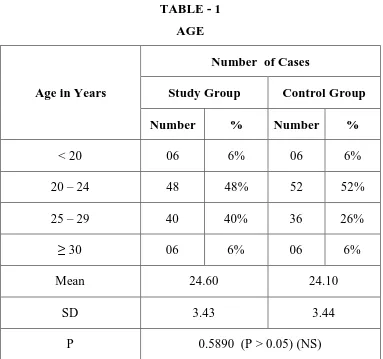
TABLE - 1 AGE
Number of Cases
Study Group Control Group Age in Years
Number % Number %
< 20 06 6% 06 6%
20 – 24 48 48% 52 52%
25 – 29 40 40% 36 26%
≥ 30 06 6% 06 6%
Mean 24.60 24.10
SD 3.43 3.44
P 0.5890 (P > 0.05) (NS)
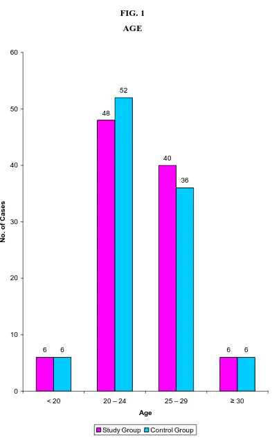
The mean age of the cases in both the groups doesn’t differ significantly.
Majority of the patients (50%) belonged to 20-24 years.
AGE
6
48
40
6 6
52
36
6
0 10 20 30 40 50 60
< 20 20 – 24 25 – 29 ≥ 30
Age
N
o
.
o
f
C
a
s
e
s
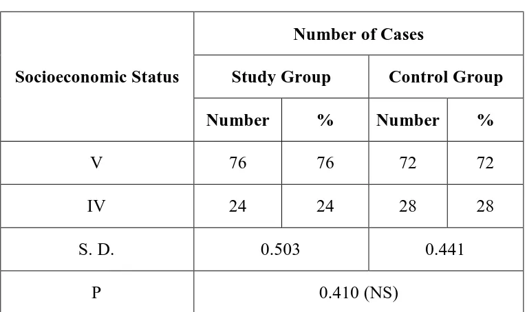
[image:57.595.124.517.97.733.2]TABLE – 2
SOCIOECONOMIC STATUS
Number of Cases
Study Group Control Group Socioeconomic Status
Number % Number %
V 76 76 72 72
IV 24 24 28 28
S. D. 0.503 0.441
P 0.410 (NS)
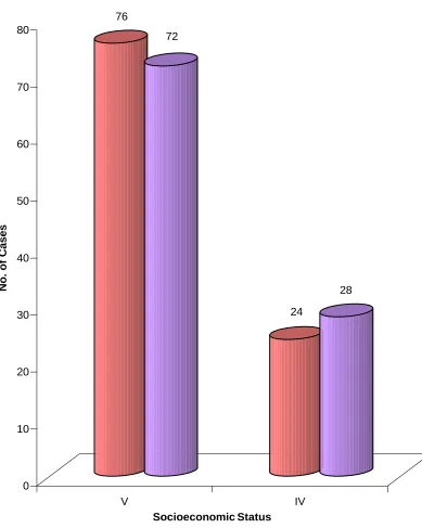
In our study no patient belonged to the class I, II or III socioeconomic status.
SOCIOECONOMIC STATUS
76
72
24
28
0 10 20 30 40 50 60 70 80
N
o
.
o
f
C
a
s
e
s
V IV
Socioeconomic Status
[image:59.595.126.515.197.684.2]TABLE – 3
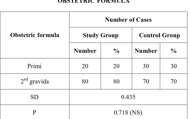
OBSTETRIC FORMULA
Number of Cases
Study Group Control Group Obstetric formula
Number % Number %
Primi 20 20 30 30
2nd gravida 80 80 70 70
SD 0.435
P 0.718 (NS)
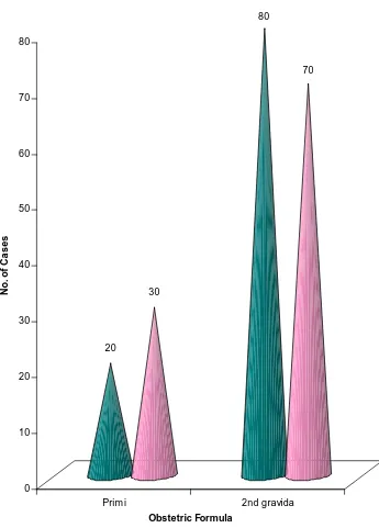
20 patients in study group and 30 patients in control group were primi gravida.
80 patients in study group and 70 patients in control group were 2nd gravida.
OBSTETRIC FORMULA
20
30
80
70
0 10 20 30 40 50 60 70 80
N
o
.
o
f
C
a
s
e
s
Prim i 2nd gravida
Obstetric Formula
[image:61.595.143.488.187.668.2]TABLE – 4
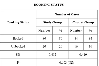
BOOKING STATUS
Number of Cases
Study Group Control Group Booking Status
Number % Number %
Booked 80 80 84 84
Unbooked 20 20 16 16
SD 0.412 0.419
P 0.603 (NS)
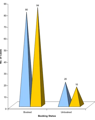
Antenatal booking doesn’t differ in both the groups significantly.
BOOKING STATUS
80
84
20
16
0 10 20 30 40 50 60 70 80 90
N
o
.
o
f
C
a
s
e
s
Booked Unbooked
Booking Status
[image:63.595.126.510.208.689.2]TABLE – 5
SUBJECTIVE CHARACTERS
Study group Control group Subjective characters
Mean S.D. Mean S.D.
P
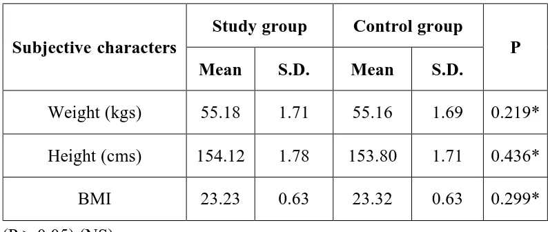
Weight (kgs) 55.18 1.71 55.16 1.69 0.219* Height (cms) 154.12 1.78 153.80 1.71 0.436* BMI 23.23 0.63 23.32 0.63 0.299* * (P > 0.05) (NS)
The average weight was 55.18kg in the study group and 55.16kg in the control group.
The average height was 154.12cm in the study group and 153.80cm in the control group.
The average BMI was 23.23kg/m2 in the study group and 23.32kg/m2 in the control group.
SUBJECTIVE CHARACTERS
55.18
154.12
23.23 55.16
153.8
23.32
0 20 40 60 80 100 120 140 160 180
Weight (kgs) Height (cms) BMI
Subjective characters
M
e
a
n
TABLE – 6
CHANGES IN VITAL PARAMETERS Number of cases
Study group Control group Parameters
Mean S.D. Mean S.D.
P
PULSE RATE / MIN
Pre delivery 82.38 2.79 82.86 2.60 Post delivery 83.86 2.64 88.24 2.61
Change +1.48 +5.38
0.0001 (S)
SYSTOLIC BP MM HG
Pre delivery 117.52 5.65 108.68 5.61 Post delivery 116.40 5.34 104.92 5.29
Change -1.12 -3.76
0.0001 (S)
DIASTOLIC BP MM HG
Pre delivery 76.80 8.87 76.92 8.47 Post delivery 76.34 8.78 73.64 8.88
Change -0.46 -3.28
0.0001 (S)
RESPIRATORY RATE / MIN
Pre delivery 17.93 0.30 17.80 0.31 Post delivery 18.14 0.98 18.06 1.07
Change -0.21 -0.26
0.147(NS)
SPO2 %
Pre delivery 99.90 12.09 99.90 10.13 Post delivery 99.66 11.94 98.79 9.64
Change -0.24 -1.11
0.0001 (S)
URINE O/P ML / HR
Pre delivery 108.10 2.02 109.70 2.12 Post delivery 106.90 1.97 106.50 1.86
Change -1.20 -3.20
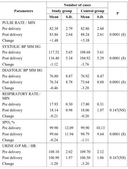
Mean increase in pulse rate was 1.48/min in study group and 5.38/min in control group.
Mean fall in systolic BP was 1.12mmHg in study group and 3.76mmHg in control group.
Mean fall in diastolic BP was 0.46mmHg in study group and 3.28mmHg in control group.
Mean increase in respiratory rate was 0.21/min in study group and 0.26/min in control group.
Mean fall in spO2 was 0.24% in study group and 1.11% in control group.
Mean fall in urine o/p was 1.20ml/min in study group and 3.20ml/min in control group.
PR increases and BP decreases significantly in control group compared with that of study group.
CHANGES IN VITAL PARAMETERS
[image:68.595.130.502.185.715.2]TABLE – 7
CHANGES IN BLOOD INDICES
Study group Control group Blood indices
Mean S. D Mean S. D
P
HB GM %
Pre delivery 8.86 1.78 8.88 1.93 Post delivery 8.84 1.77 8.73 1.91
Change -0.02 -0.15
0.0001(S)
PCV %
Pre delivery 39.04 7.04 38.72 4.56 Post delivery 38.70 7.01 37.62 4.53
Change -0.34 -1.10
0.0001(S)
Mean fall in haemoglobin was 0.02gm% in study group and 0.15gm% in control group.
Mean fall in hematocrit was 0.34% in study group and 1.10% in control group.
CHANGES IN BLOOD INDICES 8 .8 6 8 .8 8 3 9 .0 4 3 8 .7 2 8 .8 4 8 .7 3 3 8 .7 3 7 .6 2 0 5 10 15 20 25 30 35 40
HB GM % Study Group Control Group PCV % Study Group Control Group
[image:70.595.132.495.127.708.2]TABLE – 8
ONSET OF LABOUR
Number of cases
Study group Control group Onset of labour
Number % Number %
Spontaneous 52 52 56 56
Induced 48 48 44 44
SD 1.432 1.889
P 0.413 (NS)
ONSET OF LABOUR
52
56
48
44
0 10 20 30 40 50 60
N
o
.
o
f
C
a
s
e
s
Spontaneous Induced
Onset of Labour
[image:72.595.140.497.214.683.2]TABLE – 9
MODE OF DELIVERY
Number of cases
Study group Control group Mode of delivery
Number % Number %
Labour natural 72 72 60 60
Labour natural with
episiotomy 22 22 36 36
Labour natural with
lacerated perineum 06 06 04 04
SD 2.705 2.998
P 0.631 (NS)
Majority of the patients (66%) had labour natural.
MODE OF DELIVERY
72
22
6 60
36
4
0 10 20 30 40 50 60 70 80
Labour natural Labour natural with episiotomy
Labour natural with lacerated perineum
Mode of Delivery
N
o
.
o
f
C
a
se
s
[image:74.595.149.489.154.708.2]TABLE – 10
CHANGES IN DURATION OF THIRD STAGE
Duration of 3rd stage in
minutes Study group Control group
Mean 4.6 4.48
S.D. 1.428 1.854
P 0.141 (NS)
The mean duration of 3rd stage was 4.6mins in the study group and 4.48mins in the control group.
CHANGES IN DURATION OF THIRD STAGE
4.6 4.48
1.428 1.854
0 0
0 0.5 1 1.5 2 2.5 3 3.5 4 4.5 5
C
a
te
g
o
ri
e
s
Mean S.D. P (NS)
Duration of 3rd stage in minutes
[image:76.595.124.514.201.674.2]TABLE – 11
BLOOD LOSS
Study group Control group Blood loss in ml
Mean S.D. Mean S.D.
P
Time of delivery to 30 mins 81.01 50.387 191.65 49.884 0.0001(S) 30 mins to 2 hrs 27.27 6.17 51.72 9.78 0.0001(S) Time of delivery to 2 hrs 108.27 58.570 247.37 58.070 0.0001(S)
The mean blood loss at the end of 2 hrs was 108.27 ml in study group and 247.37 ml in the control group.
BLOOD LOSS
81.01
27.27
108.27 191.65
51.72
247.37
0 50 100 150 200 250 300
Time of delivery to 30 mins
30 mins to 2 hrs Time of delivery to 2
hrs
Blood loss in ml
C
a
te
g
o
ri
e
s
[image:78.595.142.488.119.727.2]TABLE – 12
ADDITIONAL UTEROTONICS
Study group Control group Additional uterotonics
Number % Number %
Yes 2 2 22 22
No 98 98 78 78
P 0.002
22% of the patients in the control group needed additional uterotonics compared to only 2% in the study group.
There was a significant difference in the requirement of uterotonics between the groups.
2 patients in the study group needed additional uterotonics (carboprost and methergin) and both needed blood transfusion.
22 patients in the control group needed additional uterotonics, of which 10 had PPH and other 12 had mild atonicity.
All the 22 patients were given 250 µg carboprost – PGF2α im. 8 patients responded to carboprost.
ADDITIONAL UTEROTONICS
2
22
98
78
0 10 20 30 40 50 60 70 80 90 100
N
o
.
o
f
C
a
s
e
s
Yes No
Additional uterotonics
[image:80.595.141.493.194.682.2]TABLE – 13
MATERNAL BLOOD TRANSFUSION
Study group Control group Blood transfusion
Number % Number %
Yes 02 02 10 10
No 98 98 90 90
Chi-Square Value 0.092 0.207
P 0.0102 (S)
10% of the patients in the control group needed blood transfusion compared to 2% in the study group.
MATERNAL BLOOD TRANSFUSION
2
10
98
90
0 10 20 30 40 50 60 70 80 90 100
N
o
.
o
f
C
a
s
e
s
Yes No
Blood transfusion
[image:82.595.139.493.198.674.2]TABLE – 14
MATERNAL COMPLICATIONS
Study group Control group Complications
Number % Number %
Vomiting 6 6 6 6
Fever 4 4 6 6
Nil 90 90 88 88
Chi-Square Value 0.211 0.213
P 0.41 (NS)
MATERNAL COMPLICATIONS
6
4
90
6 6
88
0 10 20 30 40 50 60 70 80 90 100
Vomiting Fever Nil
Complications
N
o
.
o
f
C
a
s
e
s
[image:84.595.141.495.175.695.2]TABLE – 15
APGAR SCORES
Study group Control group Apgar scores
Number % Number %
≥ 8 / 10 96 96 94 94 < 8 / 10 04 04 06 06
Chi-Square 0.211 0.212
P 0.500 (NS)
APGAR SCORES
6 6
4 6
90 88
0 10 20 30 40 50 60 70 80 90
N
o
.
o
f
C
a
s
e
s
Vomiting Fever Nil
Complications
[image:86.595.135.496.123.678.2]TABLE – 16
DURATION OF STAY
Study group Control group Duration of stay
Number % Number %
≤ 3 days 98 98 88 88
> 3 days 02 02 12 12
Chi-square 1.895 2.022
P 0.041 (S)
DURATION OF STAY
98
88
2
12
0 10 20 30 40 50 60 70 80 90 100
N
o
.
o
f
C
a
s
e
s
3 days > 3 days
Duration of stay
[image:88.595.148.482.182.664.2]DISCUSSION
As obstetric blood loss contributes to one fourth of global maternal death, death resulting from PPH should be avoided. As the fibrinolytic system gets activated after placental delivery antifibrinolytic agents can be used to reduce obstetric blood loss.
As prevention is always better than cure regarding PPH- an antifibrinolytic agent tranexamic acid was used prophylactically in our study to observe its efficacy in reducing blood loss during and after vaginal birth.
1. Maternal age
In our study, the age group of patients included varied from 18 to 35 years. Maximum percentage of patients belong to the age group of 20-24 years. 48% of study group and 52% control group were between 20-24 years. In a study conducted by Yang H, Shi C-Department of Obst & Gynaecology, first teaching hospital of Beijing University, Beijing, China in 2001 october - the mean age was 23.5 years.
2. Socio economic status
3. Obstetric formula
In our study, second gravida were more in both groups than primigravida. All were Singleton pregnancies. In the study group 20% were Primigravidas and 80% were 2nd gravidas. In the control group 30% were Primigravidas and 70% were 2nd gravidas. In a similar study conducted by Yildrium M.D at Erzincan military hospital, Turkey in april 2011 - second gravidas were 72% and primigravidas were 28%.
4. Booking status
In our study, 80% of study group and 84% of Control group were booked. 20% of study group and 16% of control group were unbooked. In a similar study conducted by Panagiotis and Rezan from Department of Obstetrics and Gynaecology, London in march 2011, 86% of study group and 88% of control group were booked. Proper antenatal care is important to identify the high risk factors in the antenatal period itself and to correct them thereby reducing the incidence of PPH.
5. Subjective characters
6. Change in Vital parameters
In our study, mean post delivery increase in PR was 1.48 in study group and 5.38 in control group. Mean fall in SBP was 1.12 in study group and 3.76 in control group. Mean fall in DBP was 0.46 in study group and 3.28 in control group. Mean increase in RR was 0.21 in group A and 0.26 in group B. mean fall in spO2 was 0.24% in the study group and 1.11% in the control group. Mean fall in urine output was 1.20ml in the study group and 3.20ml in the control group and was not statistically significant. There was a significant fall in SBP and rise in PR without any significant change in RR. In a similar study conducted by Natalia Novikova et al in 2010, there was a statistically significant change in vital parameters.
7. Changes in blood indices
In our study statistically significant fall in Hb% occurred after delivery in control group than with study group. Mean fall of Hb% was 0.02gm% in study group and 0.15gm% in control group. Mean fall in hematocrit was 0.34% in the study group and 1.10% in the control group. In a study conducted in the Department of obstetrics and gynaecology, University of Manitoba, 2010, statistically significant drop in haemoglobin was observed in the control group.
8. Onset of labour