0022-538X/93/074122-11$02.00/0
Copyright© 1993, AmericanSocietyforMicrobiology
Characterization of the
Myristylated
Polypeptide Encoded by the
ULl
Gene That Is
Conserved
in
the
Genome
of Defective
Interfering Particles of Equine
Herpesvirus
it
RONALD N.
HARTY,lt
GRETCHEN B. CAUGHMAN,2V. ROGERHOLDEN,'
ANDDENNIS J.
O'CALLAGHANI*
Department ofMicrobiology and Immunology, Louisiana State University Medical Center, 1501 Kings Highway, P.O. Box
33932,
Shreveport, Louisiana71130-3932,1
and Department of Oral Biology/Microbiology,Schoolof Dentistry, MedicalCollege of Georgia, Augusta,
Georgia
30912-11262 Received 25 January1993/Accepted 19April1993Equine herpesvirus 1 (EHV-1, KentuckyAstrain) preparations enriched for defectiveinterfering particles (DIPs) canreadilyestablishpersistentinfection. The ULl gene, which is conservedin thegenomeofDIPs that mediate persistent infection, maps between nucleotides 1418 and 2192 (258 amino acids) from the L (long) terminus. ULI has nohomologywithany known gene encoded by herpessimplexvirustype1 but has limited homology to open readingframe2ofvaricella-zoster virus and the "circ" geneof bovineherpesvirus type 1. Previous work showed that theEHV-1ULl gene belongs to the
early
kineticclassandistranscribedas a1.2-kb polyadenylated mRNA (R. N. Harty, R. R. Yalamanchili, and D. J. O'Callaghan, Virology 183:830-833, 1991). In this report, the ULl protein was identified and characterized as a 33-kDa polypeptide in EHV-1-infected cells by using rabbit polyclonal antiserum raised against aTrpE-UL1 fusion protein (amino acids 7 to 258of ULI) synthesized inEscherichia coli. Results from Western blot (immunoblot), immunopre-cipitation, indirect immunofluorescence, and biochemical analyses indicated that theULl polypeptide (i) is more abundant in cells infected with DIP-enriched virus than in cells infected with standard EHV-1, (ii) is synthesizedasearlyas3hpostinfection (p.i.)ininfection with standardvirus or ininfection with DIP-enriched virus preparations and increasesin abundanceup to 12 h p.i., (iii) appears tobe associated withtherough endoplasmicreticulum-Golgi apparatusearlyininfection (3to 4 hp.i.), whileadiffusecytoplasmic pattern of fluorescence is observedlate ininfection(7to8 hp.i.), (iv)is modifiedbymyristicacid as it contains aconsensus N-terminal myristylation site and isreadily labeled with [3H]myristic acid, and (v) is associated with mature EHV-1 virions.The standard genome of equine herpesvirus (EHV-1, Kentucky Astrain) is a linear double-stranded DNA mole-cule which is approximately 144 kbp in length (1, 2, 22, 31, 39,48).The genome iscomposedofuniquesequences(UL,
unique long; US, unique short) as well as inverted repeat (IR) sequences that bracket the US segment, thus allowing the formation of two isomeric forms of the genome. As observed for other alphaherpesviruses, EHV-1 genes are
temporally regulated into immediate-early, early, and late kinetic classes (4, 5, 13, 14).
AstandardEHV-1cytolytic infection is characterized by rapid lysis of infected cells; however, infection of cells with EHV-1preparations enriched for defective interfering parti-cles (DIPs)results inpersistent infection in which standard EHV-1 and DIPs are released continuously from infected cells (8, 17, 32, 38). SinceEHV-1 DIPhave beenimplicated in mediating persistence, identification and characterization of viralproteins encoded byEHV-1genesmaintained within the DIPgenome mayprovide a better understanding of the molecular events involved in establishing and maintaining persistent EHV-1 infection.
*Corresponding author.
tD.J.O.dedicates this papertohismentorandcolleague,Charles C. Randall, onthe occasion of his 80thbirthday and 53rd year of continuous and active research.
t Present address: Department of Microbiology, Mount Sinai School ofMedicine, NewYork, NY 10029-6574.
Recently, the UL2 gene of EHV-1 (a homolog of UL55 of herpes simplex virus type 1[HSV-1]),which is conserved in the DIP genome, was shown to direct the synthesis of a 0.9-kb mRNA and a 23-kDa polypeptide that are overex-pressed in DIP-enriched infection (18). In addition to UL2 and sequences that contain theorigin of replication and the cleavage-packaging elements (50), other sequences con-served in theDIPgenomewereshowntogenerateahybrid gene composed of sequences that encode the N-terminal portion of the IR4 protein (ICP22 homolog [23]) joined to sequences encoding the C-terminal portion of the UL3 protein (ICP27 homolog [53]). This hybrid IR4-UL3 gene is conserved in all clones of theDIP DNAanalyzedtodate(51)
and has been shownto beexpressedinDIP-enriched infec-tion (16).
Restrictionendonuclease, Southern blot,andpartialDNA
sequencing analyses of DIP DNA revealed that the ULl gene of EHV-1is also conserved in the genome of DIP(2, 16,
52). Interestingly, the ULl gene of EHV-1 (Kentucky A strain) exhibits no homologyto genes encoded by HSV-1; however, ULl does exhibit homologytogene 3 of the Ab4 strain of EHV-1 (46), open reading frame 2 (ORF2) of varicella-zoster virus (9), and the "circ" gene of bovine herpesvirus 1(BHV-1) (11, 41). The gene products encoded by these homologs of ULl have yet to be identified and characterized. Previously, the ULl gene was shown to be transcribed as a 1.2-kb polyadenylated mRNA at early stages ofinfection(20). Northern (RNA) blotandS1 nucle-ase analyseswere utilized to map the ULl transcript (20), 4122
on November 9, 2019 by guest
http://jvi.asm.org/
MYRISTYLATED POLYPEPTIDE ENCODED BY ULl IN EHV-1 DIPs 4123
andin vitro transcription-translation and hybrid-arrest trans-lation analyses were employed to identify theULl polypep-tidesynthesized from a cloned copy of theULl gene (19).
The studies described in this report have extended our characterization of the ULl protein synthesized in cells infected with standard EHV-1- or DIP-enriched EHV-1. The data revealed that the ULl polypeptide (i) has a molecular size of approximately 33 kDa and is synthesized as early as 3 h postinfection (p.i.), (ii) is modified by myristic acid (a saturatedtetradecanoic fatty acid), (iii) is stable and does not rapidly turn over during infection, (iv) appears to be associ-ated with cellular membranes, (v) is expressed more abun-dantly in cells infected with DIP-enriched virus than in cells infected with standard EHV-1, and (vi) is a component of mature EHV-1virions.
To our knowledge, this is the first report to identify a myristylated polypeptide encoded by EHV-1. However, myristylated proteins have been identified and characterized for many RNA- and DNA-containing viruses (for a review, see reference 47), including the gene product encoded by UL11of HSV-1 (26, 28). These initial studies to characterize the myristylated ULl polypeptide may provide for a better understanding of its function and potential role in persistent EHV-1 infection.
MATERIALS AND METHODS
Virus and cell culture. The Kentucky A strain of EHV-1 waspropagated in murine L-M cells and assayed by plaque titration as described previously (33, 34). The generation of DIPsof EHV-1 and the properties of cell lines persistently infected with EHV-1 have been described previously (2, 8,
21, 22,32). DIP-enriched EHV-1 preparations employed in theseexperiments include DIP passage 63 (3.9 x 10 PFU/ ml), DIP passage 64 (1.9 x 10⁶PFU/ml), and DIP passage 69 (9.4 x 10⁷ PFU/ml). Standard EHV-1 and DIP-enriched EHV-1 preparations were propagated in murine L-M cells and hamster embryo (HE) fibroblasts (33).
Generation of viral DNA clones. Plasmid pSVL1 was constructed by digesting the pGEML1 construct (described elsewhere [19])with AvrII (nucleotide [nt] 1367) and EcoRI
(nt 2513) and ligating this 1,146-bp fragment into an XbaI-EcoRI-digested pSV12 vector (43). Thus, plasmid pSVL1
contains the entire ULl ORF cloned downstream of the simian virus 40 (SV40) promoter-enhancer. Plasmid pATH 23L1 wasconstructed by digesting the pGEML1 construct
(19)withXbaI (nt 1440)and EcoRI(nt2513) and ligating this 1,073-bp fragment into an XbaI-EcoRI-digested pATH23 vector (24). Thus, the pATH23L1 construct contains a TrpE-UL1 fusion ORF inserted downstream of the inducible TrpE promoter. The ULl ORF within plasmid pATH23L1
beginswith codon 7and endswith codon 258(total number of ULl codons, 252). All clones were generated by using standard protocols (27) and were screened by restriction endonuclease analyses. Plasmid pSVL1 was maintained in
Escherichia
coli DHSa, andplasmid pATH23L1 wasmain-tained inE. coli TB1. Plasmid DNA was prepared by using the Qiagen(Chatsworth, Calif.) plasmid isolation kit.
Isolation oftheTrpE-UL1 fusion protein. The pATH23L1 construct was transformed into TB1 cells, and the
TrpE-ULl fusion proteinwas purified and isolated by using the
protocolof Koerner et al. (24) as described in detail else-where (18, 29). The TrpE-UL1 fusion protein was fraction-atedbysodiumdodecyl sulfate-polyacrylamide gel electro-phoresis (SDS-PAGE) and visualized by staining with
Coomassie brilliant blue (0.25%), and the appropriate TrpE-ULl protein band was sliced from the gel.
Preparation of TrpE-UL1 antiserum. The gel slice contain-ing the TrpE-UL1 fusion protein was mixed with an equal volume of Freund's complete adjuvant (Sigma Chemical Co., St. Louis, Mo.), and 2.0 ml of the mixture was used to immunize two male New Zealand White rabbits (0.5 ml per hind leg). Preimmune serum was collected from both rabbits before theadministration of the antigen. The administration of booster injections and the isolation of the TrpE-UL1 antiserum were done as described by Harlow and Lane (15). Transfection of COS-1 cells with pSVLl. COS-1 cells were seeded onto60-mm-diameter dishes or coverslips and trans-fected by the lipofectin (Bethesda Research Laboratories, Gaithersburg, Md.) method as described in detail elsewhere (43). Following 48 h of incubation at 37°C, the transfected cells in 60-mm-diameter dishes were lysed with 0.5 ml of
radioimmunoprecipitation assay buffer (150 mM NaCl, 50 mMTris-HCl,5 mMEDTA, 1.0% SDS, 0.5% deoxycholate, 1.0% Nonidet P-40) containing protease inhibitors (aproti-nin, 50 ,ug/ml; leupeptin, 50,ug/ml, and phenylmethylsulfo-nyl fluoride, 300 ,ug/ml; Sigma Chemical Co.). Transfected cells on coverslips were fixed for immunofluorescence as-says asdescribed below. Cell extracts were stored at -70°C until use.
Western blot and SDS-PAGE
analyses.
The protocols uti-lized for Western blot (immunoblot) and SDS-PAGE analy-seshave beendescribed in detail elsewhere (18, 19, 43). The specific dilution of the antiserum used in eachexperiment is indicated in the appropriate figure legends.Infection of cells and indirect immunofluorescence. Semi-confluentmonolayers of HE cells grown on coverslips (25 by 75mm) were mockinfected or infected with standard EHV-1 or DIP-enriched EHV-1 preparations (multiplicity of infec-tion [MOI], 10 PFU per cell) in the presence or absence of 200 ,ug of phosphonoacetic acid (PAA) per ml and main-tained at 37°C for times ranging from 1 to 7 h p.i. Coverslips were rinsed in phosphate-buffered saline (PBS), fixed in ice-cold acetone (5 min at -20°C), air dried, and stored at -20°Cuntil use.Semiconfluent EHV-1 persistently infected DI-3 cell monolayers (passage 83) were incubated for 24 h with no PAA treatment and fixed in the same manner. Coverslips were rehydrated for 10 min in PBS and incubated in a humid atmosphere for 30 min with normal goat serum (1:10 dilution in PBS) to blocknonspecific antibody binding. Coverslips were washed with PBS and incubated for 45 min with preimmune normal rabbit serum or TrpE-UL1 anti-serum (1:1,000 dilution in PBS containing 1.0% bovine serumalbumin). Thecoverslips were washed extensively in PBS, incubated for 45 min with fluorescein isothiocyanate (FITC)-conjugated goat anti-rabbit immunoglobulin G (IgG) (Grand IslandBiological Co.), and washed again in PBS. All incubations were performed at room temperature. The cov-erslips weremounted on glass slides in 50% glycerol in PBS and examined for fluorescence with a Nikon Optiphot mi-croscope equipped with an episcopic-fluorescence attach-ment and aB-1A filter block.
Immunoprecipitation
analyses
of infected cell extracts. Mock- orEHV-1-infected L-M cells (2 x 10⁶ cells per flask) were harvested in radioimmunoprecipitation assay buffer with proteaseinhibitors as stated above. Cells were radiola-beled with either[35S]methionine
(50 ,uCi/ml; New England Nuclear, Boston, Mass.) or [3H]myristic acid (20 ,Ci/ml;NewEnglandNuclear). Cell extracts were stored at -70°C until use. The protocol used for immunoprecipitation has been described in detailelsewhere (18). 14C-labeled molec-VOL.67, 1993
on November 9, 2019 by guest
http://jvi.asm.org/
UL IR
Us
TRAvril Xbal Kpni - BgIll/EcoRI
I I I I .__
I
0
I
500
I
1000
I
1500 2000 2500
I
3000 nt UL1 -258 aa UL2-200 aa
1418 2192 2225 2825 nt
_pSVL1 X pATH23L1
E
E
aa7-258
FIG. 1. Diagram of theEHV-1 genome(top)withnt1to3000 from theLterminusexpandedbelow. ThearrowsrepresenttheULland UL2ORFsof258 and 200 aminoacids(aa),respectively. The ULl ORFbeginsat nt1418 and endsatnt2192,while the UL2 ORFbegins
at nt2825and endsat nt2225.Theviral insertwithinplasmid pSVL1mapsfrom the AvrIIsite(nt1367)totheBglIIsite(nt2513).Notethat theBglIIsite in theoriginalclone(pl-111) fromwhichpSVL1andpATH23L1werederivedwasconvertedto anEcoRIsiteuponcloning. TheviralinsertwithinplasmidpATH23L1mapsfrom theXbaI site (nt1440)tothe EcoRIsite(nt2513).Thefirst six N-terminal codons of theULlORF are deletedin thepATH23L1construct.Abbreviations:UL,uniquelong; IR,inverted repeat;US,unique short; TR,terminal repeat.
ular weight standards (Bethesda Research Laboratories)
wereutilizedto determinemolecular size.
Pulse-chase analysis. Pulse-chase analysis was performed asdescribedpreviously (18). Briefly,L-Mcellmonolayers (2
x 106 cells)wereinfected withDIPpassage64atanMOI of
2.0PFUpercell. The cellswerepulselabeled with
[35S]me-thionine(50,uCi/ml;NewEngland Nuclear)for 30minat5 h p.i.and chased witha100,000-foldexcessof cold methionine
(30 mg/liter; Sigma Chemical Co.) for the times indicated. Protein concentrations were determined with the bicincho-ninic acidproteinassayreagent(Pierce, Rockford, Ill.),and equal amounts of protein were immunoprecipitated and
fractionatedby SDS-PAGE.
Purification of EHV-1 virions. EHV-1 virionswerepurified
by polyethylene glycol precipitation andrate-velocity
cen-trifugation in dextran-10 gradients as described previously (34).Toremovethe virionenvelope,theappropriateamount ofpurifiedvirions(collected by centrifugationat50,000rpm
for 30 min with a TLA 100.3 rotor in an Optima TL ultracentrifuge) was suspended in sterile PBS containing 1.0% Nonidet P-40 and 1.0%deoxycholateand incubated for 1 hat37°C.Thedisruptedvirionswerecentrifugedat27,000
rpm for 30minwith the TLA100.3rotor, and the
supema-tant represented the envelope fraction, while the pellet represented the nucleocapsid-tegument fraction. Proteins present in each fraction were fractionated by SDS-PAGE
andanalyzed byWesternblot hybridization. RESULTS
Generation ofTrpE-UL1polyclonalantiserum.To charac-terizetheULlpolypeptide synthesized duringEHV-1 infec-tion, polyclonal antiserum was raised against a TrpE-UL1
fusionprotein.TheULl ORF(Fig. 1)wasclonedpreviously intothetranscription expressionvectorpGEM-3Zto gener-ate plasmid pGEML1 (19). A portion of the ULl ORF (codons 7 through 258) was excised from pGEML1 as an
XbaI-EcoRI fragment and ligated into an
XbaI-EcoRI-di-gested pATH23 vector containing the bacterial TrpE gene
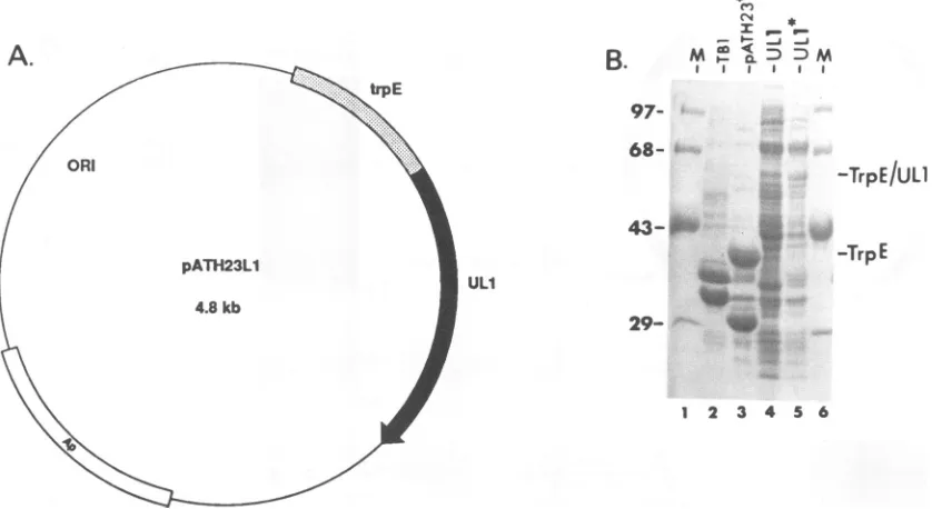
and its inducible promoter(Fig. 1). The resultantconstruct, pATH23L1 (Fig. 2A), contains the TrpE-UL1 fusion ORF under the control of the inducibleTrpEpromoter.
E. coli TB1 cellswere transformed with the pATH23L1 construct, and the TrpE-UL1 fusionproteinwas identified
by fractionatingthe proteinson anSDS-polyacrylamide gel
followed by staining with Coomassie blue (Fig. 2B). Cell extracts from TB1cells (Fig. 2B, lane 2), TB1 cells trans-formed with thepATH23vector andgrownunderinducing conditions (lane 3), and ThB cells transformed with pATH23L1 andgrownundernoninducing (lane 4)or
induc-ing (lane 5)conditionswerefractionatedbySDS-PAGE. The
37-kDaTrpE proteinsynthesizedfrom thepATH23vectoris clearly visible in extracts of TB1 cells transformed with pATH23 (lane 3), demonstrating that theTrpEpromoter is functional and inducible. A protein with the approximate sizeexpectedfortheTrpE-UL1fusionproteinwasobserved in extracts of TB1 cells transformed with pATH23L1 and
grown under inducing conditions (lane 5) but was not ob-served in extractsofeither TB1 cells (lane 2) orTB1 cells transformed with the pATH23 vector alone (lane 3). The abundanceand level ofexpressionof theTrpE-UL1 protein
were relatively low in extracts of pATH23L1-transformed cells grown under inducing conditions (lane 5). However, this level of expression was not surprising, since proteins encoded by largeORFs inserted into pATH23, such as the largeULl insert of 252codons, arenotexpressedfromthe pATH vectors as efficiently as are proteins encoded by
smaller ORFs (24).
Specificity of the TrpE-UL1 antiserum. To demonstrate that the TrpE-UL1 antiserum was specific for the ULl
polypeptide, a ULl expression vector was employed in indirect immunofluorescence and Western blot analyses. Theentire ULl ORFwasinserted downstream of theSV40 A
-9 -
-I I a I
m n m m m u Ml
I I I I I
I
I
I
on November 9, 2019 by guest
http://jvi.asm.org/
[image:3.612.136.485.76.278.2]MYRISTYLATED POLYPEPTIDE ENCODED BY ULl IN EHV-1 DIPs
* - 1- _J __
B.
II I I97-68-'
s.
-
29-ULI
-TrpE/ULl
-TrpE
[image:4.612.103.522.86.315.2]1 2 3 4 5 6
FIG. 2. Expression of a TrpE-UL1 fusion protein from the pATH23L1 construct. (A) Diagram of the pATH23L1 plasmid (4.8 kb)
containing the TrpE-UL1 fusiongene.The ULl ORF in thisconstructbegins with codon 7 andends with codon 258(252 codons).Expression
oftheTrpE-UL1 fusion proteinisdriven by theinducible TrpEpromoter.Abbreviations: ORI, origin ofreplication; Ap, ampicillin resistance
gene. (B)SDS-PAGE analysis of Coomassie blue-stained proteins fromextractsof TB1 cells (lane 2),TB1cellstransformed withpATH23
grownunderinducing (*) conditions (lane 3), TB1cells transformed with pATH23L1grownunder noninducing conditions (lane 4), and TB1
cellstransformed withpATH23L1grownunderinducing (*) conditions (lane 5). The positions of the TrpE-UL1 fusion protein(approximately
58kDa; lane 5) and theTrpE protein (approximately 37 kDa; lane 3) are indicated. Molecular weight standards (M, lanes 1 and 6), in
thousands,areindicatedontheleft.
promoter-enhancer region present in vector pSV12 to gen-erateplasmid pSVL1 (Fig. 1 and3A). Plasmid pSVL1 was
transfected intoCOS-1 cells byusing thelipofectin method (43). Indirect immunofluorescence analyses demonstrated that theTrpE-UL1 antiserumdidnotreactwithCOS-1cells transfectedwith the pSV12vector alone (Fig. 3B, top) but did react strongly with cells transfected with pSVL1 (Fig. 3B, bottom). As further evidence of the specificity of the TrpE-UL1 antiserum, Western blotanalysiswasperformed
with extracts of COS-1 cells transfected with pSV12 (Fig. 3C, lane1)orwith 5(lane 2),10 (lane 3),and15 ,ug (lane 4)
of pSVL1. The 33-kDa ULl polypeptide was detected in
extracts ofpSVL1-transfected COS-1 cells(lane 2to4) but not in pSV12-transfected COS-1 cells (lane 1). To confirm that the 33-kDa protein was synthesized from ULl-coding
sequences, pSVL1 was first linearized by digestion with either
KpnI
(which cleaves within the ULl codingse-quences) or EcoRI (which cleaves within the vector
se-quences) and then transfected into COS-1 cells. As
ex-pected, the33-kDa ULlproteinwasnotobserved in COS-1 cell extracts receiving the
KpnI-digested
pSVL1 (Fig. 3C, lane 5) butwas observed in extracts receiving the EcoRI-digestedpSVL1 (lane 6).Theseresults and thefinding (data not shown) that the ULl antiserum also reacted with the product of an in vitro-transcribed and -translated pGEM construct containing ULl sequences (pGEML1 [19]) indi-cated that the TrpE-UL1 antiserum could recognize the EHV-1 ULlpolypeptide.The TrpE-UL1 antiserum reacts with the ULl protein synthesized in EHV-1-infected cells. To detect the ULl polypeptide synthesized in vivo, murine L-M cells were
mock infected or infected with standard EHV-1 or
DIP-passage-69-enriched EHV-1. Westernblot analysisof these infected cellextracts(harvestedat8 h p.i.) revealed that the 33-kDa ULl polypeptide was present in extracts of cells infected with standard EHV-1 (Fig. 4A, lane 2) and with DIP-enriched virus (lane 1) but not in extracts of mock-infected cells(lane 3).Theintensity of the signalin the cell extract infected with DIP-passage-69-enriched EHV-1 69 (lane 1) was slightly higher than that observed in cells infectedwithstandardEHV-1 (lane 2).
Immunoprecipitation analysis was also utilized to detect the ULl protein in EHV-1-infected cells. L-M cells were
infected with standard EHV-1 in the presence of [35S]me-thionine, and the cell extracts were harvested at 8 h p.i. Equal amounts of the extracts were incubatedwith either preimmune serum (Fig. 4B, lane 1) orthe TrpE-UL1 anti-serum (lane 2), and the immunoprecipitated proteins were
fractionated by SDS-PAGE. The 33-kDa ULl protein was
immunoprecipitated readily with the TrpE-UL1 antiserum (lane 2), butnotwith preimmuneserum(lane 1).
The ULlproteinismyristylated. Computer analysisof the ULl amino acid sequencerevealed a consensus
myristyla-tion site(47) atthe N terminus of theprotein (Fig. 5A).The myristic acid moiety is linked to proteins containing the
consensus site via an amide bond to a glycine residue
immediately following the initial methionine. The third amino acid position may beoccupied by anyof the amino acidsindicated,whilepositions4and 5maybefilledbyany
amino acid. Amino acidposition6canbeoccupied byoneof the six amino acids shown, however a serine residue is preferred. Lastly,amino acidposition 7cannotbeoccupied by aproline residue. Thefirst seven amino acids are illus-trated for the ULl protein (homolog of ORF3 of EHV-1
A.
VOL.67, 1993 4125
on November 9, 2019 by guest
http://jvi.asm.org/
B.
Pi ULl
I
-33
1 2
B. C.
2Y pSVLI 5 10 15 K E
1 2 3 4 5 6
[image:5.612.55.296.69.450.2]_~~~~~~
FIG. 3. Expression of the ULl polypeptide fromanSV40 pro-moter. (A) Diagram of the pSVL1construct (3.7 kb) depicting the complete ULl ORF downstream of the SV40 promoter-enhancer element.TheAvrII-EcoRI fragment (see Fig. 1) containing the ULl
ORFwascloned intothepSV12vectordigested with XbaI-EcoRI. Ap, ampicillin resistance gene. (B) Indirect immunofluorescence analysesofCOS-1 cells transfected with either the pSV12vector
(top) or pSVL1 (bottom) and reacted with TrpE-UL1 antiserum
(1:1,000). (C) Western blot analysis of extracts of COS-1 cells
transfected withpSV12 (lane 1),5.0p.gofpSVL1 (lane 2), 10.0p.g ofpSVL1 (lane 3), 15.0 pgofpSVL1 (lane 4), 10.0 pgofpSVL1 linearizedwithKpnI(lane 5), and 10.0p,g of pSVL1 linearized with
EcoRI(lane 6). The blotwasreacted withTrpE-UL1 antiserumata
1:7,500dilution.
Ab4), the circgene productofBHV-1, and theUL11 gene
productofHSV-1. TheUL11geneproductisnotahomolog ofULl of EHV-1 but does representaknownmyristylated
herpesvirus protein(26). The circgeneproductof BHV-1 is a homolog of ULl of EHV-1 (11, 41), and although it
possesses a consensus myristylation site, the circ gene
product has not yet been shown to be myristylated. To demonstratewhether the ULlprotein ismodifiedby myris-tic acid, L-M cells were mock infected or infected with
standardorDIP-enriched EHV-1 in thepresenceof 20,uCi
of
[3H]myristic
acidperml. The cellextractswereharvestedat8 hp.i.,andequalamountsof theextractswereincubated with the TrpE-UL1 antiserum. Following fractionation of
1 2 3
FIG. 4. Expression of the ULlpolypeptide in standard EHV-1 and EHV-1 DIPinfections. (A) Western blot analysis ofextracts
harvestedat8 h p.i. from cells infected with either DIP-passage-69-enriched(lane 1)orstandard (lane 2) EHV-1-ormock-infected cells (lane 3). The blotwasreacted with TrpE-UL1antiserumatadilution
of 1:10,000. (B) Immunoprecipitation of [35S]methionine-labeled
extracts (4 x 10⁵ cell equivalents) from L-M cells infected with
standardEHV-1(8 h p.i.). Proteinswereimmunoprecipitated with
eitherpreimmuneserum(15 Al,lane 1)orTrpE-UL1antiserum (15
,ul, lane 2). The position of the 33-kDa band is indicated in each panel.
the immunoprecipitated proteins by SDS-PAGE, a
radiola-beled 33-kDa protein was detected in extracts of cells infected with standard EHV-1 (Fig. 5B, lane 3) and DIP-passage-64 (lane 4)-and-69(lane5)-enriched EHV-1 follow-ing an overnight exposure of the gel. The myristylated
33-kDa protein was not detected in mock-infected cell
ex-tracts (lane 2). This posttranslational modification may
ex-plain whythe ULlprotein synthesized in vivo migrates at approximately33kDa, althoughitscomputer-predicted
mo-lecular sizeis 28,343 Da.
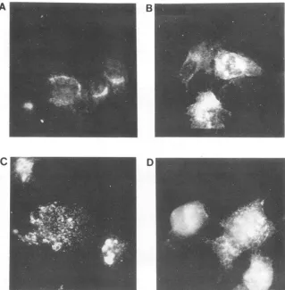
Intracellular localizationof the ULlproteininstandard and DIP-enriched EHV-1 infections. TheULl geneproductwas
firstdetected at 3 hp.i. in HE cellsinfected with standard EHV-1 as apolarized, perinuclear regionofreactivity (Fig.
6A). Thefluorescence appeared to delineatea multilayered
structure,consistentwith the idea that the ULlproteinwas
localizedtotheGolgiapparatus(Fig. 6A).Subsequently,the majority of reactivitywas associated withstructureswhich appearedtobeintracytoplasmicvesicles(Fig.6B andC).At 7 hp.i.,the ULlreactivitywasintense andevenly distrib-uted throughout the cell such that little intracellular detail
wasvisible. Thispattern ofreactivity is consistent with the ULl protein being localized in or near the cytoplasmic membrane. In DIP-enriched EHV-1-infected HEcells, the fluorescence was more intense, and the time course of the intracellulardistribution of the ULl proteinwasmorerapid
than that in standard EHV-1-infected HEcells(Fig. 7C and D). By4 hp.i., extensive, intensecytoplasmic reactivitywas
observed in DIP-enriched EHV-1-infectedcells. This
gener-alizedcytoplasmic pattern ofreactivitywas similar to that observed in standard EHV-1-infectedcells at 7 hp.i. andto
A.
A.
DI 69 STD ma a I
ULt
33-_M
Xbal(Awrll)
on November 9, 2019 by guest
http://jvi.asm.org/
[image:5.612.320.548.75.299.2]MYRISTYLATED POLYPEPTIDE ENCODED BY ULl IN EHV-1 DIPs 4127 A CONSENSUS = M - G - (ANCQGILMSTV) - X2 - (STAGCN) - {P}
M G- A
-
M G- AM G- L
- C C ---- s ---- S
R A ---- S ---- A
--- S F ---- S
----B MW STD D6DI
MWSD64 69
200- 97-68-;
-33Kd
29-i
[image:6.612.150.468.80.382.2]
_--1 2 3 4 s
FIG. 5. Myristylation of the ULl polypeptide. (A) Theconsensusamino acidsequence(single-letter code)necessaryfor myristylation of polypeptidesisshown.Theunderlined G(glycine)representsthe amino acidtowhich the myristic acid is bonded viaanamide linkage. Amino
acids withinparenthesesrepresentthose thatareallowedatthat particular position, while only proline (in braces) isnotallowedatposition
7. The S(serine)residue isunderlinedtoindicate that it is the preferred amino acidatthis position. The letter Xrepresentsanyamino acid.
ThesevenN-terminal aminoacids of the ULl protein (EHV-1 KentuckyAstrain)-ORF3 (EHV-1 strain Ab4), the circ protein ofBHV-1,and
theUL11geneproductofHSV-1arealso shown. (B) Immunoprecipitation of[3H]myristic acid-labeled cellextracts. Cellextracts(4 x 10⁵ cellequivalents) from cells thatweremock infected (m, lane 2)orinfected with standard (STD, lane 3) EHV-1orDIP-passage-64(DI 64, lane
4) DIP-passage-69 (DI 69,lane5)-enriched EHV-1wereincubated with the TrpE-UL1 antiserum (15 ,.l), and theimmunoprecipitated proteins
werefractionatedby SDS-PAGE. Molecular weightstandards (MW, lane 1) (in thousands)areindicatedonthe left; theposition of the 33-kDa
band is indicatedontheright.
that observed inpersistentlyinfected DI-3 cells(Fig. 7E).It should be noted that the presence of PAA throughout infection didnotinfluence thesynthesisof the ULlprotein
oraffect the levels detectedduringinfection(Fig. 7F). Also,
mock-infected HE cells incubated with the TrpE-UL1
anti-serumandEHV-1-infected cells incubated withpreimmune
serum did notyield any level of fluorescence above back-ground (Fig. 7A andB, respectively).
Timecourseandstabilityof the ULl polypeptide.Western blotanalysiswasutilizedtofollow thesynthesisofthe ULl polypeptide at various times p.i. Murine L-M cells were
infected with DIP-passage-64-enriched EHV-1 (MOI, 2.0 PFUpercell),and cellextractswereharvestedat2,4, 6, 8, 10, and 12 hp.i. (Fig. 8A). The 33-kDa ULl protein was
readilydetectedat6 hp.i. (Fig. 8A,lane3)andcontinuedto
increase in abundance up to 12 hp.i. (lane 6). The level of sensitivityof thisexperimentwasnotsufficienttodetectthe ULlproteinat4 hp.i., althoughitwasshowntobepresent
atearlytimes of infectionbyimmunofluorescence assays.
The stability of the ULl polypeptide was analyzed by
pulse-chase analysis (Fig. 8B). Murine L-M cells infected with DIP passage 64 (MOI, 2.0 PFU per cell) were
pulse-labeled for 30minwith [35S]methionine at 5 hp.i. and then
chased with cold methionine (100,000-fold excess) for the times indicated (Fig. 8B). Cell extracts containing equal amounts of protein were incubated with either the TrpE-ULl antiserum or preimmune serum, and the immunopre-cipitated proteinswereanalyzed bySDS-PAGE. The 33-kDa ULlproteinwas immunoprecipitated throughoutthe chase period of 48 h (Fig. 8B, lanes 3 to 7). Although a slight
decrease in theabundance of the ULlproteinwasobserved
asthe time ofchase increased(comparelanes5 and7),these resultssuggestthat the ULlprotein isnotrapidly degraded and does not undergo extensive posttranslational
process-ing.TheULlproteinwasnotdetected in mock-infected cell extracts (lane 2)orwith preimmuneserum (lane 8).
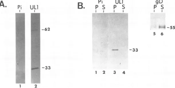
TheULlproteinin purifiedEHV-1 virions.SincetheULl protein increased in abundance late in infection and since
many myristylated viral proteins are also virion structural proteins (for a review, see reference 47), mature EHV-1 virions were assayed by Western blot analyses for the
presence of the ULl polypeptide. Purified EHV-1 virions
were solubilized in 1x Laemmli sample buffer (25) and
fractionated by SDS-PAGE (Fig. 9A). The blotted virion
proteinswerereacted with eitherpreimmuneserum(lane 1) ortheTrpE-UL1antiserum(lane 2)atadilution of1:15,000.
UL1/ORF3
BHV1-circ HSVI-ULI1
VOL.67, 1993
on November 9, 2019 by guest
http://jvi.asm.org/
A
B
Cr
_
~~~D
FIG..6.Deeto fteUlpoeni tnadEV1ifce Ecls.EV1ifce el MIo 0PUm)wr iet3()
4~,(B)6C,ad7()hpi n ece ihTp-L nieu n FT-otat-abtimngoui sdsrbdi aeil
andMtos
Indeed, aproteinofapproximately33 kDawasdetectedby
using theTrpE-UL1 antiserum(lane 2) butwas notdetected with preimmune serum (lane 1). An additional protein mi-grating at approximately 62 kDa also observed with the TrpE-UL1 antiserum(lane 2)may representacross-reactive virion proteinor an aggregate (dimer) of the ULl
polypep-tide.
Tolocalize further theULlpolypeptideinmaturevirions, purified virus was incubated at 37°C in a solution of PBS
containing 1.0% Nonidet P-40 and 1.0% deoxycholate to separatethe viralenvelopefrom thenucleocapsid-tegument regions. The viral proteins in the envelope fraction (super-natant) and those in the nucleocapsid-tegument fraction
(pellet) were fractionated by SDS-PAGE and analyzed by Western blotanalysis (Fig.9B).As anegative control, both the supernatant and pellet fractionswerereacted with pre-immune serum (lanes 1 and 2). As a positive control, both the supernatant and pellet fractions were reacted with a
peptide-specificantiserum to EHV-1 glycoproteinD(lanes 5 and6) (10). Asexpected, the majority of glycoprotein D (55 kDa) was detected in the supernatant or envelope fraction
(lane 6), while only a small amount was observed in the pellet fraction (lane 5).Incontrast, essentially all of theULl
protein wasobserved in the pellet (nucleocapsid-tegument)
fraction (lane 3), as no ULl protein was detected in the
supernatant (envelope) fraction (lane 4).
Since the UL1 protein was present in mature virions,
neutralization assayswere performedtodetermine whether the TrpE-UL1 antiserum could block EHV-1 infection of murineL-Mcells. The results ofthese studiesindicated that theTrpE-UL1 antiserumwasunableto neutralize the virus and prevent virus infection (data not shown). These data suggestthat the ULl protein is acomponentof the mature EHV-1virion and thatULl doesnotappeartobelocalized within the viral envelope asisglycoproteinD.
TheTrpE-UL1antiserum cross-reacts with a BHV-1 virion protein. Since the ULl geneexhibits homology, albeit lim-itedhomology,totherecently identified circ gene of BHV-1 (11, 41), purified BHV-1 virions were assayed by Western blot analysesfor BHV-1 proteinsthat react with the TrpE-ULl antiserum. Variousamounts (0, 1, and2 ,ul)ofpurified BHV-1virions andafixedamountofpurified EHV-1 virions
(10 p.l) were fractionated by SDS-PAGE, and the blotted virion proteins were reacted with either preimmune serum (Fig. 1OA, lanes 1 to 4) or the TrpE-UL1 antiserum (Fig. lOB, lanes 5 to 8). Both EHV-1 andBHV-1virion proteins thatwereincubatedwithpreimmuneserumyieldednosignal (Fig. 1OA, lanes 1 to4), whereasthe TrpE-UL1 antiserum
recognizedthe 33-kDa EHV-1ULl protein(Fig. lOB,lane5)
aswellas a35-kDaBHV-1virionprotein(Fig. lOB,lanes6 and 7). Whether this 35-kDa BHV-1 virion protein repre-sents the product of the BHV-1 circ gene remains to be
determined,sincethecircprotein has yettobeidentified and characterized.
on November 9, 2019 by guest
http://jvi.asm.org/
[image:7.612.151.470.104.429.2]MYRISTYLATED POLYPEPTIDE ENCODED BY ULl IN EHV-1 DIPs 4129 A
C!
B
D
E
FIG. 7. Indirectimmunofluorescenceshowingthe distribution of theULl proteinin standardEHV-1-infectedHEcellsinthepresence or absence of 200 ,ug of PAA perml,DIP-enrichedEHV-1-infectedHEcells,andpersistently infectedDI-3 cells.(A)Mock-infected HE cells reacted withTrpE-UL1antiserum; (B)DI-3 cells(passage83)reacted withpreimmunerabbit serum; (C)standardEHV-1-infectedHEcells (4hp.i.)reacted withTrpE-UL1antiserum; (D) DIP-enrichedEHV-1-infected HE cells(4hp.i.)reacted withTrpE-UL1 antiserum;(E) DI-3 cells(passage 83)reacted with theTrpE-UL1 antiserum;(F)PAA-treated standardEHV-1-infected HE cells(6hp.i.) reacted with TrpE-UL1 antiserum.
gDISCUSSION
EHV-1 genes conserved in the genome of DIPs are of
interest, sincetheir geneproductsmayfunction inpersistent
EHV-1infection. TheULlgeneof EHV-1 is includedwithin
sequences atthe L terminus thatare conserved in the DIP genome(1,2,49, 50).Inthis report, thepolypeptide encoded
bytheULl gene has been identified and characterized with regard to its time ofsynthesis, stability, intracellular local-ization, and posttranslational modification.
ULl-specific antiserum was used to identify the 33-kDa
ULl protein in cells infected with standard EHV-1 orwith DIP-enriched preparations of EHV-1. The ULl protein synthesized in cells infected with various passages of
DIP-enriched preparations of EHV-1 was more abundant than that synthesized in cells infected with standard EHV-1 as
judged by Westernblot, immunoprecipitation, and indirect
immunofluorescence analyses. These observations parallel
those observed for the UL2 protein, which is also overex-pressed in DIP-enriched infection as the UL2 gene is also conserved in the DIP genome (18). It should be noted that clonedcopiesof both theULl andUL2 geneswereutilized in transient cotransfection experiments to determine whether the ULl or UL2protein, in conjunction with the EHV-1 immediate-early protein, could regulate expression of chloramphenicol acetyltransferase (CAT) from various EHV-1 promoter-CAT constructs (44). The results from VOL.67, 1993
on November 9, 2019 by guest
http://jvi.asm.org/
[image:8.612.147.470.73.509.2]2 4 6 8 10 12 h
ULl
Pi
- 6 9 12 24 48 48 -hrs p.i. B. M Oj5 05 3.5 6.5 18.5 42.5 42.5 -hrs chase
43-I
-33 29-i
.. A333.
1234 56 1 2 3 4 6 7 8
FIG. 8. Timecourseand pulse-chase analyses of ULl polypeptide in L-M cells infected with DIP-passage-64-enrichedEHV-1. Protein
concentrationsweredetermined by using the bicinchoninic acid protein assayreagent(Pierce),andequalamountsofproteinwereanalyzed
in eachlane. (A) DIP-passage-64-enriched EHV-1 infected cellextractswereharvestedat2, 4, 6, 8, 10, and12 hp.i.,and the filtercontaining the blottedproteinswasreacted withTrpE-UL1 antiserumatadilution of1:15,000. (B)Pulse-chaseanalysisof the ULlprotein synthesized inDIP-passage-64-enriched EHV-1-infected cells. Cellswerepulsedat5 h p.i. for 30min with [35S]methionine. The duration of infection and the hours of chaseareindicated. Cellswerelabeledat5 h p.i. and harvestedat6, 12, 24,and48hp.i. The [35S]methionine-labeled proteins
wereimmunoprecipitated with either TrpE-UL1 antiserum (lanes 2to7)orpreimmuneserum(lane 8). Lane 2representsmock-infected cells. Molecularweight standards (M, lane 1) (in thousands)areindicated.
these CATassays indicated that neither the ULl norUL2
protein could significantly transactivateortransrepressCAT expressionfrom EHV-1immediate-early, early,orlate
pro-moter-CATconstructs (datanotshown).
The amino acidsequenceof ULlwasreported previously
(52), andnumerous potentialsites ofposttranslational
mod-ification within the ULl polypeptidewere noted (19). One potential site of modification includedamyristylation siteat the Nterminus of the ULlpolypeptide (19). Indeed, results from biochemical and immunoprecipitation analyses
sug-gested that the ULl polypeptide was modifiedby myristic acid in cells infected with standardorDIP-enriched EHV-1. As further evidence of myristylation, the radiolabel ([3H]myristic acid) on the ULl protein was not released followingincubation of theimmunoprecipitatedULlprotein ina1.0 M solution ofpotassium hydroxide (datanotshown). These datasuggestthat the radiolabel is associated withan
A.pi
UL1
51 -62
-33,.
..,
amide-linkedmyristicacidmoietyrather thananester-linked palmiticacidmoiety (42). Althoughthis is the first EHV-1-myristylated protein identified, a plethora of myristylated viralproteinshave been described in other viral systems(for
a review, see reference 47), including p60vsrc of Rous sarcoma virus (40), the gag proteins of many retroviruses including human immunodeficiency virus (36), pre-Sl
pro-tein ofhepatitisB(35), VP2 ofSV40 (45),VP4ofpoliovirus (6), and the UL11gene productofHSV-1 (26). TheUL11
geneof HSV-1wasshowntoencodeafamilyofmyristylated
proteins that are also structural components of the mature virion (26). Moreover, all of the viral myristylated proteins mentioned aboverepresentvirionstructuralproteinsaswell. Similarly, the ULl protein of EHV-1 was shown to be associated withmaturevirions andappearsnot tobe local-ized within the virion envelope. Lastly, it should be noted that the TrpE-UL1 antiserum also recognized a 35-kDa
Pi
ULI
B.
prsp
s
I
gD
P S
-#55 5 6
-33
1 2 3 4
1 2
FIG. 9. Westernblot analyses ofEHV-1virions with TrpE-UL1 antiserum. (A) Purified EHV-1 virionswere suspended in Laemmli
sample buffer, boiled,and fractionatedbySDS-PAGE. The blotted virionproteinswerereacted with eitherpreimmuneserum(Pi;lane1)or
TrpE-UL1antiserum(UL1;lane2)ata1:15,000dilution.(B)Purified EHV-1 virionswereincubated inadetergentbuffer(seeMaterials and
Methods)toseparatetheenvelope from thenucleocapsid-tegument. Proteins within theenvelopefraction(S)and within the
nucleocapsid-tegumentfraction(P)wereanalyzed by SDS-PAGE,and the blottedproteinswerereacted withpreimmuneserum(1:15,000,lanes1 and2), TrpE-UL1 antiserum (1:15,000, lanes 3 and 4),orantipeptide antiserum(1:800)raisedagainstEHV-1glycoproteinD(gD,lanes 5 and 6[10]).
A.
on November 9, 2019 by guest
http://jvi.asm.org/
[image:9.612.137.488.76.227.2] [image:9.612.159.464.511.664.2]MYRISTYLATED POLYPEPTIDE ENCODED BY UL1 IN EHV-1 DIPs 4131
B.
E
B2
Be
I I I I
2 1 0
E
B
B
B
I I I I
infection. However, elucidation of the role of the ULl protein in EHV-1 cytolytic and persistent infections and its kd precise location within the EHV-1 virion awaits further
investigations. --35
-33
1 2 3 4 5 6 7 8
FIG. 10. Western blot analyses of purified EHV-1 and BHV-1
virions. Purified EHV-1 virions (E; lane 1) and 2 p.1(B2; lane 2),1P1
(B'; lane 3),or0,ul(BO;lane 4) of BHV-1 virionswerefractionated by SDS-PAGE following disruption in RIPA buffer and boiling for 5 min. The filter containing the blotted virion proteins wasreacted
with either preimmuneserumata1:2,000 dilution (A)orTrpE-UL1
antiserumata1:10,000 dilution (B).
BHV-1 virion protein that may represent the BHV-1 circ
gene product, ahomologofULlof EHV-1 (11, 41).
Myristylation isthought to occur eithercotranslationally
orimmediately followingtranslation ofthe protein (47). The cellular enzyme responsible for myristylation, N-myristyl
transferase,wasshowntobehaveasaperipheral membrane
proteininculturedmurinecells andmayexistaspartof the ribosome complex (47). Several proposed functions for the myristic acidmoietyinclude(i) facilitating membrane
asso-ciationorfusionevents,(ii) enhancing protein-protein inter-actions, (iii) ensuringthecorrectfoldingof theprotein, and (iv) localizingtheproteinin cellularmembranesor
juxtapo-sitioning of the myristylated protein with other membrane-associated proteins (47). The importance and relevance of myristylation have been made clearrecently. For example, themyristate-protein interactions of VP4 ofpolioviruswere
shown tobe critical for stability andproper capsid confor-mation ofmaturevirions(30). Secondly, myristylationof the alpha subunit of the G-binding protein was shown to be absolutelyessential for itspropersignalingand transforma-tion functransforma-tions (12). Lastly, myristylation of viral
oncopro-teins (v-Src andv-Abl)was shown to berequired for their fibroblast-transforming functions (3, 7, 37). Moreover,
my-ristylation of these oncoproteins appearstobenecessarybut notsufficient for localization tocellular membranes(7, 37). The results from indirect immunofluorescence analyses indicated that the myristylated ULl protein is associated with cellular membranes in EHV-1-infected cells and
con-firmed that the ULl gene productisoverexpressed in cells infected with DIP-enriched EHV-1 preparations compared with cells infected with standard EHV-1. Since the ULl protein was also detected in mature virions, it may play a
role in theassemblyof the virus. AlthoughtheULlprotein
wasnotobserved in the nucleusbyindirect
immunofluores-cence,it didappeartobe associatedattheperinuclear region (rough endoplasmic reticulum-Golgi apparatus) during early stagesof infection andmaybe associated with the nuclear membraneatlatestagesof infection. Furtherstudies, includ-ing cellular fractionation, will be necessary to determine whether ULl ispresentin the nuclei of infected cells late in infection. It istemptingtospeculate that if theULlprotein functions in viral assembly, the DIPs of EHV-1 may con-serve this gene since overexpression of the ULl gene
productmayhelptoensurethat the DIPgenomeispackaged and/orthat DIPsareassembled andperhapsenrichedduring
ACKNOWLEDGMENTS
We thank Suzanne Zavecz and Scarlett Flowers for excellent technical support, Vikram Misra for thegenerousgift of purified BHV-1virions, and Alan Jenkins and Yeini Thompson for
assis-tancewith the immunofluorescence assays. We also thank David McNabb for fruitful discussions and C. Clay Flowers for theuseof
gD antiserum.
Thisinvestigationwassupported by Public Health Researchgrant
AI 22001 to D.J.O. and Al 22894 to G.B.C. from the National InstitutesofHealth, aGrayson-Jockey Club Research Foundation
Inc. research grant to D.J.O., and grants 89-37266-4735 and 92-37204-8040 from the U.S. Department of Agriculture Animal Mo-lecularBiology ProgramtoD.J.O.
REFERENCES
1. Baumann, R. P., S. A. Dauenhauer, G. B. Caughman, J. Staczek, and D. J. O'Callaghan. 1984. Structure andgenetic complexity of thegenomesof herpesvirus defective interfering
particles associated with oncogenic transformation and
persis-tentinfection. J. Virol. 50:13-21.
2. Baumann, R. P., J. Staczek, and D. J. O'Callaghan. 1986. Cloningand finemappingthe DNA ofequine herpesvirustype1
defectiveinterfering particles. Virology 153:188-200.
3. Buss,J. E., C. J. Der, and P. A. Solski. 1988. The six amino-terminal amino acids ofp6O"' aresufficienttocause
myristyla-tionofp21l'-.Mol. Cell.Biol. 8:3960-3963.
4. Caughman, G. B.,A. T.Robertson,W. L.Gray,D. C.Sullivan, and D.J. O'Callaghan.1988.Characterizationofequine herpes-virustype1 immediate-early proteins. Virology 163:563-571.
5. Caughman, G. B., J. Staczek, and D. J. O'Callaghan. 1985. Equine herpesvirustype1 infected cellpolypeptides: evidence
for immediate-early/early/late regulation of viralgene
expres-sion. Virology145:49-61.
6. Chow, M., J. F. E. Newman, D. Filman, D. M. Hogle, D.J. Rowlands, and F. Brown. 1987. Myristylation ofpicornavirus capsid proteinVP4 and its structuralsignificance.Nature (Lon-don) 327:482-486.
7. Daley,G.Q.,R. A. VanEtten,P. K.Jackson,A.Bernards,and D.Baltimore. 1992.NonmyristoylatedAblproteinstransforma
factor-dependent hematopoietic cell line. Mol. Cell. Biol. 12: 1864-1871.
8. Dauenhauer, S. A., R. A. Robinson, and D. J. O'Callaghan. 1982. Chronic production ofdefective-interfering particles by
hamster embryo cultures ofherpesvirus persistently infected andoncogenicallytransformed cells. J. Gen. Virol. 60:1-14. 9. Davison, A. J., and J. E. Scott. 1986. The complete DNA
sequenceof varicella-zoster virus. J. Gen. Virol. 67:597-611. 10. Flowers,C.C.,and D.J. O'Callaghan.1992.Equine herpesvirus
1glycoproteinD: mappingof thetranscriptand neutralization
epitope.J. Virol. 66:6451-6460.
11. Fraefel, C., B. Vogt, and M. Schwyzer. 1992. ProgramAbstr.
17th InternationalHerpesvirus Workshop,abstr. 90.
12. Gallego, C., S. K. Gupta, S. Winitz,B.J. Eisfelder,and G. L.
Johnson.1992. Myristylationof theG-alpha2polypeptide,aG
protein alpha subunit,isrequiredfor itssignalingand
transfor-mationfunctions. Proc.Natl.Acad. Sci. USA 89:9695-9699. 13. Gray, W.L., R.P.Baumann,A. T.Robertson,G. B.Caughman,
D.J. O'Callaghan, andJ. StaczeL 1987. Regulationofequine herpesvirustype 1geneexpression: characterization of
imme-diate-early, early,and latetranscription. Virology158:79-87. 14. Gray,W. L., R. P. Baumann,A. T. Robertson, D.J.
O'Cal-laghan,andJ.Staczek. 1987. Characterization andmappingof
equine herpesvirus type 1 immediate-early, early, and late
transcripts.Virus Res. 8:233-244.
15. Harlow, E.,and D. Lane. 1988. Antibodies: alaboratory
man-ual.ColdSpringHarborLaboratory,ColdSpring Harbor,N.Y.
A.
VOL. 67,1993
on November 9, 2019 by guest
http://jvi.asm.org/
[image:10.612.59.300.76.184.2]16. Harty, R. N., M. Chen, V. R. Holden, Y. Zhao, and D. J. O'Callaghan. Unpublished data.
17. Harty, R. N., W. L. Gray, and D. J.O'Callaghan. Unpublished data.
18. Harty, R. N., V. R. Holden, and D. J. O'Callaghan. 1993. Transcriptional and translational analyses of the UL2 gene of equine herpesvirus 1: a homolog of UL55 of herpes simplex virus type 1 that is maintained in the genome of defective interferingparticles. J. Virol. 67:2255-2265.
19. Harty, R. N., and D. J. O'Callaghan. 1992. Identification and expression of the ULl gene product of equine herpesvirus 1. Virus Res. 25:105-116.
20. Harty, R. N., R. R. Yalamanchili, and D. J. O'Callaghan. 1991. Transcriptional analysis of theULl gene of equine herpesvirus 1: a gene conserved in the genome of defective interfering particles. Virology183:830-833.
21. Henry,B. E., W. W. Newcomb, and D. J. O'Callaghan. 1979. Biological and biochemical properties of defective interfering particlesofequine herpesvirus type 1. Virology 92:495-506. 22. Henry,B.E., R. A.Robinson, S. A.Dauenhauer, S. S. Atherton,
G. S. Hayward, and D. J. O'Callaghan. 1981. Structure of the genomeof equine herpesvirus type 1. Virology 115:97-114. 23. Holden, V. R., R. R. Yalamanchili, R. N. Harty, and D. J.
O'Callaghan. 1992. ICP22 homolog of equine herpesvirus 1: expression from early and late promoters. J. Virol. 66:664-673. 24. Koerner, T. J., J. E. Hill, A. M. Myers, and A. Tzagoloff. 1991. High-expression vectors with multiple cloning sites for con-structionof trpE fusion genes: pATH vectors. Methods Enzy-mol. 194:477-490.
25. Laemmli, U. K. 1970. Cleavage of structural proteins during the assembly of the head ofbacteriophage T4. Nature (London) 227:680-685.
26. MacLean, C. A., B. Clark, and D. J. McGeoch. 1989. Gene UL11 ofherpes simplex virus type 1 encodesavirion protein which ismyristylated. J. Gen.Virol. 70:3147-3157.
27. Maniatis, T., E. F. Fritsch, and J. Sambrook. 1982.Molecular cloning: alaboratorymanual. ColdSpringHarborLaboratory, ColdSpring Harbor, N.Y.
28. McGeoch, D. J., M. A. DalIrymple, A. J. Davison, A. Dolan, M.C. Frame, D. McNab, L. J.Perry,J. E.Scott, and P. Taylor. 1988.The complete DNA sequence of the long unique region of the genome of herpes simplex virus type 1. J. Gen. Virol. 69:1531-1574.
29. McNabb, D. S., and R. J. Courtney. 1992. Identification and characterization of the herpes simplex virus type 1 virion protein encoded by the UL35 open reading frame. J. Virol. 66:2653-2663.
30. Moscufo, N., and M. Chow. 1992.Myristate-proteininteractions in poliovirus: interactionsof VP4threonine 28 contribute to the structuralconformation ofassemblyintermediates andthe sta-bility of assembledvirions. J. Virol. 66:6849-6857.
31. O'Callaghan, D. J., and R. N. Harty. General features of the equine herpesviruses. In R. G. Webster and A. Granoff (ed.), Encyclopediaofvirology, in press. Saunders Scientific Publica-tions, London.
32. O'Callaghan, D. J., B. E. Henry,J. H.Wharton, S. A. Dauen-hauer, R. B. Vance, J. Staczek, and R. A. Robinson. 1981. Equine herpesvirus: biochemical studiesongenomicstructure,
DIparticles, oncogenictransformation and persistent infection, p. 387-418. In Y. Becker (ed.), Developments in molecular virology, vol. 1. Herpesvirus DNA. Martinus Nijhoff, The Hague,TheNetherlands.
33. O'Callaghan, D. J., J. M. Hyde, G. A. Gentry, and C. C. Randall. 1968. Kinetics of viral deoxyribonucleic acid, protein, andinfectious particle production and alterations in host mac-romolecularsynthesis in equine abortion (herpes) virus-infected cells. J. Virol. 2:793-804.
34. Perdue, M. L., M. C. Kemp, C. C. Randall, and D. J. O'Cal-laghan. 1974. Studies on the molecular anatomy of L-M cell
strain of equineherpesvirus type 1. Proteins of the nucleocap-sidsand intact virions.Virology 50:201-216.
35. Persing, D. H., H. E. Varmus, and D. Ganem. 1987. The preSl protein of hepatitis B virus isacylatedatitsaminoterminuswith myristic acid. J. Virol. 61:1672-1677.
36. Ratner, L., W. Haseltine, R. Patarca, K. J. Livak, B. Starcich, S. F. Josephs, E. R. Doran, J. A.Rafalsji, E. A. Whitehorn, K. Baumeister, L. Ivanoff, S. R.Petteway, M. L. Pearson, J. A. Lautenberger, T. S. Papas, J. Ghrayeb, N. T. Chang, R. C. Gallo, and F. Wong-Staal. 1985. Completenucleotide sequence of the AIDS virus, HTLV-III. Nature(London)313:277-284. 37. Resh, M. D.1989. Specificandsaturable bindingof pp6Ov-src to
plasma membranes: evidence for amyristyl-src receptor. Cell 58:281-286.
38. Robinson, R. A., P. W. Tucker, S. A. Dauenhauer, and D. J. O'Callaghan. 1981. Molecular cloning of equine herpesvirus type 1DNA: analysis of standardand defective viralgenomes andviral sequences in oncogenically transformed cells. Proc. Natl. Acad. Sci. USA78:6684-6688.
39. Ruyechan, W. T., S. A. Dauenhauer, and D. J. O'Callaghan. 1982. Electronmicroscopic study of equine herpesvirus type 1 DNA. J. Virol. 42:297-300.
40. Schultz, A. M., L. E. Henderson, S. Oroszlan, E. A. Garber, and H.Hanafusa. 1985. Amino terminalmyristylation of the protein kinase p6Osrc, a retroviral transforming protein. Science 227: 427-429.
41. Schwyzer, M. (UniversityofZurich). 1992. Personal communi-cation.
42. Sefton, B. M., I. S. Trowbridge, and J. A. Cooper. 1982. The transforming proteins of Rous sarcoma virus, Harvey sarcoma virus, and Abelson virus contain tightly bound lipid. Cell 31:465-474.
43. Smith, R. H., G. B. Caughman, and D. J. O'Callaghan. 1992. Characterizationof the regulatoryfunctions of the equine her-pesvirus1immediate-earlygeneproduct. J. Virol. 66:936-945. 44. Smith, R. H., R. N. Harty, and D. J. O'Callaghan.Unpublished
data.
45. Streuli, C. H., and B. E. Griffin. 1987. Myristic acid is coupled to a structural protein of polyoma virus and SV40. Nature (London) 326:619-622.
46. Telford, E. R., M. S. Watson, K. McBride, and A. J. Davison. 1992. The DNA sequence of equine herpesvirus-1. Virology 189:304-316.
47. Towler, D., J. I.Gordon, S. P. Adams, and L. Glaser. 1988. The biology and enzymologyof eukaryotic protein acylation. Annu. Rev.Biochem. 57:69-99.
48. Whalley, J. M., G. R. Robertson, and A. J. Davison. 1981. Analysisof the genome of equine herpesvirus type 1: arrange-mentof cleavage for restriction endonuclease EcoRI,BglII,and BamHI. J. Gen. Virol.57:307-323.
49. Yalamanchili, R. R., and D. J. O'Callaghan. 1990. Sequence and organization of the genomic termini of equine herpesvirus type 1.Virus Res. 15:149-162.
50. Yalamanchili, R. R., and D. J. O'Callaghan. 1990.Organization and functionof the ORIs sequence in the genome of EHV-1 DI particles.Virology179:867-870.
51. Yalamanchili, R. R., B. Raengsakulrach,R. P. Baumann, and D.J.O'Callaghan. 1990. Identification and nucleotide sequence analysis of the site of recombination that generates the defective genomesofequineherpesvirus type 1. Virology 175:448-455. 52. Yalamanchili, R. R., B.Raengsakulrach,and D. J.O'Callaghan.
1990. Equine herpesvirus 1 sequence near the left terminus codes for two open reading frames. Virus Res. 18:109-116. 53. Zhao, Y., V. R. Holden, R. N.Harty, and D. J.O'Callaghan.
1992.Identificationandtranscriptional analyses of the UL3 and UL4genesofequineherpesvirus1,homologs of the ICP27 and glycoprotein K genes of herpes simplex virus. J. Virol. 66:5363-5372.