EVALUATION OF THE TENSILE AND PEEL BOND STRENGTH OF THE SILICONE AND ACRYLIC SOFT LINER FOLLOWING DENTURE BASE SURFACE TREATMENT– AN IN VITRO STUDY
A Dissertation Submitted to
The TamilNadu Dr. M.G.R. Medical University, Chennai
in partial fulfillment of the requirements for the degree of
MASTER OF DENTAL SURGERY
BRANCH VI – PROSTHODONTICS
CERTIFICATE
This is to certify that the dissertation titled EVALUATION OF THE TENSILE AND PEEL BOND STRENGTH OF THE SILICONE AND
ACRYLIC SOFT LINER FOLLOWING DENTURE BASE SURFACE TREATMENT– AN IN VITRO STUDY” is a bonafide record of work carried out under our guidance by Dr. V.HARISHNATH during the
period of 2004-2007. This dissertation is submitted in partial fulfillment
of the requirements for the degree of Master of Dental Surgery awarded
by The TamilNadu Dr. M.G.R Medical University, Chennai in the branch
of Prosthodontics. It has not been submitted partially or fully for the
award of any other degree or diploma.
Dr. E.Subramaniam, M.D.S,
Professor and Head, Department of Prosthodontics, TamilNadu Government Dental College and Hospital,
ACKNOWLEDGEMENT
I express my deep sense of gratitude to Dr.E.Subramaniam, M.D.S, Professor and Head, Department of Prosthodontics, TamilNadu Government Dental College and Hospital, Chennai-600003, for his
constant encouragement, valuable advice and guidance throughout the
period of the course and this study.
I wish to thank Dr. C. Kumaravelu, M.D.S, Principal, TamilNadu Government Dental College and Hospital, Chennai-600 003, for
permitting me to utilize the facilities available in the college for the study.
It is with great pleasure that I express my sincere thanks and
gratitude to my guide, Dr. C. Thulasingam, M.D.S, Professor, Department of Prosthodontics, TamilNadu Government Dental College
and Hospital, Chennai-600 003, for his wholehearted support and help,
expertise advice, guidance and untiring discussions with out which this
study would not have been possible.
My sincere thanks to Dr.K.S.G.A. Nasser, M.D.S, Professor, Department of Prosthodontics, TamilNadu Government Dental College
and Hospital, Chennai-600 003, for all his valuable advice, guidance, and
I wish to thank Dr. T. Jeyanthikumari, M.D.S, TamilNadu Government Dental College and Hospital, Chennai-600 003, for the
cooperation and endless support rendered throughout the study.
I express my sincere thanks to Assistant Professors,
Dr.C. Sabarigirinathan, M.D.S, Dr. K. Vinayagavel M.D.S,
and Dr. V. Balaji M.D.S, Department of Prosthodontics, TamilNadu Government Dental College and Hospital, Chennai-600 003, for their
constant support throughout this study.
I thank Mrs. Shanthi, B.E, Annamalai University for helping me with the SEM analysis
I wish to thank Mr. P. Paranthaman at C.I.P.E.T for the personal interest taken and help offered.
My profound thanks to Dr.R.Ravanan. Reader Presidency College for helping with the statistical analysis.
CONTENTS
SL. NO
TITLE
PAGE NO
1 INTRODUCTION
1
2
REVIEW OF LITERATURE
6
3
MATERIALS AND METHODS
28
4 RESULTS
45
5 DISCUSSION
58
6 SUMMARY
65
7 CONCLUSION
67
INTRODUCTION
Denture soreness with associated pain is one of the most alarming
situations seen in complete denture wearers. The predisposing causes for
denture soreness may vary and depends upon fabrication of the prosthesis
and bio-acceptability of denture bearing and supporting tissues. Technical
errors such as over extension and under extension of prosthesis can be
well managed by correcting these errors.
Denture soreness, which occur due to tissue intolerance to the
denture particularly in chronic systemic disorders such as diabetes
mellitus. Mere correction of the technical errors alone will not be much
helpful to treat the denture soreness due to hard acrylic resin bases in
debilitating patient. Hard acrylic denture bases are liable to stress the
mucosa beyond its physiological levels of tolerance leading to
inflammation and resorption. Excessive resorption of the residual ridges
may cause impairment of stability of the denture bases, which is
accompanied by soreness, pain and discomfort to the patient masticatory
system.
Hence it becomes mandatory to the Prosthodontist and dentist to
introduce a suitable material on the tissue surface of the denture to
overcome undesirable clinical situation of denture wearers. Soft resilient
complete denture patients particularly those who are medically
compromised.
The use of resilient soft liners are helpful in fabricating removable
complete and partial dentures due to their ability to alleviate inflamed
mucosa, resulting in a more equal distribution of functional load to the
denture bearing tissues and improving the retention of the prosthesis16,24.
Soft resilient liners have been used for more than a three decades
in dentistry; the resilient liners can be categorized according to their
chemical structures as plasticized acrylic resin using chemical or heat
polymerization, vinyl resins, polyurethane, and polyphosphazine and
silicone rubbers1,8.
The basic chemical natures of silicone soft liners are entirely
different from acrylic soft liners. Silicone soft liners are not dependent on
leachable Plasticizer and therefore it retains their elastic properties for
prolonged periods. The soft liners have a key role in modern dentistry
because they act as a cushion for denture bearing mucosa through
absorption and redistribution of forces8 transmitted to the stress bearing
areas of the edentulous ridges42. They provide comfort42 to the patients who suffer from compromised residual ridges and other debilitating
osteoporetic conditions, sharp bony spicules, thin atrophic mucosa, bony
undercuts and poor fit of the denture base.
The longevity of the soft liner varies ranges from 6 months to 5
years depending on the type of material used. If a soft liner serves for
more than two years it can be considered as successful one for prevention
of denture soreness and preservation of supporting hard and soft
structures.
However one of their major drawbacks in silicone soft liners is the
lack of durable bond to the denture base resin. The bond between the heat
polymerized acrylic resin and the silicone soft liners failed quite often40
requiring repeated relines. This failure results when the soft lining
material swells due to water sorption leading to stress build up between
the interfaces of denture base and liner or the viscoelastic properties of
the materials may change. The material becomes brittle and transfers the
external load to the bonding area thus leading to bond failure. This
weakened bond between the silicone reliner and denture base resin
encourages the ingress of oral fluids and microorganisms at their junction
and leads to staining, compromised denture hygiene and facilitates the
detachment of liner from the denture base.
Hence understanding the chemistry of the bonding of soft liners
bond failure will help us in overcoming the problem and rendering better
service to patients who are to be rehabilitated by removable prosthesis.
Various workers have done evaluation of the bond strength of the
soft liner by subjecting them to a variety of studies, which include peel,
tensile, shear bond strength and creep test.
The present study was conducted to evaluate the peel and tensile
bond strength of some of the commercially available soft liners following
denture base resin surface pretreatment with methyl metha acrylate. In
addition to this to evaluate the surface topography of the heat cure acrylic
denture base samples before and after surface pretreatment with methyl
AIMS OF THE STUDY
1. To study the tensile and peel bond strength of the auto polymerized
acrylic and silicone based soft liners bonded to the heat cure acrylic
denture base whose surface is pretreated with methyl metha
acrylate.
2. To study the tensile bond strength of the auto polymerized acrylic
and silicone based soft liners bonded to the heat cure acrylic
denture base.
3. To study the peel bond strength of the auto polymerized acrylic and
silicone based soft liners bonded to heat cure acrylic denture base.
4. To study the surface topography of the heat cure acrylic denture
base before and after surface pretreatment with methyl metha
REVIEW OF LITERATURE
Complete denture wearers are commonly prone for denture
soreness following the insertion of complete denture. The predisposing
causes for denture soreness may vary and depends upon fabrication of the
prosthesis and bio-acceptability of denture bearing and supporting tissues;
Technical errors such as over extension and under extension of prosthesis
can be well managed by correcting these errors.
Denture soreness which occurs due to tissue intolerance to the
denture particularly in chronic systemic disorders such as diabetes
mellitus have to be treated with different approach. Mere corrections of
technical errors will not be much helpful to treat the soreness.
Hence it became mandatory to the Prosthodontist and dentist to
introduce a suitable material on the tissue surface of the denture to over
come this undesirable situation of denture wearers. Different materials
such as velum rubbers tissue conditioners and soft resilient liners, which
are of soft constancy were tried to solve this problem. Several workers
involved themselves to find out a material to meet the clinical demands.
In (1977)14 Gonzalez JB. The application of elastomer polymers in the
prevention and treatment of chronic tissue irritation from dentures is an
preserving the health of the remaining denture-supporting tissues. Wider
applications will be found in the future once the present shortcomings of
the available materials are overcome, whether by improving these
materials or by developing new ones. Specific uses for these materials
have been outlined with awareness that the readers may be able to add
other applications to the list. At the same time, it is not the intent of this
article to imply that the use of elastomer polymers is the panacea for all
prosthodontic problems or that fundamental principles can be neglected.
In (1983)47 William F. Schmidt and Dale E. Smith conducted a six- years
retrospective study investigation into the serviceability of
Molloplast-B-lined dentures. Within limits of this study, the authors concluded that the
longevity of the soft liner is dependent on correct processing procedures
and proper home care, the resiliency of the liner is dependent on its
thickness and the resiliency of the liner did not decrease with time.
In (1992)36 Polyzois GL compared the adhesive strength of three resilient
denture-lining materials with different chemical compositions when
bonded to visible light-cured (VLC) denture base resin. Within limits of
this study, he concluded that all of the lining materials were acceptable
for clinical use but that water storage reduced their bond strength to VLC
strength to traid resin but that their water storage reduced the bond
strength of liner.
In (1992)41 Sinobad D evaluated the bond strength and rupture properties
of three soft acrylic liners. The results of the study indicated that denture
soft liners had variable water sorption values depending on their basic
structure, and some properties changed after immersion in water, a
finding that is of relevance to prosthodontic practice
In (1993)39 Robert W. Loney evaluated the effect of finishing and
polishing on surface roughness of a processed resilient denture liner.
Within limits of this study, the authors concluded that without polishing,
the burs produced rougher surfaces than stones and required longer times
for reduction, and bur samples also remained rougher than stone samples
after pumicing but no significant differences were found between
treatments or controls after the use of either a combination of pumice and
tin oxide or tin oxide alone.
In (1993)22 Jepson et al evaluated the viscoelastic properties of a widely
used temporary soft lining material have been monitored in vivo and in
vitro using a force distance probe. They observed that over a period of 8
reduction in compliance with time, the reduction being particularly rapid
over the first week and all reductions in compliance were significantly
less than those seen clinically.
In (1994)17 Hiroki Nikawa et al investigated the deterioration of six
commercially available resilient denture-lining materials immersed in
seven groups of denture cleansers. Their results suggested that various
components of denture cleansers and soft lining materials, particularly
peroxides, in cleansers & gel formation components of soft liners, played
an important role in the deterioration of soft liners caused by liners.
In (1994)35 Omer Kutay, evaluated the bond strength characteristics of
resilient liners by means of 1800 Peeling and butt tensile strength testing.
He found that the mode of failure of Molloplast-B and Novus liners were
significantly differs between the tensile bond and peel bond test methods.
Within the limitations and based upon the results of the study, the authors
came to conclusion that bond strength characteristics varied according to
the test method used.
In (1994)10 Fumiaki et al evaluated the cushioning effect of soft denture
liners with the use of a free drop test with an accelerometer. The materials
tested included Super soft, Kurepeet-Dough, Molteno soft and
Molloplast-B and Molten soft materials showed excellent shock
absorption. They also concluded that the aging of all materials also
affects the cushioning effect.
In (1994)27 Moodhy et al compared the bond strength of some of the
commercially available heat cured denture soft lining materials to various
denture base resins. Their result showed that the bond strength of Coe
super soft (acrylic soft liner) and Molloplast-B (silicone soft liner) were
greater than the shear strength. Coe super soft specimens had the highest
shear strength values indicating high bond strength. The bond strength of
Novus (fluro elastomer) was dependent on the denture base material, and
was greatest with Ts 1195(denture base resin).
In (1994)46 von Fraunhofer JA, The physical and viscoelastic properties
of two resilient denture liners, the polyphosphazine-based Novus and
silicone-based Molloplast b, have been characterized. The two materials
were found to have comparable tensile strengths and frictional properties
but differed in their tear strengths, water sorptions, and solubilities.
Novus had a greater tear strength and lower solubility, but greater water
sorption, than Molloplast b. Compressibility studies indicated that
significantly less force was required to compress 2- and 3-mm
mechanical analysis indicated that Novus should have a greater
propensity for energy/impact absorption.
In (1995)7 Danielle Buch compared the viscoelastic properties of
permaflex to other soft lining materials. Their test provided practical
instructions for the use of permaflex, which shoed good adaptive
properties to stress and surface condition. He found that the application of
varnish showed good adaptive properties to stress.
In (1995)45 Thomas J.Emmer et al studied the adhesive and cohesive
strength of different soft tissue liners bonded to the denture base resin by
use of new technique. They concluded that significant differences were
observed in the bonding of liners to the denture base resin and that light
cure systems exhibited the greatest amount of stress needed for failure.
In (1996)26 Moodhy et al compared the peel, tensile and shear bond
strength values of a commonly used heat cured denture soft lining
material (Molloplast-B) bonded to a polymethyl methacrylate denture
base material and also evaluated the effect of liner thickness and
deformation rate on the bond strength. Within limits of this study, the
authors concluded that the measured bond strength of Molloplast-B
type of test method, the measured bond strength and mode of failure was
affected by both liner thickness and the deformation rate.
In (1996)30 Nanette E.Dominguez found that the life of soft liners could
be extended by the use of polymethyl methacrylate coating material
(Monopoly). The monopoly coating also prevents the water absorption
and Plasticizer loss from an underlying tissue conditioner.
In (1997)11 Fumiaki Kawano et al conducted an invitro study to compared
the bond strength of six soft resilient liners processed against polymerized
and unpolymerized polymethyl methacrylate surface. The bond strength
was evaluated by a two-phase tensile test. Four of six liners demonstrated
increased bond strength when processed against polymerized polymethyl
methacrylate. Within the limitations and based upon the results of the
study, the authors came to conclusion that the bonding could be
influenced by processed method.
In (1997)49 Yutaka Takahasi conducted a study to evaluate the flexural
strength of denture base material relined with four different types of
denture reline materials. He also found that the flexural strength at
proportioned limit (PLf) of the reline denture base progressively
In (1997)20 Iwao Hayakawa et al examined the intra oral changes of the
elastic properties and roughness of tissue conditioners after treatment
with fluorinated copolymer coating agent. Within the limitations and
based upon the results of the study, the authors came to conclusion that
the coating provided an improved glossy surface to the conditioner and
may increase its life.
In (1997)29 Nancy et al conducted an invitro study to evaluated the effects
of a specific sand blasted or lased preparation on the interfacial bonding
of polymethyl methacrylate, silicone and polyethylmethacrylate resilient
liners. Within the limitations and based upon the results of the study, the
authors came to conclusion that the altering the polymethyl methacrylate
surface by sand blasting significantly reduced the peel strengths of the
polymethyl methacrylate/polyethylmethacrylate and polymethyl
methacrylate/silicone specimens. They also concluded that the
mechanical surface preparation of denture bases before application of a
resilient liner might not be warranted.
In (1997)12 Fumiaki Kawano et al evaluated the cushioning effect of soft
denture liners by using a free drop test with an accelerometer. Within the
limits of this study they concluded that accelerated aging favorably
In (1998)3 Aylin Baysan et al conducted an invitro study to determine
whether using microwave energy to activate the polymerization of a
silicone rubber denture soft lining material affected its properties. Within
the limitations and based upon the results of the study, the authors came
to conclusion that the method of polymerization does not compromise the
strength of a soft lining materials and its adhesion to polymethyl
methacrylate.
In (1998)28 Murata.H et all evaluated the setting behavior and viscoelastic
properties of various types of resilient denture liners and the changes in
viscoelasticity with the passage of time. They concluded that significant
differences were found in the setting behavior of the autopolymerizing
materials. The acrylic resin materials exhibited the greatest changes in
viscoelastic properties over time when compared with silicone,
polyolephin, and fluoroethylene materials.
In (1998)13 Furukawa KK et al conducted an two phases of study, in the
first phase of study evaluated the effectiveness of 3 minute chlorine
dioxide spray and immersion disinfection procedures on 2 denture liners
(Coe Soft and Coe Comfort) and stainless steel specimens used as
controls. The second phase evaluated the effectiveness of spray
disinfection at time intervals of 1,3,10 minutes. Within the limitations and
the Coe Soft and Coe Comfort denture liners should be removed before
entering the laboratory. These materials contain sufficient viable bacteria
after routine disinfection procedures to cause contamination of the “clean
laboratory.”
In (2000)2 Amany EL-Hadary & James L.Drummond, evaluated and
compared the water sorption, solubility and tensile bond strength of a
newly introduced silicone (Luci-sof) based soft liner and a plasticized
acrylic resin soft liner (Permaoft). The results of comparison of the
materials in this study indicated that the silicone based soft liner was
superior, based on the properties investigated. Its lower water sorption
and solubility together with its higher tensile bond strength may provide
for better clinical use.
In (2000)34 Olan – Rodrigues L et al evaluated the effect of 2 dentures
sealer agents on the microbial colonization of a newly placed soft interim
denture liner during a period of 14 days. Within the limitations and based
upon the results of the study, the authors came to conclusion that the
Coating of Coe Soft denture liner with either palaseal or Mono - Poly
In (2000)32 H.Nikawa et al evaluated the interaction between thermal
cycled resilient denture lining materials, salivary and serum pellicles and
candida albicansin. Within the limitations and based upon the results of
the study, the authors suggest that the ageing of the materials and the
biological fluids of the host promote yeast colonization on the resilient
lining materials.
In (2000)31 Nesrin Anil et al investigated microleakage at the interface of
various soft liners and base materials. Within the limits of the study, the
authors concluded that silianization of soft liners may be beneficial in
reducing microleakage between the soft liner material and the acrylic
resin base. However, the reduction effect of sealant on microleakage may
change after aging.
In (2001)37 R.N.Rached & A.A.Del-Bel Curyconducted an invitro study
to evaluate the influence of chemical surface treatments in the repair
strength of a heat cured acrylic resin (Lucitone 550 (LU)). In this study
for surface treatment they were used 30seconds methyl methacrylate
monomer dipping, 30seconds acetone dipping, 15seconds acetone
dipping + blast of air + 15seconds methyl methacrylate monomer dipping
and untreated repair surface. Within the limitations and based upon the
results of the study, the authors came to conclusion that the all surface
resin, acetone dipping achieved the highest transverse strength when
compared with acetone-monomer association and no surface treatment
and LU exhibited different surface textures under the treatments studied.
In (2001)25 Leles et al evaluated the effect of six different surface
treatments with chemical etchants (1. methyl methacrylate monomer, 2.
isobutyl methacrylate monomer, 3. chloroform, 4. acetone 5.
experimental adhesive and 6. no surface treatment) on the bond strength
between a hard chair sides reline acrylic resin and a heat-cured acrylic
resin. Within the limitations and based upon the results of the study, the
authors came to conclusion that treating the surface with acetone 550
monomer or chloroform improves the sites bonding, and promoted the
highest transverse bond mean values.
In (2001)51 Yutaka Takahashi & John Chai, conducted an invitro study to
characterize the shear bond strength between four denture relining
materials and four denture base polymers. The denture base polymers
were one conventional heat processed, one microwave energy processed,
one pour type autopolymerizing and one light activated denture base
polymer. The reline polymers wee two autopolymerizing and two light
activated denture reline polymers. Within the limitations and based upon
the results of the study, the authors came to conclusion that bond strength
generally lower than those with other denture base polymers and this may
be attributed to the highly cross linked nature of this material. The
authors also state that these results were not observed in earlier studies
and the difference in the method of testing bond strength probably
explains the different results.
In (2001)50 Yutaka Takahashi & John Chai, conducted an invitro study to
evaluate the effect of five surface treatments on the bond strength
established between three denture reline materials (Kooliner, Trade VLC
Reline and GC Reline) and a denture base resin (Lucitone 199). . Within
the limitations and based upon the results of the study, the authors came
to conclusion that the bond strength of dichloromethane-treated kooliner
was significantly lower than those achieved with Traid–Traid bonding
agent and GC reline-denture base monomer combinations. These
combinations achieved the highest bond strengths among the various
surface treatments of the respective reline materials. Thus it is advisable
that Trade bonding agent and denture base monomer be used on the
respective reline materials when relining the denture base resin used in
this study.
In (2001)43 N. Taguchi et al evaluated the influence of viscoelastic
properties of resilient denture liners on the pressures under dentures, a
simplified mandibular edentulous model and denture model. They
concluded that (i) T he use of the resilient denture liners is effective for
stress relief under dentures. (ii) The thickness increase of each denture
liners causes the effect of stress relaxation. (iii) The material exhibited
viscoelastic behavior after applying the stress and has the ability to
distribute stress or stress relaxation.
In (2002)40 A. Sertgoz et al conducted an invitro study of the effect of
thermocycling on peel strength of six commercially available silicone
resilient lining materials of which four were of autopolymerizable type
(Mollosil, Ufigel P, Ufigel C and Permaquick) and two were heat
polymerizable type (Molloplast-B, Permaflex). The specific objectives of
this study aimed at developing a peeling method to characterize the
failure modes to evaluate the bonding and/ or the cohesive strength of
selected permanent soft reline materials bonding to a denture base
material. Within the limitations and based upon the results of the study,
the authors came to conclusion that peel strength of all soft lining
materials increased a result of thermocycling except for U figel P and U
figel C demonstrated mixed or cohesive mode or failures, with the latter
In (2002)23 Jose Renato Ribeiro Pinto et al conducted an invitro study to
evaluate the effect of thermocycling on the bond strength and elasticity of
4 long-term soft denture liners (two silicone and two acrylic) to acrylic
resin bases. The result of this invitro study indicated that bond strength
and permanent deformity values of 4 soft denture liners tested varied
according to their chemical composition. Within the limitations of this
study the tensile test indicated that thermocycling had a deleterious effect
on the bond strengths of the soft liners. The permanent deformation test
indicated that, regardless of thermocycling, acrylic soft lining materials
have more permanent deformation than silicone materials. Thermocycling
had a deleterious effect on the permanent deformation of acrylic soft
lining materials and did not have deleterious effect on the permanent
deformation of silicone soft lining materials.
In (2002) 44 Tamura F et al evaluated the viscoelastic characteristics of a
group of soft denture liners by means of a creep test. Within the
limitations of this study the authors came to conclusion that the silicone
rubber was as soft as the tissue conditioner and softer than the polyolefin
liner. The stiffer the material, the lower the permanent deformation
observed.
In (2002)19 Igor J. Pesun et al conducted an invitro study tomeasured the
denture base material after different finishing and polishing procedures
were performed. The surface smoothness of the 2 liner materials also was
evaluated. Based upon the results of this study the authors observed that
larger average gaps were found in the experimental liner (SL-702-2-M,
heat polymerized methyl siloxane-resin based material) than in
Molloplast-B.
In (2002)21 Jagger RG investigate the effect of roughening the denture
base surface on the tensile and shear bond strengths of a
poly(dimethylsiloxane) resilient lining material (Molloplast-B) bonded to
a heat-cured acrylic resin denture base material. They concluded that the
roughening the denture base surface prior to the application of
Molloplast-B had a statistically significant weakening effect on tensile
bond strength compared with the smooth surface and the acrylic resin
dough.
In (2003)48 Yasemin Kulak-Ozkan et al conducted a study to investigate
the effect of thermocycling on the tensile bond strength of six commonly
used silicone based soft lining materials (Ufigel C, Ufigel P, Mollosil,
Molloplast-B, Prmafix and Permaflex). The bond strength was
determined in tension after processing to PMMA. Within the limitations
and based upon the results of the study, the authors came to conclusion
change the mode of failure to adhesive failures in resilient liner materials.
The results showed that the force for failure was 4.5 kg/cm2, which is
acceptable for clinical use. Considering this criterion, all materials tested
had also satisfactory bond strength to the polymerized PMMA denture
base resin after thermocycling.
In (2003)38 Renata C.M. Rodrigues et al evaluated the effects of a denture
cleanser on weight change, roughness, and tensile bond strength on 2
denture resilient lining materials. Within the limitations of this in vitro
study, specimens immersed in polident demonstrated increased weight
changes of resilient liners when compared with tap water, but surface
roughness and tensile bond strength were unaffected.
In (2003) 33 K.Ohtani et al evaluated the effects of denture surface
roughness on peel bond strengths of silicone denture liners. They were
used three silicone denture liners, Reline-Soft (GC), Permafix (Kohler),
and Mollosil plus (DETAX). Fifty-four acrylic denture base specimens
(60x10x10mm) were divided into three groups, and each group received
surface treatments including polishing with #2000 grit SiC (Control),
grinding with #120 grit SiC (Ground), and air-abrasion with Al2O3
(Abrasion). Each denture liner was bonded to the specimens with the
evaluated using an Instron universal testing machine at a 50mm/min
crosshead speed. Based on the study results the authors concluded that the
increase of surface roughness on a denture base might cause a decrease of
bond strength of silicone denture liners.
In (2004)18 Hong G The purpose of this study was to determine the
influence of plasticizer content on the tensile bond strength of heat-cured
acrylic soft denture liners to a denture base resin. Differences among
materials were significant, except for 100 wt% Dibutyl Sebacate (DBS)
and 80 wt% DBS of tensile bond strength. The bond strength of all
materials to the denture base increased with an increase in thermal cycles
significantly except for 40 wt% DBS. The tensile bond strength of soft
denture liners to the denture base resin significantly decreased with an
increase of plasticizer contents. Differences were found among the
difference plasticizer contents in failure types between the denture base
resin and soft denture liners. The results suggest that the tensile bond
strengths of heat-cured acrylic soft denture liners to the denture base resin
were lower with an increase in plasticizer content.
In (2004)42 Y. Sinasi Sarac et al conducted an in vitro study to investigate
the effect of 2 surface treatments, airborne-particle abrasion and wetting
silicone-based resilient liner and denture base resin using a gamma camera
imaging technique. Based upon this study, the authors concluded that in
all experimental groups microleakage was not prevented only reduced
microleakage of fluid between a silicone based resilient liner and denture
base resin, wetting the PMMA surface with methyl methacrylate
monomer was significantly more effective than either airborne particle
abrasion with Al2o3 particles or resilient liner application without any
surface treatment and only adhesive application.
In (2005)5 A.V. Naik & J. L. Jabade conducted an in vitro study to
determine the tensile bond strength of three commercially available soft
liners to a polymethyl methacrylate denture base resin, for to help the
clinicians to select the liner for their patients and to provide a
comparative database when new materials are introduced. Within the
limitations and based upon the results of the study, the authors came to
conclusion that tensile bond strength of heat cured acrylic soft liner was
better than the silicone soft liners.
In (2005)4 Ayse Mese et al evaluated the effect of storage duration on
tensile bond strength of acrylic and silicone based soft denture liners to a
processed denture base polymer. The denture liners investigated were
cured), Molloplast-B (silicone based, heat cured). Within limits of this
study, the authors concluded that the bond strength of all lining materials
decreases with storage duration; the decrease being greatest for the
acrylic based soft liners. The decrease in bond strength of the auto-cured
material is greater than that of the heat cured products. Comparison of the
materials in this study indicates that the silicone based, heat cured soft
liner is superior, based on the tensile bond strength property. Use of
silicone based, heat cured soft liners may provide better clinical success
over a long period.
In (2005)9 Fujii K et all evaluated the ease of manipulation and durability
of 11 commercially available silicone-based resilient denture liners,
extrusion force, hardness, weight change, and bond strength were
determined. They concluded that materials exhibited good handling
properties--for example, mixing and spreading of material could be done
easily. However, some materials exhibited inadequate durability for
clinical service, because hardness increased during storage and/or bond
strength decreased after thermal cycling.
In (2006)8 Duygu Sarac, Y. Sinasi sarac evaluated the effects of denture
base resin surface pretreatments with different chemical etchants
leakage and bond strength. 42 polymethyl methacrylate denture base
specimens consisting of two plates measuring 30 x 30 x2 mm were
prepared and were divided in to seven groups according to the surface
pretreatments which they received prior to the bonding of the silicone
liner. Specimen groups were treated by immersion in acetone for 30
seconds and 45seconds, methyl methacrylate monomer for 180 seconds,
and methylene chloride for 5, 15 and 30 seconds. The group which did
not receive any kind of surface pretreatment constituted the control
group. Subsequently the silicone liner was bonded to the acrylic resin and
tracer activity as a parameter for micro leakage was measured using a
gamma camera. For bond strength measurements 84 rectangular P MMA
specimens of dimensions 10 x 10 x 40mm were surface smoothed for
bonding and treated with different chemical etchants using the same
previously described group configuration. Tensile bond strength was
measured in a universal testing machine at a crosshead speed of 5
mm/min. The specimens were then observed in a stereomicroscope and
failure was recorded as cohesive, adhesive or mixed. Within the
limitations and based upon the results of the study, the authors came to
conclusion that treating the denture base resin surface with chemical
etchants increased the bond strength of the silicone based resilient denture
materials and the use of methacrylate monomer for 180 seconds was
found to be the most chemical effective treatment.In (2006) 6 Cal E et all
investigated the hardness and microbiologic adherence of four permanent
soft denture-lining materials. In addition, the adherence of Candida
albicans and Staphylococcus aureus was studied in vitro by quantitative
culture method and scanning electron microscopy (SEM). Surface
properties of the materials also were observed with SEM. They concluded
MATERIALS AND METHODS
The present study was conducted to evaluate the peel and tensile
bond strength of some of the commercially available soft liners with
denture base resin and the effect of surface pretreatment of acrylic
denture base with methyl methacrylate. In addition to this the surface
topography of the heat cure acrylic denture base is observed before and
after surface pretreatment with methyl metha acrylate by scanning
electron microscope MATERIALS USED Sl.No Materials Commercial Name Type of polymerization
Form of the materials
Manufacturers Name
1
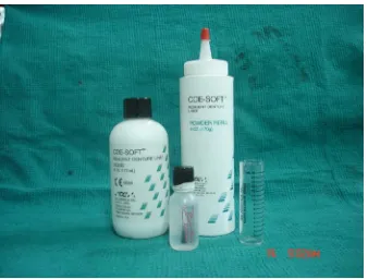
G.C RELINE TM SOFT (Fig.I) Auto polymerized silicone soft liner Supplied as cartridge (base & catalyst) G.C corporation Tokyo. Japan
2 COE – SOFTTM
RESILIENT DENTURE LINER (Fig.IA)
Auto
polymerized acrylic soft liner
Powder & Liquid
G.C America inc. Made in U.S.A
3 ACRYLAN–H Heat cure Powder &
Liquid
Asian acrylates
4 ACRYLAN–H Heat cure Liquid (For
pretreatment procedure)
TENSILE BOND STRENGTH
A total of 40 acrylic blocks of dimensions 40 x 10 x 10mm were
prepared in heat cure denture base resin. The polymethyl metha acrylate
blocks were ground with 320-grit silicone carbide paper to remove
surface irregularities and excess material. These 40 blocks were then
divided into 4 groups
1. Group A
2. Group B
3. Group C
4. Group D
Each group contains 5samples and each sample consists of two acrylic
blocks with soft liner interposed.
• Group A and C samples surfaces were not treated with methyl
metha acrylate.
• Group B and D samples surface were treated with methyl metha
acrylate for 180 seconds.
• Auto polymerized silicone soft liner were bonded to group A and B
PREPARATION OF THE TEST SAMPLES
DETAILS OF THE METAL DIES (FIG. I):
Two rectangular steel dies for a size of 40 x 10 x 10mm and one
steel die for a size of 10 x 10 x 3mm were prepared and all these surfaces
were smooth and flat with sharp edges. Theses steel dies are used to
fabricate the acrylic blocks and soft liners blocks respectively.
PREPARATION OF HEAT CURED ACRYLIC BLOCKS:
Mold space was created from steel dies size of 40 x 10 x 10mm by
using addition silicon putty material (Fig. II). Wax blocks were prepared
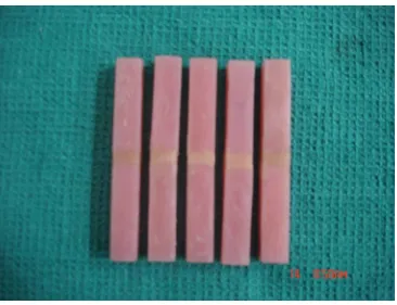
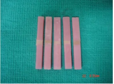
by pouring the molten wax into the mold space. A total number of 40 wax
patterns were prepared (Fig. III).
The wax patterns were then flasked by conventional technique.
After the dewaxing procedure, heat cure resin was packed into the mold
space as per the manufacturer instruction to prepare resin blocks. These
720to 800 centigrade for a period of 9 hours and bench cooled. The
polymethyl metha acrylate blocks were then ground with 320-grit silicone
carbide paper to remove surface irregularities and excess material.
PREPARATION OF MOLD SPACE FOR LINER PLACEMENT:
The two rectangular shaped acrylic blocks of 40 x 10 x 10mm and
one steel die of 10 x 10 x 3mm were flasked with type II gypsum as
follows. The 10 x 10 x 3mm steel die placed in-between the two acrylic
blocks of 40 x 10 x 10mm (Fig. III). After the flasking procedure the die
was removed to create a mold space (Fig.III) for soft liner.
PREPARATION OF GROUP A SAMPLES (FIG. IV):
The surfaces of the acrylic blocks to be bonded with soft liner were
coated with primer R and dried with clean air, and then it was placed in
the mold space. In-between the two acrylic blocks the auto polymerized
silicone soft liners were packed into the mold space (Fig.III) as per the
manufacturer instructions. After the packing procedure the flask was kept
under bench press for 10 minutes to allow the liner to cure completely
then the samples were removed from the mold space and the excess
PREPARATION OF GROUP B SAMPLES (Fig. V):
The surface of the acrylic blocks to be bonded with soft liner were
pretreated with methyl metha acrylate for 180 seconds, and then the
specimens were left to dry for 2minutes. The primer R was applied gently
to the methyl methacrylate treated surfaces with a brush and dried with
clean air, and then acrylic blocks were placed into the mold space.
In-between the two acrylic blocks as per the manufacturer instructions the
auto polymerized silicone soft liners were mixed and packed into the
mold space. After the packing procedure the flask was kept under bench
press for 10 minutes to allow the liner to cure completely then the
samples were removed from the mold space and the excess material were
removed with sharp scalpel blade.
PREPARATION OF GROUP C SAMPLES (Fig.VI):
The polymethyl metha acrylate blocks were placed into the mold
space. In-between the two acrylic blocks as per the manufacturer
instructions the auto polymerized acrylic soft liners were packed into the
mold space. After the packing procedure the flask was kept under bench
press for 10 minutes to allow the liner to cure completely then the
samples were removed from the mold space and the excess material were
PREPARATION OF GROUP D SAMPLES (Fig.VII):
The surface of the acrylic blocks to be bonded with soft liner were
pretreated with methyl metha acrylate for 180 seconds, then the acrylic
blocks were left to dry for 2minutes and then acrylic blocks were placed
into the mold space. In-between the two acrylic blocks as per the
manufacturer instructions the auto polymerized acrylic soft liners were
packed into the mold space. After the packing procedure the flask was
kept under bench press for 10 minutes to allow the liner to cure
completely then the samples were removed from the mold space and the
PEEL BOND STRENGTH
A total of 20 acrylic plates of dimensions 75 x 25 x 3mm were prepared
in heat cured denture base resin. The polymethyl metha acrylate plates
were ground with 320-grit silicone carbide paper to remove surface
irregularities and excess material. These twenty plates were then divided
into five groups.
1. Group E
2. Group F
3. Group G
5. Group H
Each group contains 5 samples and each sample consists of one acrylic
plate bonded with soft liner.
• Group E and G samples surfaces were not pretreated with methyl
metha acrylate.
• Group F and H samples surfaces were pretreated with methyl
metha acrylate for 180 seconds.
• Auto polymerized silicone soft liner were bonded to group E and F
PREPARATION OF THE TEST SAMPLES:
Two rectangular steel dies were prepared and it has two parts.
1. LID (Fig. IX)
2. BASE (Fig. IX)
LID:
The dimension of the lid is 85mm length X 35mmwidth X 4mm
thickness. The surfaces of the lid were smooth, flat and the corners were
BASE:
Base metal die has two sides.
1. One side of the steel die, mold space was prepared for the
dimension of 75mm length x 5mm depth x 25mm width. The
surfaces of the steel die mold space were smooth, flat and with
sharp edges. This mold space was used for soft liner attachment to
2. Other side of the steel die (Fig.X) has elevated rectangular slab
having the dimension of 75mm length x 25mm width X 3mm
height. The surfaces of the steel die mold space were smooth, flat
and with sharp edges. This elevated side of the die was used for the
PREPARATION OF ACRYLIC PLATES:
A modeling wax sheet was used to box the elevated side of the
steel die (Fig.XI). Vaseline was applied over the die and die stone was
mixed with recommended water powder ratio as per the manufacturer
instruction and it was poured to create a mold space. A total number of
twenty-die stone mold space (Fig.XII) having a dimension of 75mm x
25mm x 3mm were prepared. Molten wax was poured into the mold
space and flasking was done by conventional technique. After dewaxing
heat cure resin was packed into the mold space as per the manufacturer
instruction to prepare resin plates. These heat cure resin plates were cured
as per the manufacturer instruction at 720 to 80 centigrade for a period of
9 hours and bench cooled. The polymethyl metha acrylate plates were
retrieved from the flask and ground with 320-grit silicone carbide paper
to remove surface irregularities and excess material(Fig.XIII). Out of the
total surface area of the acrylic plate, the space having the dimensions of
50mm length X 25mm width was covered by polyethylene sheet and
remaining portion of acrylic plate having the dimensions of 25mm length
X 25mm width was left uncovered to facilitate the bonding of soft liner
PREPARATION OF GROUP E SAMPLES:
The part of the acrylic plates to be bonded with soft liner were
coated with primer R and dried with clean air then it was placed in the
mold space of steel die. The acrylic plate occupies the mold space of
75mm length X 25mm width X 3mm depth and the rest of the mold space
was left for the auto polymerized silicone soft liner which is packed over
the acrylic plates as per the manufacturer instructions (Fig.XIV) and the
soft liner was covered by polyethylene sheet over that lid was placed and
it was compressed for 10 minutes under the bench press, the excess
materials were removed by scalpel blade. In the prepared sample out of
total dimension of 75mm length X 25mm width X 2mm thickness only
25mm length X 25mm width of the liner was bonded to the acrylic plate.
The remaining part of the soft liner was not bonded (Fig. XV), to
facilitate the attachment with testing machine.
PREPARATION OF GROUP F SAMPLES:
The surface of the acrylic plates to be bonded with soft liner were
pretreated with methyl metha acrylate for 180 seconds, and then the
over the treated surfaces with a brush and dried with clean air and then
polymethyl metha acrylate plate was placed in the mold space of steel
die. The acrylic plates occupies the space of 75mm length X 25mm width
X 3mm depth and the rest of the mold space was left for the auto
polymerized silicone soft liner, which is packed over the acrylic plates as
per the manufacturer instructions and the soft liner was covered by
polyethylene sheet over that lid was placed and it was compressed for 10
minutes under the bench press, the excess materials were removed by
scalpel blade. In the prepared sample out of total dimension of 75mm
length X 25mm width X 2mm thickness only 25mm length X 25mm
width of the liner was bonded to the acrylic plate. The remaining part of
the soft liner was not bonded, to facilitate the attachment with testing
machine.
PREPARATION OF GROUP G SAMPLES:
The acrylic plate was placed in the mould space of steel die and it
occupied the space of 75mm length X 25mm width X 3mm depth and the
rest of the mold space was left for the auto polymerized acrylic soft liner
which is packed over the acrylic plates as per the manufacturer
instructions and the soft liner was covered by polyethylene sheet over that
press, the excess materials were removed by scalpel blade. In the
prepared sample out of total dimension of 75mm length X 25mm width X
2mm thickness only 25mm length X 25mm width of the liner was bonded
to the acrylic plate. The remaining part of the soft liner was not bonded,
to facilitate the attachment with testing machine.
PREPARATION OF GROUP H SAMPLES:
The surface of the acrylic plates to be bonded with soft liner were
pretreated with methyl metha acrylate for 180 seconds, and then the
specimens were left to dry for 2minutes and it was placed in the mold
space of steel die. It occupied the space of 75mm length X 25mm width
X 3mm depth and the rest of the mold space was left for the auto
polymerized acrylic soft liner which is packed over the acrylic plates as
per the manufacturer instructions and the soft liner was covered by
polyethylene sheet over that lid was placed and it was compressed for 10
minutes under the bench press, the excess materials were removed by
scalpel blade. In the prepared sample out of total dimension of 75mm
length X 25mm width X 2mm thickness only 25mm length X 25mm
width of the liner was bonded to the acrylic plate. The remaining part of
the soft liner was not bonded, to facilitate the attachment with testing
SAMPLES PREPARATION FOR SCANNING ELECTRON
MICROSCOPE
Mold space was created from one steel die having the dimension of
10mm length X 10mm width X 3mm thickness by using addition silicone
putty material. Wax blocks were prepared by pouring the molten wax into
the silicone mould space. A total of two wax patterns were prepared,
The wax patterns were then flasked by conventional technique. After
dewaxing heat cure resin was packed into the mold space as per the
manufacturer instruction to prepare acrylic resin blocks. These heat cure
resin blocks were cured as per the manufacturer instruction at 720to 800
centigrade for a period of 9 hours and bench cooled. The polymethyl
metha acrylate blocks were then ground with 320-grit silicone carbide
paper to remove surface irregularities and excess material.
• One-heat cure resin block was treated by methyl metha acrylate for
PREPARATION OF SAMPLES FOR SCANNING ELECTRON
MICROSCOPE
Specimens requiring to be studied under scanning electron
microscope should be made electro conductive. In order to make the
specimen’s electro conductive, the specimens were gold sputtered with
the help of sputter coating machine before subjecting them to screening in
a scanning electron microscope.
TESTING THE SAMPLES
Tensile and peel bond strength tests were carried out with a
universal testing machine named Lloyed instrument. The universal testing
machine was connected to an IBM computer. In peel test, the stress is
limited to a line at the edge of the joint as the fibers of the soft liners are
stretched and pulled away whereas in the tensile test the whole cross
sectional area of the bonded surface is under stress.
TENSILE BOND STRENGTH
The specimen was fixed to the grip of the Lloyed machine and
pulled in either way at a crosshead speed of 5mm/minute was used for
recorded for each specimen. Tensile bond strength was calculated by the
following formula20
Maximum load (N)
Tensile bond strength = ---
Cross sectional area (mm2)
The crosshead speed was same for all samples in order to standardize the
procedure.
PEEL BOND STRENGTH
The specimen was placed in Lloyed universal testing machine at
180-degree angle with the polymethyl metha acrylate plate portion in the
lower clamp and the soft liner was in the upper clamp. The machine was
operated at crosshead speed of 5mm/minute (Fig.XVI ). The maximum
load and the soft liner stretched length before failure was recorded for
each specimen. The peel bond strength was calculated by the following
formula20
F 1+λ n
Peel bond strength = ---- --- +1 ---
W 2 mm
The crosshead speed was same for all samples in order to standardize the
FIGURE I – G. C Reline Soft - SILICONE SOFT LINER
[image:51.612.157.495.392.648.2]TENSILE BOND
FIGURE IV – GROUP A SAMPLES
[image:54.612.141.507.404.685.2]FIGURE VI - GROUP C SAMPLES
[image:55.612.140.509.407.680.2]PEEL BOND STRENGTH
FIGURE IX
FIGURE XI
FIGURE XIII
FIGURE XV
RESULTS
An invitro study was conducted to evaluate the peel and tensile
bond strength of some of the commercially available soft liners with
acrylic denture base and the effect of surface pretreatment of denture base
with methyl metha acrylate. In addition to this the surface topography of
the heat cure denture base is observed before and after surface
pretreatment with methyl metha acrylate by scanning electron
microscope. Tensile and peel bond strength tests were carried out with a
universal testing machine named Lloyed instrument. Five samples from
each group were tested at a constant cross head speed of 5mm/min12. The
tensile and peel bond strength were recorded. All data’s were tabulated
and statistical comparisons were made by one-way ANOVA variance and
Tukey-HSD multiple range comparison test.
TENSILE BOND STRENGTH:
TABLE - I
GROUP A - SILICONE SOFT LINER (Untreated with methyl
methacrylate)
Sample No. Peak load (N) Tensile bond strength (N/mm)
1. 132 1.32
2. 130 1.30
3. 131 1.31
4. 133 1.33
5. 134 1.34
Mean 132 1.32
TABLE - II
GROUP B - SILICONE SOFT LINER (Treated with methyl methacrylate)
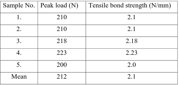
Sample No. Peak load (N) Tensile bond strength (N/mm)
1. 210 2.1
2. 210 2.1
3. 218 2.18
4. 223 2.23
5. 200 2.0
TABLE –III
GROUP C - ACRYLIC SOFT LINER (Untreated with methyl
methacrylate)
Sample No. Peak load (N) Tensile bond strength (N/mm2)
1. 23 0.23
2. 24 0.24
3. 23 0.23
4. 25 0.25
5. 24 0.24
Mean 23.8 0.24
TABLE – IV
GROUP D - ACRYLIC SOFT LINER (Treated with methyl
methacrylate)
Sample No. Peak load (N) Tensile bond strength (N/mm2)
1. 26 0.26
2. 28 0.28
3. 28 0.28
4. 23 0.23
5. 28 0.28
TABLE - V
STATISTICAL RESULT OF TENSILE BOND STRENGTH
Sl. no Group Mean SD P- value
1 A 1.32b 0.02
2 B 2.12c 0.09
3 C 0.24 a 0.01
4 D 0.27 a 0.02
<0.001**
Note: 1. ** Denotes significant at 1% level
2.Different alphabet between the groups denotes significant at 5%
level
PEEL BOND STRENGTH
TABLE – VI
GROUP E - SILICONE SOFT LINER (Untreated with methyl
methacrylate)
Sample
No.
Peak load
(N)
Soft liner stretched
length (mm)
Peel bond strength (N/mm)
1 40 57 3.31
2 42 62 3.76
3 39 62 3.30
4 40 60 3.36
5 41 57 3.39
TABLE – VII
GROUP F - SILICONE SOFT LINER (Treated with methyl
methacrylate)
Sample
No.
Peak load
(N)
Soft liner stretched
length (mm)
Peel bond strength (N/mm)
1 50 62 4.24
2 55 60 4.62
3 52 57 4.30
4 50 60 4.20
5 50 62 4.24
Mean 51.4 60.2 4.32
TABLE - VIII
GROUP G - ACRYLIC SOFT LINER (Untreated with methyl
methacrylate)
Sample
No.
Peak load
(N)
Soft liner stretched
length (mm)
Peel bond strength (N/mm)
1 10 110 1.04
2 12 100 1.20
3 11 120 1.18
4 12 97 1.18
5 10 127 1.10
TABLE - IX
GROUP H - ACRYLIC SOFT LINER (Treated with methyl
methacrylate)
Sample
No.
Peak load
(N)
Soft liner stretched
length (mm)
Peel bond strength (N/mm)
1 10 110 1.04
2 12 123 1.31
3 9 127 0.99
4 9 95 0.88
5 11 97 1.08
Mean 10.2 110.4 1.06
TABLE – X
STATISTICAL RESULTS OF PEEL BOND STRENGTH
Sl. no Group Mean SD P- value
1 E 3.42b 0.19
2 F 4.32c 0.17
3 G 1.06a 0.16
4 H 1.14 a 0.07
<0.001**
Note: 1. ** Denotes significant at 1% level
2. Different alphabet between the groups denotes significant at 5%
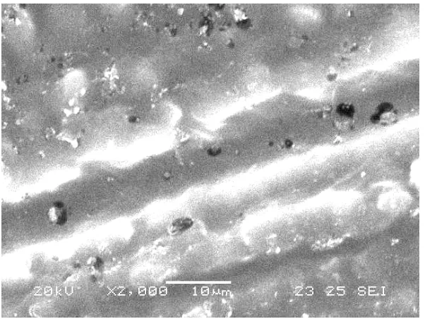
SCANNING ELECTRON MICROSCOPE RESULTS
Fig-A untreated surface of the denture base resin ground with 320-grit silicone carbide paper.
[image:67.612.187.482.145.367.2] [image:67.612.174.475.442.668.2]
BAR DIAGRAM
TENSILE BOND STRENGTH
1.32
2.1
0.24 0.27
0 0.5 1 1.5 2 2.5
A B C D
MATERIALS
Mean value (n/mm)
A
B
C
D
PEEL BOND STRENGTH
1.06(4/mm) 4.32(4/mm) 3.42(4/mm) 1.14(4/mm) 0 0.5 1 1.5 2 2.5 3 3.5 4 4.5 5E F G H
Table – I show the peak load 132(N) tensile bond strength and mean
value 1.32 N/mm2 of group-A samples of silicone soft liner without
surface treatment of denture base.
Table – II show the peak load 212(N) tensile bond strength and mean
value 2.1 N/mm2 of group-B samples of silicone soft liner with surface
treatment of denture base.
Table – III show the peak load 23.8(N) tensile bond strength and mean
value 0.24N/mm2 ofgroup-C samples of acrylic soft liner without surface
treatment of denture base.
Table – IV show the peak load 26.6(N) tensile bond strength and mean
value 0.27N/mm2 of group-C samples of acrylic soft liner with surface
treatment of denture base.
Table – V shows the statistical analysis of tensile bond strength of
silicone and acrylic soft liner bonded to treated and untreated surface of
denture base. ** Denotes significant at 1% level and different alphabet
Table – VI show the peak load 40.4(N), stretched length 59.6mm and
peel bond strength mean value3.42 N/mm2 ofgroup-E samples of silicone
soft liner without surface treatment of denture base.
Table – VII show the peak load 51.4(N), stretched length 60.2mm and
peel bond strength mean value 4.32 N/mm2 of group-E samples of
silicone soft liner with surface treatment of denture base.
Table – VIII show the peak load 11(N), stretched length 110.8mm and
peel bond strength mean value 1.14 N/mm2 ofgroup-G samples of acrylic
soft liner without surface treatment of denture base.
Table – IX show the peak load 10.2(N), stretched length 110.4mm and
peel bond strength mean value 1.06 N/mm2 ofgroup-H samples of acrylic
soft liner with surface treatment of denture base.
Table – X shows the statistical analysis of peel bond strength of silicone
and acrylic soft liner bonded to treated and untreated surface of denture
base. ** Denotes significant at 1% level and different alphabet between
Fig-A scanning electron microscope investigation result shows scratches,
pores and depressions.
Fig-B scanning electron microscope investigation result shows the
prominent pores and smoother surface texture than the figure-A. This
may be attributed to swelling of the superficial layer of the denture base.
DESCRIPTION OF BAR DIAGRAM
Fig-C : Bar diagram represents Tensile bond strength of acrylic and
silicone softliner bonded to treated and untreated surface of the denture
base tested in this study.
Fig-D : Bar diagram represents Peel bond strength of acrylic and silicone
softliner bonded to treated and untreated surface of the denture base
tested in this study.
STATISTICAL ANALYSIS
• Overall comparison of groups was done using one-way analysis of
variance (ANOVA) with significant at 1%. Level.
• Comparison with in the groups was done using multiple range tests
INTERPRETATION OF RESULTS
• Group A samples show lesser value than the group B samples,
which is statistically significant
• Group A samples show higher values than the group C and D
samples, which are statistically significant
• Group B samples show higher values than the group A, group C
and group D samples, which are statistically significant.
• From the results it was found that between the group C and group
D samples, the values are not statistically significant.
• Group E samples show lesser value than the group F samples,
which is statistically significant
• Group E samples show lesser value than the group F samples and
higher values than the group G and H, which are statistically
significant
• Group F samples show higher values than the group E, group G
and group H samples, which are statistically significant
• From the results it was found that between the group G and group
DISCUSSION
Resilient soft liners are widely used in prosthetic dentistry as an
adjunct to removable prosthesis to restore the health of the inflamed and
abused denture supporting tissues17. Use of these materials as an adjunct
in the successful treatment proved appreciable prognosis of patients with
complete and removable partial dentures42. These materials are
commonly used for patients with resorbed mandibular alveolar ridge, thin
and nonresilient mucosal tissue, maxillofacial defect, patients unable to
tolerate the hardness of heat-polymerized acrylic resin denture base42 and
medically compromised individuals. Excess and uneven pressure on
mental foramen; sharp ridges (knife edge); thin, atrophic mucosa; bony
undercuts. In addition to this irregular bone resorption; poor fit of the
denture base; Bruxism and /or debilitating diseases23(diabetes mellitus)
are also can be included.
Resilient soft liners are used to distribute functional loads by
optimizing adaptation of the denture base to residual ridges, to reduce the
stress concentration on residual ridge and to make dentures more
However one of the major drawbacks of soft liners is the lack of
durable bond to the denture base. The bond between the heat polymerized
acrylic denture base and the soft liners not found to be long lasting and
requiring repeated relines.
Hence understanding the chemistry of the bonding of soft liners
with acrylic resin along with the nature of the bond and mechanism of
bond failure will help us to overcome the problem and render better
service to the patients to be rehabilitated by removable prosthesis.
Various workers have done elaborate study on the bond strength of
the soft liners with acrylic denture base by subjecting them to a variety of
studies, which include peel, tensile, shear bond strength and creep test,
currently most of the clinicians prefer the autopolymerized soft lining
materials as an alternative to heat cured soft liners because of their
chairside usage, easy application and less laboratory procedures.
The present study was under taken to evaluate the tensile & peel
bond strength of two commercially available soft liners G.C reline
soft-Auto polymerized silicone soft liner & Coe-soft-soft-Auto polymerized
acrylic soft liner with heat activated acrylic denture base samples. The
surfaces of acrylic denture base was also evaluated in this study. In
addition to this the surface topography of the heat cure acrylic denture
base before and after surface pretreatment with methyl metha acrylate for
180 seconds was evaluated with scanning electron microscope. The
surface of acrylic denture base samples were pretreated with methyl
methacrylate for about 180 seconds to improve the bonding ability with
soft liners.
From the result of this tensile and peel bond strength test, it was
found that the silicone soft liner bonded to the treated surface of the
denture base resin with methyl methacrylate for 180 seconds exhibited
higher bonding ability than the silicone soft liner bonded to the untreated
surface of the acrylic denture base. Mode of bond failure was also
observed among the groups of the treated and untreated surface of the
denture base. The pretreated surface of the denture base demonstrated
primarily cohesive type of failure and the untreated surface shows
primarily adhesive type of failure. Both the samples, which are subjected
to the bond tests, were abraded with 320 grit silicone carbide paper.
The scanning electron microscope study on the treated and untreated