Open Access
Technical Note
Determination of quantitative and site-specific DNA methylation of
perforin by pyrosequencing
Supraja Narasimhan, Virginia R Falkenberg, Maung M Khin and
Mangalathu S Rajeevan*
Address: Division of Viral and Rickettsial Diseases, National Center for Zoonotic Vector-Borne and Enteric Diseases, Centers for Disease Control and Prevention, Atlanta, GA 30333, USA
Email: Supraja Narasimhan - [email protected]; Virginia R Falkenberg - [email protected]; Maung M Khin - [email protected]; Mangalathu S Rajeevan* - [email protected]
* Corresponding author
Abstract
Background: Differential expression of perforin (PRF1), a gene with a pivotal role in immune surveillance, can be attributed to differential methylation of CpG sites in its promoter region. A reproducible method for quantitative and CpG site-specific determination of perforin methylation is required for molecular epidemiologic studies of chronic diseases with immune dysfunction.
Findings: We developed a pyrosequencing based method to quantify site-specific methylation levels in 32 out of 34 CpG sites in the PRF1 promoter, and also compared methylation pattern in DNAs extracted from whole blood drawn into PAXgene blood DNA tubes (whole blood DNA) or DNA extracted from peripheral blood mononuclear cells (PBMC DNA) from the same normal subjects. Sodium bisulfite treatment of DNA and touchdown PCR were highly reproducible (coefficient of variation 1.63 to 2.18%) to preserve methylation information. Application of optimized pyrosequencing protocol to whole blood DNA revealed that methylation level varied along the promoter in normal subjects with extremely high methylation (mean 86%; range 82–92%) in the distal enhancer region (CpG sites 1–10), a variable methylation (range 49%–83%) in the methylation sensitive region (CpG sites 11–17), and a progressively declining methylation level (range 12%–80%) in the proximal promoter region (CpG sites 18–32) of PRF1. This pattern of methylation remained the same between whole blood and PBMC DNAs, but the absolute values of methylation in 30 out of 32 CpG sites differed significantly, with higher values for all CpG sites in the whole blood DNA.
Conclusion: This reproducible, site-specific and quantitative method for methylation determination of PRF1 based on pyrosequencing without cloning is well suited for large-scale molecular epidemiologic studies of diseases with immune dysfunction. PBMC DNA may be better suited than whole blood DNA for examining methylation levels in genes associated with immune function.
Published: 12 June 2009
BMC Research Notes 2009, 2:104 doi:10.1186/1756-0500-2-104
Received: 12 May 2009 Accepted: 12 June 2009
This article is available from: http://www.biomedcentral.com/1756-0500/2/104 © 2009 Rajeevan et al; licensee BioMed Central Ltd.
BMC Research Notes 2009, 2:104 http://www.biomedcentral.com/1756-0500/2/104
Page 2 of 9 (page number not for citation purposes)
Background
Perforin (PRF1), a major immune surveillance gene, is a candidate gene for the identification of methylation mark-ers, and for investigating the pathophysiology of a number of chronic diseases with suspected immune dys-function [1-9]. Earlier studies to assess PRF1 promoter methylation used bisulfite sequencing of cloned PCR products [5]. Cloning PCR products for quantitative site-specific determination of CpG methylation is laborious, and can be inaccurate due to under-representation of the number of clones analyzed. On the other hand, pyrose-quencing technology offers a convenient way for quanti-tative site-specific determination of CpG methylation but this technology has not yet been developed for the study of PRF1. Pyrosequencing determines the ratio of C/T base changes after bisulfite treatment, reflecting the proportion of unmethylated and methylated cytosines at each CpG site in the original sequence [10].
Several factors like sample collection, bisulfite treatment, and PCR can affect the reproducibility of methylation lev-els determined by pyrosequencing. Blood is a non-inva-sive source of sample for molecular epidemiologic studies. Usually, blood is drawn into blood collection tubes followed by separation of peripheral blood mono-nuclear cells (PBMCs) to extract DNA for molecular genetic studies including methylation. Recently, to mini-mize variability in primary blood samples from multi-center studies and to enable transport and storage, blood is drawn into specialized tubes like PAXgene blood DNA tubes containing additives for cell lysis. These PAXgene blood DNA tubes are then transported to laboratories where DNA is extracted directly from the whole blood without PBMC separation. DNA extracted from PAXgene blood DNA tubes thus represents the contribution of all blood cell types and have been successfully used for gen-otyping. However, the impact of these blood collection procedures for DNA (whole blood vs PBMC DNA) on methylation levels has not been reported.
In this study, we developed a pyrosequencing based meth-ylation assay for a total of 32 out of 34 CpG sites in the promoter region of PRF1. The assay is highly reproducible with respect to bisulfite treatment, and PCR. Examination of methylation levels in normal subjects showed that methylation levels differ significantly between DNAs extracted from PBMCs and whole blood for all 32 CpG sites except two, although the methylation pattern remained similar over the 1.4 kb promoter region. These results show that site-specific assessment of CpG methyl-ation in PRF1 can be reproducibly assessed by pyrose-quencing without cloning, and that PBMC DNA may be better suited than whole blood DNA for molecular epide-miologic studies on methylation levels of genes associated with immune dysfunction.
Methods
Pyrosequencing strategy
Bisulfite converted sequences were simulated by a previ-ously reported Microsoft Word bisulfite macro [11] fol-lowed by pyrosequencing assay design using Assay Design Software (Biotage, Uppsala, Sweden). Both top and bot-tom strands were used for designing primers to cover all the 34 CpG sites in 1.4 kb PRF1 promoter. Similarly, both forward and reverse sequencing designs were employed with 5'biotinylation of the reverse or forward PCR primer respectively. All primers were synthesized at the Biotech-nology Core Facility, Centers for Disease Control & Pre-vention, Atlanta, GA.
DNA samples and bisulfite treatment
Blood drawn in PAXgene blood DNA tubes (Qiagen, CA) for DNA extraction were obtained from 20 subjects who took part in the Chronic Fatigue Syndrome Study at the Centers for Disease Control and Prevention, Atlanta, GA. From five of these subjects classified as normal, blood was also drawn in BD Vacutainer CPT tubes (BD, NJ) followed by separation of PBMC for DNA extraction. DNA from PAXgene blood tubes was extracted using PAXgene blood DNA kit (Qiagen) whereas DNA from PBMCs was extracted using Roche DNA isolation kit for mammalian blood (Roche Applied Science, IN). DNA was quantified using NanoDrop ND-100 Spectrophotometer (Thermo Fisher Scientific, MA) prior to bisulfite treatment. DNA (200 ng/reaction) was bisulfite treated using the EZ DNA Methylation Kit (Zymo Research, CA), according to the manufacturer's instructions. Bisulfite treated DNA (BST-DNA) was eluted twice in 10 μl of the manufacturer's M-Elution Buffer (20 μl final volume), and was stored in aliquots at -20°C until use.
PCR and pyrosequencing
200
9,
2
:10
4
http://ww
w.b
iomed
central.com/1
756
-0
500
Page
(page number not for citation purposes)
(bp) position∞
A (236) FW: TTG ATT TTA TAG GTG AGG AAA TTA
RV: TTC CAA CTA TCA CCC ATA ACC TAA
TTT TAT AGG TGA GGA AAT TA
AYGTTTAGAAAGGGGGTTGATATTTATYGTYGTGAGGTATAYGG ATT TTT ATG TTT TTT AAA T
YGGTTTTTTTGTTA
1, -1365; 2, -1339; 3, -1336; 4, -1325 6, -1231
B (226) FW: ATG TTG AGG TTG TGA GGA GTT TT
RV: AAA TTC CAA AAT CCT CTC TTT AAT
GAA GTT TTG TAA AGT ATT TG YGGGAAAAGAG
5, -1267
C (196) FW: TTG GAA AGT GAT TAG GAG GTT GTA
RV: CAA ACT CCA AAC CAC ATA TAA CAT
AGA GGG TGG GGA TATT GYGGAGAGAAGATGGGGTTAGATTTYG GTT TTT GTT TTT GTA AGA GT AGGGAYGGAAGTAGGGATATAAAYG
7, -1110; 8, -1088 9, -1045; 10, -1027
D (91) FW: GAG GTT TTT ATG GGT GGA GTG AT
RV:CAC CTC CTC CCT TAC CCA ACT A
TTG GGG GGT AAA ATT ATAYGGTTTT T
11, -876
E (256) FW: TTT TGA GTG GGA GAA GAG AGA TGT
RV:CCC CAC CCT AAC CTC AAA CA
GTG AGA GTG GTT TGG TAG TATYGGAGG
TTG GTT TTA GTT TTG TTG A GGTYGTGGGT
AGG ATA GTT AGT GGT TTT TA YGTTGGTTTTAGTTTTGTTG
12, -776 13, -774 14, -720
F (167) FW: TGG AGG TTA TTG GTT GTT TTT ATA
RV: TAA CCA TTC CCT CCT CCC TAA ATA
GGT TAT TGG TTG TTT TTA TA
AAGYGAGGAGTAGGAGTTTTTGTTYGAGGAATATGTTTGGAGTTYGG
15, -691; 16, -670; 17, -650
G (300) FW: TTA GGG AGG AGG GAA TGG TTA TAG
RV: AAC CAA CAA AAC CAT CTC CTT ACT
TAG TTT ATA TTG TTG GTG TA TAATYGAGTTGTTTAAGTTTYGGYGGTTTGGYG
18, -397; 19, -381; 20, -378; 21, -370
H (300) FW: TTT AGG GAG GAG GGA ATG GTT ATA
RV: ACC AAC AAA ACC ATC TCC TTA CTT
TAC TTC TAA TAC ACA ACA TC RCATATATAAAATATAAAAAACAAACAAAAACRACRAC
24, -313; 23, -345; 22, -348
I (233) FW: GGG GAT TTA GGG TAT ATA GG
RV: AAA CCC TAC CAA TCC ACA CTA CT
GGG GAT TTA GGG TAT AT
AGGYGGAGGAGGGYGGGGYGTTGAGGATTTTGAGATTYGGT
28, -219; 27, -229; 26, -234; 25, -253
J (237) FW: TGG TTT TGT TGG TTT GTT TAT TAA
RV: CCC CAA CTA TAA TCA CAA ATC CTT
CAA AAC CAA AAA CTC ATC T ACCRAATAAAACTACTAAAACTCR CCT CAA CCC TCA TCC
RACTCCCCACTAACAACCCTCAAAAAACRAAC
30, -180; 29, -200 32, -122; 31, -150
K (97) FW: TTG AGG ATA GGG TGG GTG TT
RV: CCA CCA CTC ACA TCA CTT CTA CTT
GGA TAG GGT GGG TGT T
YGTGGGAGGGGAGAGTATAAAGGATTTGTGATTATAGTTGGGGGY
33, -92; 34, -48
[image:3.792.87.743.76.523.2]BMC Research Notes 2009, 2:104 http://www.biomedcentral.com/1756-0500/2/104
Page 4 of 9 (page number not for citation purposes)
Pyrosequencing was performed according to the manufac-turer's instructions. The PyroGold kit was used in con-junction with the PSQ 96MA instrument (Biotage), and each pyrosequencing reaction used 20 μl of PCR product. Biotinylated single strand DNA was separated by immobi-lizing the PCR product on streptavidin coated Sepharose High Performance beads (Amersham Biosciences, NJ) and strand separated by denaturation with 0.2 N NaOH. After denaturation, the biotinylated strand was annealed to sequencing primer (0.5 μM final concentration), and sub-jected to sequencing using an automatically generated nucleotide dispensation order for the "sequence to ana-lyze" corresponding to each reaction (Table 1). Pyrograms were analyzed in "Allele Quantification" mode in order to determine the percentage of C/T, corresponding to the percentages of methylated and unmethylated C at each of the CpG sites. As a positive control for PCR and pyrose-quencing, we used previously reported PCR and sequenc-ing primers to detect two CpG sites in CDNK2A [13].
Assay reproducibility 1. Bisulfite reproducibility
The impact of bisulfite treatment on assay reproducibility was tested using the PCR and pyrosequencing conditions for CpG sites 18–21 in amplicon G with whole blood DNA from 20 subjects. The same DNA was bisulfite treated on three separate days. Each day, 20 individual bisulfite reactions were set up with 200 ng DNA/reaction. BST-DNA from each of the three days was simultaneously subjected to PCR in the same thermocycler to eliminate PCR-to-PCR variability. All PCR products were used for pyrosequencing in duplicate, giving a mean of 6 pyrogram readings per CpG site for all three sets of BST-DNA per subject. Day-to-day variation in bisulfite reaction was expressed as coefficient of variation (CV) calculated as the percentage of standard deviation to its mean
2. Touchdown PCR reproducibility
The impact of PCR-to-PCR variability, expressed in terms of CV, was also tested for CpG sites 18–21 in amplicon G on three different days using the same BST-DNA. Touch-down PCR reproducibility was done with whole blood DNA from 10 subjects. Each PCR product was subjected to pyrosequencing in duplicate, yielding a mean of 6 values per CpG site for PCR from all three days per subject.
Evaluation of methylation pattern in the PRF1 by pyrosequencing
A small pilot study consisting of five normal subjects was conducted to evaluate the robustness of the optimized conditions to determine the methylation level/pattern in the PRF1 promoter for 32 CpG sites. This pilot study also examined the impact of DNA from whole blood or PBMC on PRF1 methylation profile in normal subjects. For each subject, two separate bisulfite reactions were set up,
fol-lowed by corresponding PCR and pyrosequencing reac-tions yielding a mean of 2 values per CpG site. Both whole blood and PBMC DNAs were treated similarly to generate the PRF1 methylation profiles.
Results
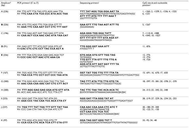
The PRF1 promoter spans about 1.4 kb (-1411 to +1) upstream of the transcriptional start site and harbors a number of transcription factor binding sites [14,15]. Out of a total of 34 CpG sites in the PRF1 promoter, 10 are in the distal promoter region containing enhancer elements (CpG sites 1–10), 17 are in the proximal promoter region harboring repetitive elements (CpG sites 18–34), and seven (CpG sites 11–17) are in the methylation sensitive region (MSR) located between the distal and proximal promoter regions (Figure 1). Eleven amplicons A to K and 16 sequencing primers were designed to cover all 34 CpG sites (Figure 1; Table 1).
We compared the performance of PCR with single anneal-ing temperature and touchdown PCR and found that PCR cycling program with a single annealing temperature often generated multiple bands, particularly with ampli-con E, which is rich in AT ampli-content (70%). On the other hand, touchdown PCR consistently generated specific products of expected size with all amplicons except ampli-con F, for which PCR with single annealing temperature consistently resulted in specific product than touchdown PCR. Thus, we used touchdown PCR to generate all ampli-cons, except amplicon F which was generated with PCR following single annealing temperature. Pyrosequencing reactions for all CpG sites were successful except CpG sites 33 and 34 in amplicon K, although amplicon K was suc-cessfully generated by the touchdown PCR.
Variability due to bisulfite treatment was tested for four CpG sites (sites 18–21) in amplicon G using whole blood DNA from 20 individual subjects. Variability due to bisulfite treatment was found to be minimal for all four CpG sites in amplicon G, (mean CV of 2.18%, and CVs for individual CpG sites ranged from 1.97% to 2.37%, Figure 2). Variability due to touchdown PCR was also assessed for CPG sites 18–21 using whole blood DNA from 10 sub-jects and was found to be very low for all four CpG sites in amplicon G, with a mean CV of 1.63%, and with CVs for individual CpG sites ranging from 1.34% to 1.86% (Fig-ure 3).
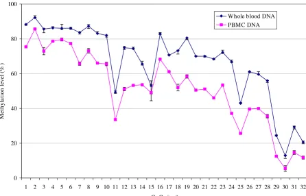
appeared to vary up and down from a low of 49% (CV = 1.49%) at CpG site 11 to a high of 83% (CV = 0.84%) at CpG site 16 (mean methylation level for sites 11–17 = 67%). Interestingly, methylation levels in the proximal promoter region covering CpG sites 18–32 progressively declined from a high of 73–80% at sites 18–19 to as low as 12–20% at sites 30 and 32 (mean methylation at sites 18–32 = 54%). Among specific sites, CpG sites 11, 15, 25, 29 and 30 showed sharp decline in methylation levels compared to adjacent CpG sites. Perforin methylation profile over the same 32 CpG sites in the same five sub-jects using PBMC DNA appeared very similar i.e., extremely high methylation in the distal enhancer region, a variable methylation in the MSR, and progressively declining methylation in the proximal promoter region. However, except for CpG sites 5 and 15, the absolute methylation levels at each CpG site as determined from the whole blood and PBMC DNAs differed significantly, with higher values for all CpG sites in the whole blood DNA (Figure 4).
Discussion
Analytical procedures for sensitive and site-specific quan-titative determination of CpG methylation are influenced
by several factors, all of which require careful experimen-tal optimization and validation before the assay can be applied to valuable, and often limited clinical samples in large-scale molecular epidemiologic studies. A number of PCR-based methylation assays utilize bisulfite treatment to preserve the methylation signature of the original DNA. Bisulfite treatment selectively converts unmethylated cytosines to uracil. This base modification results in reduced sequence complexity, which often makes the development of methylation assays difficult due to dimin-ished flexibility in designing quality PCR and sequencing primers, allele-specific PCR biases, and reduced signal strength or abnormally shaped sequencing reactions [16,17]. We considered these factors while developing a pyrosequencing assay to determine the methylation status of PRF1, an important gene in immune surveillance.
[image:5.612.67.542.98.337.2]Pyrosequencing assay was designed for all 34 CpG sites in the PRF1 promoter, of which seven CpG sites form a func-tionally significant subset in the MSR of the promoter. We successfully optimized several steps of the pyrosequenc-ing technology to obtain highly reproducible methylation levels for each of the 32 out of 34 CpG sites. Bisulfite treat-ment resulted in minimal day-to-day variation (CV The perforin (PRF1) promoter (1411 bp) with locations of 34 CpG and several putative transcription factor binding sites Figure 1
The perforin (PRF1) promoter (1411 bp) with locations of 34 CpG and several putative transcription factor
BMC Research Notes 2009, 2:104 http://www.biomedcentral.com/1756-0500/2/104
Page 6 of 9 (page number not for citation purposes)
2.18%) that was comparable to the minimal reaction-to-reaction variation in bisulfite treatment as determined by fluorescent probe based quantitative version of methyla-tion-sensitive PCR (MSP, MethyLight assay) [18]. A touch-down PCR approach has been recommended for overcoming PCR biases in reactions using BST-DNA tem-plate [12], but the reproducibility of touchdown PCR has not been examined with BST-DNA. We found that touch-down PCR helped to overcome problems with PCR prod-uct specificity, and resulted in highly reproducible results from PCR-to-PCR on different days (CV 1.63%).
In this study, the PRF1 methylation pattern identified using both whole blood and PBMC DNAs from normal subjects appeared similar throughout the promoter region in terms of extremely high methylation in distal enhancer region, variable methylation in the MSR, and progres-sively declining methylation in the proximal promoter region. Except CpG sites 5 and 15, the absolute levels of methylation at each of these CpG sites, however, differed between whole blood and PBMC DNAs, with higher methylation levels obtained using whole blood DNA. We have not investigated the factors that contribute to this Reproducibility of bisulfite treatment determined using amplicon G
Figure 2
similar pattern but different levels in methylation along the PRF1 promoter between whole blood and PBMC DNAs but it is likely to be related to differences in cell types between these samples from the same subject. In a previous study that used bisulfite sequencing followed by cloning, CpG site 14 was found to be the most highly methylated (> 50%) in the MSR of CD4+ T and CD8+ T
cells of normal subjects [5]. On the contrary, the most highly methylated CpG site in the MSR in normal subjects in this study was found to be CpG site 16 located 70 bases away from CpG site 14. These differences may be related to differences in the subjects, cell populations or quantita-tive nature of the assays employed between the studies. The pysosequencing assay described in this report is highly suitable to quantify differences in the methylation
of PRF1 in the various sub-populations of cells in blood from normal and subjects with chronic diseases of immune dysfunction. Further, the study suggests that blood drawn in PAXgene blood DNA tubes may be inap-propriate for DNA methylation studies of genes with immune function.
We have not compared the estimates of PRF1 methylation determined by pyrosequencing with other method such as MSP or combined bisulfite restriction analysis since our reproducibility data agree with other reports supporting the reliability of pyrosequencing for quantitative and effi-cient screening of multiple CpG sites in candidate genes studies [13,19-21]. Further, pyrosequencing has been used as a reference method for validation of other meth-Reproducibility of touchdown PCR determined using amplicon G
Figure 3
BMC Research Notes 2009, 2:104 http://www.biomedcentral.com/1756-0500/2/104
Page 8 of 9 (page number not for citation purposes)
ods and has been shown to have excellent specificity and sensitivity for diagnostic uses [22,23]. A number of dilu-tion and titradilu-tion experiments with known amounts of methylated and unmethylated DNA or cells with methyl-ated viral DNA in the background of uninfected cells also support the reliability of pyrosequencing for the sensitive and specific determination of methylation at multiple CpG sites [10,13,18,23,24].
Conclusion
In conclusion, we have developed a highly reproducible procedure based on pyrosequencing technology for the site-specific and quantitative determination of methyla-tion levels in 32 CpG sites in the PRF1 promoter. In nor-mal subjects, methylation levels along the 1.4 kb promoter varied similarly in DNAs from whole blood and PBMC but differed in absolute methylation levels, sug-gesting collection of blood in devices like PAXgene blood DNA tube may not be appropriate for methylation studies of immune function related genes. This method can be applied to clinical samples in order to systematically eval-uate the significance of multiple CpG sites in perforin
which will improve our understanding of the pathophys-iology of chronic disorders with alterations in immune function.
Competing interests
The authors declare that they have no competing interests.
Authors' contributions
SN developed the pyrosequencing strategy, carried out the experiments and participated in manuscript writing. VRF contributed to experimental designs, interpretation of results and participated in the manuscript writing. MMK participated in the design, implementation of laboratory experiments, and manuscript writing. MSR conceived of this study and as Principle Investigator participated in all aspects of this study and drafting of this manuscript. All authors have read and approved the manuscript.
Acknowledgements
[image:8.612.66.513.105.391.2]Support for S. Narasimhan and VR. Falkenberg was provided by the research participation program at the National Center for Zoonotic Vec-tor-Borne and Enteric Diseases, Centers for Disease Control and Preven-tion (CDC), Division of Viral and Rickettsial Diseases, administered by the The pattern of methylation in 32 CpG sites in 1.4 kb PRF1 promoter using DNA from whole blood and PBMCs from normal subjects
Figure 4
The pattern of methylation in 32 CpG sites in 1.4 kb PRF1 promoter using DNA from whole blood and PBMCs from normal subjects. Y-axis, methylation level (%) ± SD. X-axis, CpG sites 1–32. Methylation levels at each CpG site rep-resent mean of 5 normal subjects.
0 20 40 60 80 100
1 2 3 4 5 6 7 8 9 10 11 12 13 14 15 16 17 18 19 20 21 22 23 24 25 26 27 28 29 30 31 32
CpG site #
M
et
h
yl
a
ti
on
leve
l (
%
)
Publish with BioMed Central and every scientist can read your work free of charge "BioMed Central will be the most significant development for disseminating the results of biomedical researc h in our lifetime."
Sir Paul Nurse, Cancer Research UK
Your research papers will be:
available free of charge to the entire biomedical community
peer reviewed and published immediately upon acceptance
cited in PubMed and archived on PubMed Central
yours — you keep the copyright
Submit your manuscript here:
http://www.biomedcentral.com/info/publishing_adv.asp
BioMedcentral Oak Ridge Institute for Science and Education through an interagency
agreement between the U.S. Department of Energy and the CDC. The find-ings and conclusions in this report are those of the authors and do not nec-essarily represent the views of the funding agency.
References
1. Gulan G, Ravlic-Gulan J, Strbo N, Sotosek V, Nemec B, Matovinovic D, et al.: Systemic and local expression of perforin in lym-phocyte subsets in acute and chronic rheumatoid arthritis. J Rheumatol 2003, 30:660-670.
2. Kaplan MJ, Lu Q, Wu A, Attwood J, Richardson B: Demethylation of promoter regulatory elements contributes to perforin overexpression in CD4+ lupus T cells. J Immunol 2004, 172:3652-3661.
3. Kastelan M, Prpic ML, Gruber F, Zamolo G, Zauhar G, Coklo M, et al.: Perforin expression is upregulated in the epidermis of psoriatic lesions. Br J Dermatol 2004, 151:831-836.
4. Li M, Yang Q, Zhang Y: Effects of CD134 monoclonal antibody on hemolysis activities and expression of perforin in periph-eral blood mononuclear cells of systemic lupus erythemato-sus patients. Hybridoma (Larchmt) 2007, 26:191-200.
5. Lu Q, Wu A, Ray D, Deng C, Attwood J, Hanash S, et al.: DNA methylation and chromatin structure regulate T cell per-forin gene expression. J Immunol 2003, 170:5124-5132. 6. Maher KJ, Klimas NG, Fletcher MA: Chronic fatigue syndrome is
associated with diminished intracellular perforin. Clin Exp Immunol 2005, 142:505-511.
7. Skarpa I, Rubesa G, Moro L, Manestar D, Petrovecki M, Rukavina D: Changes of cytolytic cells and perforin expression in patients with posttraumatic stress disorder. Croat Med J 2001, 42:551-555.
8. Steinau M, Unger ER, Vernon SD, Jones JF, Rajeevan MS: Differen-tial-display PCR of peripheral blood for biomarker discovery in chronic fatigue syndrome. J Mol Med 2004, 82:750-755. 9. Trapani JA, Voskoboinik I: The complex issue of regulating
per-forin expression. Trends Immunol 2007, 28:243-245.
10. Rajeevan MS, Swan DC, Duncan K, Lee DR, Limor JR, Unger ER: Quantitation of site-specific HPV 16 DNA methylation by pyrosequencing. J Virol Methods 2006, 138:170-176.
11. Singal R, Grimes SR: Microsoft Word macro for analysis of cyto-sine methylation by the bisulfite deamination reaction. Bio-techniques 2001, 30:116-120.
12. Shen L, Guo Y, Chen X, Ahmed S, Issa JP: Optimizing annealing temperature overcomes bias in bisulfite PCR methylation analysis. Biotechniques 2007, 42:48. 50, 52.
13. Colella S, Shen L, Baggerly KA, Issa JP, Krahe R: Sensitive and quan-titative universal Pyrosequencing methylation analysis of CpG sites. Biotechniques 2003, 35:146-150.
14. Lichtenheld MG, Podack ER: Structure of the human perforin gene. A simple gene organization with interesting potential regulatory sequences. J Immunol 1989, 143:4267-4274. 15. Zhang J, Scordi I, Smyth MJ, Lichtenheld MG: Interleukin 2
recep-tor signaling regulates the perforin gene through signal transducer and activator of transcription (Stat)5 activation of two enhancers. J Exp Med 1999, 190:1297-1308.
16. Gharizadeh B, Akhras M, Nourizad N, Ghaderi M, Yasuda K, Nyren P, et al.: Methodological improvements of pyrosequencing technology. J Biotechnol 2006, 124:504-511.
17. Mashayekhi F, Ronaghi M: Analysis of read length limiting fac-tors in Pyrosequencing chemistry. Anal Biochem 2007, 363:275-287.
18. Ogino S, Kawasaki T, Brahmandam M, Cantor M, Kirkner GJ, Spiegel-man D, et al.: Precision and performance characteristics of bisulfite conversion and real-time PCR (MethyLight) for quantitative DNA methylation analysis. J Mol Diagn 2006, 8:209-217.
19. Dupont JM, Tost J, Jammes H, Gut IG: De novo quantitative bisulfite sequencing using the pyrosequencing technology. Anal Biochem 2004, 333:119-127.
20. Mikeska T, Bock C, El-Maarri O, Hubner A, Ehrentraut D, Schramm J, et al.: Optimization of quantitative MGMT promoter meth-ylation analysis using pyrosequencing and combined bisulfite restriction analysis. J Mol Diagn 2007, 9:368-381.
21. Tost J, Gut IG: DNA methylation analysis by pyrosequencing. Nat Protoc 2007, 2:2265-2275.
22. Irizarry RA, Ladd-Acosta C, Carvalho B, Wu H, Brandenburg SA, Jed-deloh JA, et al.: Comprehensive high-throughput arrays for rel-ative methylation (CHARM). Genome Res 2008, 18:780-790. 23. White HE, Durston VJ, Harvey JF, Cross NC: Quantitative analysis
of SNRPN(correction of SRNPN) gene methylation by pyro-sequencing as a diagnostic test for Prader-Willi syndrome and Angelman syndrome. Clin Chem 2006, 52:1005-1013. 24. Uhlmann K, Brinckmann A, Toliat MR, Ritter H, Nurnberg P: