R E S E A R C H A R T I C L E
Open Access
Phase I clinical trial of the combination of
eribulin and everolimus in patients with
metastatic triple-negative breast cancer
Jin Sun Lee
1†, Susan E. Yost
1†, Suzette Blanchard
2, Daniel Schmolze
3, Hongwei Holly Yin
3, Raju Pillai
3,
Kim Robinson
1, Aileen Tang
1, Norma Martinez
1, Jana Portnow
1, Wei Wen
4, John H. Yim
4, Heather Ann Brauer
5,
Yuqi Ren
5, Thehang Luu
6, Joanne Mortimer
1*and Yuan Yuan
1*Abstract
Background:Alteration of the PI3K/AKT/mTOR pathway is a common genomic abnormality detected in triple-negative breast cancer (TNBC). Everolimus acts synergistically with eribulin in TNBC cell lines and xenograft models. This phase I trial was designed to test the safety and tolerability of combining eribulin and everolimus in patients with metastatic TNBC.
Methods:The primary objective of this study was to evaluate the safety and toxicities of the combination. Patients with metastatic TNBC who had up to four lines of prior chemotherapies were enrolled. The combination of eribulin and everolimus was tested using three dosing levels: A1 (everolimus 5 mg daily; eribulin 1.4 mg/m2days 1 and 8 every 3 weeks), A2 (everolimus 7.5 mg daily; eribulin 1.4 mg/m2, days 1 and 8 every 3 weeks), and B1 (everolimus 5 mg daily; eribulin 1.1 mg/m2days 1 and 8 every 3 weeks).
Results:Twenty-seven patients with median age 55 years were enrolled. Among 8 evaluable patients who received dose level A1, 4 had dose-limiting toxicities (DLTs). Among 3 evaluable patients treated with dose level A2, 2 had DLTs. Among 12 evaluable patients who received dose level B1, 4 had DLTs. The DLTs were neutropenia, stomatitis, and hyperglycemia. Over the study period, 59% had a≥grade 3 toxicity, 44% had≥grade 3 hematologic toxicities, and 22% had grade 4 hematologic toxicities. The most common hematological toxicities were neutropenia, leukopenia, and lymphopenia. Thirty-three percent had grade 3 non-hematologic toxicities. The most common non-hematological toxicities were stomatitis, hyperglycemia, and fatigue. The median number of cycles completed was 4 (range 0–8). Among 25 eligible patients, 9 patients (36%) achieved the best response as partial response, 9 (36%) had stable disease, and 7 (28%) had progression. The median time to progression was 2.6 months (95% CI [2.1, 4.0]), and median overall survival (OS) was 8.3 months (95% CI [5.5, undefined]).
Conclusion:Eribulin 1.1 mg/m2days 1 and 8 every 3 weeks with everolimus 5 mg daily was defined as the highest dose with acceptable toxicity (RP2D). The combination is safe, and efficacy is modest. A post hoc analysis showed that participants that used dexamethasone mouthwash stayed on treatment for one additional cycle.
Trial registration:ClinicalTrials.gov,NCT02120469. Registered 18 April 2014
Keywords:Phase I trial, Eribulin, Everolimus, Metastatic TNBC
© The Author(s). 2019Open AccessThis article is distributed under the terms of the Creative Commons Attribution 4.0 International License (http://creativecommons.org/licenses/by/4.0/), which permits unrestricted use, distribution, and reproduction in any medium, provided you give appropriate credit to the original author(s) and the source, provide a link to the Creative Commons license, and indicate if changes were made. The Creative Commons Public Domain Dedication waiver (http://creativecommons.org/publicdomain/zero/1.0/) applies to the data made available in this article, unless otherwise stated. * Correspondence:jmortimer@coh.org;yuyuan@coh.org
†Jin Sun Lee and Susan E. Yost contributed equally to this work. 1Department of Medical Oncology & Therapeutics Research, City of Hope
National Medical Center and Beckman Research Institute, 1500 E. Duarte Road, Duarte, CA 91010, USA
Background
Triple-negative breast cancer (TNBC) is an aggressive subtype of breast cancer characterized by a lack of estro-gen receptor, progesterone receptor, and human epider-mal growth factor receptor 2 (HER-2) amplification and accounts for approximately 15 to 20% of all breast cancer [1–3]. TNBC is a heterogeneous disease comprised of sev-eral molecular subtypes defined by gene expression profil-ing [4–7]. TNBCs often become chemotherapy-resistant upon relapse; hence, clinical outcome for patients with metastatic TNBC (mTNBC) is particularly poor with a median overall survival (OS) of approximately 13–16 months, largely due to the lack of effective targeted ther-apy [2,8,9]. There have been recent breakthrough therap-ies in subpopulations of patients with mTNBC, such as poly ADP ribose polymerase (PARP) inhibitors olaparib and talazoparib in patients with germline BRCA1 or BRCA2 mutation [10–12]. Immune checkpoint inhibitors targeting programmed death 1 (PD-1) or programmed death ligand 1 (PD-L1) showed efficacy in PD-L1-positive TNBCs [13–18]. Despite the progress being made, there is an unmet need for novel targeted therapies to improve the outcome of patients with mTNBC.
Phosphatidylinositol 3-kinase (PI3K)/AKT pathway alter-ations are some of the most common genomic alteralter-ations identified in breast cancer, and recently, the FDA granted approval of PI3Kα-specific inhibitor alpelisib plus fulves-trant in PIK3CA-altered ER-positive metastatic breast can-cer [19]. The PI3K/AKT pathway alterations include the loss-of-function of the tumor suppressor phosphatase and tensin homolog (PTEN) [20, 21]. Overall, the activating mutations of PI3K catalytic subunit alpha (PIK3CA)/AKT/ PTEN-altered tumors account for approximately 35% of pa-tients with TNBC [21,22]. It is also noted that the alter-ation of this pathway increases in metastatic disease, which suggests that PI3K pathway alterations may be associated with chemotherapy resistance in TNBC [23,24].
Eribulin mesylate is a synthetic analog of halichondrin B, a natural product isolated from the marine sponge okadai kadota[25].It shows antitumor activity via a tubulin-based anti-mitotic mechanism, which leads to G2/M cell cycle ar-rest, disruption of mitotic spindles, and apoptotic cell death. Eribulin inhibits cell growth in a wide range of cancer cell lines including breast, colon, prostate, and ovarian cancer [26]. Eribulin suppresses metastasis of breast cancer cells by reversing the phenotype from epithelial-mesenchymal tran-sition (EMT) to mesenchymal-epithelial trantran-sition (MET) states [27]. In the EMBRACE trial, eribulin showed OS benefit compared with conventional single-agent chemo-therapy of physician’s choice in heavily pretreated patients with metastatic breast cancer [28]. This result led to FDA approval in 2011.
Everolimus inhibits cytokine and growth factor-dependent cell proliferation by inhibiting the mammalian target of
rapamycin (mTOR), a key protein involved in the PI3K pathway [29]. In both preclinical models and early clinical trials in breast cancer, everolimus demonstrated promising efficacy [30,31]. Furthermore, a randomized phase III study has indicated better clinical outcomes for the treatment of metastatic hormone receptor-positive breast cancer in com-bination with an aromatase inhibitor [32]. Paradoxically, single-agent mTOR inhibition with everolimus enhances up-stream receptor tyrosine kinase activity and activates down-stream AKT activity [33,34], which may explain the lack of activity as a single agent in breast cancer. Increased efficacy has been observed when everolimus is combined with HER2-targeted therapy [35–37] or carboplatin [38,39].
Our preclinical studies demonstrated the synergistic effect of eribulin and everolimus in TNBC cell lines and murine models [34]. In TNBC cell lines MDA-MB-468 and BT549, phosphorylation of AKT was suppressed by eribulin. The combination of eribulin and everolimus re-sulted not only in an increased reduction of p-S6K1 and p-S6, but also a synergistic inhibition of cell survival in vitro and enhanced suppression of tumor growth in two mouse models. These findings provide a preclinical foundation for targeting both the microtubule cytoskel-eton and the PI3K/AKT/mTOR pathway in the treat-ment of refractory TNBC [34].
We hypothesize that the combination of eribulin and everolimus may be effective in patients with metastatic TNBC resistant to anthracyclines and taxanes. Here, we report the results of a phase I trial of everolimus and eri-bulin in patients with mTNBC.
Methods
Study design
This study was a single-center phase I trial conducted in patients with mTNBC resistant to anthracycline and/or taxane. The primary objective was to evaluate the safety and tolerability of everolimus and eribulin and to deter-mine the recommended phase 2 dose (RP2D) of the com-bination in patients with mTNBC who had received up to four lines of prior chemotherapy for metastatic disease. The secondary objective was to assess the activity based on the response rate (RR) and progression-free survival (PFS). Study treatment was provided until disease progres-sion or unacceptable toxicity. The study was conducted with the Declaration of Helsinki and Good Clinical Prac-tice. This study is registered at the clinical trial website
ClinicalTrials.govunder number NCT02723877.
Patient population
the following: age ≥18 years; life expectancy of ≥3 months; ECOG performance status 0–2; histologically confirmed TNBC (ER < 10%, PR < 10%, HER-2neu-negative defined by IHC 0 or 1, or FISH-HER-2neu-negative); 0–3 lines of prior chemotherapy for metastases; prior treat-ment with anthracycline and/or taxane therapy including (neo) adjuvant setting; adequate bone marrow reserve (hemoglobin ≥9 g/dL, absolute neutrophil count (ANC) ≥1500/mm3, platelet count ≥100,000/mm3); adequate renal function defined by creatinine ≤1.5× the upper limit of normal (ULN); adequate liver function defined by total bilirubin ≤1.5× ULN, alanine transaminase (ALT), and aspartate transaminase (AST)≤2.5× ULN (≤ 5× ULN in patients with liver metastases); able to obtain baseline computed tomography (CT), bone scan, or posi-tron emission tomography (PET)/CT; and able to pro-vide written informed consent. The mandatory use of dexamethasone mouthwash (alcohol-free 0.5 mg/5 mL dexamethasone solution) was amended at time of dose level B1 to reduce oral mucositis based on SWISH study results [40].
The main exclusion criteria included the following: chemotherapy or radiotherapy within 2 weeks prior to study entry; persisting ≥grade 2 AE defined by NCI CTCAE v4.0 at screening visit except alopecia; untreated or unstable brain metastases; prior eribulin or everoli-mus use; HIV; chronic hepatitis B or C (known from the existing medical record); concomitant use with strong or moderate CPY3A4/PgP inhibitors and CPY3A4/PgP in-ducers; uncontrolled current illness including, but not limited to, ongoing or active infection (≥grade 2 based on the NCI CTCAE v4.0); symptomatic congestive heart failure, unstable angina pectoris, or myocardial infarction within the past 6 months; cardiac ventricular arrhyth-mias requiring anti-arrhythmic therapy; unable to take oral medication; history of non-compliance to medical regimens; and psychiatric illness or social situations that would limit compliance with study requirements.
Baseline assessments
At screening visit, physical examination, vital signs, and labs including complete blood cell count (CBC) and dif-ferential, complete metabolic panels (CMP), and liver function test (LFT) were performed and repeated every 3 weeks on day 1 of each cycle. A baseline electrocardio-gram (EKG) was also obtained and repeated as clinically indicated. All patients were screened for hepatitis B and C infection. Urine or serum pregnancy test was also per-formed at screening. For tumor response assessment, CT scan of the chest/abdomen/pelvis and bone scan were performed for response evaluation criteria in solid tumor (RECIST1.1) evaluation. Brain MRI with contrast was used if clinically indicated.
Study treatment
Three dosing levels of the combinations were tested. For dose level A1, everolimus 5 mg daily and eribulin 1.4 mg/m2days 1 and 8 every 3 weeks were given. For dose level A2, everolimus 7.5 mg daily and eribulin 1.4 mg/m2 days 1 and 8 every 3 weeks were given. In dose level B1, everolimus 5 mg daily and eribulin 1.1 mg/m2days 1 and 8 every 3 weeks were given. A mandatory dexametha-sone mouthwash consisting of 10 mL of alcohol-free dexamethasone 0.5 mg/5 mL oral solution (swish for 2 min and spit, four times daily) was added to the protocol in an amendment in 2017 when dose level B1 was opened [40]. Other supportive medications, including anti-emetics, were used according to the current stand-ard of care guidelines. Patients were treated until pro-gression or unacceptable toxicity occurred.
Statistical methods
The study utilized the toxicity equivalence range (TEQR) design with a target equivalence range for dose-limiting toxicities (DLTs) of 0.20–0.35. Toxicity levels of ≥0.51 were considered too toxic, and the dose that achieve this level was closed. Patients entered the protocol in cohorts of 3. This trial was considered complete when 12 evalu-able patients were studied at a single dose level with a toxicity level of < 0.51. The RP2D was determined as the dose closest to the target of 0.25 below 0.51 based on isotonic regression [39].
DLT was defined as (i) grade 3 febrile neutropenia, grade 3 neutropenia lasting for > 7 days, grade 4 neutropenia, and grade 4 thrombocytopenia; (ii) any non-hematological tox-icity≥grade 3, controllable grade 3 nausea and vomiting, < 5 days of grade 3 fatigue, triglycerides < 1500 mg/dL which recovers in 1 week, grade 3 lab abnormalities that are cor-rectable to grade 2 or less with 24 h, and grade 3 hypergly-cemia that is controlled to grade 2; (iii) treatment delays > 2 weeks as a result of unresolved toxicity during the first cycle of therapy; and (iv) failure to complete at least 75% of planned dose of either drugs during cycle 1 due to toxicity. Toxicity was evaluated based on the National Cancer Insti-tute Common Terminology Criteria for Adverse Events (NCI CTCAE) v4.0.
Descriptive statistics were used to summarize the pa-tient demographic characteristics and adverse events. Rates and exact Clopper-Pearson 95% confidence inter-vals were provided for DLTs at the RP2D and disease re-sponse. PFS and OS were described using Kaplan-Meier methods.
Tumor genomic profiling
[41] (n= 9). For tumor RNA profiling, NanoString nCounter® PanCancer Pathway (n= 20) and Breast Can-cer 360™Panel (BC360™) (n= 11) were performed for pa-tients who had sufficient baseline FFPE tumor tissue. PanCancer multiplex gene expression analysis was per-formed using 770 genes from cancer-associated canon-ical pathways including: MAPK, STAT, PI3K, RAS, cell cycle, apoptosis, Hedgehog, Wnt, DNA damage control, transcriptional regulation, chromatin modification, and TGF-β. BC360™ panel analysis was performed for 770 genes across 24 key breast cancer pathways and pro-cesses (PAM50 signature, TNBC signature, claudin-low signature, tumor inflammation signature, and 38 unique signatures with well-established roles in breast cancer and immuno-oncology) [42]. Tumor sections were mi-crodissected from the patients’ unstained slides, and RNA was extracted using miRNeasy FFPE kit (Qiagen). RNA concentration was assessed with the Nanodrop spectrophotometer ND-1000 and Qubit 3.0 fluorometer (Thermo Scientific, CA). RNA fragmentation and quality control were further determined by 2100 Bioanalyzer (Agilent, CA). RNA was hybridized with codeset from gene panel at 65 °C for 16–20 h. Post-hybridization probe-target mixture was purified and quantified with nCounter Digital Analyzer, and all data analysis was per-formed on nSolver (NanoString Technologies, WA). All raw data from expression analysis were first aligned with internal positive and negative controls then normalized to the selected housekeeping genes included in the assay. Differential gene expression patterns as well as pathway and cell type scores with statistical analyses were per-formed with nSolver software (NanoString Technologies, WA). BC360™signatures including PAM50 breast cancer intrinsic subtype classifier and the tumor inflammation signature were analyzed by NanoString. NanoString data was analyzed for statistical significance using nSolver analysis software. Box plots of differential expression with associatedpvalue calculation were generated using Graphpad Prism 5.0 software (La Jolla, CA, USA).
Results
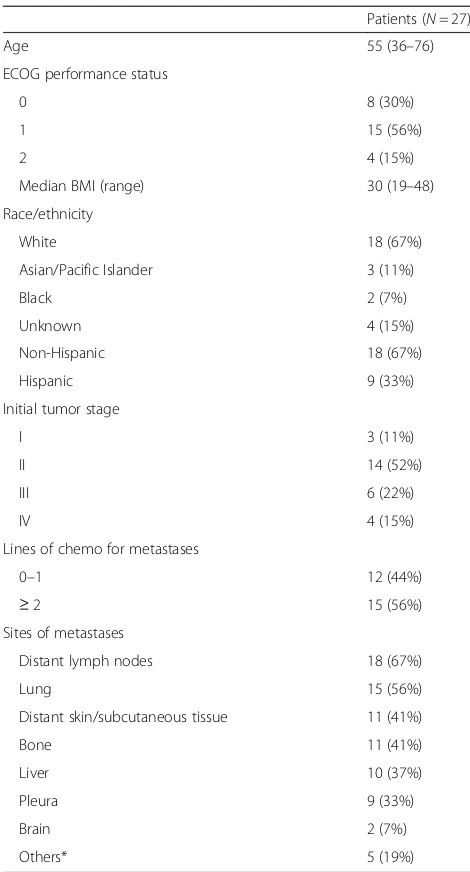
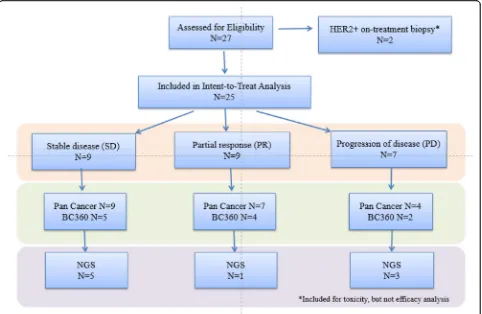
A total of 27 patients were enrolled and received treat-ment from November 2014 to March 2019. The median age of patients was 55 (range 36–76). Sixty-seven per-cent (18/27) of participants were non-Hispanic and 33% (9/27) were Hispanic (Table1). Sites of metastases were distant lymph nodes 67% (18/27), lung 56% (15/27), dis-tant skin/subcutaneous tissue 41% (11/27), bone 41% (11/27), liver 37% (10/27), pleura 33% (9/27), brain 7% (2/27), and others 19% (5/27). The number of patients receiving 0–1 lines of chemotherapy for metastases was 44% (12/27), and the number of patients receiving ≥2 lines of chemotherapy was 56% (15/27). Two of 27 pa-tients were found to be HER-2/neu-amplified at the time
of confirmatory biopsy and were taken off study. These two patients were not included in efficacy analysis but were included in toxicity analysis (Fig.1).
Treatment
[image:4.595.303.538.96.532.2]Three dose levels of everolimus and eribulin were evalu-ated: level A1 (everolimus 5 mg daily; eribulin 1.4 mg/m2 on days 1 and 8 every 3 weeks), level A2 (everolimus 7.5 mg daily; eribulin 1.4 mg/m2 on days 1 and 8 every 3 weeks), and level B1 (everolimus 5 mg daily; eribulin 1.1 mg/m2 on days 1 and 8 every 3 weeks) (Table 2). Ini-tially, 4 patients received treatment on dose level A1. One patient was not evaluable for dose escalation, due to receiving < 75% drug as a result of an event that was Table 1Patient characteristics
Patients (N= 27)
Age 55 (36–76)
ECOG performance status
0 8 (30%)
1 15 (56%)
2 4 (15%)
Median BMI (range) 30 (19–48)
Race/ethnicity
White 18 (67%)
Asian/Pacific Islander 3 (11%)
Black 2 (7%)
Unknown 4 (15%)
Non-Hispanic 18 (67%)
Hispanic 9 (33%)
Initial tumor stage
I 3 (11%)
II 14 (52%)
III 6 (22%)
IV 4 (15%)
Lines of chemo for metastases
0–1 12 (44%)
≥2 15 (56%)
Sites of metastases
Distant lymph nodes 18 (67%)
Lung 15 (56%)
Distant skin/subcutaneous tissue 11 (41%)
Bone 11 (41%)
Liver 10 (37%)
Pleura 9 (33%)
Brain 2 (7%)
Others* 5 (19%)
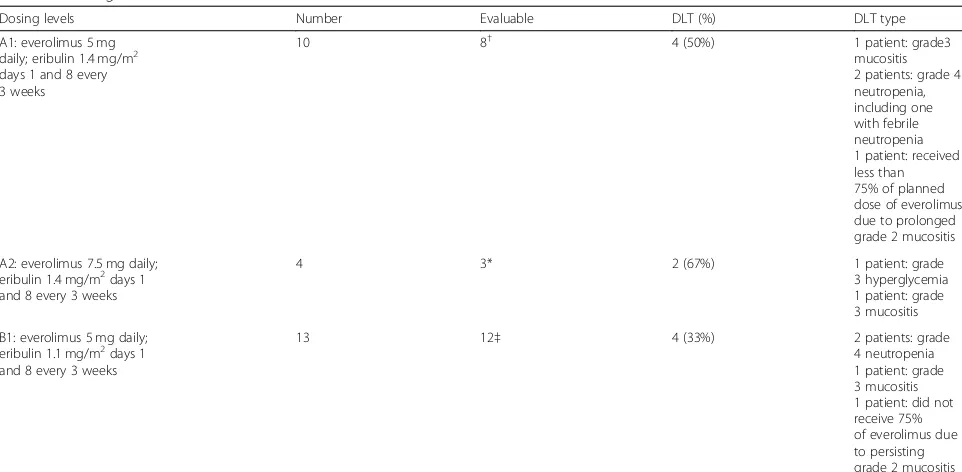
unrelated to treatment. There were no DLTs in 3 evalu-able patients, so the dose was escalated based on the guidelines for the TEQR design. Four patients were treated on dose level A2. One patient was found to be HER2+ and was taken off study. A total of 2/3 evaluable patients experienced DLTs in cycle 1, so dose level A2 was de-escalated to dose level A1 according to TEQR guidelines. Six additional patients were treated at dose level A1. Of those, 1 patient progressed without com-pleting the first cycle and did not have a DLT, hence unevaluable for dose escalation. Of the 5 evaluable pa-tients, 4 experienced DLTs. Of the 8 patients evaluable at dose level 1, 4/8 had DLTs (50%). The DLTs included 2 patients with grade 4 neutropenia, 1 patient with grade 3 mucositis, and 1 patient received less than 75% of planned dose of everolimus due to persisting grade 2 mucositis. The protocol was amended to add a lower dose level of B1, eribulin at 1.1 mg/m2on days 1 and 8 every 3 weeks and everolimus 5 mg daily. Thirteen pa-tients were treated at dose level B1. One patient was in-eligible due to HER2+ disease on repeat of biopsy. Of the 12 patients treated, the DLT rate at dose level B1
was 33% (4/12) with a 95% CI of 0.1, 0.65, thus complet-ing the phase 1 part of the trial (Table 2). Dose level B1 (everolimus 5 mg daily and eribulin 1.1 mg/m2on days 1 and 8 every 3 weeks) was determined to be the RP2D doses. The median number of cycles completed was 4 (0–18).
Dose modification
Sixty-eight percent (17/25) of participants had a dose modification or hold, including 56% (14/25) for eribulin and 60% (15/25) for everolimus.
Toxicities
Of the 27 patients, 96% (26/27) had a grade≥2 toxicity, and 59% (16/27) had grade 3–4 toxicity attributed to treatment. There were no grade 5 toxicities attributed to treatment.
Hematologic toxicities
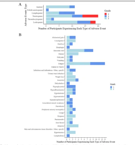
Of the 27 patients, 44% (12/27) had grade 3 and above hematologic toxicities, including neutropenia (n= 10), lymphopenia (n= 6), and leukopenia (n= 7) (Fig.2a).
[image:5.595.58.540.88.402.2]Non-hematologic toxicities
Of the 27 patients, 9/27 (33%) had grade 3 non-hematologic toxicities, including oral mucositis (n= 3), hyperglycemia (n= 3), and fatigue (n= 5). The counts of maximum grade 2 and above for each participant were listed for each event type where either a grade 3 or above toxicity was experienced, or 2 participants experi-enced a grade 2 (Fig. 2b). When dose level B1 was opened, a mandatory dexamethasone (Dex) mouthwash was added to reduce oral mucositis based on principle of best medical practice and clinical trial data [40]. Of the 12 evaluable participants enrolled to dose level B1, 2 did not use Dex mouthwash and 3 of the patients in the A1 dose used Dex mouthwash. A post hoc analysis was per-formed to compare the number of cycles that patients received in the Dex group with the non-Dex group. The median number of cycles was higher by 1 cycle (from 3 to 4 cycles) in the participants that took Dex. This was statistically significant using a Wilcoxon rank sum test (Dex:n= 13; non-Dex:n= 12;p= 0.046).
Antitumor activity and survival
Of the 25 evaluable patients, 9/25 (36%) had a partial re-sponse (PR), 9/25 (36%) achieved a best rere-sponse of stable disease (SD), and 7/25 (28%) had progression of disease (PD). A total of 21/25 (84%, 95%CI [64%, 95%]) experi-enced progression by RECIST1.1 or showed clinical pro-gression. The median PFS was 2.6 months (95%CI [2.1, 4.0]). Nineteen of 25 (76%, 95% CI [55%, 91%]) eligible
participants had event of death at the time of data cutoff (25 March 2019), and the median OS was 8.3 months (95% CI [5.5, undefined]). The cause of death was disease progression for 18 and failure to thrive for 1. Kaplan-Meier curves for PFS and OS are shown in Fig.3. Of the 24 participants that were off treatment, 17 (71%) were off for progression, 4 (17%) for clinical progression, 2 (8%) for toxicity, and 1 (4%) for lack of insurance coverage.
mRNA expression and response
NanoString PanCancer Pathway profiling was performed for 20 patients who had sufficient FFPE from metastatic biopsy. PanCancer Pathway with the grouping of pa-tients’best response analysis revealed a total of 22 differ-entially expressed genes (DEGs) comparing PR (n= 7) vs. SD+PD (n= 13) (linear fold change > 2 andp< 0.05). Five DEGs (CDKN2A, WNT5A, CNTFR, DDIT4, and SPP1) were downregulated in PR, and 17 DEGs were up-regulated in PR. Of the 22 genes, 9 DEGs are involved in the PI3K pathway (Table3). It should be noted that sev-eral immune-related genes including CD19, IL7R, IL6, and CCR7 were upregulated in partial responders. A vol-cano plot with differentially expressed genes (linear fold change > 2 and p< 0.05) was used to compare SD+PD and PR (Fig. 4a). Decreased CDKN2A expression (p= 0.02) (Fig.4b) and increased CALML5 (p= 0.01) (Fig.4c) were associated with better response to therapy.
[image:6.595.57.538.96.332.2]Further analysis using NanoString BC360™ analysis was performed for 11 patients (due to limited sample). Table 2Dosing levels and DLTs
Dosing levels Number Evaluable DLT (%) DLT type
A1: everolimus 5 mg daily; eribulin 1.4 mg/m2
days 1 and 8 every 3 weeks
10 8† 4 (50%) 1 patient: grade3
mucositis 2 patients: grade 4 neutropenia, including one with febrile neutropenia 1 patient: received less than 75% of planned dose of everolimus due to prolonged grade 2 mucositis
A2: everolimus 7.5 mg daily; eribulin 1.4 mg/m2days 1
and 8 every 3 weeks
4 3* 2 (67%) 1 patient: grade
3 hyperglycemia 1 patient: grade 3 mucositis
B1: everolimus 5 mg daily; eribulin 1.1 mg/m2days 1
and 8 every 3 weeks
13 12‡ 4 (33%) 2 patients: grade
4 neutropenia 1 patient: grade 3 mucositis 1 patient: did not receive 75% of everolimus due to persisting grade 2 mucositis †One patient progressed without completion of the first 2 cycles of therapy, hence unevaluable for DLT; 1 patient did not receive planned dose due to grade 3
hypoglycemia attributed to diabetes
Biologically significant pathways and single gene expres-sion results are shown in Additional file 1: Figure S1. Samples are grouped based on the initial stage (stage II, stage III, and de novo stage IV), lines of therapy > 2, PAM50 molecular subtypes (basal, HER2+, lu-minal A, and lulu-minal B), and TNBC subtypes (BLIA, BLIS, LAR, and MES). BC360™ analysis revealed
diverse tumor and tumor microenvironment (TME) pathway signatures with no observable pattern (Add-itional file 2: Figure S2A). There was no clear associ-ation of these variables and response to therapy, with the exception of HER2-enriched subtype which was associated with treatment resistance (p= 0.02) (Add-itional file 2: Figure S2B).
[image:7.595.62.536.85.597.2]Genomic mutation profiles
Nine patients had genomic mutation analysis through FoundationOne®. Additional file 3: Figure S3 shows the most frequent genomic alterations identified. Of the nine patients analyzed, two patients (3 and 16) carried muta-tions in the PI3K-Akt-mTOR pathway. Due to the
limited sample size, the association of genomic alteration with clinical response or survival could not be assessed.
Discussion
Activation of the PI3K/AKT pathway contributes to the resistance to anti-cancer agents including
[image:8.595.58.537.86.624.2]targeting agents. Despite the high frequency of alter-ations of PIK3CA/AKT/mTOR pathway, the presence of these mutations does not translate to a significant re-sponse to single agent PI3K inhibitors in early clinical trials [43, 44]. This is likely attributed to multiple by-pass signaling pathways. We hypothesized that targeting both the microtubule cytoskeleton and the PI3K/AKT/ mTOR pathway would lead to a synergistic anti-tumor effect. Our previous work showed synergistic inhibition of the PI3K/AKT/mTOR pathway, which resulted in an increased reduction of p-S6K1 and p-S6. The synergistic suppression of cell survival was found in a number of breast cancer cell lines in vitro and breast cancer mouse models in vivo [34].
The current study demonstrated that the combination of eribulin and everolimus is feasible and an effective
treatment for patients with mTNBC. Everolimus 5 mg daily with eribulin 1.1 mg/m2days 1 and 8 every 3 weeks was determined to be the RP2D doses [45]. Disease re-sponse rate was 36%. Tolerability of the combination im-proved after mandatory Dex mouthwash was added for dose level B1, which reconfirmed the SWISH data. Since 50% of DLTs in dose levels A1 and A2 were oral muco-sitis, the mandatory use of dexamethasone mouthwash may reduce DLTs in both dose levels.
Eribulin has been reported to have anti-tumor activity with a manageable tolerability profile with side effects consisting of neutropenia, fatigue, alopecia, nausea, and anemia. In addition, there was a low incidence of periph-eral neuropathy [46–48]. The Eisai Metastatic Breast Cancer Study Assessing Physician’s Choice Verses E7389 (EMBRACE) clinical trial was a phase III trial of patients with heavily pretreated metastatic breast cancer. Partici-pants received eribulin (E7389) monotherapy or treat-ment of physician’s choice (TPC). The patients in the trial received a median of four prior therapies. Improve-ment was seen in OS with HR 0.81, 95% CI [0.66–0.99], p= 0.041. Median OS was 13.1 months in patients re-ceiving eribulin vs. 10.6 months in TPC [28]. This study led to the FDA approval of eribulin mesylate for the treatment of breast cancer in patients who had failed taxane- or anthracycline-based therapies.
The mTOR inhibitor everolimus has emerged as a po-tential combination therapy drug for the treatment of cancer unresponsive to conventional therapy [49]. When used alone, everolimus can induce increased levels of p-AKT via a negative feedback loop leading to resistance of cells to mTOR inhibitors [50, 51]. Dual blockade of mTOR and other PI3K pathway inhibitors results in syn-ergistic decrease in cancer cell growth [33, 51, 52]. The PI3K pathway has been shown to play a critical role in TNBC. However, downstream of PI3K, mTOR inhibitor alone does not demonstrate clinical benefit. Studies combining chemotherapy with PI3K/AKT/mTOR path-way inhibitors have shown efficacy [38,53,54].
In a phase II trial of everolimus and carboplatin in metastatic TNBC, clinical benefit rate (CBR) was 36% and medical PFS was 3 months [38]. Other clinical trials have targeted different inhibitors in the PI3K pathway. Recently, the LOTUS trial studied the oral AKT inhibi-tor ipatasertib in TNBC. In this study, combination of ipatasertib and paclitaxel showed longer PFS compared to paclitaxel alone [53]. This improved PFS was observed particularly in patients with PIK3CA/AKT1/PTEN -al-tered tumors. The IPATunity130 trial is underway to confirm these findings [55].
[image:9.595.55.292.111.526.2]In the NanoString analysis, we identified the following immune-related genes which were upregulated in pa-tients who achieved partial responses: CD19, IL7R, IL6, and CCR7. Due to the limited sample size, the result is Table 3NanoString PanCancer Pathways® differentially
expressed genes mRNA Log2 fold change Standard error (log2) Linear fold change
pvalue Gene sets
SPP1 −2.96 0.804 0.129 0.0017 PI3K
CDKN2A −2.05 0.805 0.242 0.0211 Cell cycle, apoptosis, tumor suppressor gene
CNTFR −1.98 0.702 0.253 0.0111 JAK-STAT
DDIT4 −1.65 0.557 0.319 0.0848 PI3K
WNT5A −1.49 0.702 0.355 0.0474 Hedgehog, Wnt
ITGA9 1.01 0.36 2.02 0.0116 PI3K
LAT 1.07 0.453 2.1 0.0297 Ras
IL7R 1.08 0.427 2.11 0.0214 JAK-STAT, PI3K
PPARGC1A 1.21 0.519 2.32 0.0312 Chromatin
modification
TSLP 1.23 0.544 2.34 0.0379 JAK-STAT
ID4 1.31 0.353 2.47 0.00163 TGF-β
TNR 1.33 0.602 2.52 0.0408 PI3K
RASGRP2 1.42 0.614 2.68 0.0336 MAPK, Ras
HNF1A 1.49 0.591 2.8 0.0223 Driver gene
COL2A1 1.59 0.606 3.01 0.0178 PI3K
EFNA2 1.69 0.626 3.22 0.0154 PI3K, Ras
CCR7 1.7 0.68 3.25 0.0229 Transcriptional
misregulation
IL2ORB 1.71 0.607 3.26 0.0116 JAK-STAT
WNT16 1.87 0.777 3.66 0.0277 Hedgehog,
transcriptional misregulation, Wnt
CALML5 2.07 0.749 4.21 0.0127 Ras
IL6 2.51 0.707 5.71 0.00244 JAK-STAT, PI3K,
transcriptional misregulation
hypothesis-generating and requires further verification. This finding is consistent with the observation that up-regulation of immune-related genes in TNBC correlates with better response to chemotherapy and improved survival [56,57].
The current dose-defining study did not reveal the underlying mechanism predicting response to the combination of eribulin and everolimus. mRNA pro-filing and genomic analyses were performed, but with limited sample size. Although no clear conclusions were drawn, we observed an association between HER2-enriched subtype and poor response to therapy. Future studies targeting the PI3K-AKT-mTOR path-way will provide more insight into the molecular pre-dictors of response.
Conclusion
The combination of eribulin and everolimus is feasible and an effective treatment in metastatic TNBC. This
phase I clinical trial defines the RP2D as eribulin 1.1 mg/ m2(days 1 and 8) and everolimus 5 mg daily for further study. A post hoc analysis showed that participants that used dexamethasone mouthwash stayed on treatment for one additional cycle.
Supplementary information
Supplementary informationaccompanies this paper athttps://doi.org/10. 1186/s13058-019-1202-4.
Additional file 1: Figure S1.BC360™analysis (n=11): A) Relevant gene signatures and biologically significant single genes are shown. Samples were grouped based on stage (stage II, stage III, and de novo stage IV), lines of therapy >2, molecular subtypes (PAM50: basal, HER2+, luminal A, and luminal B); and TNBC subtypes: BLIA, BLIS, LAR, and MES. Signatures scores are mapped to quantiles of TCGA with a 0.5 value approximating the median TCGA value.
Additional file 2: Figure S2.BC360™analysis (n=11): A) Forest plot showing differentially expressed signatures comparing SD+PD and PR groups; B) HER2-enriched signature is up-regulated in SD+PD compared with PR group (P=0.02).
Fig. 4NanoString PanCancer Pathways® analysis (n= 20).aVolcano plot showing differentially expressed genes with linear fold change > 2 and
[image:10.595.61.539.85.460.2]Additional file 3: Figure S3.Tile plot showing genomic mutations in mTNBC patients (n=9).
Additional file 4: Table S1.Normalized mRNA expression data for NanoString PanCancer Pathways® cohort (n=20).
Additional file 5: Table S2.Normalized mRNA expression data for NanoString BC360™cohort (n=11).
Additional file 6: Table S3.Genomic alteration data for TNBC patients with genomic reports (n=9).
Abbreviations
TNBC:Triple-negative breast cancer; HER-2: Human epidermal growth factor receptor amplification; mTNBC: Metastatic TNBC; OS: Overall survival; PARP: Poly ADP ribose polymerase; 1: Programmed death 1; PD-L1: Programmed death ligand 1; PIK3CA: Phosphatidylinositol-4,
5-bisphosphate 3-kinase, catalytic subunit, alpha; PTEN: Phosphatase and tensin homolog; EMT: Epithelial-mesenchymal transition; MET: Mesenchymal-epithelial transition; mTOR: Mammalian target of rapamycin;
RP2D: Recommended phase II dose; RR: Response rate; PFS: Progression-free survival; ANC: Absolute neutrophil count; UNL: Upper limit of normal; TEQR: Toxicity equivalence range; DLTs: Dose-limiting toxicities; MTD: Maximum tolerated dose
Acknowledgements
The authors gratefully acknowledge Novartis and Eisai for the funding and Eisai for the analysis of correlatives. The research reported in this publication includes the work performed at the Pathology and Biostatistics Core of City of Hope National Cancer Center, supported by the National Cancer Institute (NCI) under award number P30CA033572. The content is solely the responsibility of the authors and does not necessarily represent the official views of the NCI.
Authors’contributions
YY made substantial contributions to the conception and design, acquisition of the data, analysis and interpretation of the data, drafting and revising of the manuscript, and final approval for publication. JSL contributed to the data analysis and manuscript preparation. SEY contributed to the database management, chart review, data analysis, and manuscript preparation and revision. SB performed the statistical analysis. DS performed the biomarker measurements. HY and RP performed the NanoString analysis. KR, AT, and NM contributed to the data collection. JP, TL, and JM assisted with the patient identification and manuscript revision. WW and JHY performed the preclinical studies. HAB and YR assisted in the NanoString analysis. YY is the guarantor and agrees to be accountable for all aspects of the work. All authors read and approved the final manuscript.
Authors’information
Not applicable
Funding
This study was supported by the STOP Cancer Foundation (Yuan Yuan), NIH K-12 Career Development Award K12CA001727 (Joanne Mortimer MD).
Availability of data and materials
Normalized mRNA expression data for NanoString PanCancer cohort is available in Additional file4: Table S1, normalized mRNA expression data for the NanoString BC360 cohort is available in Additional file5: Table S2, and available genomic alteration data is available in Additional file6: Table S3.
Ethics approval and consent to participate
All procedures performed in studies involving human participants were in accordance with the ethical standards of the institutional and/or national research committee and with the 1964 Helsinki Declaration and its later amendments or comparable ethical standards. Informed consent was obtained from all participants included in the study. All tumor specimens were identified through a City of Hope IRB-approved retrospective protocol from patients consented to City of Hope Biorepository Protocol.
Consent for publication
Not applicable
Competing interests
Dr. Yuan has contracted research sponsored by Merck, Eisai, Novartis, Puma, Genentech, and Pfizer; is a consultant for Puma; and is on the Speakers Bureau for Eisai. The other authors declare that they have no competing interests.
Author details
1
Department of Medical Oncology & Therapeutics Research, City of Hope National Medical Center and Beckman Research Institute, 1500 E. Duarte Road, Duarte, CA 91010, USA.2Department of Biostatistics, City of Hope
National Medical Center and Beckman Research Institute, Duarte, CA, USA.
3
Department of Pathology, City of Hope National Medical Center and Beckman Research Institute, Duarte, CA, USA.4Department of Surgery, City of
Hope National Medical Center and Beckman Research Institute, Duarte, CA, USA.5NanoString Technologies, Inc., Seattle, WA, USA.6OncoGambit, Irvine,
CA, USA.
Received: 8 July 2019 Accepted: 13 September 2019
References
1. Carey LA, Perou CM, Livasy CA, Dressler LG, Cowan D, Conway K, Karaca G, Troester MA, Tse CK, Edmiston S. Race, breast cancer subtypes, and survival in the Carolina Breast Cancer Study. JAMA. 2006;295(21):2492–502. 2. Tomao F, Papa A, Zaccarelli E, Rossi L, Caruso D, Minozzi M, Vici P, Frati L,
Tomao S. Triple-negative breast cancer: new perspectives for targeted therapies. OncoTargets Ther. 2015;8:177.
3. Howlader N, Noone A, Krapcho M, Miller D, Bishop K, Kosary C, Yu M, Ruhl J, Tatalovich Z, Mariotto A. SEER Cancer Statistics Review, 1975–2014. Bethesda: National Cancer Institute; 2017.
4. Lehmann BD, Bauer JA, Chen X, Sanders ME, Chakravarthy AB, Shyr Y, Pietenpol JA. Identification of human triple-negative breast cancer subtypes and preclinical models for selection of targeted therapies. J Clin Invest. 2011;121(7):2750–67.
5. Metzger-Filho O, Tutt A, de Azambuja E, Saini KS, Viale G, Loi S, Bradbury I, Bliss JM, Azim HA Jr, Ellis P, et al. Dissecting the heterogeneity of triple-negative breast cancer. J Clin Oncol. 2012;30(15):1879–87.
6. Masuda H, Baggerly KA, Wang Y, Zhang Y, Gonzalez-Angulo AM, Meric-Bernstam F, Valero V, Lehmann BD, Pietenpol JA, Hortobagyi GN. Differential response to neoadjuvant chemotherapy among 7 triple-negative breast cancer molecular subtypes. Clin Cancer Res. 2013;19(19):5533–40. 7. Burstein MD, Tsimelzon A, Poage GM, Covington KR, Contreras A, Fuqua SA,
Savage MI, Osborne CK, Hilsenbeck SG, Chang JC. Comprehensive genomic analysis identifies novel subtypes and targets of triple-negative breast cancer. Clin Cancer Res. 2015;21(7):1688–98.
8. Liedtke C, Mazouni C, Hess KR, André F, Tordai A, Mejia JA, Symmans WF, Gonzalez-Angulo AM, Hennessy B, Green M. Response to neoadjuvant therapy and long-term survival in patients with triple-negative breast cancer. J Clin Oncol. 2008;26(8):1275–81.
9. Rodler E, Korde L, Gralow J. Current treatment options in triple negative breast cancer. Breast Dis. 2011;32(1–2):99–122.
10. Robson M, Im S-A, Senkus E, Xu B, Domchek SM, Masuda N, Delaloge S, Li W, Tung N, Armstrong A. Olaparib for metastatic breast cancer in patients with a germline BRCA mutation. N Engl J Med. 2017;377(6):523–33. 11. Robson ME, Im S-A, Senkus E, Xu B, Domchek SM, Masuda N, Delaloge S, Li
W, Tung NM, Armstrong A. OlympiAD: phase III trial of olaparib monotherapy versus chemotherapy for patients (pts) with HER2-negative metastatic breast cancer (mBC) and a germline BRCA mutation (gBRCAm): J Clin Oncol. 2017;35(18_suppl).
12. Litton JK, Rugo HS, Ettl J, Hurvitz SA, Gonçalves A, Lee K-H, Fehrenbacher L, Yerushalmi R, Mina LA, Martin M. Talazoparib in patients with advanced breast cancer and a germline BRCA mutation. N Engl J Med. 2018;379(8): 753–63.
13. Nanda R, Chow LQ, Dees EC, Berger R, Gupta S, Geva R, Pusztai L, Pathiraja K, Aktan G, Cheng JD. Pembrolizumab in patients with advanced triple-negative breast cancer: phase Ib KEYNOTE-012 study. J Clin Oncol. 2016; 34(21):2460–7.
triple-negative breast cancer: cohort A of the phase II KEYNOTE-086 study. Ann Oncol. 2019;30(3):397–404.
15. Adams S, Loi S, Toppmeyer D, Cescon D, De Laurentiis M, Nanda R, Winer E, Mukai H, Tamura K, Armstrong AC. KEYNOTE-086 cohort B: pembrolizumab monotherapy for PD-L1–positive, previously untreated, metastatic triple-negative breast cancer (mTNBC). Clin Cancer Res. 2018;78(4 Supplement). 16. Emens LA, Braiteh FS, Cassier P, Delord J-P, Eder JP, Fasso M, Xiao Y, Wang Y,
Molinero L, Chen DS. Inhibition of PD-L1 by MPDL3280A leads to clinical activity in patients with metastatic triple-negative breast cancer (TNBC): AACR. Cancer Res. 2015;75(9 Supplement):2015.
17. Emens LA, Cruz C, Eder JP, Braiteh F, Chung C, Tolaney SM, Kuter I, Nanda R, Cassier PA, Delord J-P. Long-term clinical outcomes and biomarker analyses of atezolizumab therapy for patients with metastatic triple-negative breast cancer: a phase 1 study. JAMA Oncol. 2019;5(1):74–82.
18. Schmid P, Adams S, Rugo HS, Schneeweiss A, Barrios CH, Iwata H, Diéras V, Hegg R, Im S-A, Shaw Wright G. Atezolizumab and nab-paclitaxel in advanced triple-negative breast cancer. N Engl J Med. 2018;379(22):2108–21. 19. Andre F, Campone M, Ciruelos EM, Iwata H, Loibl S, Rugo HS, Wilke C, Mills
D, Chol M, Longin A-S. Alpelisib for PIK3CA-Mutated, Hormone Receptor– Positive Advanced Breast Cancer. N Engl J Med 2019;380:1929–40. 20. Millis SZ, Gatalica Z, Winkler J, Vranic S, Kimbrough J, Reddy S,
O’Shaughnessy JA. Predictive biomarker profiling of > 6000 breast cancer patients shows heterogeneity in TNBC, with treatment implications. Clin Breast Cancer. 2015;15(6):473–481. e473.
21. Network CGA. Comprehensive molecular portraits of human breast tumours. Nature. 2012;490(7418):61.
22. Leroy C, Ramos P, Cornille K, Bonenfant D, Fritsch C, Voshol H, Bentires-Alj M. Activation of IGF1R/p110β/AKT/mTOR confers resistance toα-specific PI3K inhibition. Breast Cancer Res. 2016;18(1):41.
23. Wein L, SJTB L. Mechanisms of resistance of chemotherapy in early-stage triple negative breast cancer (TNBC). Breast. 2017;34:S27–30.
24. Jensen JD, Laenkholm A-V, Knoop A, Ewertz M, Bandaru R, Liu W, Hackl W, Barrett JC, HJCCR G. PIK3CA mutations may be discordant between primary and corresponding metastatic disease in breast cancer. Clin Cancer Res. 2011;17(4):667–77.
25. Hirata Y, Uemura D. Halichondrins-antitumor polyether macrolides from a marine sponge. Pure Appl Chem. 1986;58(5):701–10.
26. Kuznetsov G, Towle MJ, Cheng H, Kawamura T, TenDyke K, Liu D, Kishi Y, Melvin JY, Littlefield BA. Induction of morphological and biochemical apoptosis following prolonged mitotic blockage by halichondrin B macrocyclic ketone analog E7389. Cancer Res. 2004;64(16):5760–6. 27. Yoshida T, Ozawa Y, Kimura T, Sato Y, Kuznetsov G, Xu S, Uesugi M,
Agoulnik S, Taylor N, Funahashi Y. Eribulin mesilate suppresses experimental metastasis of breast cancer cells by reversing phenotype from epithelial– mesenchymal transition (EMT) to mesenchymal–epithelial transition (MET) states. Br J Cancer. 2014;110(6):1497.
28. Cortes J, O’Shaughnessy J, Loesch D, Blum JL, Vahdat LT, Petrakova K, Chollet P, Manikas A, Diéras V, Delozier T. Eribulin monotherapy versus treatment of physician’s choice in patients with metastatic breast cancer (EMBRACE): a phase 3 open-label randomised study. Lancet. 2011;377(9769):914–23.
29. Boulay A, Lane HA. The mammalian target of rapamycin kinase and tumor growth inhibition. In: Targeted interference with signal transduction events: Springer; Recent Results Cancer Res. 2007;172:99–124.
30. Ellard SL, Clemons M, Gelmon KA, Norris B, Kennecke H, Chia S, Pritchard K, Eisen A, Vandenberg T, Taylor M, et al. Randomized phase II study comparing two schedules of everolimus in patients with recurrent/ metastatic breast cancer: NCIC Clinical Trials Group IND.163. J Clin Oncol. 2009;27(27):4536–41.
31. Boulay A, Rudloff J, Ye J, Zumstein-Mecker S, O’Reilly T, Evans DB, Chen S, Lane HA. Dual inhibition of mTOR and estrogen receptor signaling in vitro induces cell death in models of breast cancer. Clin Cancer Res. 2005;11(14):5319–28.
32. Baselga J, Campone M, Piccart M, Burris HA 3rd, Rugo HS, Sahmoud T, Noguchi S, Gnant M, Pritchard KI, Lebrun F, et al. Everolimus in
postmenopausal hormone-receptor-positive advanced breast cancer. N Engl J Med. 2012;366(6):520–9.
33. O’Reilly KE, Rojo F, She Q-B, Solit D, Mills GB, Smith D, Lane H, Hofmann F, Hicklin DJ, Ludwig DL. mTOR inhibition induces upstream receptor tyrosine kinase signaling and activates Akt. Cancer Res. 2006; 66(3):1500–8.
34. Marcinkowski E, Luu T, Yuan Y, Mortimer J, Leong L, Portnow J, Xing Q, Wen W, Yim J. Abstract P6-13-17: the combination of eribulin and everolimus results in enhanced suppression of tumors in mouse models of triple negative breast cancer. Cancer Res. 2016;76(4 Supplement). 35. Jerusalem G, Fasolo A, Dieras V, Cardoso F, Bergh J, Vittori L, Zhang Y,
Massacesi C, Sahmoud T, Gianni L. Phase I trial of oral mTOR inhibitor everolimus in combination with trastuzumab and vinorelbine in pre-treated patients with HER2-overexpressing metastatic breast cancer. Breast Cancer Res Treat. 2011;125(2):447–55.
36. Hurvitz SA, Andre F, Jiang Z, Shao Z, Mano MS, Neciosup SP, Tseng L-M, Zhang Q, Shen K, Liu D. Combination of everolimus with trastuzumab plus paclitaxel as first-line treatment for patients with HER2-positive advanced breast cancer (BOLERO-1): a phase 3, randomised, double-blind, multicentre trial. Lancet Oncol. 2015;16(7):816–29.
37. Morrow PK, Wulf GM, Ensor J, Booser DJ, Moore JA, Flores PR, Xiong Y, Zhang S, Krop IE, Winer EP. Phase I/II study of trastuzumab in combination with everolimus (RAD001) in patients with HER2-overexpressing metastatic breast cancer who progressed on trastuzumab-based therapy. J Clin Oncol. 2011;29(23):3126.
38. Singh JC, Novik Y, Stein S, Volm M, Meyers M, Smith J, Omene C, Speyer J, Schneider R, Jhaveri K. Phase 2 trial of everolimus and carboplatin combination in patients with triple negative metastatic breast cancer. Breast Cancer Res. 2014;16(2):R32.
39. Schwarzlose-Schwarck S, Scholz CW, Regierer AC, Martus P, Neumann C, Habbel P, Liu H, Zang C, Schefe J-H, Schulz C-O. The mTOR inhibitor everolimus in combination with carboplatin in metastatic breast cancer–a phase I trial. Anticancer Res. 2012;32(8):3435–41.
40. Rugo HS, Seneviratne L, Beck JT, Glaspy JA, Peguero JA, Pluard TJ, Dhillon N, Hwang LC, Nangia C, Mayer IA. Prevention of everolimus-related stomatitis in women with hormone receptor-positive, HER2-negative metastatic breast cancer using dexamethasone mouthwash (SWISH): a single-arm, phase 2 trial. Lancet Oncol. 2017;18(5):654–62.
41. Parsons HA, Beaver JA, Cimino-Mathews A, Ali SM, Axilbund J, Chu D, Connolly RM, Cochran RL, Croessmann S, Clark TA, et al. Individualized molecular analyses guide efforts (IMAGE): a prospective study of molecular profiling of tissue and blood in metastatic triple-negative breast cancer. Clin Cancer Res. 2017;23(2):379–86.
42. Chumsri S, Asleh K, Brauer H, Mashadi-Hossein A, Lauttia L, Lindman H, Nielsen T, Joensuu H, Thompson E. Abstract P3-11-05: predictive gene signatures of adjuvant capecitabine benefit in triple negative breast cancer in the FinXX trial. Cancer Res.2019;79(4 Supplement).
43. Arsenic R, Lehmann A, Budczies J, Koch I, Prinzler J, Kleine-Tebbe A, Schewe C, Loibl S, Dietel M, Denkert C. Analysis of PIK3CA mutations in breast cancer subtypes. Appl Immunohistochem Mol Morphol. 2014;22(1):50–6. 44. LoRusso PM. Inhibition of the PI3K/AKT/mTOR pathway in solid tumors. J
Clin Oncol. 2016;34(31):3803–15.
45. Blanchard MS, Longmate JA. Toxicity equivalence range design (TEQR): a practical phase I design. Contemp Clin Trials. 2011;32(1):114–21. 46. Vahdat LT, Pruitt B, Fabian CJ, Rivera RR, Smith DA, Tan-Chiu E, Wright J, Tan
AR, Dacosta NA, Chuang E, et al. Phase II study of eribulin mesylate, a halichondrin B analog, in patients with metastatic breast cancer previously treated with an anthracycline and a taxane. J Clin Oncol. 2009;27(18):2954–61. 47. Cortes J, Vahdat L, Blum JL, Twelves C, Campone M, Roche H, Bachelot T,
Awada A, Paridaens R, Goncalves A, et al. Phase II study of the halichondrin B analog eribulin mesylate in patients with locally advanced or metastatic breast cancer previously treated with an anthracycline, a taxane, and capecitabine. J Clin Oncol. 2010;28(25):3922–8.
48. Aogi K, Iwata H, Masuda N, Mukai H, Yoshida M, Rai Y, Taguchi K, Sasaki Y, Takashima S. A phase II study of eribulin in Japanese patients with heavily pretreated metastatic breast cancer. Ann Oncol. 2012;23(6):1441–8. 49. Seto B. Rapamycin and mTOR: a serendipitous discovery and implications
for breast cancer. Clin Transl Med. 2012;1(1):29.
50. Wan X, Helman LJ. The biology behind mTOR inhibition in sarcoma. Oncologist. 2007;12(8):1007–18.
51. Wang X, Yue P, Kim YA, Fu H, Khuri FR, Sun SY. Enhancing mammalian target of rapamycin (mTOR)-targeted cancer therapy by preventing mTOR/ raptor inhibition-initiated, mTOR/rictor-independent Akt activation. Cancer Res. 2008;68(18):7409–18.
53. Kim S-B, Dent R, Im S-A, Espié M, Blau S, Tan AR, Isakoff SJ, Oliveira M, Saura C, Wongchenko MJ. Ipatasertib plus paclitaxel versus placebo plus paclitaxel as first-line therapy for metastatic triple-negative breast cancer (LOTUS): a multicentre, randomised, double-blind, placebo-controlled, phase 2 trial. Lancet Oncol. 2017;18(10):1360–72.
54. Dent RA, Kim S-B, Im S-A, Espie M, Blau S, Tan AR, Isakoff S, Oliveira M, Saura C, Wongchenko M. LOTUS (NCT02162719): a double-blind placebo (PBO)-controlled randomized phase II trial of first-line ipatasertib (IPAT)+ paclitaxel (P) for metastatic triple-negative breast cancer (TNBC). J Clin Oncol. 2018; 36(15_suppl):1008.
55. Dent R, Kim S-B, Oliveira M, Isakoff SJ, Barrios CH, O’Shaughnessy J, Lu X, Wongchenko M, Bradley D, Mani A. IPATunity130: a pivotal randomized phase III trial evaluating ipatasertib (IPAT)+ paclitaxel (PAC) for PIK3CA/ AKT1/PTEN-altered advanced triple-negative (TN) or hormone receptor-positive HER2-negative (HR+/HER2–) breast cancer (BC). J Clin Oncol. 2018; 36(15_suppl).
56. Denkert C, von Minckwitz G, Brase JC, Sinn BV, Gade S, Kronenwett R, Pfitzner BM, Salat C, Loi S, Schmitt WD, et al. Tumor-infiltrating lymphocytes and response to neoadjuvant chemotherapy with or without carboplatin in human epidermal growth factor receptor 2-positive and triple-negative primary breast cancers. J Clin Oncol. 2015;33(9):983–91.
57. Yuan Y, Yost S, Chang C, Yoh K, Johnson R, Schmolze D, Liang J, Hutchinson K. Abstract PD5-07: comprehensive profiling of poor-risk paired primary and recurrent triple-negative breast cancers reveals immune phenotype shifts. Cancer Res. 2019; 79(4 Supplement).
Publisher’s Note
![Fig. 3 Kaplan-Meier survival analysis. a Median PFS was 2.6 months (95% CI [2.1, 4.0])](https://thumb-us.123doks.com/thumbv2/123dok_us/8262054.278188/8.595.58.537.86.624/fig-kaplan-meier-survival-analysis-median-pfs-months.webp)


