R E S E A R C H A R T I C L E
Open Access
An ELISA-based high throughput protein
truncation test for inherited breast cancer
Mark J Lim
1*, Gabriel J Foster
1, Sadanand Gite
2, Heather P Ostendorff
1, Steven Narod
3, Kenneth J Rothschild
1,4Abstract
Introduction:Breast cancer is the most diagnosed and second leading cause of cancer deaths in the U.S. female population. An estimated 5 to 10 percent of all breast cancers are inherited, caused by mutations in the breast cancer susceptibility genes (BRCA1/2). As many as 90% of all mutations are nonsense mutations, causing a truncated polypeptide product. A popular and low cost method of mutation detection has been the protein truncation test (PTT), where target regions ofBRCA1/2are PCR amplified, transcribed/translated in a cell-free protein synthesis system and analyzed for truncated polypeptides by sodium dodecyl sulfate polyacrylamide gel
electrophoresis (SDS-PAGE) and autoradiography. We previously reported a novel High Throughput Solid-Phase PTT (HTS-PTT) based on an enzyme-linked immunosorbent assay (ELISA) format that eliminates the need for
radioactivity, SDS-PAGE and subjective interpretation of the results. Here, we report the next generation HTS-PTT using triple-epitope-tagged proteins and demonstrate, for the first time, its efficacy on clinical genomic DNA samples forBRCA1/2analysis.
Methods:Segments of exons 11 ofBRCA1/2open reading frames were PCR amplified from either blood derived
genomic DNA or cell line mRNA. PCR primers incorporate elements for cell-free transcription/translation and epitope tagging. Cell-free expressed nascent proteins are then antibody-captured onto the wells of a microtiter plate and the relative amount of truncated polypeptide measured using antibodies against the N- and C-terminal epitope tags in an ELISA format.
Results:100% diagnostic sensitivity and 96% specificity for truncating mutations in exons 11 ofBRCA1/2were achieved on one hundred blood-derived clinical genomic DNA samples which were previously assayed using the conventional gel based PTT. Feasibility of full gene coverage forBRCA1/2using mRNA source material is also demonstrated.
Conclusions:Overall, the HTS-PTT provides a simple, quantitative, objective, low cost and high throughput method for analysis of truncating mutations as an alternative to gel based PTT forBRCAanalysis. The technology is readily accessible to virtually any laboratory, with the only major instrumentation required being a PCR
thermocycler and a basic micro-well plate reader. When compared to conventional gel based PTT, the HTS-PTT provides excellent concordance.
Introduction
Breast cancer is the most diagnosed and second leading cause of cancer deaths in the US female population, with approximately 200,000 new cases and approximately 40,000 deaths reported annually [1]. Ovarian cancer is the second ranking gynecological cancer, with approxi-mately 22,000 cases and approxiapproxi-mately 15,000 deaths annually [1]. Approximately 3% to 5% of all breast
cancers and approximately 10% of ovarian cancers are due to inherited mutations in the breast and ovarian can-cer susceptibility 1 or 2 genes (BRCA1orBRCA2) [2-4]. Lifetime risks ofBRCA1/2mutation carriers have been estimated at approximately 80% for breast cancer and approximately 40% and approximately 20% for ovarian cancer (BRCA1andBRCA2, respectively) [5].
Nonsense or frameshift mutations, which result in the truncated gene product (and presumably non-functional or dysfunctional BRCA protein), account for approxi-mately 90% of the clinically importantBRCA1/2entries * Correspondence: mlim@ambergen.com
1AmberGen, Inc., 313 Pleasant Street, Watertown, MA 02472, USA
Full list of author information is available at the end of the article
in the Breast Cancer Information Core (BIC) database and approximately 50% fall within the large exon 11 (of BRCA1andBRCA2) alone [6]. Mutation-specific techni-ques are rendered impractical because of the sheer number of mutations (more than 850 clinically impor-tant mutations can be found in BRCA1and more than 750 can be found inBRCA2 [6]).
In addition to inherited breast and ovarian cancers, a variety of inherited diseases, including familial adenoma-tous polyposis (APC) [7], hereditary non-polyposis colon cancer (MSH2/MLH1) [8], polycystic kidney disease (PKD1) [9], neurofibromatosis (NF1 andNF2) [10,11], and Duchenne muscular dystrophy (DMD) [12], are caused by chain truncations as the primary mode.
Direct automated intra-exonic DNA sequencing is the ‘gold standard’scanning method for detection ofBRCA1/2 mutations (Myriad Genetics, Salt Lake City, UT, USA). A variety of other approaches, including several electro-phoretically based assays that essentially resolve the mutant DNA or protein by mobility differences, are possi-ble. These include the protein truncation test (PTT), con-formation-sensitive gel electrophoresis (CSGE), and two-dimensional gene scanning (TDGS) as well as variants of these basic methods. Analogous to the electrophoretic methods, denaturing high-performance liquid chromato-graphy (DHPLC) is a highly sensitive method to detect mutant-WT DNA heteroduplexes. High-throughput or multiplexed mutation-specific methods (that is, requiring a prioriknowledge of possible mutations) such as DNA microarrays and reverse transcription-polymerase chain reaction (RT-PCR) are also possible. A recent review of BRCA1/2mutation detection methods [2] found that, whereas the specificity was essentially 100% for most methods, the sensitivity varied significantly: the sensitivity of the different electrophoretic mobility-based DNA assays ranged from 50% to 100%, that of the PTT was 75%, and DHPLC was the top performer with a sensitivity of 100% in all cases.
However, methods such as DNA sequencing, DNA microarrays, and DHPLC are expensive to perform or require expensive and specialized instrumentation or both. Thus, despite its imperfect sensitivity, the PTT assay is popular for its simplicity, cost-effectiveness, and general accessibility to virtually any laboratory [5]. Con-ventional PTT begins with PCR amplification of target gene segments from the patient source material (for example, genomic DNA or mRNA from blood). The PCR primer pair incorporates additional sequences into the amplicons required for subsequent protein produc-tion, including an RNA polymerase promoter, a Kozak (ribosome-binding) site, and start and stop codons. The amplified DNA is then added to a cell-free transcrip-tion-translation extract along with radioactive amino acids (35S-methione or 14C-leucine), and the expressed
protein is then analyzed by SDS-PAGE and autoradio-graphy. Chain truncation mutations are detected by the presence of a lower-molecular-weight (increased mobi-lity) species relative to the wild-type (WT) protein band. More recent variants of the SDS-PAGE-based PTT include non-radioactive versions based on Western blot-ting [13] or, as reported by us, using tRNA-mediated protein engineering to fluorescently label the expressed protein [14]. Additional benefits of the PTT include (a) the potential to be one of the highest throughput muta-tion scanning methods since 1 to 3 kb of DNA sequence can be scanned from a single PCR, compared with 200 to 300 base pairs in many other methods [15,16], and (b) the ability to detect large genomic deletions and insertions as well as mRNA splicing errors that can be missed by methods such as direct DNA sequencing and DHPLC [16,17].
However, as with all of the electrophoresis-based methods, PTT is inherently low-throughput and difficult to automate, and results (which are based on visual inspection of the gel) can be difficult to interpret, thereby making them subject to human error [2]. Furthermore, mutations in the 5′end of theBRCAgene which result in small truncation fragments are missed (not resolved on gel) [16] and this affects the diagnostic sensitivity.
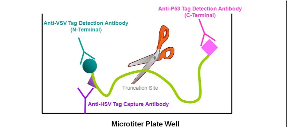
To overcome these limitations, we previously developed [14] the first high-throughput solid-phase protein trunca-tion test (HTS-PTT), which uses an industry-standard microtiter-plate enzyme-linked immunosorbent assay (ELISA) format, which can be implemented in any clinical laboratory with minimal, inexpensive, and widely used instrumentation. This assay, which we previously applied to the detection of familial adenomatous polyposis [14], used a triple-tag system comprised of N-terminal and C-terminal epitope tags for normalization and mutation detection, respectively, as well as directly incorporated bio-tin labels as the capture tag for protein immobilization on the ELISA plate. Unfortunately, biotin incorporation dur-ing translation usdur-ing tRNA-mediated protein engineerdur-ing occurs at very low efficiency, thereby adversely affecting mutation detection capabilities and diagnostic sensitivity (data not shown). Du and colleagues [18] later introduced a two-tag variant of our assay as applied to the genetic dis-order ataxia-telangiectasia; however, this variant lacks the ability to normalize for the amount of total nascent pro-tein that is expressed and captured on the ELISA plate, critical for the reproducibility and accuracy required in a clinical or diagnostic setting or both.
segments from patient blood. The amplified DNA is cell-free expressed and simultaneously captured and purified by epitope capture onto the ELISA plate, and the remaining two tags enable N- and C-terminal detec-tion (Figure 1). We have applied this improved assay for the first time to the detection of truncation mutations in exons 11 of the breast cancer susceptibility genes BRCA1/2and have evaluated it using 100 clinical geno-mic DNA test samples.
Materials and methods
Human samples were obtained through the Women’s College Research Institute (ref. no. 2007-0036-B) for these experiments and for the collaboration with AmberGen, Inc. (Watertown, MA, USA). All study sub-jects provided informed written consent. For HTS-PTT analysis, purified genomic DNA samples were provided to AmberGen, Inc., in de-identified form, marked only with non-descriptive alphanumeric codes along with only theBRCA1/2mutation status/designation, and no identifying information or clinical annotation was pro-vided to AmberGen, Inc.
DNA and polymerase chain reaction
One hundred human genomic clinical DNA samples isolated from blood and previously characterized by the conventional gel-based PTT (method as described in [19]) were used for HTS-PTT. The genomic DNA was extracted from peripheral blood leukocytes by means of the Gentra Puregene Blood Kit (Gentra
Systems, Minneapolis, MN, USA). mRNA samples for HTS-PTT were extracted from several BRCA mutant cell lines (B-lymphocytes from blood) obtained from the Coriell Cell Repository (Camden, NJ, USA); sam-ples were extracted by means of the Qiagen Rneasy Mini Kit (Qiagen, Germantown, MD, USA) and con-verted to cDNA by means of the SMART™ PCR cDNA Synthesis Kit (Clontech, Mountain View, CA, USA). PCR amplification was carried out with 50 ng of geno-mic DNA or cDNA, 0.245μM each of the forward and reverse primers, 2.5 mM MgCl2, 0.2 mM dNTPs
(each), and the Phusion Hot Start DNA Polymerase with the HF Buffer (New England Biolabs, Ipswitch, MA, USA). Amplification was performed as follows: an initial denaturation at 95°C for 2.5 minutes, 40 cycles of denaturation at 95°C for 30 seconds, annealing at 67.5°C for 45 seconds, extension at 72°C for 3.5 min-utes, and a final extension step at 72°C for 10 minutes. The gene-specific PCR primers used on genomic DNA or cDNA templates were as follows:
Forward primer: 5′-ggATCC TAATACgACTCACTA-TAgggAgACCACCATgTACACCgACATCgAgATgA ACCgCCTgggCAAgggAggA CAgCCTgAACTCgCTC-CAgAggATCCggAAgAT[gene-specific hybridization region]-3′.
[image:3.595.60.539.469.681.2]Forward primer key: The italicized nucleotides corre-spond to the T7 promoter, the underlined ATg is the initiation codon, the boldface nucleotide region codes for the N-terminal detection tag (VSV-G; YTDIEMNRLGK), the underlined boldface nucleotide region codes for the
N-terminal capture tag (HSV; QPELAPEDPED), the bracketed nucleotide sequence codes for the BRCA -specific complementary region (see below), and the remaining nucleotide sequences correspond to the Kozak (ribosome binding) and spacer regions.
Reverse primer: 5′-TTATTA CAgCAgCTTgTg-CAggTCgCTgAAggT [gene-specific hybridization region]-3′.
Reverse primer key: The boldface nucleotides code for the C-terminal detection tag (p53-derived tag; TFSDLHKLL), the underlined TTATTA is two succes-sive stop codons, and the bracketed nucleotide sequence codes for theBRCA-specific complementary region (see below).
Gene-specific hybridization regions and sequence of the primers are provided in Table 1. After amplification, the quality and quantity of the PCR products were ana-lyzed by agarose gel electrophoresis. Refer to Figure 2 for an example of agarose gel analysis of the five differ-ent PCR segmdiffer-ents, and refer to Figure 3 for the names of the aforementioned cell lines used for the mRNA samples.
Cell-free protein synthesis
The rabbit reticulocyte cell-free reaction mixture con-tained 8.5 μL of TNT T7 Quick Rabbit Reticulocyte Lysate for PCR DNA (Promega, Madison, WI, USA), 0.5μL of a complete amino acid mix (50 μM final for each), and 1 μL of PCR-amplified DNA (approxi-mately 100 to 200 ng). The coupled transcription-translation reaction was allowed to proceed for 45 minutes at 30°C.
HTS-PTT
MicroLite 2 + 96-well microtiter plates (Thermo Fisher Scientific, Waltham, MA, USA) were pre-coated with
[image:4.595.305.538.87.341.2]0.5 ng/μL of an anti-HSV tag monoclonal antibody (EMD Biosciences, La Jolla, CA, USA) solution in 50 mM sodium carbonate at a pH of 9.5. The plates were then washed four times with Tris-buffered saline (TBS) containing 0.05% (vol/vol) Tween-20 (TBS-T) and blocked for 30 minutes with TBS-T containing 1% (wt/vol) bovine serum albumin (BSA). After protein synthesis, the reaction mixture was diluted 100-fold
Table 1BRCA1/2exon 11 primers and segments for high-throughput solid-phase protein truncation test
Segment Nucleotides Segment size, base pairs Primer sequence (5’to 3’) F, forward; R, reverse; subscript‘I’, intronic primer
BRCA1a
1 123,126-125,033 1,908 F: gCTTgTgAATTTTCTgAgACggAT
R: TgTATTCTgCAAATACTgAgCATCAAg
2 124,617-126,578 1,962 F: gTCAATCCTAgCCTTCCAAgAgAA
RI: gggCAAACACAAAAACCTggTTCC
BRCA2a
1 25,758-27,458 1,701 FI: TTTTgTCACTTTgTgTTTTTATgTTTAgg
R: gCCAgCAAACTTCCgTTTAATTTC
2 27,174-29,330 2,157 F: AACCATAATTTAACACCTAgCCAAAAg
R: TgAAgAgCTAgTCACAAgTTCCTC
3 29,181-30,743 1,563 F: gATTCTggTATTgAgCCAgTATTgAAg
RI: CACgAAAggTAAAAATgAACACTTACC
a
[image:4.595.66.537.551.713.2]National Center for Biotechnology Information (NCBI) reference sequences: Ng_005905.2 forBRCA1and Ng_012772.1 forBRCA2, both numbered from whole sequence entry.BRCA1/BRCA2, breast and ovarian cancer susceptibility gene 1/2.
with TBS-T further supplemented with 0.1% Triton X-100, 1% (wt/vol) BSA, and one of either the N- or C-terminal detection antibodies (anti-VSV-G-HRP at 40 ng/mL, Clone P5D4, Roche Applied Science, Indiana-polis, IN, USA; or anti-p53-HRP at 80 ng/mL, Clone BP53-12, Santa Cruz Biotechnology, Inc., Santa Cruz, CA, USA; HRP = horseradish peroxidase). Alternatively, both the anti-VSV-G-HRP antibody and an anti-p53-AP antibody (Santa Cruz Biotechnology, Inc.; AP = alkaline phosphatase) could be used in the same solution (same concentrations as above) to ultimately allow both signals to be detected in the same well using this dual-reporter system [14]. In either case, 100μL of the diluted reac-tion mixture was then added to each well of the afore-mentioned antibody-coated microtiter plates and incubated for 45 minutes on an orbital shaker. The plate was washed four times with TBS-T and developed
using a chemiluminescent HRP substrate (Super Signal Femto; Pierce Chemicals, Rockford, IL, USA). If the dual-reporter system was used, a chemiluminescent AP substrate was added first (Roche Applied Science), and after signal readout, the plates were washed four times with TBS-T and developed using the HRP substrate described above. All signals were read using a Lumi-Count luminescent plate reader (Packard Biosciences, Meriden, CT, USA). The background levels correspond to cell-free protein synthesis reactions that lacked only the added DNA in the reaction mixture.
Results and Discussion The basic HTS-PTT
[image:5.595.59.537.90.408.2]segments using PCR (corresponding to roughly 50- to 75-kDa overlapping protein segments within exons 11 of BRCA1 andBRCA2; Table 1). As detailed in the Materi-als and methods section, the PCR Materi-also incorporates all of the needed expression sequences (for example, pro-moter) and epitope tag sequences, as required by the HTS-PTT method. After cell-free coupled transcription/ translation of the PCR products, the nascent proteins are analyzed by HTS-PTT for truncation mutations. The N-terminal capture tag (HSV epitope) is used for concurrent immobilization/purification of the cell-free expressed protein onto antibody-coated microtiter-well ELISA plates. The N- and C-terminal detection tags (VSV-G and p53 epitopes, respectively) are sub-sequently used for measuring the relative level of shor-tened protein produced by the chain truncation mutation (Figure 1). Detection is achieved using HRP-labeled epitope tag antibodies and a highly sensitive che-miluminescent readout in separate replicate wells of the ELISA plate. Alternatively, if tighter control is desired, a per-well normalization is possible, in which N- and C-terminal epitope tag antibodies (each carrying a different enyzyme reporter) are used in the same wells [14].
Polymerase chain reaction optimizations onBRCA1/2for HTS-PTT
Although lengthy sequences need to be added by the PCR primers in the HTS-PTT process, we have fully optimized the PCR conditions so that this can be achieved in a single reaction (one-step PCR) with a sin-gle primer pair (approximately 130-mer forward and approximately 60-mer reverse) as opposed two sequen-tial PCRs commonly used to add sequences of this length to an amplicon. This is important since two sequential PCRs are not compatible with standard clini-cal PCR clean-room practices (that is, separate pre- and post-PCR rooms). A significant problem we encountered is that the lengthy one-step PCR primers are prone to form non-specific or primer dimer extension products (or both) that produce substantial background in the HTS-PTT since both can be expressed as protein and can contain the in-frame epitope tags. Optimizing factors such as primer concentration, annealing tem-perature, magnesium concentration, and the use of a heat-activated‘hot start’DNA polymerase were found to be critical in avoiding this problem (data not shown). Extreme care should be taken to avoid the primer dimer extension products in the one-step and even the two-step PCR methodologies as these products are ideal expression templates and can interfere with HTS-PTT even when present at levels near or below the detection limit of conventional ethidium bromide agarose gel elec-trophoresis. For instance, the quantity and quality of the genomic DNA input into the PCR should be closely
monitored (discussed later) as, in general, less cognate template means that more primer dimer extension pro-ducts will be formed. All experiments described in this article use this optimized one-step PCR method (see Figure 2 for an example of agarose gel analysis of five different PCR segments).
Linearity of the HTS-PTT assay
After the HTS-PTT assay (Figure 1), the C-terminal sig-nal arising from full-length protein in a given sample is normalized against its N-terminal signal representing total nascent protein produced and is captured on the ELISA plate. These C-terminal-to-N-terminal signal ratios (C/N ratios) of the test samples are calculated as a percentage of the ratio arising from knownBRCAWT control samples (% C/N). Thus, in theory, BRCA WT samples would have a 100% C/N ratio and heterozygous BRCAmutants would have a 50% ratio.
In practice, however, the % C/N ratios of the BRCA heterozygous mutants deviate from 50% in either direc-tion (for example, Figure 3a and 3b; red bars for various segments), a phenomenon that can be explained by sev-eral possible factors. All of these factors essentially derive from the coexistence of two protein species in the heterozygous mutant samples (full-length and trun-cated) as opposed to a single protein species in the WT samples (full-length). One likely factor is skewed ratios of the actual mutant and WT proteins within a hetero-zygous sample, either in the expressed sample itself or ultimately what is captured on the ELISA plate, and this could occur through various mechanisms. For example, the enhanced expression or plate-binding kinetics of the mutant protein, owing to its decreased size compared with the WT [14], could increase the ratio of mutant protein actually bound to the ELISA plate to greater than 50% and therefore skew the % C/N ratio to less than 50%. Similarly, nucleotide composition differences between the mutant and WT can ultimately alter the mRNA secondary structure and hence result in differ-ences in expression efficiency between the mutant and WT, and this may be particularly applicable to larger deletions or insertions and could skew the % C/N in either direction. Differences in the physical accessibility (that is, steric hindrance) of the N-terminal epitope tag between the mutant and WT proteins could be caused by different secondary and tertiary folding structures between the two protein species and thereby could skew the ratio of mutant and WT protein captured on the ELISA plate.
tends to have a unique % C/N ratio for the mutants. For example,BRCA2exon 11 segment 1 mutants fall in the range of 30% ± 6% C/N, whereasBRCA2exon 11 seg-ment 2 mutants fall in the range of 61% ± 5% C/N (Fig-ure 3b). However, intra-segment variation in the % C/N ratio for the mutants is also apparent.
Figure 3c shows an example of linearity of the HTS-PTT assay as a function of different amounts of trans-lated protein input into the ELISA assay. The standard curves in Figure 3c are for the N- and C-terminal detec-tion tags of the WT reference sample for BRCA2 exon 11 segment 1 (n= 7). Importantly, although this verified excellent linearity of the assay over an approximate 10-fold range (R2 = 0.99), the y-intercepts of these N-and C-terminal signal curves do not pass precisely though zero. A critical consequence of this is that, in order to correctly compare the % C/N ratio of the WT reference sample with that of the test samples, the input amount of translated protein (for the ELISA) must be standardized among all of the samples. This is easily and typically achieved by normalizing the input amount of PCR product into the cell-free protein expression reaction and can be quality-controlled by monitoring the raw N-terminal signals (total expressed and captured protein).
Clinical validation of a genomic DNA-based HTS-PTT on
BRCA1/2using a 100-member training sample set
To evaluate the HTS-PTT for BRCA mutation analy-sis on clinical samples, we designed primer pairs to fully cover the large exons 11 of both BRCA1 and BRCA2, dividing them into two and three overlapping segments, respectively; each segment was roughly 2 kb in size. One hundred clinical genomic DNA sam-ples collected from blood were analyzed by means of HTS-PTT (25 BRCA1 mutants, 25 BRCA2 mutants, and 50 normal controls). The mutation status of the samples had been previously determined by conven-tional gel-based PTT. HTS-PTT results are shown in Figure 3a and 3b. The range of mutations covered is detailed in Supplemental table S1 in Additional file 1. This sample cohort contained deletions as large as 40 bases, all of which were detected by our HTS-PTT method.
Despite the aforementioned segment-specific % C/N ratios for heterozygous mutants in HTS-PTT, a fixed method based on the variability within the WT cohort was still able to be employed for setting the diagnostic scoring cutoffs. HTS-PTT scoring cutoffs for a positive protein truncation were fixed at 3 standard deviations below the mean of the 50 normal (WT) samples (on a segment-by-segment basis) for an approximately 99% confidence inter-val. With these cutoffs (Figure 3a and 3b; green bar and green line), the sensitivity and specificity of the HTS-PTT
(as compared with conventional PTT) were 100% and 96%, respectively. Note that the % C/N ratios for the only two false-positive calls (Figure 3a and 3b; black bars) were just slightly below the cutoff and were likely due to assay variance (approximately 1/100 false-positive calls are sta-tistically expected with the aforementioned 99% confi-dence interval used). Finally, it is worth noting that, in the HTS-PTT assay, raw signal-to-noise ratios of the N- and C-terminal detection were never less than 20:1 and in most cases were greater than 100:1.
An mRNA-based HTS-PTT forBRCA1/2
To obtain fullBRCA1/2(open reading frame) coverage using a genomic DNA test, roughly 50 HTS-PTT seg-ments per patient would be required because of the large number of small exons in both genes, outside of the large exons 11. The high-throughput ELISA-based HTS-PTT is certainly more amenable to such large segment numbers than the conventional gel-based PTT. However, one way to further alleviate this issue is to design an mRNA-based assay, which would allow theBRCA1/2transcripts to be divided evenly into approximately 10 HTS-PTT segments of roughly 1 to 2 kb in size (an effective size for the HTS-PTT). To demonstrate the feasibility of this approach, we performed experiments on cDNA created from cell lines (obtained from the Coriell Cell Repository) having known BRCA1/2truncating mutations (Coriell Institute for Medi-cal Research, Camden, NJ, USA). Figure 4 showsBRCA1 results for one gene segment on 13 cell lines (1 mutant) and again demonstrates 100% accuracy. Importantly, non-sense-mediated decay of the mutant transcripts was not found to be a problem in this small set, but should the problem occur, mutation detection sensitivity as low as 25% is possible with HTS-PTT [14].
Conclusions
Additional material
Additional file 1: Supplemental table S1 -BRCA1/2Mutations Covered by the HTS-PTT. This table lists the mutation designations for the 50 patient genomic DNA samples tested which were positive for BRCA1/2truncation mutations. HTS-PTT segments containing the mutation and the measured % C/N ratios are also listed.
Abbreviations
BIC: Breast Cancer Information Core;BRCA1/BRCA2: breast and ovarian cancer susceptibility gene 1/2; BSA: bovine serum albumin; C/N: C-terminal to N-terminal signal (ratio); DHPLC: denaturing high-performance liquid chromatography; ELISA: enzyme-linked immunosorbent assay; HRP: horseradish peroxidase; HTS-PTT: high-throughput solid-phase protein truncation test; PCR: polymerase chain reaction; PTT: protein truncation test; TBS: Tris-buffered saline; TBS-T: Tris-buffered saline containing 0.05% (vol/vol) Tween-20; WT: wild-type.
Acknowledgements
This work was funded in part by Phase I and Phase II Small Business Innovation Research grants (CA110403) from the National Cancer Institute to AmberGen, Inc.
Author details
1AmberGen, Inc., 313 Pleasant Street, Watertown, MA 02472, USA.2First Light
Biosciences, Inc., 1 Oak Park Drive, Floor 2, Bedford, MA 01730, USA.3Centre for Research in Women’s Health, Women’s College Hospital, University of Toronto, 790 Bay Street, Toronto, ON, M5G 1N8, Canada.4Molecular Biophysics Laboratory, Department of Physics and Photonics Center, Boston University, Boston, MA 02215, USA.
Authors’contributions
MJL and KJR helped conceive of the HTS-PTT assay and its application to BRCAanalysis, participated in the design and coordination of all studies in this article, and contributed significantly to the drafting of the manuscript. SG helped conceive of the HTS-PTT assay and its application toBRCA analysis and participated in the design and coordination of all studies in this article. SN supervised and coordinated the isolation and gel-PTT analysis of all clinical genomic DNA samples used in this article and contributed
significantly to the drafting of the manuscript. GJF was responsible for performing and designing PCR amplifications, cell-free protein expressions, and HTS-PTT assays for optimization purposes and for the final analyses of the clinical samples. HPO performed advanced PCR assays that contributed to a better understanding of the effects of primer dimers and non-specific PCR extension products on the HTS-PTT assay forBRCAanalysis and contributed significantly to the drafting of the manuscript. All authors read and approved the final manuscript.
Competing interests
MJL, HPO, and GJF are current employees of AmberGen, Inc., a developer of commercial diagnostic assays. SG is a former employee (within the last 5 years), and KJR is a co-founder of the company. This project was financed by AmberGen, in part using Small Business Innovation Research grant funds (from the National Institutes of Health) awarded to AmberGen (see Acknowledgements). AmberGen is the assignee on issued patents related to the commercial use of the HTS-PTT technologies described in this article. However, these patents do not preclude the use of the HTS-PTT by the general research community. In the article, we disclose in full detail the methodology required for researchers to perform HTS-PTT, which needs no specialized reagents or instrumentation (and requires nothing to be purchased from AmberGen).
Received: 16 April 2010 Revised: 4 August 2010 Accepted: 4 October 2010 Published: 4 October 2010
References
1. Worman HJ, Courvalin JC:Antinuclear antibodies specific for primary biliary cirrhosis.Autoimmun Rev2003,2:211-217.
2. Gerhardus A, Schleberger H, Schlegelberger B, Gadzicki D:
Diagnostic accuracy of methods for the detection of BRCA1 and BRCA2 mutations: a systematic review.Eur J Hum Genet2007,
15:619-627.
3. Claus EB, Schildkraut JM, Thompson WD, Risch NJ:The genetic attributable risk of breast and ovarian cancer.Cancer1996,77:2318-2324.
4. Rebbeck TR, Couch FJ, Kant J, Calzone K, DeShano M, Peng Y, Chen K, Garber JE, Weber BL:Genetic heterogeneity in hereditary breast cancer: role of BRCA1 and BRCA2.Am J Hum Genet1996,59:547-553. 5. Narod SA, Foulkes WD:BRCA1 and BRCA2: 1994 and beyond.Nat Rev
Cancer2004,4:665-676.
[image:8.595.58.540.88.293.2]6. Breast Cancer Information Core (BIC) Database.[http://research.nhgri.nih. gov/bic/].
7. Powell SM, Petersen GM, Krush AJ, Booker S, Jen J, Giardiello FM, Hamilton SR, Vogelstein B, Kinzler KW:Molecular diagnosis of familial adenomatous polyposis.N Engl J Med1993,329:1982-1987.
8. Luce MC, Marra G, Chauhan DP, Laghi L, Carethers JM, Cherian SP, Hawn M, Binnie CG, Kam-Morgan LN, Cayouette MC, Koi M, Boland CR:In vitro transcription/translation assay for the screening of hMLH1 and hMSH2 mutations in familial colon cancer.Gastroenterology1995,109:1368-1374. 9. Peral B, Gamble V, Strong C, Ong AC, Sloane-Stanley J, Zerres K,
Winearls CG, Harris PC:Identification of mutations in the duplicated region of the polycystic kidney disease 1 gene (PKD1) by a novel approach.Am J Hum Genet1997,60:1399-1410.
10. Heim RA, Kam-Morgan LN, Binnie CG, Corns DD, Cayouette MC, Farber RA, Aylsworth AS, Silverman LM, Luce MC:Distribution of 13 truncating mutations in the neurofibromatosis 1 gene.Hum Mol Genet1995,
4:975-981.
11. Parry DM, MacCollin MM, Kaiser-Kupfer MI, Pulaski K, Nicholson HS, Bolesta M, Eldridge R, Gusella JF:Germ-line mutations in the
neurofibromatosis 2 gene: correlations with disease severity and retinal abnormalities.Am J Hum Genet1996,59:529-539.
12. Roest PA, Roberts RG, van der Tuijn AC, Heikoop JC, van Ommen GJ, den Dunnen JT:Protein truncation test (PTT) to rapidly screen the DMD gene for translation terminating mutations.Neuromuscul Disord1993,3:391-394. 13. Kahmann S, Herter P, Kuhnen C, Muller KM, Muhr G, Martin D,
Soddemann M, Muller O:A non-radioactive protein truncation test for the sensitive detection of all stop and frameshift mutations.Hum Mutat 2002,19:165-172.
14. Gite S, Lim M, Carlson R, Olejnik J, Zehnbauer B, Rothschild K:A high-throughput nonisotopic protein truncation test.Nat Biotechnol2003,
21:194-197.
15. Garvin AM:A complete protein truncation test for BRCA1 and BRCA2.Eur J Hum Genet1998,6:226-234.
16. Andrulis IL, Anton-Culver H, Beck J, Bove B, Boyd J, Buys S, Godwin AK, Hopper JL, Li F, Neuhausen SL, Ozcelik H, Peel D, Santella RM, Southey MC, van Orsouw NJ, Venter DJ, Vijg J, Whittemore AS:Comparison of DNA-and RNA-based methods for detection of truncating BRCA1 mutations.
Hum Mutat2002,20:65-73.
17. Ozcelik H, Nedelcu R, Chan VW, Shi XH, Murphy J, Rosen B, Andrulis IL:
Mutation in the coding region of the BRCA1 gene leads to aberrant splicing of the transcript.Hum Mutat1999,14:540-541.
18. Du L, Lai CH, Concannon P, Gatti RA:Rapid screen for truncating ATM mutations by PTT-ELISA.Mutat Res2008,640:139-144.
19. Evans J, Reuben A, Craft J:PBC 95k, a 95-kilodalton nuclear autoantigen in primary biliary cirrhosis.Arthritis Rheum1991,34:731-736.
doi:10.1186/bcr2722
Cite this article as:Limet al.:An ELISA-based high throughput protein truncation test for inherited breast cancer.Breast Cancer Research2010
12:R78.
Submit your next manuscript to BioMed Central and take full advantage of:
• Convenient online submission
• Thorough peer review
• No space constraints or color figure charges
• Immediate publication on acceptance
• Inclusion in PubMed, CAS, Scopus and Google Scholar
• Research which is freely available for redistribution