RESEARCH NOTE
Phenotypic and genotypic
characterization of macrolide, lincosamide
and streptogramin B resistance among clinical
isolates of staphylococci in southwest of Iran
Reza Khashei
1*, Yalda Malekzadegan
1, Hadi Sedigh Ebrahim‑Saraie
1and Zahra Razavi
2Abstract
Objective: The present study aimed to determine the phenotypic and genotypic profile of macrolide, lincosamide and streptogramin B (MLSB) resistance in clinical isolates of staphylococci.
Results: This cross‑sectional study was conducted on 164 non‑duplicated staphylococci isolates collected during August 2015 to February 2016 from two tertiary care hospitals in Shiraz, southwest of Iran. Of the 164 isolates, 86 erythromycin‑resistant isolates consist of 35 Staphylococcus aureus and 51 coagulase negative staphylococci (CoNS) were included in the study. Of the 35 S. aureus, the prevalence of cMLS (constitutive), iMLS (inducible), and MS phenotypes were found 82.9%, 8.6% and 8.6%, respectively. Among 51 CoNS, the frequencies of cMLS, iMLS, and MS phenotypes were detected 66.7%, 11.8% and 21.6%, respectively. Among S. aureus isolates, the predominant genes were ermC in 82.9% isolates, followed by ermA in 57.1% and msrA in 28.6% of isolates. Among CoNS isolates, the most frequent genes were diagnosed ermC in 70.6% isolates followed by msrA in 68.6% and ermA in 11.8% of isolates. In conclusion, regarding the presence of MLSB resistance in our region, diagnosis of this resistance type on a routine basis in staphylococcal clinical isolates is of particular importance.
Keywords: Inducible resistance, Staphylococcus, Clindamycin, Erythromycin, erm genes
© The Author(s) 2018. This article is distributed under the terms of the Creative Commons Attribution 4.0 International License (http://creat iveco mmons .org/licen ses/by/4.0/), which permits unrestricted use, distribution, and reproduction in any medium, provided you give appropriate credit to the original author(s) and the source, provide a link to the Creative Commons license, and indicate if changes were made. The Creative Commons Public Domain Dedication waiver (http://creat iveco mmons .org/ publi cdoma in/zero/1.0/) applies to the data made available in this article, unless otherwise stated.
Introduction
Staphylococci are amongst the most frequent causes of nosocomial and community-acquired infections worldwide [1, 2]. The emergence of antibiotic resistant Staphylococcus aureus, particularly methicillin resistant Staphylococcus aureus (MRSA) pose difficulties in treat-ment of related infections [3]. Macrolide, lincosamide and streptogramin B (MLSB) antibiotics are one the avail-able options for treating staphylococcal infections [4]. These antibiotics are used in treatment of a wide range of bacterial infections; however, frequently used as drug of choice to treat staphylococcal skin and soft tissue infec-tions (SSTIs) [5]. Recently, the increasing prevalence of
methicillin resistance and other chemotherapeutic agents among staphylococci become a global health concern [6].
Resistance to MLS antibiotics in S. aureus and coagu-lase negative staphylococci (CoNS), can resulted in the target site modification encoded by erm genes [7, 8]. The methylation of the 23S rRNA conferred by erm genes prevents the binding of antibiotic to its ribosomal tar-get [9]. Other mechanisms are efflux pumps encoded by msrA gene which mediated resistance to MSB, and the drug modification encoded by lnu gene [10, 11].
MLSB phenotype can be expressed into forms of con-stitutive (cMLSB) or inducible (iMLSB) [12]. Constitu-tive resistance is related to S. aureus strains which are resistant to both erythromycin and clindamycin [13]. Inducible strains define those bacteria which are actu-ally resistant to erythromycin and clindamycin, but are susceptible to clindamycin by routine susceptibility tests
Open Access
*Correspondence: re.khashei@gmail.com; khasheir@sums.ac.ir 1 Department of Bacteriology and Virology, School of Medicine, Shiraz University of Medical Sciences, Shiraz, Iran
[13]. Constitutive MLSB resistance do not require specific method for routine detection, whereas iMLSB resistance is not recognized using the standard susceptibility meth-ods [14]. Erythromycin is the effective inducer of iMLSB resistance than clindamycin. Owed to this fact, iMLSB resistance can be determined by special disk approxi-mation tests that incorporate erythromycin induction of clindamycin resistance (D-zone effect) [14].
Clindamycin treatment in patients with iMLSB resist-ance may lead to development of cMLSB resistant strains and subsequently therapeutic failure [12]. Therefore, it is important to identify actual MLSB resistance for estab-lishing appropriate therapy in infected patients. Due to the lack of data on the prevalence and characteristics of MLSB resistant strains in our region, we aimed to deter-mine the resistance rates and predominant resistance mechanisms toward MLSB antibiotics among clinical iso-lates of staphylococci obtained from Iranian patients.
Main text
Methods
Study design and identification of isolates
This cross-sectional study was conducted on 164 non-duplicated staphylococci isolates collected from two major teaching hospitals, Nemazee and Faghihi, in Shi-raz, southwest of Iran. Staphylococci isolates were recov-ered from different body sites such as blood, wound, sputum, urine and other clinical specimens between August 2015 and February 2016. Standard microbio-logical techniques, including colony morphology, Gram stain, catalase test, tube coagulase test, DNase test and growth on mannitol salt agar were used for identification of S. aureus and CoNS isolates.
Antibiotic susceptibility testing
Antibiotic susceptibility test was performed on Muller Hinton agar (Merck, Germany) using the disk diffusion method according to Clinical and Laboratory Standards Institute (CLSI) guidelines [15]. Methicillin resistance was primary detected based on resistance to cefoxitin (30 μg) disk (Rosco, Denmark) by CLSI recommended disk diffusion method. The isolates which were initially susceptible to clindamycin (2 μg) and resistant to eryth-romycin (15 μg) were examined for inducible clinda-mycin resistance using the D-test according to CLSI recommendations [15]. Briefly, erythromycin (15 μg) and clindamycin (2 μg) disks were placed 15–20 mm apart (edge to edge) and then incubated at 35–37 °C for 18 h. Staphylococcal isolates showing resistance to erythromy-cin but being susceptible to clindamyerythromy-cin and produerythromy-cing a D-shaped zone of inhibition around the clindamycin disk on the side facing the erythromycin disk were consid-ered as iMLS resistance phenotype. Moreover, resistance
to both erythromycin and clindamycin was indicated a cMLS resistance phenotype. Staphylococcal isolates showing resistance to erythromycin while being suscep-tible to clindamycin with no blunting zone were classified as the MS resistance phenotype. S. aureus ATCC 25923 was used as a control strain for antibiotic susceptibility testing.
DNA extraction and polymerase chain reaction (PCR) assay
Bacterial whole DNAs were extracted from Staphylococ-cal isolates using the boiling method and used as PCR templates. All S. aureus isolates, including methicillin sensitive Staphylococcus aureus (MSSA) and MRSA were subsequently tested for the presence of femA and mecA genes by a set of previously described primers [16, 17]. PCR reaction was performed for detection of erythro-mycin resistance determinants (ermA, ermB, ermC, and msrA genes) [18], and two major virulence factors of S. aureus (tst-1, and pvl genes) with specific primers [17,
19]. The reference strains were provided from our col-league previous work and used as positive controls in PCR experiments [20]. PCR amplifications were per-formed in a DNA Thermal Cycler 5530 (Ependrof master, Germany). PCR products were mixed with 1 µl loading buffer solution and were loaded into the wells of aga-rose gel (1.5%) carefully and electrophoresed at 75 V for 90 min. The gel was then stained with KBC (Merck, Ger-many) solution for 15 min and observed under the UV trans-illuminator.
Statistical analysis
Data were analyzed using SPSS™ software (IBM corp., USA) version 21.0. The results were presented as descrip-tive statistics in terms of reladescrip-tive frequency. Chi–square or Fisher’s exact tests were used to estimate any statisti-cal association for quantitative variables. Values were expressed as the mean ± standard deviation (continu-ous variables) or percentages of the group (categori-cal variables). Statisti(categori-cal significance was regarded as P-values < 0.05.
Results
(RTIs) 12 (14%), eye infections 3 (3.5%), abdominal infec-tions 1 (1.2%), and bone and joint infecinfec-tions 1 (1.2%). The mecA gene screening showed that 32 of the tested isolates were MRSA and 44 were methicillin-resistant coagulase-negative staphylococci (MRCNS).
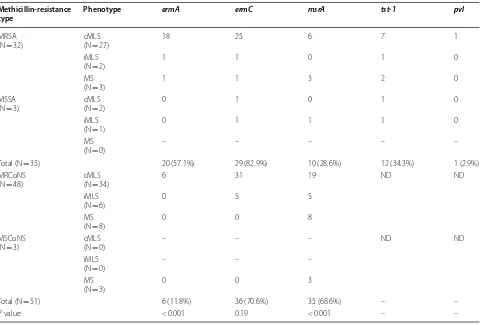
Of 35 S. aureus, the overall prevalence of cMLS, iMLS, and MS phenotypes were 29 (82.9%), 3 (8.6%), and 3 (8.6%), respectively. Among 51 CoNS, the overall prevalence of cMLS, iMLS, and MS phenotypes were 34 (66.7%), 6 (11.8%), and 11 (21.6%), respectively. As shown in Table 1, among S. aureus isolates the predominant genes were ermC in 29 (82.9%) isolates, followed by ermA in 20 (57.1%) and msrA in 10 (28.6%) isolates. Among CoNS isolates the most prevalent genes were ermC in 36 (70.6%) isolates, followed by msrA in 35 (68.6%) and ermA in 6 (11.8%) isolates. Meanwhile, ermB gene was not found among all of the tested isolates. Interestingly, in one of the MSSA isolates with cMLS phenotype any of investigated genes was not found.
The details of macrolides resistance phenotypic and genotypic related to the source of infections are
presented in Table 2. Moreover, in urinary tract, eye and abdominal originated specimens no ermA gene was found. On the other hand, the most common isola-tion sites of staphylococci with iMLS phenotype were bloodstream, skin and soft tissue, urinary tract, and eye.
The prevalence of two virulence factors of S. aureus, namely toxic shock syndrome toxin (TSST-1), and Pan-ton–Valentine leukocidin (PVL) in 35 erythromycin-resistant isolates by detection of tst-1 and pvl genes were 34.3% and 2.9%, respectively. As shown in Table 2, the main source of toxins producing S. aureus was SSTIs. Moreover, the proportion of these toxins in all 97 S. aureus isolates were 37.1% (36/97) and 3.1% (3/97) for TSST-1 and PVL, respectively.
The combination patterns of genes responsible for resistance to MLS antibiotics in studied isolates are depicted in Table 3. As be shown, 7 different combi-nations were detected. The most prevalent pattern in S. aureus and CoNS were ermA/ermC (37.1%) and ermC/msrA (39.2%), respectively.
Table 1 The frequency of phenotypic and genotypic resistance to MLS according to type of isolates
ND, not determined Methicillin-resistance
type Phenotype ermA ermC msrA tst-1 pvl
MRSA
(N = 32) cMLS(N = 27) 18 25 6 7 1
iMLS
(N = 2) 1 1 0 1 0
MS
(N = 3) 1 1 3 2 0
MSSA
(N = 3) cMLS(N = 2) 0 1 0 1 0
iMLS
(N = 1) 0 1 1 1 0
MS
(N = 0) – – – – –
Total (N = 35) 20 (57.1%) 29 (82.9%) 10 (28.6%) 12 (34.3%) 1 (2.9%)
MRCoNS
(N = 48) cMLS(N = 34) 6 31 19 ND ND
iMLS
(N = 6) 0 5 5
MS
(N = 8) 0 0 8
MSCoNS
(N = 3) cMLS(N = 0) – – – ND ND
iMLS
(N = 0) – – –
MS
(N = 3) 0 0 3
Total (N = 51) 6 (11.8%) 36 (70.6%) 35 (68.6%) – –
[image:3.595.59.540.379.704.2]Discussion
Multi-resistant strains of staphylococci have been reported to frequently acquire resistance to macrolides and related antibiotics, which can led to difficulties in the treatment of infections [21]. Hence, due to variety in the prevalence of these strains in different geographi-cal areas, constant surveillance can be helpful to con-trol their spread and providing information regarding their current epidemiology. Results of the present study showed new information regarding the occurrence of the MLS resistance in this era of high incidence of MRSA in Iran [22].
In the current study, the overall frequency of iMLS phe-notype among S. aureus and CoNS were found to be 8.6%, and 11.8%, respectively. Despite the discrepancies, our find-ing was consistent with previous reports from Iran which had shown low prevalence of iMLS phenotype in staphy-lococci ranged from 4.1 to 14.9% [11, 23–26]. However, much higher rates (≥ 30%) was also reported by Moosavian et al. and Saffar et al. from Ahvaz and Tehran, respectively [14, 35]. Moreover, the prevalence reported in our study is higher than those reported among staphylococci isolated from Brazil (S. aureus, 0%) [12], and Mexico (CoNS, 8%) [27], whereas it is lower than those reported from Japan (S.
aureus, 91%) [28], Korea (S. aureus, 34%; CoNS, 90%) [29], Poland (CoNS, 18.7%) [7], Nepal (S. aureus, 11.5%) [30], and India (S. aureus, 10.8%) [31].
Resistance to MLS antibiotics in staphylococci is mainly mediated by methyltransase encoded by erm genes [32]. The distribution of these genes mostly depends on the bacterial species [7]. In our findings, ermA gene was more prevalent in S. aureus compared to CoNS (P < 0.001). Previ-ously, similar results have been cited by authors from Iran and other countries [9, 23, 33]. In contrast, we found msrA gene was more abundant in CoNS (P < 0.001). In agree-ment with our findings, this gene is the most frequently reported gene in CoNS isolates exhibiting the MLS resist-ance phenotypes [33, 34]. We did not find any isolates car-rying ermB gene. Despite the similar reports indicating the prevalence of ermB gene in low rates [23, 35], it has been noted that ermB is more characteristic of beta-haemolytic streptococci or staphylococci with animal origin [7, 12]. In our study, in one erythromycin-resistant isolate with cMLS phenotype, none of the investigated genes were found. Previously, such discordance among MLS phenotypes and erythromycin resistance genes due to a mutation in the coding or promoter region of targeted genes was reported [32].
In conclusion, regarding the presence of different types of MLSB resistance in our region, diagnosis of these resist-ance types on a routine basis in staphylococcal clinical isolates is of particular importance. These results suggest that the empiric use of MLSB antibiotics for staphylococcal infections should be prescribed in a logical manner by our physicians.
Limitations
Finally, as the main limitations of the present study, the lack of evaluation of genes expression by real time-PCR, and genetic relatedness of erythromycin-resistant isolates by a molecular typing method should be acknowledged.
Table 2 Macrolides resistance phenotypic and genotypic characterization in relation to source of infections
BSIs, bloodstream infections; SSTIs, skin and soft tissue infections; UTIs, urinary tract infections; RTIs, respiratory tracts infections; EIs, eye infections; AIs, abdominal infections, BJIs, bone and joint infections
a The proportion estimated among 35 erythromycin-resistant S. aureus
Phenotype ermA ermC msrA tst-1 pvl cMLS iMLS MS
BSIs (40) 11 29 23 2 0 28 5 7
SSTIs (16) 7 12 10 4 1 11 2 3
UTIs (13) 0 11 8 3 0 11 1 1
RTIs (12) 7 10 2 1 0 11 0 1
EIs (3) 0 2 1 2 0 1 1 1
AIs (1) 0 0 1 0 0 0 0 0
BJIs (1) 1 1 0 0 0 1 0 0
[image:4.595.57.541.101.220.2]Total 26 (30.2) 65 (75.6) 45 (52.3) 12 (34.3%)a 1 (2.9%)a 63 (73.3%) 9 (10.5%) 14 (16.3)
Table 3 The combination patterns of genes responsible for resistance to MLS antibiotics in studied isolates
Pattern S. aureus
(N = 35) CoNS(N = 51) Total(N = 86)
ermA 3 (8.6) 2 (3.9) 5 (5.8)
ermC 8 (22.9) 12 (23.5) 20 (23.3)
msrA 2 (5.7) 13 (25.5) 15 (17.4)
ermA/ermC 13 (37.1) 2 (3.9) 15 (17.4) ermC/msrA 4 (11.4) 20 (39.2) 24 (27.9) ermA/ermC/msrA 4 (11.4) 2 (3.9) 6 (7)
[image:4.595.57.290.301.415.2]Abbreviations
MRSA: methicillin resistant Staphylococcus aureus; MLSB: macrolide, lincosa‑ mide and streptogramin B; SSTIs: skin and soft tissue infections; CoNS: coagu‑ lase negative staphylococci; MRCNS: methicillin‑resistant coagulase‑negative staphylococci; cMLSB: constitutive macrolide, lincosamide and streptogramin B resistance; iMLSB: inducible macrolide, lincosamide and streptogramin B resistance; CLSI: Clinical and Laboratory Standards Institute.
Authors’ contributions
RK and HSE‑S: conceived the study. RK, YM, HSE‑S, ZR: participated in the design of the study and performed the statistical analysis. YM, HSE‑S, ZR: interpreted the data. FS: obtained ethical clearance and permission for study. RK, YM, ZR: Supervised data collectors. RK, YM, HSE‑S: Drafting the article or revisiting it critically for important intellectual content. RK and YM were project leaders and primary investigators of the study. All authors read and approved the final manuscript.
Author details
1 Department of Bacteriology and Virology, School of Medicine, Shiraz Uni‑ versity of Medical Sciences, Shiraz, Iran. 2 Student Research Committee, Shiraz University of Medical Sciences, Shiraz, Iran.
Acknowledgements
We thank all participants and personals at the studied hospital for their friendly cooperation.
Competing interests
The authors declare that they have no competing interests.
Availability of data and materials
The datasets used and/or analyzed during the current study are available from the corresponding author on reasonable request.
Consent for publication Not applicable.
Ethics approval and consent to participate
This study was in accordance with the declaration of Helsinki and an ethical permission was sought from the institutional Ethics Committee of Shiraz Uni‑ versity of Medical Sciences (Approval No. IR.SUMS.REC.1394.S348). However, because we only used leftovers from clinical specimens, the local ethics com‑ mittee waived the need for informed consent.
Funding support
This study was supported by Shiraz University of Medical Sciences, Shiraz, Iran with Grant No. 93‑8690.
Publisher’s Note
Springer Nature remains neutral with regard to jurisdictional claims in pub‑ lished maps and institutional affiliations.
Received: 17 August 2018 Accepted: 3 October 2018
References
1. Nejabat M, Khashei R, Bazargani A, Sedigh Ebrahim‑Saraie H, Motamedi‑ far M. Evaluation of High‑Level of Mupirocin Resistance among Clinical Isolates of Methicillin‑Resistant Staphylococcus aureus from Shiraz, Iran (2008–2009). Pharm Sci. 2015;21(4):225–8.
2. Zomorodian K, Rahimi MJ, Taheri M, Ghanbari Asad A, Khani S, Ahrari I, et al. The cutaneous bacterial microflora of the bodybuilders using anabolic‑androgenic steroids. Jundishapur J Microbiol. 2015;8(1):e12269.
https ://doi.org/10.5812/jjm.12269 .
3. Rasmussen RV, Fowler VG Jr, Skov R, Bruun NE. Future challenges and treatment of Staphylococcus aureus bacteremia with emphasis on MRSA. Future Microbiol. 2011;6(1):43–56. https ://doi.org/10.2217/fmb.10.155. 4. Saribas Z, Tunckanat F, Pinar A. Prevalence of erm genes encod‑
ing macrolide‑lincosamide‑streptogramin (MLS) resistance among
clinical isolates of Staphylococcus aureus in a Turkish university hospital. Clin Microbiol Infect. 2006;12(8):797–9. https ://doi.org/10.111 1/j.1469‑0691.2006.01486 .x.
5. Adaleti R, Nakipoglu Y, Ceran N, Tasdemir C, Kaya F, Tasdemir S. Prevalence of phenotypic resistance of Staphylococcus aureus isolates to macrolide, lincosamide, streptogramin B, ketolid and linezolid antibiotics in Turkey. Braz J Infect Dis. 2010;14(1):11–4.
6. Dadashi M, Nasiri MJ, Fallah F, Owlia P, Hajikhani B, Emaneini M, et al. Methicillin‑resistant Staphylococcus aureus (MRSA) in Iran: a systematic review and meta‑analysis. J Glob Antimicrob Resist. 2018;12:96–103. https ://doi.org/10.1016/j.jgar.2017.09.006.
7. Juda M, Chudzik‑Rzad B, Malm A. The prevalence of genotypes that determine resistance to macrolides, lincosamides, and streptogramins B compared with spiramycin susceptibility among erythromycin‑resistant Staphylococcus epidermidis. Mem Inst Oswaldo Cruz. 2016;111(3):155–60.
https ://doi.org/10.1590/0074‑02760 15035 6.
8. Filippin L, Roisin S, Nonhoff C, Vandendriessche S, Heinrichs A, Denis O. Evaluation of the automated Vitek 2 system for detection of various mechanisms of macrolide and lincosamide resistance in Staphylococcus aureus. J Clin Microbiol. 2014;52(11):4087–9. https ://doi.org/10.1128/ jcm.01617 ‑14.
9. Hess S, Gallert C. Resistance behaviour of inducible clindamycin‑resistant staphylococci from clinical samples and aquatic environments. J Med Microbiol. 2014;63(Pt 11):1446–53. https ://doi.org/10.1099/jmm.0.07708 1‑0.
10. Li L, Feng W, Zhang Z, Xue H, Zhao X. Macrolide‑lincosamide‑strepto‑ gramin resistance phenotypes and genotypes of coagulase‑positive Staphylococcus aureus and coagulase‑negative staphylococcal isolates from bovine mastitis. BMC Vet Res. 2015;11:168. https ://doi.org/10.1186/ s1291 7‑015‑0492‑8.
11. Sedaghat H, Esfahani BN, Mobasherizadeh S, Jazi AS, Halaji M, Sadeghi P, et al. Phenotypic and genotypic characterization of macrolide resistance among Staphylococcus aureus isolates in Isfahan, Iran. Iran J Microbiol. 2017;9(5):264–70.
12. Pereira JN, Rabelo MA, Lima JL, Neto AM, Lopes AC, Maciel MA. Phe‑ notypic and molecular characterization of resistance to macrolides, lincosamides and type B streptogramin of clinical isolates of Staphylococ-cus spp. of a university hospital in Recife, Pernambuco, Brazil. Braz J Infect Dis. 2016;20(3):276–81. https ://doi.org/10.1016/j.bjid.2016.03.003. 13. Lall M, Sahni AK. Prevalence of inducible clindamycin resistance in
Staphylococcus aureus isolated from clinical samples. Med J Armed Forces India. 2014;70(1):43–7. https ://doi.org/10.1016/j.mjafi .2013.01.004. 14. Saffar H, Rajabiani A, Abdollahi A, Habibi S, Baseri Z. Frequency of induc‑
ible clindamycin resistance among gram‑positive cocci in a tertiary hospital, Tehran, Iran. Iran J Microbiol. 2016;8(4):243–8.
15. Wayne P. Performance Standards for Antimicrobial Susceptibility Testing. Clinical and Laboratory Standards Institute (CLSI). 2016; 26th Informa‑ tional Supplement.
16. Zhang K, McClure JA, Elsayed S, Louie T, Conly JM. Novel multiplex PCR assay for characterization and concomitant subtyping of staphylo‑ coccal cassette chromosome mec types I to V in methicillin‑resistant Staphylococcus aureus. J Clin Microbiol. 2005;43(10):5026–33. https ://doi. org/10.1128/jcm.43.10.5026‑5033.2005.
17. Mehrotra M, Wang G, Johnson WM. Multiplex PCR for detection of genes for Staphylococcus aureus enterotoxins, exfoliative toxins, toxic shock syndrome toxin 1, and methicillin resistance. J Clin Microbiol. 2000;38(3):1032–5.
18. Martineau F, Picard FJ, Grenier L, Roy PH, Ouellette M, Bergeron MG. Mul‑ tiplex PCR assays for the detection of clinically relevant antibiotic resist‑ ance genes in staphylococci isolated from patients infected after cardiac surgery. The ESPRIT Trial. J Antimicrob Chemother. 2000;46(4):527–34. 19. Wong H, Louie L, Lo RY, Simor AE. Characterization of Staphylococcus
aureus isolates with a partial or complete absence of staphylococcal cas‑ sette chromosome elements. J Clin Microbiol. 2010;48(10):3525–31. https ://doi.org/10.1128/jcm.00775 ‑10.
20. Alfatemi SM, Motamedifar M, Hadi N, Saraie HS. Analysis of virulence genes among methicillin resistant Staphylococcus aureus (MRSA) strains. Jundishapur J Microbiol. 2014;7(6):e10741. https ://doi.org/10.5812/ jjm.10741 .
•fast, convenient online submission
•
thorough peer review by experienced researchers in your field
• rapid publication on acceptance
• support for research data, including large and complex data types
•
gold Open Access which fosters wider collaboration and increased citations maximum visibility for your research: over 100M website views per year
•
At BMC, research is always in progress.
Learn more biomedcentral.com/submissions
Ready to submit your research? Choose BMC and benefit from: macrolides and lincosamides in Serbia. Front Public Health. 2017;5:200.
https ://doi.org/10.3389/fpubh .2017.00200 .
22. Shahini Shams‑Abadi M, Halaji M, Hoseini‑Alfatemi SM, Gholipour A, Mojtahedi A, Sedigh Ebrahim‑Saraie H. Epidemiology of toxic shock syndrome toxin‑1 harboring Staphylococcus aureus obtained from clinical samples in Iran: a systematic review and meta‑analysis. Ann Ig. 2018;30(5):391–400. https ://doi.org/10.7416/ai.2018.2239.
23. Emaneini M, Eslampour MA, Sedaghat H, Aligholi M, Jabalameli F, Shahsavan S, et al. Characterization of phenotypic and genotypic induc‑ ible macrolide resistance in staphylococci in Tehran, Iran. J Chemother. 2009;21(5):595–7. https ://doi.org/10.1179/joc.2009.21.5.595.
24. Abdollahi S, Ramazanzadeh R, Khiabani ZD, Kalantar E. Epidemiological and inducible resistance in coagulase negative staphylococci. Glob J Health Sci. 2015;8(4):109–19. https ://doi.org/10.5539/gjhs.v8n4p 109. 25. Ghanbari F, Ghajavand H, Havaei R, Jami MS, Khademi F, Heydari L, et al.
Distribution of erm genes among Staphylococcus aureus isolates with inducible resistance to clindamycin in Isfahan, Iran. Adv Biomed Res. 2016;5:62. https ://doi.org/10.4103/2277‑9175.17918 4.
26. Rahbar M, Hajia M. Inducible clindamycin resistance in Staphylococcus aureus: a cross‑sectional report. Pak J Biol Sci. 2007;10(1):189–92. 27. Castro‑Alarcon N, Ribas‑Aparicio RM, Silva‑Sanchez J, Calderon‑Navarro
A, Sanchez‑Perez A, Parra‑Rojas I, et al. Molecular typing and characteriza‑ tion of macrolide, lincosamide and streptogramin resistance in Staphylo-coccus epidermidis strains isolated in a Mexican hospital. J Med Microbiol. 2011;60(Pt 6):730–6. https ://doi.org/10.1099/jmm.0.02784 7‑0.
28. Shoji K, Shinjoh M, Horikoshi Y, Tang J, Watanabe Y, Sugita K, et al. High rate of inducible clindamycin resistance in Staphylococcus aureus isolates–a multicenter study in Tokyo, Japan. J Infect Chemother. 2015;21(2):81–3. https ://doi.org/10.1016/j.jiac.2014.10.003.
29. Lim HS, Lee H, Roh KH, Yum JH, Yong D, Lee K, et al. Prevalence of induc‑ ible clindamycin resistance in staphylococcal isolates at a Korean tertiary care hospital. Yonsei Med J. 2006;47(4):480–4. https ://doi.org/10.3349/ ymj.2006.47.4.480.
30. Adhikari RP, Shrestha S, Barakoti A, Amatya R. Inducible clindamycin and methicillin resistant Staphylococcus aureus in a tertiary care hospital, Kath‑ mandu, Nepal. BMC Infect Dis. 2017;17(1):483. https ://doi.org/10.1186/ s1287 9‑017‑2584‑5.
31. Abbas A, Srivastava P, Nirwan PS. Prevalence of MLSB resistance and observation of erm A & erm C genes at a Tertiary Care Hospital. J Clin Diagn Res. 2015;9(6):Dc08–10. https ://doi.org/10.7860/jcdr/2015/13584 .6112.
32. Zmantar T, Chaieb K, Ben Abdallah F, Ben Kahla‑Nakbi A, Ben Hassen A, Mahdouani K, et al. Multiplex PCR detection of the antibiotic resistance genes in Staphylococcus aureus strains isolated from auricular infections. Folia Microbiol (Praha). 2008;53(4):357–62. https ://doi.org/10.1007/s1222 3‑008‑0055‑5.
33. Teodoro CR, Mattos CS, Cavalcante FS, Pereira EM, dos Santos KR. Characterization of MLS(b) resistance among Staphylococcus aureus and Staphylococcus epidermidis isolates carrying different SCCmec types. Microbiol Immunol. 2012;56(9):647–50. https ://doi.org/10.111 1/j.1348‑0421.2012.00481 .x.
34. Zmantar T, Kouidhi B, Miladi H, Bakhrouf A. Detection of macrolide and disinfectant resistance genes in clinical Staphylococcus aureus and coagulase‑negative staphylococci. BMC Res Notes. 2011;4:453. https :// doi.org/10.1186/1756‑0500‑4‑453.