R E S E A R C H A R T I C L E
Open Access
Targeted next-generation sequencing
identifies clinically relevant somatic
mutations in a large cohort of
inflammatory breast cancer
Xu Liang
1,2, Sophie Vacher
2, Anais Boulai
2, Virginie Bernard
3, Sylvain Baulande
4, Mylene Bohec
4, Ivan Bièche
2,5,
Florence Lerebours
6and Céline Callens
2*Abstract
Background:Inflammatory breast cancer (IBC) is the most aggressive form of primary breast cancer. Using a custom-made breast cancer gene sequencing panel, we investigated somatic mutations in IBC to better understand the genomic differences compared with non-IBC and to consider new targeted therapy in IBC patients.
Methods: Targeted next-generation sequencing (NGS) of 91 candidate breast cancer-associated genes was performed on 156 fresh-frozen breast tumor tissues from IBC patients. Mutational profiles from 197 primary breast tumors from The Cancer Genome Atlas (TCGA) were used as non-IBC controls for comparison analysis. The mutational landscape of IBC was correlated with clinicopathological data and outcomes.
Results: After genotype calling and algorithmic annotations, we identified 392 deleterious variants in IBC and 320 variants in non-IBC cohorts, respectively. IBC tumors harbored more mutations than non-IBC (2.5 per sample vs. 1.6 per sample,p< 0.0001). Eighteen mutated genes were significantly different between the two cohorts, namelyTP53, CDH1,NOTCH2,MYH9,BRCA2,ERBB4,POLE,FGFR3,ROS1,NOTCH4,LAMA2,EGFR,BRCA1,TP53BP1,ESR1,THBS1,CASP8, and NOTCH1. In IBC, the most frequently mutated genes wereTP53(43.0%),PIK3CA(29.5%),MYH9(8.3%),NOTCH2(8.3%), BRCA2(7.7%),ERBB4(7.1%),FGFR3(6.4%),POLE(6.4%),LAMA2(5.8%),ARID1A(5.1%),NOTCH4(5.1%), andROS1(5.1%). After grouping 91 genes on 10 signaling pathways, we found that the DNA repair pathway for the triple-negative breast cancer (TNBC) subgroup, the RTK/RAS/MAPK and cell cycle pathways for the HR–/HER2+subgroup, the DNA repair, RTK/RAS/MAPK, and NOTCH pathways for the HR+/HER2–subgroup, and the DNA repair, epigenome, and diverse pathways for the HR+/HER2+subgroup were all significantly differently altered between IBC and non-IBC. PIK3CAmutation was independently associated with worse metastasis-free survival (MFS) in IBC since the median MFS for thePIK3CAmutant type was 26.0 months and for thePIK3CAwild type was 101.1 months (p= 0.002). This association was observed in TNBC (p= 0.04) and the HR–/HER2+subgroups (p= 0.0003), but not in the HR+/HER2–subgroup of IBC.
Conclusions:Breast cancer-specific targeted NGS uncovered a high frequency of deleterious somatic mutations in IBC, some of which may be relevant for clinical management.
Keywords:Inflammatory breast cancer, Targeted NGS, Somatic mutation, Prognosis
* Correspondence:celine.callens@curie.fr
2Pharmacogenomic Unit, Department of Genetics, Curie Institute, PSL Research University, 26 rue d’Ulm, 75005 Paris, France
Full list of author information is available at the end of the article
Background
Inflammatory breast cancer (IBC) is a breast adenocarcoma defined by a rapid onset of inflammatory signs in-volving at least one-third of the breast, such as erythema and edema (also known as ‘peau d’orange’) [1]. Although IBC is rare, constituting 1–5% of breast cancer cases, it harbors aggressive behavior with poor a prognosis and accounts for roughly 10% of breast cancer mortality annu-ally [2]. Compared to non-IBC, IBC frequently presents resistance to conventional therapies and early recurrence. Although therapeutic progress in the past two decades in the context of non-IBC has also had a positive impact in women with IBC, with a more than 22-month improve-ment in median breast cancer-specific survival (BCSS) and a 14% improvement in 2-year BCSS [3], IBC is still a challenge for breast cancer physicians because of poor survival and lack of specific treatment. The clinical pres-entation and outcome of IBC are obviously different from those of non-IBC but there is no significant difference in treatment between IBC and stage III non-IBC. The poor understanding of the specific biological and molecular characteristics of IBC precludes specific therapeutic inter-ventions. We urgently need to identify how and why IBC is distinct from non-IBC.
The ability to exploit the genetic information of a tumor for any clinical potential has only recently become evident. In this evidence-based precision medicine, genetic data have been exploited to identify therapies appropriate for an indi-vidual and has led to changes in drug oversight policy and the way certain drugs have been designated. As a special case of breast cancer mostly defined by clinical symptoms, IBC genome-specific maps are barely understood. Thus far, in previous studies, the IBC gene expression profiles dem-onstrated high transcriptional heterogeneity and heavy over-all mutation burden compared with non-IBC [4, 5]. The largest molecular biology research on IBC mainly focused on the transcriptome and demonstrated the presence of molecular subtypes similar to those of non-IBC tumors, al-though with over-representation of human epidermal growth factor receptor 2 (HER2)-enriched tumors and a low prevalence of Luminal A tumors, and suggested the de-regulation of the expression of few genes in IBC compared with non-IBC, in particular those involved in cell motility, invasion, inflammatory pathways, and transforming growth factor (TGF)Beta signaling [6–8]. Recently, some studies re-ported a higher frequency of TP53, PIK3CA, and ERBB2 mutations in IBC than in non-IBC [9,10], but these studies were performed in a small series and need further study to draw any conclusions.
Therefore, there is a need to extensively describe the genomic alterations in IBC to identify pathways involved in metastatic processes and drug resistance and to gener-ate new treatment strgener-ategies for IBC patients. We have de-signed a breast cancer and targeted treatment-associated
gene panel and performed targeted next-generation sequencing (NGS) in a large cohort of 156 IBC samples. Using the clinicopathological data and long-term survival follow-up, the association of the IBC mutational landscape with clinical outcomes was studied.
Methods Samples
Tumor samples were collected from 156 women with IBC who underwent core biopsies at the Curie Institute/ Rene Huguenin Hospital (Saint-Cloud, France) between 1988 and 2012. Each patient signed a written informed consent form and the study was approved by the Curie Institute/Rene Huguenin Hospital ethics committee. Tumor samples were immediately stored in liquid nitro-gen after biopsy or surgery until DNA extraction. The samples analyzed contained more than 70% tumor cells.
Criteria for the diagnosis of IBC were the simultan-eous presence of diffuse erythema and edema (peau d’ or-ange) involving at least one-third of the breast with or without a measurable breast mass (staged T4d according to the AJCC classification) [1]. All patients with IBC tumors prospectively collected between 1988 and 2012 (with only 27 IBC samples collected between 1988 and 2003) received anthracycline-based ± taxane induction chemotherapy associated after 2003 with trastuzumab for HER2-positive tumors. Thirty-two of 43 HER2-positive patients (74.4%) received trastuzu-mab combined chemotherapy. Mastectomy with axillary node dissection was performed in all nonme-tastatic patients following first-line systemic therapy. Radiation therapy was performed in all patients and hormone therapy was administered when indicated.
Public data for 197 invasive breast carcinomas from The Cancer Genome Atlas (TCGA) were used as a non-IBC dataset. This cohort was obtained using http://
www.cbioportal.org [11, 12]. We extracted stage III and
stage IV patients from 1105 samples in the clinical file downloaded from the TCGA data matrix (Breast invasive carcinoma, TCGA Provisional 2016), and we filtered out male patients, patients without completed cancer status information, and inflammatory breast cancer which was shown to be T4d. Of the 226 patients selected, somatic mutations detected by whole-exome sequencing were available for 197 patients and the clinical characteristics are shown in Additional file1(Table S1).
Identification of breast cancer subtypes
defined as ER and/or PR positivity. The HER2 status was determined by IHC staining and fluorescence in-situ hybridization (FISH). HER2 positivity was defined as 3+ IHC staining intensity (strong membranous staining in 10% of cells) or gene amplification (a HER2/CEP17 gene copy ratio of 2.0) using FISH. The subtypes were assessed for the primary tumor site.
Somatic mutation data collection
Targeted NGS was applied to a custom-made panel of 91
‘breast cancer-specific’genes selected for their involvement in breast cancer. This BreastCurie panel was made up of the most frequently mutated genes (mutation frequency greater than 1%) in breast cancer from TCGA [13] and genes with potential therapeutic-targeted mutation based on the agreement of biologic specialists of the Institut Curie. The BreastCurie panel includes 91 genes (Additional file2: Table S2) which were grouped into nine different sig-naling pathways: PIK3CA/AKT/mTOR, RTK/RAS/MAPK, cell cycle and apoptosis, DNA repair, NOTCH, ER, extra-cellular matrix, transcription, and epigenome. The genes
KEAP1, LDLRAP1, STMN2, MYO3A, VHL, AGTR2,
CTNNB1,APC,SF3B1, andMYH9all have different func-tionalities and were thus grouped into a pathway called
“diverse”.
For each sample, coding exons and intron-exon boundaries of all genes were amplified using two ultra-high-multiplex polymerase chain reaction (PCR) primer pools (4834 amplicons) based on Ion AmpliSeq Targeted Sequencing Technology (ThermoFisher Scien-tific, USA). DNA libraries were prepared using the TruSeq nano DNA kit (Illumina, San Diego, CA, USA). Targeted NGS was performed on an Illumina Hiseq2500 sequencer according to the manufacturer’s instructions using the paired-end 120 nucleotide (PE120) sequencing mode. Sequence data were aligned to the human refer-ence genome (hg19) using the Bowtie2 algorithm. The single nucleotide variants (SNVs) and indels were called using the GATK UnifiedGenotyper with default parame-ters. Known variants found in dbsnp129 and dbsnp137 with a variant allele frequency (VAF) superior to 1% (1000 g or ESP6500) were removed. Filtered retained variants had to have a total coverage depth of greater than or equal to 100 reads and a VAF of at least 5%. The sequencing results were analyzed for base substitutions, short insertions and deletions (INDELs), and copy num-ber alterations (focal amplifications and homozygous deletions) as previously described [14, 15]. Further con-firmation of detected variants was performed with a comparison to public databases (cbioportal, tumorpor-tal), and potential pathogenicity was evaluated with four different public algorithms (Polyphen2, Sift, Mutation Assessor, Mutation Taster). We annotated all variants detected with the ‘treatment algorithms’ as previously
described [14, 16], and which was performed in SHIVA [17], SAFIR01 [18], and SAFIR02 (trial in progress). Briefly, only hotspots missense, splice-site mutation re-vealing in-frame exon skipping, in-frame micro-deletions, or micro-insertions that were well established to be acti-vating mutations should be considered functionally rele-vant for oncogenes. Meanwhile, for tumor suppressor gene, nonsense mutations, splice-site mutation, or frame-shift insertion/deletions were considered pathogenic; mis-sense mutations were considered relevant if they were established inactivating mutations in silico or in the litera-ture [14]. Eventually, detected mutations were classified as pathogenic variants, unknown pathogenic variants, and nonpathogenic variants. The mutational profile of the 197 non-IBC cohort from TCGA was obtained after exclusion of all genes that were not included in our 91 genes panel. The same algorithms and classification were used to anno-tate the somatic variants of non-IBC cohort from TCGA.
DNA copy number estimation
We estimated the copy number variations (CNV) using ONCOCNV, a computed method and software tool for high-quality base counting of the sequenced genes, as previously described [15]. Only mapping and base qual-ity more than 20 were considered. The total number of reads covering each gene area was summarized and then normalized twice: first, by the total number of reads cov-ering the analyzed sample, and then by the median coverage of the 155 other samples. We considered marked amplification if 80% of the captured gene areas had a normalized count of 2 or more. We considered homozygous deletion if 80% of the captured gene areas had a normalized count of 0.5 or less.
Statistical analysis
prognostic significance in the multivariate analysis and the results are presented as hazard ratios. All statistical tests were two sided at the 5% level of significance.
Results
Patients characteristics
Pathological and clinical characteristics available for 156 IBC are provided in Table 1. All patients were female. Median age at IBC diagnosis was 53 years (age range
23–84 years). Median follow-up was 50.4 months (range 3.0 to 212.4 months) for IBC patients. Thirty-six of the 156 IBC patients (23.1%) had stage IV disease at diagno-sis. Metastases were detected in 62.8% (98/156) of IBC patients and 26.9% (53/197) of non-IBC patients (p< 0.0001). Patients from the IBC and non-IBC cohorts were respectively classified into four subgroups accord-ing to hormone receptor (HR) and HER2 status. We also combined HR+/HER2–and HR+/HER2+to be HR+(72 pa-tients, 46.2%), triple-negative breast cancer (TNBC) and HR–/HER2+ to be HR– (72 patients, 53.8%), and HR–/ HER2+and HR+/HER2+to be HER2+subtypes (43 patients, 27.6%) during the IBC analysis.
IBC mutation profiling
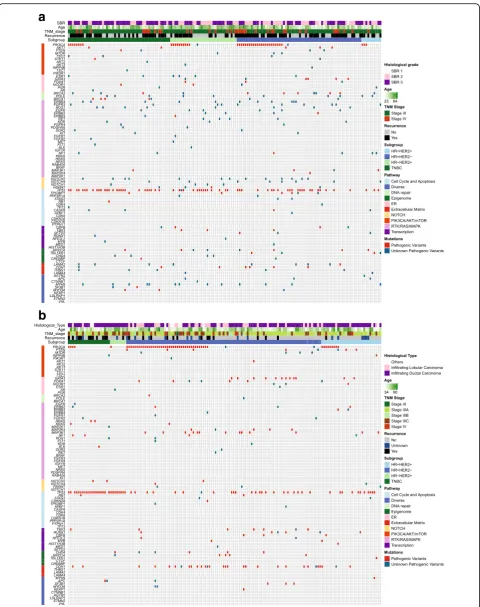
Among the 91 sequenced genes from 156 IBC samples, we detected 777 somatic mutations comprising 733 point mutations and 44 indels. Point mutations included 666 missense, 59 nonsense, and 8 splice-site mutations, and the 44 indels included 28 frame-shift and 16 in-frame. They corresponded to 516 different mutations in 84 genes, and 152 samples exhibited at least one mu-tation. After annotation of all these mutations, 392 mutations in 73 genes on 144 samples were predicted to have high probability of being deleterious, and were thus classified as pathogenic variants and unknown patho-genic variants (Fig. 1a). In the non-IBC, a total of 320 deleterious mutations in 56 genes on 146 samples was identified (Fig.1b). All pathogenic variants and unknown pathogenic variants were considered as mutations for further research.
Comparison of mutation frequency between IBC and non-IBC
The average number of mutations per sample was higher in IBC than in non-IBC (2.5 vs. 1.6, p= 0.0009). We observed a significantly different mutation frequency be-tween IBC and non-IBC patients for the following genes:
TP53, CDH1, NOTCH2, MYH9, BRCA2, ERBB4, POLE,
FGFR3, ROS1, NOTCH4, LAMA2, EGFR, BRCA1,
TP53BP1,ESR1,THBS1,CASP8, andNOTCH1. All genes, exceptCDH1 which was less frequently mutated in IBC, were more frequently mutated in IBC compared with non-IBC (Fig. 2). The most frequently mutated genes in IBC were TP53 (43.0%), PIK3CA (29.5%), MYH9 (8.3%),
NOTCH2 (8.3%), BRCA2 (7.7%), ERBB4 (7.1%), FGFR3 (6.4%), POLE (6.4%), LAMA2 (5.8%), ARID1A (5.1%),
[image:4.595.56.291.232.701.2]NOTCH4(5.1%), andROS1(5.1%) (Fig.2). A comparison was made of the mutated genes between IBC and non-IBC according to the four subgroups. Gene mutation frequencies were not significantly different in the triple-negative IBC and non-IBC subgroups (Additional file3: Figure S1a). In the HR–/HER2+subgroup, less mu-tations on TP53were detected in IBC (Additional file3:
Table 1Pathological and clinical characteristics of the inflammatory breast cancer (IBC) cohort
IBC
Total,n(%) 156 (100)
Age (years)
Median 53
Range 23–84
≤50,n(%) 63 (40.5)
>50,n(%) 93 (59.5)
Sex,n(%)
Female 156 (100)
Male 0 (0)
Stage,n(%)
III 120 (76.9)
IV 36 (23.1)
ER status,n(%)
Negative 89 (57.1)
Positive 67 (42.9)
PR status,n(%)
Negative 111 (71.2)
positive 45 (28.8)
Her2 status,n(%)
Negative 113 (72.4)
Positive 43 (27.6)
Subgroups,n(%)
TNBC 51 (32.7)
HR–/Her2+ 33 (21.2)
HR+/Her2– 62 (39.7)
HR+/Her2+ 10 (6.4)
Distant metastases,n(%)
Yes 98 (62.8)
No 58 (37.2)
SBR histological grade,n(%)
I 2 (1.3)
II 63 (40.4)
III 91 (58.3)
a
b
[image:5.595.58.539.85.693.2]Figure S1b). In the HR+/HER2–subgroup (Additional file3: Figure S1c), genes such asBRCA2,NOTCH2,ERBB4,FGFR3, andLAMA2(allpvalue less than 0.01), andTP53,NOTCH4,
TP53BP1,MYH9, andEGFR(allpvalue less than 0.05) were more frequently mutated in IBC. In the HR+/HER2+ sub-group (Additional file3: Figure S1d),POLEwas found to be more significantly highly mutated in IBC than in non-IBC.
For a better understanding of IBC tumorigenesis, the 91 genes were categorized in 10 different signaling path-ways and the pathway was considered altered when at least one gene of the pathway was mutated (Fig.3). Al-teration in DNA repair, RTK/RAS/MAPK, NOTCH, cell cycle and apoptosis, and diverse pathways showed sig-nificant differences between the two cohorts (p< 0.0001,
(See figure on previous page.)
Fig. 1Deleterious somatic mutations of IBC tumors and non-IBC tumors.aDeleterious somatic mutations of 156 IBC tumors.bDeleterious somatic mutations of 197 non-IBC tumors. Tumors with available mutation data are grouped by four molecular subtypes along thexaxis, with thexaxis also showing clinic characteristics for each tumor; the 91 genes of BreastCurie panel are enriched to 10 signaling on theyaxis. The somatic mutations of each tumor are indicated by colored boxes: red boxes indicate pathogenic variants, and green boxes indicate unknown pathogenic variants. ER estrogen receptor, HER2 human epidermal growth factor receptor 2, HR hormone receptor, SBR Scarff Bloom Richardson classification, TNBC triple-negative breast cancer, TNM tumor node metastasis
[image:6.595.59.538.276.704.2]p< 0.0001, p< 0.0001, p= 0.02, and p= 0.002, respect-ively). Furthermore, we compared alterations of these pathways according to subgroups (Additional file 4: Figure S2). In TNBC, the signaling pathway of DNA re-pair was more frequently altered in IBC than in non-IBC (p= 0.02) (Additional file 4: Figure S2a). In the HR–/ HER2+ subgroup, the RTK/RAS/MAPK pathway was significantly more altered in IBC (p= 0.02), but the cell cycle and apoptosis pathway was more altered in non-IBC (p= 0.03) (Additional file 4: Figure S2b). IBC harbored more mutations in the DNA repair, RTK/RAS/ MAPK, NOTCH, and cell cycle pathways in the HR+/ HER2–subgroup than in non-IBC (p= 0.0001,p= 0.003,
p= 0.001, and p= 0.04, respectively) (Additional file 4: Figure S2c), and DNA repair, epigenome and diverse pathways were also found to be more altered in the HR+/HER2+ subgroup of IBC (p= 0.007, p= 0.02, andp = 0.02, respectively) (Additional file4: Figure S2d).
Several pathways such as PIK3CA/AKT/mTOR, DNA repair, RTK/RAS/MAPK, NOTCH, and ER in-clude targeted genes. The well-known targeted genes of RTKs and NOTCH families were frequently mu-tated in IBC. Intriguingly, more unknown pathogenic variants of ERBB4, FGFR3, ERBB3, and PDGFRA were detected on the catalytic domain of tyrosine kinase, and we also detected unknown pathogenic variants of NOTCH4 on the ankyrin repeat domain in several IBC patients (Fig. 1a). Our results thus demonstrated a genomic instability of the IBC cell surface, and we are continuing to explore whether
those frequent unknown pathogenic mutation of RTKs on ERBB4, FGFR3, EGFR, and ERBB2 are functional and provide potential therapeutic targets in further research.
DNA copy number alterations in IBC
We applied ONCOCNV to calculate the DNA copy num-ber alterations (CNAs) for 156 IBC samples (Additional file 5: Figure S3). In 44 IBC samples with HER2-positive status detected via IHC or FISH, 38 samples were called as havingERBB2 amplification with ONCOCNV calcula-tion (86.4% concordance was achieved). The rate of
ERBB2 and MED1 coamplification was 57.1% in IBC, which is similar to that reported in a non-IBC study [19]. The other frequently amplified genes were observed in
FGFR1in 10.8%,EGFRin 4.4%, andDDR2in 3.2% of IBC samples. The frequent deletions were found in RB1 in 5.1%,STAG2in 3.8%, andCDKN2AandMAP3K1in 3.2% of IBC.
Survival analysis of IBC
We compared the gene mutation frequency in 10 pathways between 98 metastatic IBC and 58 non metastatic IBC (Fig. 4). PIK3CA/AKT/mTOR was the only pathway showing a significant difference, which was more frequently altered in metastatic IBC compared with nonmetastatic IBC (48.0% vs. 24.1%, p = 0.003). Regarding subgroups, a significant difference was found for this pathway in HR+/HER2– (58.1% vs. 26.3%, p= 0.02). A higher mutation trend is shown in
[image:7.595.60.538.87.317.2]the three other subgroups. Among the 11 genes of the PIK3CA/AKT/mTOR pathway, PIK3CA mutations were highly enriched in the metastatic IBC group (38.8% vs. 13.8%, p= 0.0009).
Among 120 stage III IBC patients, MFS was avail-able for 115 patients, and one patient was excluded because of disease progression within 6 months. We assessed the relation of PIK3CA mutation status with MFS in 114 patients. The median survival was 83.1 months (range 6.2 to 212.1 months). Univariate and multivariate analyses are reported in Table 2. The
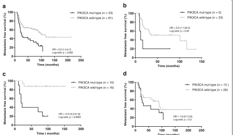
PIK3CA genotype was the only marker significantly associated with MFS in IBC patients (hazard ratio = 2.6, 95% confidence interval (CI) 1.4–4.7, p= 0.002; Fig. 5a). The median MFS for the PIK3CA mutant-type was 26.0 months and for the PIK3CA wild-type it was 101.1 months. Regarding the sub-groups, PIK3CA mutation was associated with MFS in triple-negative (Fig. 5b) and HR–/HER2+ (Fig. 5c) sub-groups, with hazard ratios for PIK3CA mutant-type of 5.6 and 12.9, respectively. A high frequency of
PIK3CA mutation was observed in the HR+/HER2– IBC subgroup; however, no association between
[image:8.595.56.538.88.292.2]PIK3CA mutant-type and MFS was found (hazard ra-tio = 1.5, 95% CI 0.7–3.6, p= 0.3; Fig. 5d). The num-ber of cases was too small to interpret the results for the HR+/HER2+ subgroup. We combined subgroups for further analysis of prognostic impact of PIK3CA mutation, and the PIK3CA mutant-type was found to significantly associated with worse MFS in HR– and HER2+ subgroups; no association was found in the HR+ subgroup (Additional file 6: Figure S4).
Table 2Correlation ofPIK3CAmutation and classic clinical characteristics with metastasis-free survival (MFS) in primary inflammatory breast cancer (IBC) patients with stage III disease (n= 114)
Parameters n Univariate analysis Multivariate analysis
HR (95% CI) pa HR (95% CI) pb
Age 0.8 (0.5–1.1) 0.6 1.0 (0.5–1.7) 0.9
≤50 years 48 >50 years 66
ER status 0.9 (0.6–1.6) 0.8 1.0 (0.5–2.0) 1.0
Negative 67
Positive 47
PR status 1.3 (0.8–2.3) 0.3 1.7 (0.8–3.5) 0.1
Negative 82
Positive 32
Her2 status 1.7 (1.0–2.8) 0.1 2.0 (1.1–3.8) 0.03
Negative 79
Positive 35
SBR grade 1.2 (0.7–2.0) 0.2 1.2 (0.7–2.2) 0.5
III 71
I and II 43
PIK3CA 2.6 (1.4–4.7) 0.002 2.7 (1.5–4.7) 0.001 Mut-type 33
Wild-type 81
CIconfidence interval,ERestrogen receptor,Her2human epidermal growth factor receptor 2,HRhazard ratio,PRprogesterone receptor,SBRScarf Bloom Richardson classification
a
Log-rank test b
Cox multivariate analyses
[image:8.595.305.538.410.689.2]Discussion
A key purpose of precision cancer medicine is to tailor clinical management based on the specific events that are relevant to tumor development and progression. The high frequency of clinically relevant genomic alterations in IBC when sequenced with a targeted NGS raises the possibility that targeted therapies may be developed for patients with this highly aggressive form of breast can-cer. First, a heavy mutation burden was found in IBC tu-mors. This could be the hallmark of increased genomic instability correlating with tumor aggressiveness. TP53 was the most frequently mutated gene, in accordance with previous studies on IBC [7, 10, 20]. There were 12 genes with more than 5% mutation frequencies in IBC:
TP53, PIK3CA, MYH9, NOTCH2, BRCA2, ERBB4,
FGFR3, POLE, LAMA2, ARID1A, NOTCH4, and ROS1. For TP53, PIK3CA, and BRCA2, high mutation rates in IBC have been also reported by other groups. In con-trast, we did not detect higher frequent mutation of
ERBB2,RB1, orNOTCH1[7,9,10].
Comparative analysis of biology pathways between IBC and non-IBC revealed high mutation frequencies of genes in DNA repair, NOTCH, and RTK/RAS/MAPK pathways that could be clinically relevant. The alteration of BRCA1/BRCA2/POLE genes of the DNA repair
pathway was independent of molecular subtypes, so PARP inhibitor may be especially evaluated in IBC [21]. To the best of our knowledge, this is the first time that
POLEhas been detected frequently mutated in IBC. The correlation of POLE mutation with PD1/PD-L1 immunotherapy in colorectal cancer and endometrial cancer [22–24] leads us towards further research to ex-plore whether there is a treatment option for immuno-therapy inPOLE-mutated IBC. Note that POLE-detected variants are not hotspots pathogenic variants that have already been described.NOTCH1/2/4andFBWX7genes were more frequently mutated in each subgroup of IBC compared with non-IBC. A preclinical study in IBC showed that a gamma secretase inhibitor, RO4929097, was able to block the Notch signaling and to attenuate the stem-like phenotype of IBC cells and regulate the in-flammatory environment [25]. Targeting the Notch path-way might be an option for IBC treatment. Receptor tyrosine kinases (RTKs) are frequently activated in can-cer cells and therefore have become the target of numer-ous treatments. The BreastCurie gene panel included most targetable RTK genes and we found that IBC car-ried higher frequencies of unknown pathogenic variants of RTKs than non-IBC. Higher gene instability due to DNA repair dysfunction may promote variants of
0 50 100 150 200 250
0 20 40 60 80
100 PIK3CA mut-type (n = 33)
PIK3CA wild-type (n = 81)
HR = 2.6 (1.4-4.7) Log-rank,p= 0.002
a
Time (months) M et as tas is fr e es u rv iva l( % )0 50 100 150
0 20 40 60 80
100 PIK3CA mut-type (n = 5)
PIK3CA wild-type (n = 33)
HR = 5.6 (1.1-29.3) Log-rank,p= 0.04
b
Time (months) M e ta s ta s isf re es u rv iv a l (% )0 50 100 150 200
0 20 40 60 80
100 PIK3CA mut-type (n = 10)
PIK3CA wild-type (n = 16)
HR = 12.9 (3.2-51.6) Log-rank,p= 0.0003
c
Time ( months)
M e ta st a s is fr ee s u rv iv al (% )
0 50 100 150 200 250
0 20 40 60 80 100
PIK3CA mut-type (n = 15 ) PIK3CA wild-type (n = 26)
d
HR = 1.6 (0.7-3.6) Log-rank,p= 0.3
Time (months) M et as tasi s fr e e su rvi va l (% )
[image:9.595.59.542.90.368.2]unknown significance in IBC but we cannot exclude that other unknown mechanisms are also implicated. Activa-tion of downstream pathways of RTKs, such as ERBB2, EGFR, and IGF1R, has been proven to be related to tumor cell anoikis resistance, and IBC cells have been associated with more evasion of anoikis [26,27] which is consistent with our findings. We now aim to explore whether the frequent unknown pathogenic mutations of RTKs onERBB4,FGFR3, EGFR, andERBB2reported in the present study are potential therapeutic targets.
Compared with the breast cancer literature [10, 12,
13, 28], we did not find IBC-specific amplified genes with our gene panel. Unfortunately, some frequent CNAs of breast cancer (e.g., MYC, CCND1 amplifica-tion) reported previously were not included in our gene panel. We detected STAG2 deletion, a tumor suppressor gene coding cohesion protein, in 3.7% of IBC. STAG2 loss of function was reported in different cancers but not in IBC [29]. However, ONCOCNV did not compute allele frequencies, which may affect the precision of the method in admixed data [15].
Our study demonstrates that PIK3CA gene mutations and PIK3CA/AKT/mTOR pathway alteration were very common events in IBC. PIK3CA gene mutations were especially observed in luminal and HER2-positive sub-types, and mainly located in hotspots of the helical do-main and the catalytic dodo-main, similar to non-IBC in previous reports [13, 30]. Recently, a large pooled analysis of more than 10,000 early-stage breast cancer patients reported that PIK3CA-mutated tumors are as-sociated with a better prognosis [31]. However, this good prognostic effect was observed in HR+/HER2– and TNBC subtypes, but not in the HER2+ subtype where
PIK3CA mutations were associated with a worse overall survival. Interestingly, in our IBC cohort,PIK3CA muta-tion was a poor prognostic factor for MFS for HER2+ and TNBC subtypes, whereas no prognostic value was found in the HR+/HER2–subtype. Of note, the prognos-tic effect was weak in the TNBC subtype of IBC since
PIK3CA mutations were rare in this subtype and our TNBC cohort was small. For the HER2+ subtype, previ-ous studies reported thatPIK3CA mutations were asso-ciated with adverse prognosis in non-IBC, but results were not conclusive [31–34]. AsPIK3CAmutation could lead to resistance to anti-HER2 treatments [34, 35], we checked that the percentage of patients in our IBC cohort receiving trastuzumab combined with chemo-therapy was balanced in both PIK3CAgenotypes (71.4% in mutant type, 75.9% in wild type). Therefore, the asso-ciation betweenPIK3CAmutation and worse MFS in IBC may be reliable. For the HR+/HER2– subtype, the prog-nostic difference regarding PIK3CA genotype between IBC and non-IBC may reflect the influence of the PI3K pathway in the two distinct biological environments of
IBC and non-IBC, and we presume interactions between ER and PI3K pathways are different. We know that PI3K inhibitors have been investigated in many breast cancer trials and have shown promising results in ER-positive endocrine therapy-refractory breast cancer [36], but no clinical trials have been performed specifically in IBC to date. The association of PIK3CA mutations with worse MFS in IBC should draw our attention to the role of the PI3K pathway in this aggressive and treatment-refractory form of breast cancer. Further experimental research to explore the PI3K pathway in IBC is therefore required.
Conclusions
Overall, IBC is the most aggressive form of breast cancer frequently refractory to conventional therapy and suffers from the lack of a specific treatment. Our study using targeted NGS analysis revealed a high frequency of som-atic mutations, in particular in DNA repair, Notch signaling, and RTKs genes, that may guide switching from conventional therapy to targeted agents in IBC. In contrast to non-IBC patients, PIK3CA mutation was associated with a poor outcome in IBC patients. These findings encourage clinical trials with targeted therapies that may provide clinical benefit to IBC patients.
Additional files
Additional file 1:Table S1.Pathological and clinical characteristics of non-IBC cohorts. (PDF 169 kb)
Additional file 2:Table S2.BreastCurie gene panel for targeted NGS. (PDF 411 kb)
Additional file 3:Figure S1.Comparison of somatic mutation frequency between IBC and non-IBC in four subgroups. (a) The percentage of samples with somatic mutation in the TNBC subgroup; (b) the percentage of samples with somatic mutation in the HR–/HER2+subgroup; (c) the percentage of sam-ples with somatic mutation in the HR+/HER2–subgroup; (d) the percentage of samples with somatic mutation in the HR+/HER2+subgroup. The gray bars indicate non-IBC, the black bars indicate IBC; *p< 0.05, **p< 0.01, ***p< 0.001. (PDF 52 kb)
Additional file 4:Figure S2.Comparison of biological pathway between IBC and non-IBC in four subgroups. (a) The percentage of sam-ples with alteration on 10 biological pathways in the TNBC subgroup; (b) the percentage of samples with alteration on 10 biological pathways in the HR–/HER2+subgroup; (c) the percentage of samples with alteration on 10
biological pathways in the HR+/HER2–subgroup; (d) the percentage of samples with alteration on 10 biological pathways in the HR+/HER2+ subgroup. The gray bars indicate non-IBC, the black bars indicate IBC; *p< 0.05, **p< 0.01, ***p< 0.001. (PDF 41 kb)
Additional file 5:Figure S3.DNA copy number alterations in the IBC cohort. The genes with DNA copy number alterations are grouped along thexaxis, the percentage of samples with DNA copy number alterations shown on theyaxis, DNA amplifications are indicated by black bars above thexaxis, and DNA deletions are indicated by gray bars below the xaxis. (PDF 164 kb)
of the HER2+subgroup, (c) Kaplan-Meier estimates of MFS according to PIK3CAmutations in patients of the HR+subgroup. (PDF 42 kb)
Abbreviations
BCSS:Breast cancer-specific survival; BRCA1/2: Breast cancer 1/2; ER: Estrogen receptor; FISH: Fluorescence in-situ hybridization; HER2: Human epidermal growth factor receptor 2; HR: Hormone receptor; IHC: Immunohistochemical; MFS: Metastasis-free survival; mTOR: Mammalian target of rapamycin; NGS: Next-generation sequencing; NOTCH1/2/4: Neurogenic locus notch homolog protein 1/2/4; PI3K: Phosphatidylinositol 3-kinase;
PIK3CA: Phosphatidylinositol 3-kinase: catalytic: alpha polypeptide gene; POLE: Polymerase (DNA) epsilon; PR: Progesterone receptor; RTK: Receptor tyrosine kinase; SBR: Scarff Bloom Richardson classification; TCGA: The Cancer Genome Atlas; TNBC: Triple-negative breast cancer
Acknowledgments
We thank Odette Mariani from Institut Curie for providing frozen samples.
Funding
This study was supported by l’association pour la recherche de Saint Cloud (ARCS). High-throughput sequencing has been performed by the ICGex NGS platform of the Institut Curie supported by the grants ANR-10-EQPX-03 (Equipex) and ANR-10-INBS-09-08 (France Génomique Consortium) from the Agence Nationale de la Recherche (“Investissements d’Avenir”program), by the Canceropole Ile-de-France, and by the SiRIC-Curie program SiRIC Grant
“INCa-DGOS- 4654”.
Availability of data and materials
All data generated and/or analyzed during this study are included in this published article.
Authors’contributions
XL, FL, IB, and CC made substantial contributions to conception and design, and revising the manuscript, and gave final approval for publication. XL, SV, MB, AB, and CC contributed to acquisition of data, analysis and interpretation of data, and drafted the manuscript. XL, AB, SB, and VB performed data analysis for the study. XL, SV, FL, IB, and CC participated in manuscript preparation and revision. XL, FL, and CC contributed to data collection and database management for the study. All other authors made substantial contributions to the acquisition of data, revising the manuscript, and final approval.
Ethics approval and consent to participate
This study was approved by the Curie Institute/Rene Huguenin Hospital ethics committee. Participating patients completed an informed consent process.
Consent for publication Not applicable.
Competing interests
The authors declare that they have no competing interests.
Publisher’s Note
Springer Nature remains neutral with regard to jurisdictional claims in published maps and institutional affiliations.
Author details
1Department of Breast Oncology, Key Laboratory of Carcinogenesis and Translational Research (Ministry of Education), Peking University Cancer Hospital & Institute, Beijing, China.2Pharmacogenomic Unit, Department of Genetics, Curie Institute, PSL Research University, 26 rue d’Ulm, 75005 Paris, France.3Clinic bioinformatic Unit, Department of Biopathology, Curie Institute, PSL Research University, Paris, France.4Institut Curie Genomics of Excellence (ICGex) Platform, Curie Institute, PSL Research University, Paris, France.5EA7331, Paris Descartes University, Sorbonne Paris Cité, Faculty of Pharmaceutical and Biological Sciences, Paris, France.6Department of Medical Oncology, Curie Institute, René Huguenin Hospital, Saint-Cloud, France.
Received: 20 February 2018 Accepted: 18 June 2018
References
1. Dawood S, Merajver SD, Viens P, Vermeulen PB, Swain SM, Buchholz TA, et al. International expert panel on inflammatory breast cancer: consensus statement for standardized diagnosis and treatment. Ann Oncol. 2011;22: 515–23.https://doi.org/10.1093/annonc/mdq345.
2. Robertson FM, Bondy M, Yang W, Yamauchi H, Wiggins S, Kamrudin S, et al. Inflammatory breast cancer: the disease, the biology, the treatment. CA Cancer J Clin. 2010;60:351–75.https://doi.org/10.3322/caac.20082. 3. Dawood S, Lei X, Dent R, Gupta S, Sirohi B, Cortes J, et al. Survival of
women with inflammatory breast cancer: a large population-based study. Ann Oncol. 2014;25:1143–51.https://doi.org/10.1093/annonc/mdu121. 4. Bertucci F, Finetti P, Rougemont J, Charafe-Jauffret E, Cervera N, Tarpin C, et
al. Gene expression profiling identifies molecular subtypes of inflammatory breast cancer. Cancer Res. 2005;65:2170–8. https://doi.org/10.1158/0008-5472.CAN-04-4115.
5. Van Laere S, Van der Auwera I, Van den Eynden G, Van Hummelen P, van Dam P, Van Marck E, et al. Distinct molecular phenotype of inflammatory breast cancer compared to non-inflammatory breast cancer using Affymetrix-based genome-wide gene-expression analysis. Br J Cancer. 2007; 97:1165–74.https://doi.org/10.1038/sj.bjc.6603967.
6. Bertucci F, Ueno NT, Finetti P, Vermeulen P, Lucci A, Robertson FM, et al. Gene expression profiles of inflammatory breast cancer: correlation with response to neoadjuvant chemotherapy and metastasis-free survival. Ann Oncol. 2014;25:358–65.https://doi.org/10.1093/annonc/mdt496. 7. Bertucci F, Finetti P, Vermeulen P, Van Dam P, Dirix L, Birnbaum D, et al.
Genomic profiling of inflammatory breast cancer: a review. Breast. 2014;23: 538–45.https://doi.org/10.1016/j.breast.2014.06.008.
8. Van Laere SJ, Ueno NT, Finetti P, Vermeulen P, Lucci A, Robertson FM, et al. Uncovering the molecular secrets of inflammatory breast cancer biology: an integrated analysis of three distinct affymetrix gene expression datasets. Clin Cancer Res. 2013;19:4685–96.https://doi.org/10. 1158/1078-0432.CCR-12-2549.
9. Matsuda N, Lim B, Wang Y, Krishnamurthy S, Woodward W, Alvarez RH, et al. Identification of frequent somatic mutations in inflammatory breast cancer. Breast Cancer Res Treat. 2017;163:263–72.https://doi.org/10.1007/ s10549-017-4165-0.
10. Ross JS, Ali SM, Wang K, Khaira D, Palma NA, Chmielecki J, et al. Comprehensive genomic profiling of inflammatory breast cancer cases reveals a high frequency of clinically relevant genomic alterations. Breast Cancer Res Treat. 2015;154:155–62. https://doi.org/10.1007/s10549-015-3592-z.
11. Gao J, Aksoy BA, Dogrusoz U, Dresdner G, Gross B, Sumer SO, et al. Integrative analysis of complex cancer genomics and clinical profiles using the cBioPortal. Sci Signal. 2013;6:pl1.https://doi.org/10.1126/ scisignal.2004088.
12. Cerami E, Gao J, Dogrusoz U, Gross BE, Sumer SO, Aksoy BA, et al. The cBio cancer genomics portal: an open platform for exploring multidimensional cancer genomics data. Cancer Discov. 2012;2:401–4.https://doi.org/10.1158/ 2159-8290.CD-12-0095.
13. Cancer Genome Atlas N. Comprehensive molecular portraits of human breast tumours. Nature. 2012;490:61–70.https://doi.org/10.1038/nature11412. 14. Le Tourneau C, Kamal M, Tsimberidou AM, Bedard P, Pierron G, Callens C, et
al. Treatment algorithms based on tumor molecular profiling: the essence of precision medicine trials. J Natl Cancer Inst. 2016;108https://doi.org/10. 1093/jnci/djv362.
15. Boeva V, Popova T, Lienard M, Toffoli S, Kamal M, Le Tourneau C, et al. Multi-factor data normalization enables the detection of copy number aberrations in amplicon sequencing data. Bioinformatics. 2014;30:3443–50. https://doi.org/10.1093/bioinformatics/btu436.
16. Lefebvre C, Bachelot T, Filleron T, Pedrero M, Campone M, Soria JC, et al. Mutational profile of metastatic breast cancers: a retrospective analysis. PLoS Med. 2016;13:e1002201.https://doi.org/10.1371/journal.pmed.1002201. 17. Le Tourneau C, Delord JP, Goncalves A, Gavoille C, Dubot C, Isambert N, et
18. Andre F, Bachelot T, Commo F, Campone M, Arnedos M, Dieras V, et al. Comparative genomic hybridisation array and DNA sequencing to direct treatment of metastatic breast cancer: a multicentre, prospective trial (SAFIR01/UNICANCER). Lancet Oncol. 2014;15:267–74.https://doi. org/10.1016/S1470-2045(13)70611-9.
19. Lamy PJ, Fina F, Bascoul-Mollevi C, Laberenne AC, Martin PM, Ouafik L, et al. Quantification and clinical relevance of gene amplification at chromosome 17q1q21 in human epidermal growth factor receptor 2-amplified breast cancers. Breast Cancer Res. 2011;13:R15.https://doi.org/ 10.1186/bcr2824.
20. Goh G, Schmid R, Guiver K, Arpornwirat W, Chitapanarux I, Ganju V, et al. Clonal evolutionary analysis during HER2 blockade in HER2-positive inflammatory breast cancer: a phase II open-label clinical trial of afatinib +/− vinorelbine. PLoS Med. 2016;13:e1002136.https://doi.org/10.1371/journal. pmed.1002136.
21. Lord CJ, Ashworth A. PARP inhibitors: synthetic lethality in the clinic. Science. 2017;355:1152–8.https://doi.org/10.1126/science.aam7344. 22. Castellucci E, He T, Goldstein DY, Halmos B, Chuy J. DNA polymerase
varepsilon deficiency leading to an ultramutator phenotype: a novel clinically relevant entity. Oncologist. 2017;22:497–502.https://doi.org/10. 1634/theoncologist.2017-0034.
23. Gargiulo P, Della Pepa C, Berardi S, Califano D, Scala S, Buonaguro L, et al. Tumor genotype and immune microenvironment in POLE-ultramutated and MSI-hypermutated endometrial cancers: new candidates for checkpoint blockade immunotherapy? Cancer Treat Rev. 2016;48:61–8.https://doi.org/ 10.1016/j.ctrv.2016.06.008.
24. Bourdais R, Rousseau B, Pujals A, Boussion H, Joly C, Guillemin A, et al. Polymerase proofreading domain mutations: new opportunities for immunotherapy in hypermutated colorectal cancer beyond MMR deficiency. Crit Rev Oncol Hematol. 2017;113:242–8.https://doi.org/10.1016/ j.critrevonc.2017.03.027.
25. Debeb BG, Cohen EN, Boley K, Freiter EM, Li L, Robertson FM, et al. Pre-clinical studies of notch signaling inhibitor RO4929097 in inflammatory breast cancer cells. Breast Cancer Res Treat. 2012;134:495–510.https://doi. org/10.1007/s10549-012-2075-8.
26. Buchheit CL, Weigel KJ, Schafer ZT. Cancer cell survival during detachment from the ECM: multiple barriers to tumour progression. Nat Rev Cancer. 2014;14:632–41.https://doi.org/10.1038/nrc3789.
27. Buchheit CL, Schafer ZT. BIM-EL localization: the key to understanding anoikis resistance in inflammatory breast cancer cells. Mol Cell Oncol. 2016; 3:e1011474.https://doi.org/10.1080/23723556.2015.1011474.
28. Curtis C, Shah SP, Chin SF, Turashvili G, Rueda OM, Dunning MJ, et al. The genomic and transcriptomic architecture of 2,000 breast tumours reveals novel subgroups. Nature. 2012;486:346–52.https://doi.org/10.1038/ nature10983.
29. Hill VK, Kim JS, Waldman T. Cohesin mutations in human cancer. Biochim Biophys Acta. 2016;1866:1–11.https://doi.org/10.1016/j.bbcan.2016.05.002. 30. Stemke-Hale K, Gonzalez-Angulo AM, Lluch A, Neve RM, Kuo WL, Davies M,
et al. An integrative genomic and proteomic analysis of PIK3CA, PTEN, and AKT mutations in breast cancer. Cancer Res. 2008;68:6084–91.https://doi. org/10.1158/0008-5472.CAN-07-6854.
31. Zardavas D, Te Marvelde L, Milne RL, Fumagalli D, Fountzilas G, Kotoula V, et al. Tumor PIK3CA genotype and prognosis in early-stage breast cancer: a pooled analysis of individual patient data. J Clin Oncol. 2018;36:981–90. https://doi.org/10.1200/JCO.2017.74.8301.
32. Mukohara T. PI3K mutations in breast cancer: prognostic and therapeutic implications. Breast Cancer. 2015;7:111–23.https://doi.org/10.2147/BCTT. S60696.
33. Cizkova M, Susini A, Vacher S, Cizeron-Clairac G, Andrieu C, Driouch K, et al. PIK3CA mutation impact on survival in breast cancer patients and in ERalpha, PR and ERBB2-based subgroups. Breast Cancer Res. 2012;14:R28. https://doi.org/10.1186/bcr3113.
34. Loibl S, Majewski I, Guarneri V, Nekljudova V, Holmes E, Bria E, et al. PIK3CA mutations are associated with reduced pathological complete response rates in primary HER2-positive breast cancer: pooled analysis of 967 patients from five prospective trials investigating lapatinib and trastuzumab. Ann Oncol. 2016;27:1519–25.https://doi.org/10.1093/annonc/mdw197. 35. Bianchini G, Kiermaier A, Bianchi GV, Im YH, Pienkowski T, Liu MC, et al.
Biomarker analysis of the NeoSphere study: pertuzumab, trastuzumab, and docetaxel versus trastuzumab plus docetaxel, pertuzumab plus trastuzumab, or pertuzumab plus docetaxel for the neoadjuvant treatment of
HER2-positive breast cancer. Breast Cancer Res. 2017;19:16.https://doi.org/10. 1186/s13058-017-0806-9.